
34
I. OVERVIEW
The adrenal (suprarenal) glands provide the bloodborne signals of stress, epinephrine, and cortisol. The sounding of the body's alarms and defenses helps an individual survive physical threats, endure pain, and tap the body's physical and metabolic reserves. In modern humans, stress is often more mental and social in nature, but such events elicit very similar stress responses as does climbing a tree to escape a pack of wolves. Besides stress, adrenal glands regulate plasma Na+ via aldosterone and certain secondary sex characteristics by the adrenal androgens. Stress, salt, and sex are heavy responsibilities for this small (~1.5 by 7.5 cm and weighing ~8–10 g) set of glands located just above each kidney. Each gland can be divided into two main sections: the cortex (~90% of gland weight) and the medulla (~10%) as shown in Figure 34.1. The cortex is controlled and regulated, in part, by the hypothalamic–pituitary axis and is further divided into the zona glomerulosa, zona fasciculata, and zona reticularis (see Figure 34.1). The zona glomerulosa produces and secretes aldosterone, which regulates plasma volume by controlling how much Na+ is retained by the kidney. Cortisol is primarily produced and secreted by the zona fasciculata and increases metabolism and catabolism as well as suppresses inflammation and immunity. The adrenal androgens, which are dehydroepiandrosterone (DHEA), dehydroepiandrosterone sulfate (DHEAS), and androstenedione, are primarily produced and secreted by the zona reticularis and participate in secondary sex characteristics (e.g., hair growth) during puberty and adolescence. The adrenal medulla is controlled and regulated by the sympathetic nervous system (SNS), and its major hormonal product is epinephrine (adrenaline). Similar to the SNS “fight-or-flight” response, epinephrine provides a rapid stress signal but delivered via the circulation rather than the nervous system.
II. HYPOTHALAMIC–PITUITARY–ADRENAL AXIS
The adrenal cortex is controlled and regulated by an endocrine axis, providing a multitiered response that allows for both gross and fine hormonal adjustment. Axis control is directed primarily at the zona fasciculata and reticularis. The zona glomerulosa is regulated primarily by other hormones (angiotensin II [Ang-II]) and ions (K+).
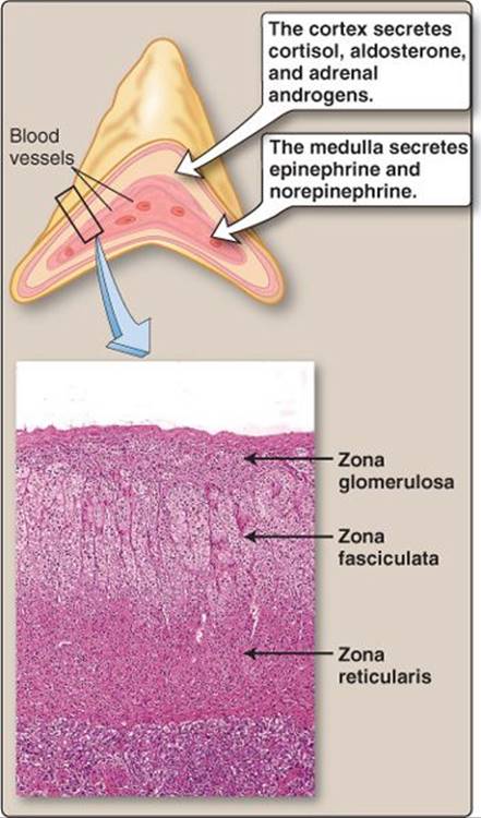
Figure 34.1
Adrenal gland structure.
A. Hypothalamus
Corticotropin-releasing hormone (CRH) is synthesized (see Figure 7.11) in the paraventricular nucleus and released into hypophyseal portal circulation for carriage to the anterior pituitary (Figure 34.2). A number of higher brain centers stimulate CRH release during physical, biochemical (e.g., low blood glucose), and emotional stress. CRH release follows a circadian rhythm, peaking just before waking and then pulsing throughout the day, based on the above stressors. The paraventricular nucleus also produces antidiuretic hormone (ADH), which can further regulate CRH release and stimulate corticotropes.
B. Pituitary gland
CRH binds to corticotrope type 1 corticotropin- releasing hormone receptor (CRH-R1), which is part of the G protein–coupled receptor (GPCR) superfamily that acts primarily through the adenylyl cyclase (AC) second-messenger system. CRH-R1 binding activates transcription factors to express the preproopiomelanocortin (POMC) gene, which encodes adrenocorticotropic hormone (ACTH), which is released into the bloodstream. ACTH's target is the adrenal cortex.
C. Adrenal cortex
Adrenocortical hormone (i.e., aldosterone, cortisol, DHEA, DHEAS, and androstenedione) synthesis begins with cholesterol. A small amount of cholesterol is synthesized by the cortex, but the majority is taken up from blood and then stored in a cytosolic pool. Cortical activity is stimulated by ACTH from the pituitary, acting via melanocortin 2 receptors, which are part of the GPCR superfamily. These receptors act primarily through the AC second messenger system (Figure 34.3) to activate enzymes that aid cholesterol uptake as well as a specialized side-chain cleavage enzyme complex (sometimes termed cholesterol desmolase, or cytochrome P450 SCC). Side-chain cleavage enzyme complex is one of the key rate-limiting steps for adrenal cortex hormone production. There are a number of common enzymes and intermediates in the synthesis of cortex hormones (Figure 34.4). The activation or inhibition or even the presence of one enzyme but not another can preferentially shunt the production to cortisol rather than an adrenal androgen, or vice versa.
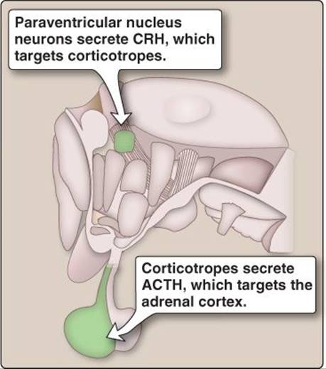
Figure 34.2
Hypothalamus and pituitary gland.
ACTH = adrenocorticotropic hormone;
CRH = corticotropin-releasing hormone.
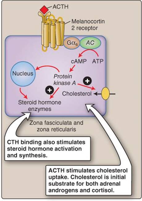
Figure 34.3
Melanocortin 2–receptor signaling.
AC = adenylyl cyclase; ACTH = adrenocorticotropic hormone; ATP = adenosine triphosphate; cAMP = cyclic adenosine monophosphate;
CRH = corticotropin-releasing hormone.
III. ALDOSTERONE
Aldosterone is synthesized in the zona glomerulosa. This is the only cortical region to express aldosterone synthase (and other CYP11B2 gene product enzymes), which facilitates the final step in the conversion of cholesterol into aldosterone. Once released into the circulation, aldosterone binds with low affinity to corticosteroid-binding protein and albumin. The hormone has a half-life of ~20 min.
A. Function
Aldosterone increases Na+ and water reabsorption as well as K+ secretion from renal tubules (see 27·IV). Aldosterone also increases Na+ reabsorption by intestinal enterocytes, which increases the body's Na+stores. The effect of aldosterone on ions (minerals) is reflected in its class name, mineralocorticoid. Aldosterone acts through cytosolic mineralocorticoid receptors in target cells to facilitate Na+ and water reabsorption in the kidney and absorption in the gastrointestinal (GI) system (see Figure 27.12).
Clinical Application 34.1: Addison Disease
Primary adrenal insufficiency (Addison disease) commonly results from an autoimmune response that destroys the adrenal cortex. Symptoms include fatigue, dehydration, hyponatremia, and hypotension associated with loss of glucocorticoids and mineralocorticoids. Adrenal hormone deficiency stimulates corticotropin-releasing hormone release and preproopiomelanocortin gene expression through a negative feedback pathway, which increases the circulating levels of adrenocorticotropic hormone (ACTH). Hyperpigmentation of hands, feet, nipples, axillae, and the oral cavity occurs because of the elevated ACTH. Treatment involves fluid replacement and exogenous glucocorticoids such as hydrocortisone. Once symptoms have stabilized, mineralocorticoid replacement therapy can be implemented until the postural drop in blood pressure can be adequately controlled.
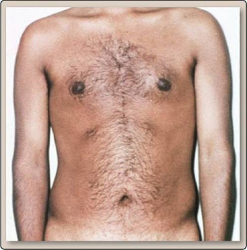
Bronze skin and nipple hyperpigmentation.

Figure 34.4
Cortex hormone biosynthesis. DHEA = dehydroepiandrosterone; DHEAS = dehydroepiandrosterone sulfate.
B. Secretion
Aldosterone synthase produces aldosterone from corticosterone. Aldosterone synthase is the gatekeeper of aldosterone production and is regulated by Ang-II and plasma K+ levels. Ang-II, a hormone within the renin–angiotensin–aldosterone system, is stimulated by low circulating fluid volume, low pressure in the glomerulus, and increases in SNS activity (see 28·III·C). An increase in ACTH, which is vital for regulation of other renal cortex hormones, must be present but is less of a stimulator for the final step in aldosterone synthesis. Feedback for aldosterone secretion is not aldosterone itself, but rather comes in the form of its effects of decreasing fluid volume and plasma K+levels (Figure 34.5).
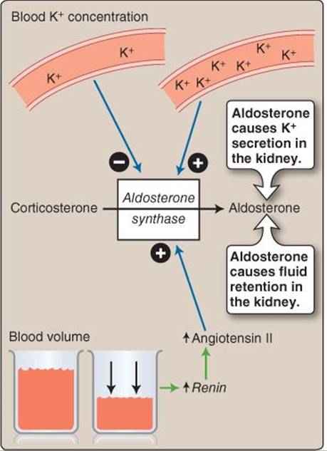
Figure 34.5
Aldosterone regulation.
IV. ANDROGENS
The adrenal androgens (DHEA, DHEAS, and androstenedione) are often produced as a cohort rather than individually (see Figure 34.4). Adrenal androgens are synthesized and secreted primarily by the zona reticularis and, to a lesser extent, by the zona fasciculata (see Figure 34.1). In the blood, DHEA and androstenedione bind with low affinity to albumin and other blood globulins and have a half-life of 15–30 minutes. In contrast, DHEAS has a higher affinity for albumin and has a half-life of 8–10 hours, thereby demonstrating that carrier proteins are able to extend the half-lives of hormones because less free (unbound) hormone is cleared from the blood and can serve as a small temporary storage facility for a hormone. Why do some hormones need a carrier protein? Think of a carrier protein as an additive that binds oil, so that it does not separate from water, allowing it to be transported anywhere water is.
Clinical Application 34.2: 21α-Hydroxylase Deficiency
Because the pathways in Figure 34.4 are interconnected, a deficiency in one of the enzymes can bias the pathway so that one hormone is overproduced and another not produced. 21α-Hydroxylase deficiency is a condition in which a mutation in CYP21A2 gene products results in nonfunctioning 21α-hydroxylase. Thus, there is a lack of mineralocorticoids (aldosterone) and glucocorticoids (like cortisol) but an overproduction of adrenal androgens. Infants with 21α-hydroxylase deficiency present with 1) hypotension and dehydration from the lack of aldosterone and the inability to adequately retain Na+, 2) hypoglycemia from the lack of cortisol-induced energy substrate release, and 3) excess virilization and ambiguous genitalia (in females) is a result of androgen overproduction.

Ambiguous genitalia.
A. Function
DHEA and DHEAS are less potent than androgens produced by the gonads but do have functional effects on secondary sex characteristics and are involved in development during childhood and adolescence. The beginning of androgen release (adrenarche) during development stimulates axillary and pubic hair growth. DHEA can be converted into androstenedione, which can then be converted to more potent androgens, such as testosterone and estrogens, in peripheral tissues. 17-Ketosteroid reductase is a key enzyme in facilitating the conversion of androstenedione to testosterone. This androgen conversion is an important source of testosterone in women.
B. Secretion
DHEA, DHEAS, and androstenedione are controlled by the negative feedback loops of CRH and ACTH (Figure 34.6). These multiple feedback loops provide a finer regulation of hormone production than does a single feedback loop. Events that trigger the release of ACTH facilitate synthesis and release of adrenal androgens. The input rhythms associated with growth and development during puberty and across the lifespan affect ACTH production and release.
V. CORTISOL
Cortisol and corticosterone are synthesized and secreted primarily by the zona fasciculata and, to a lesser extent, by the zona reticularis (see Figure 34.1). Cortisol synthesis, in contrast to adrenal androgen synthesis, requires two hydroxylases (21α-hydroxylase and 11β- hydroxylase) to eventually convert progesterone and 17-hydroxyprogesterone into their final products (see Figure 34.4). In the blood, cortisol binds corticosteroid-binding protein with a high affinity and has a half-life of ~60 minutes.
A. Function
Cortisol and corticosterone prepare the body for stress. Cortisol diffuses across the cell membrane and binds to a cytosolic glucocorticoid receptor. The hormone–receptor complex translocates to the nucleus and binds a glucocorticoid response element on DNA. Cortisol also binds with low affinity to mineralocorticoid receptors and, thus, induces some minor collateral aldosterone-like responses. Cortisol causes a number of physiologic effects (Figure 34.7).
1. Metabolic: Cortisol increases plasma glucose and free fatty acid concentration in order to provide energy substrates to body tissues for their response to the stressful event that stimulated cortisol production.
a. Increased catabolism: Cortisol increases skeletal muscle protein catabolism, liberating amino acids that are then converted to glucose via gluconeogenesis in the liver (see Chapters 32·IV·A and 33·III·A·2). This glucose response is part of the origin of the glucocorticoid classification of cortisol.

Figure 34.6
Hypothalamic-pituitary-adrenal regulation. ACTH = adrenocorticotropic hormone; CRH = corticotropin-releasing hormone.
b. Increased lipolysis: Cortisol stimulates white adipose tissue to undergo lipolysis to liberate free fatty acids and triglycerides. The fatty acids and triglycerides are then transported in the blood for use as an energy source by other tissues.
c. Increased intake: Cortisol stimulates appetite. Acutely, this is beneficial to provide energy substrates to respond to the stressful event. However, if the stressful event does not involve physical work, then this increased appetite can lead to weight gain.
2. Immune: Cortisol suppresses both immune responses and inflammation.1 Although this response may seem counterproductive in stressful conditions, when the life of the organism is in danger, fighting illness with the immune system becomes less important than immediate survival. The mechanisms by which this immunosuppression is accomplished are via decreased production of lymphocytes and interleukins 1 and 6 (IL-1 and IL-6) and T-cell suppression. The anti-inflammatory effects of cortisol are due to decreases in capillary permeability as well as reductions in both prostaglandin and leukotriene synthesis that mediate increases in local blood flow.
3. Musculoskeletal: Cortisol increases bone resorption and decreases Ca2+ absorption from the GI tract and reabsorption from the kidney. Chronic high levels of cortisol can lead to osteoporosis. Cortisol decreases collagen formation throughout the body. Protein catabolism to increase plasma glucose levels can eventually lead to muscle weakness and early fatigue onset during physical activity.
4. Cardiovascular: Cortisol increases erythropoietin release, which stimulates red blood cell production. Cortisol potentiates vasoconstrictor responses by blocking local vasodilators, such as nitric oxide and prostaglandins, and through glucocorticoid receptors in vascular smooth muscle by altering Ca2+ homeostasis within these cells. Glucocorticoids increase the effectiveness of catecholamine actions, such as inotropy and vasoconstriction, through the upregulation of adrenergic receptors.
B. Secretion
Cortisol and corticosterone release are controlled by the negative feedback loops of CRH and ACTH (see Figure 34.6). Section II, Hypothalamic–Pituitary–Adrenal Axis, described how physical, emotional, and biochemical stress stimulate the release of CRH, ACTH, and cortisol. The control of CRH is primarily for regulation of cortisol and less so for adrenal androgens or aldosterone.
 1Immune suppressive and anti-inflammatory effects of glucocorticoids can be exploited pharmacologically. Drugs like prednisone, which is structurally similar to cortisol, can be used as immune suppressants for autoimmune diseases. For more information, see LIR Pharmacology, 5e, p. 334.

Figure 34.7
Glucocorticoid effects.
Clinical Application 34.3: Cushing Syndrome
Patients with Cushing syndrome may present with muscle weakness, osteoporosis, hypertension, diabetes, and weight gain with fat redistribution. These symptoms reflect chronic elevations in glucocorticoid levels. Muscle weakness results from skeletal muscle protein catabolism, osteoporosis by Ca2+ resorption in bone, hyper-tension by cortisol's mineralocorticoid effects on Na+ retention, diabetes by increases in plasma glucose, weight gain by increases in appetite, and fat redistribution by unutilized fatty acid release. For less-understood reasons, unutilized fatty acids are redeposited in the face and upper back, causing a “moon face” appearance and the development of “buffalo hump.” The cause of the excess cortisol secretion is often a pituitary adenoma, which causes an excess secretion of adrenocorticotropic hormone.
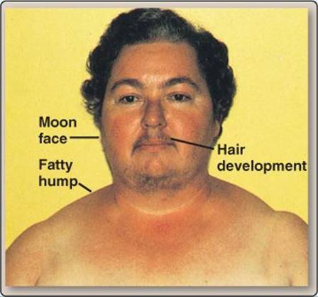
Female with Cushing syndrome.
VI. CATECHOLAMINES
The adrenal medulla is derived from the neural crest rather than the mesodermal mesenchyme, which forms the cortex. In practice, this means that the medulla functions as an extension of the SNS. The medulla is composed of small clusters of chromaffin cells (medullary cells), which synthesize catecholamines from the amino acid tyrosine (see Figure 5.7). Dopamine is synthesized in the cytosol, and a catecholamine-H+ exchanger (VMAT1, for vesicular monoamine transporter 1) transports it into secretion vesicles. Dopamine is then converted to norepinephrine via dopamine β- hydroxylase. Unlike postganglionic adrenergic nerves of the SNS, chromaffin cells contain phenylethanolamine N-methyltransferase. This enzyme is located in the cytosol and facilitates the conversion of norepinephrine to epinephrine. Therefore, norepinephrine must be transported back into the cytosol to be converted to epinephrine, which is in turn transported back into the secretion vesicle. Epinephrine and norepinephrine are then stored with chromogranin (binding protein) in preparation for vesicle exocytosis and hormonal release (Figure 34.8). Chromaffin cells secrete norepinephrine and epinephrine in an approximate 1:4 ratio into the fenestrated medullary capillary network for delivery to various body tissues. Catecholamine half-lives range from 10–90 s. Although seemingly short, they are longer than the SNS release and clearance of norepinephrine in the synaptic cleft. This allows for a more sustained SNS response.
A. Function
Epinephrine (adrenaline), produces classic fight-or-flight responses, or the “adrenaline rush.” Thus, the key actions of epinephrine and norepinephrine are similar to those of the SNS (see Figure 7.4). Delivery via the circulation means that responses to hormones, although typically slower, are wider ranging because they can reach receptor populations that are not specifically located within a SNS synaptic cleft. The functional effects are related to the amount secreted and the tissue responsiveness.
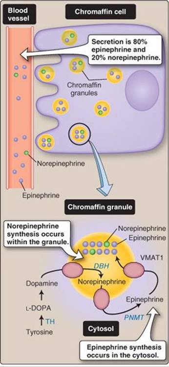
Figure 34.8
Chromaffin cell. DBH = dopamine β-hydroxylase; L-DOPA = L-3,4-dihydroxyphenylalanine; PNMT = phenylethanolamine N-methyltransferase; TH = tyrosine hydroxylase; VMAT1 = vesicular monoamine transporter 1.
Clinical Application 34.4: Pheochromocytoma
Pheochromocytomas are catecholamine-producing tumors located in the adrenal medulla or preganglionic neurons. The classic triad of symptoms includes headaches, palpitations (tachycardia), and profuse sweating. Palpitations and sweating are caused by high circulating epinephrine and norepinephrine levels. Headaches can be caused by direct vasoconstriction of cerebral blood vessels or the high blood pressure (hypertension) induced by peripheral vasoconstriction. These symptoms can be episodic or sustained, depending on the nature of the catecholamine release caused by the tumor.
B. Secretion
Catecholamine release is regulated by the SNS rather than the hypothalamic–pituitary–adrenal axis. Thus, secretion is increased during stresses to homeostasis; strong emotions, such as anger and fear; and exercise. Cholinergic preganglionic SNS neurons stimulate secretion from chromaffin cells via nicotinic type 2 acetylcholine receptors to increase chromaffin granule secretion (see Figure 7.5).
C. Regulation
Adrenergic receptor expression is dynamic. With high levels of circulating catecholamines, such as during continual stress, membrane receptors can be internalized, thereby reducing responsiveness to subsequent catecholamine stimulation. Conversely, tissue catecholamine responses may be increased by cortisol and triiodothyronine (for example), by increased receptor synthesis or increased receptor trafficking to the cell membrane.
Chapter Summary
• The hypothalamic–pituitary–adrenal axis involves the secretion of corticotropin-releasing hormone from the hypothalamus, which stimulates the secretion of adrenocorticotropic hormone (ACTH) from the anterior pituitary. ACTH then stimulates secretion of glucocorticoids and adrenal androgens from the adrenal cortex.
• The mineralocorticoid, aldosterone, is only under minor control of the hypothalamic–pituitary–adrenal axis. The major regulators of aldosterone are angiotensin II and plasma K+. Aldosterone increases Na+ and water reabsorption to preserve circulating fluid volume.
• Adrenal androgens (dehydroepiandrosterone, dehydroepiandrosterone sulfate, and androstenedione) participate in the development of secondary sex characteristics and serve as substrates in peripheral conversion of androgens to testosterone and estrogens.
• Glucocorticoids (cortisol and corticosterone) increase blood glucose and suppress immunity and inflammation, among other physiologic responses.
• Catecholamines (epinephrine and norepinephrine) are produced and secreted from the adrenal medulla by chromaffin cells, which are regulated by the sympathetic nervous system. Catecholamines prepare the body to face stressful events by increasing heart rate and inotropy and by converting stored energy sources to usable metabolic substrates.