
37
I. OVERVIEW
Pregnancy and birth are exceptional phenomena that place extreme demands on both mother and fetus. Although the likelihood of success seems improbable once the complexity of the underlying physiology is appreciated, the current global population of ~7 billion shows it to be a highly reliable way of perpetuating the species. A successful pregnancy requires that several challenges be met. After fertilization, the developing embryo must implant in the uterine endometrium. The placenta must assume hormonal control of uterine growth to create an environment that allows the fetus to develop undisturbed for the next several months. An interface must be established between maternal and fetal circulations that allows for exchange of nutrients and waste products. The mother's body must adapt to meet the needs of the growing fetus. Finally, at term, the link between mother and fetus must be broken in a manner that allows both individuals to survive and thrive. Pregnancy begins with fertilization of the ovum and ends at parturition (childbirth). Pinpointing the moment of fertilization is typically difficult, so the progression of pregnancy is usually measured with reference to the first day of a woman's last menstrual period. By this measure, pregnancy lasts approximately 40 weeks.
II. IMPLANTATION
Fertilization typically occurs within the fallopian tube ampulla (Figure 37.1). The fallopian tube is lined with motile cilia that sweep the newly fertilized ovum downward toward the uterine cavity. The embryo remains free in the mother's reproductive tract for 6 or 7 days, during which time it undergoes a series of rapid divisions to form a blastocyst with a fluidfilled cavity (blastocoele) at its center. A thin layer of trophoblast cells around the central cavity's outer edge ultimately becomes the placenta and the membranes that enclose and protect the developing embryo. The embryo develops from an inner cell mass. By the time the blastocyst is ready to attach to and invade the uterine wall, the endometrium (decidua) has been readied for implantation (decidualization) under the influence of progesterone from the corpus luteum. During implantation, the trophoblast layer enzymatically digests and invades the maternal uterine endometrium (Figure 37.2). Decidual cells within the endometrium sustain the embryo with glycogen and other nutrients until the placenta is formed and functional. Erosion of capillaries by the invading trophoblast allows blood to escape the maternal vasculature. Small pools (lacunae) ultimately coalesce to form a lake of maternal blood that fills the space between maternal and fetal placenta.

Figure 37.1
Embryo development and implantation.

Figure 37.2
Implantation.
III. PLACENTA
The placenta is a disk-shaped organ that represents an interface between a fetus and its mother (Figure 37.3). The placenta has three important functions. First, it anchors the fetus to the uterus. Second, it brings fetal and maternal blood into close apposition to facilitate exchange of materials between the two circulations. Third, it is an endocrine organ that manipulates maternal reproductive physiology in order to sustain the pregnancy.
A. Structure
The placenta comprises a fetal placenta and a maternal placenta.
1. Fetal: The fetal placenta is attached to the fetus by the umbilical cord, a ropelike, muscular tether containing two umbilical arteries and an umbilical vein (see Figure 37.3). O2-poor blood is carried from the fetus to the placenta by umbilical arteries. They penetrate the placental chorionic plate and then branch and distribute arterial blood to 60–70 villous trees that are assembled in groups called cotyledons(15–20 per placenta). The villous trees are branching structures covered with chorionic villi. Villi contain fetoplacental capillaries and are the primary site of exchange between maternal and fetal circulations. O2- and nutrient-rich blood is carried from the villi to the fetus by the umbilical vein. Some villi are structural. They are physically attached to the maternal placenta and serve as placental anchors.

Figure 37.3
Placental anatomy and blood flow.
2. Maternal: The maternal placental bed (the area below the fetal placenta) resembles an egg carton. The endometrium is hollowed out to form an array of blood-filled sinuses in which the villous trees hang (see Figure 37.3). The intervillous space between the fetal placenta and endometrium is filled with ~500 mL of maternal blood. Blood enters this space via uteroplacental vessels, which are the remnants of spiral arteries that have been eroded by the fetal trophoblast layer during implantation and placental development. Blood drains from the space via uterine veins located in the floor of the maternal placental bed.
Although fetal and maternal circulations are brought into close proximity with each other to facilitate exchange, the vascular contents do not mix appreciably under normal circumstances.
B. Exchange
Everything the fetus needs to develop and grow must cross the fetoplacental barrier separating maternal and fetal circulations. Most materials cross by simple or facilitated diffusion, driven by concentration gradients. Small amounts of material may cross by pinocytosis. Three features of placental design optimize transfer: the minimal nature of the barrier, its large surface area, and positioning of villous trees above maternal blood vessels (Figure 37.4).
1. Barrier: Blood flowing through the maternal placenta is outside the usual confines of the maternal vasculature. In practice, this means that the barrier between maternal and fetal blood comprises a single endothelial cell layer (fetal capillary wall), a thin layer of connective tissue (basal lamina), and a thin layer of syncytiotrophoblast. By the end of gestation (“term”), this barrier has thinned to <5 μm and represents a minimal barrier to diffusion.
2. Surface area: Fetal villi hang from the villous trees like bunches of bananas. The syncytiotrophoblast's apical (maternal) surface is densely packed with microvilli, which greatly amplifies the surface area available for diffusion. By term, total villous surface area amounts to ~10–12 m2.
3. Villi: Villi develop directly above and around plumes of blood spewing from an eroded maternal artery (see Figure 37.3). In practice, this means that they are washed continually by arterial blood. Constancy of flow is important for maintaining the steep concentration gradients that drive diffusional exchange of nutrients and waste products between maternal and fetal blood.
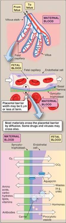
Figure 37.4
Exchange across the placental barrier.
C. Endocrine functions
A nonpregnant female's uterus sheds its lining (the outer endometrial layer) every 4 weeks and then begins the menstrual cycle anew, the cycle's timing being controlled by female reproductive hormones (see 36·VI·B). A successful pregnancy requires that the menstrual cycle be interrupted and the fetus left undisturbed for ~9 months. Cycle interruption is accomplished by the fetal placenta, which secretes several key hormones that manipulate maternal reproductive physiology, including human chorionic gonadotropin (hCG), progesterone, and estrogens (Figure 37.5).
1. Human chorionic gonadotropin: The syncytiotrophoblast (the fetoplacental precursor) begins secreting hCG within days of fertilization. hCG signals the corpus luteum that fertilization has occurred and impels it to sustain progesterone and estrogen production. These hormones prevent the uterus from shedding its lining and prepare it for implantation. The corpus luteum continues releasing progesterone and estrogen at ever-increasing levels in response to placental hCG, until the placenta takes over hormonal control around week 10 (see Figure 37.5).

Figure 37.5
Placental hormones.
The rapid rise in hCG that follows fertilization provides the basis for home pregnancy tests, which detect the presence of hCG in maternal urine. Test strips typically have a detection threshold of 25–50 mIU/mL, levels that are not achieved until after implantation has occurred (up to 10 days after ovulation).
2. Progesterone: The placental syncytiotrophoblast produces large amounts of progesterone. The hormone initially helps prepare the endometrium for implantation. Progesterone also reduces myometrial excitability, thereby preventing contractions that might expel the developing embryo. Progesterone also stimulates breast development.
3. Estrogens: The placenta produces several estrogens, the main one being estriol. Estrogens stimulate growth and development of the mother's uterus and breasts. The placenta does not have all of the necessary substrates (e.g., cholesterol) and enzymes (e.g., 17α-hydroxylase) to synthesize steroids (see 34·II·C), relying on both the fetus and its mother to provide pathway intermediates.
IV. MATERNAL PHYSIOLOGY
The fetus relies on its mother to supply it with O2 and nutrients and to dispose of CO2, heat, and other metabolic waste products. Meeting the fetal demands taxes all maternal organ systems and puts the maternal cardiovascular system under considerable stress. By term, maternal cardiac output (CO) and circulating blood volume have risen by 40%–50% (2 L). Much of this capacity increase is needed to perfuse the maternal placenta, but flow to the skin, kidneys, liver, and gastrointestinal (GI) tract increases substantially also.
A. Uterine blood flow
A nonpregnant female's uterus receives <5% of total CO. The main source of uterine vascular resistance is the highly muscular spiral arteries (Figure 37.6), which are resistance vessels that contract and relax to modulate blood flow in response to changing uterine metabolic needs (i.e., autoregulation; see 20·II·B·1). The developing fetal placenta erodes and invades the spiral arteries (see Figure 37.6B). Arterial walls are remodeled, the smooth muscle layers being replaced with fibrous material to create wide tortuous vessels with very high flow rates. The functional advantages of this remodeling to the fetus are obvious. Blood now pulses directly from the uterine supply arteries at a pressure of >70 mm Hg and washes over the fetoplacental villi, bringing with it needed O2 and nutrients (see Figure 37.6C). The cardiovascular consequences for the mother are profound, both in terms of flow increase and inability to control flow through these vessels.
1. Flow: Resistance vessels are flow regulators that limit the amount of blood that a tissue receives to its prevailing metabolic needs (see 20·II). Spiral artery erosion and widening allows blood to flow unhindered into the placental lake, and, hence, overall uterine blood flow increases dramatically during pregnancy. Flow rises in direct proportion to falling resistance, increasing from ~50 mL/min at 10 weeks’ gestation to >500 mL/min at term.

Figure 37.6
Erosion and invasion of spiral arteries during placentation.
Clinical Application 37.1: Preeclampsia
Preeclampsia is a syndrome characterized by hypertension (systolic blood pressure [SBP] of ≥140 mm Hg or diastolic blood pressure of ≥90 mm Hg) and proteinuria (≥0.3 g/24 hr) that develops after 20 weeks’ gestation. Other symptoms may include severe headaches, visual disturbances, epigastric pain, and abnormal liver function. These symptoms all reflect a generalized endothelial dysfunction that causes increased vascular tone; increased vascular permeability; and a coagulopathy affecting all organs, including the brain, kidneys, liver, and placenta. Although the underlying molecular mechanisms are not yet known, preeclampsia is believed to result from incomplete remodeling of the spiral arteries during placental development. Maternal placental vessels are narrowed as a result, causing placental hypoperfusion and impaired nutrient delivery to the fetus. Hypoperfusion causes the placenta to release factors that inhibit angiogenesis and disrupt normal maternal endothelial function. Severe preeclampsia (SBP ≥160 mm Hg) carries a significant risk of maternal stroke and death. Immediate delivery regardless of gestational age is generally indicated.
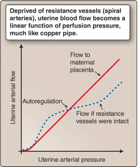
Figure 37.7
Uterine perfusion pressure–blood flow relationship during pregnancy.
2. Regulation: Eradication of maternal resistance vessels maximizes flow to the placental site but simultaneously limits the uterine vascular control system's ability to regulate blood flow. Thus, flow becomes a direct function of uterine arterial pressure, as predicted by the hemodynamic equivalent of the Ohm law (Flow = Pressure ÷ Resistance) as shown in Figure 37.7 (also see 19·IV).
Loss of uterine blood flow control mechanisms puts the mother in grave risk of massive blood loss in the event of premature placental detachment. Hemorrhage is a leading cause of pregnancy-related death in the United States (Table 37.1).
B. Hemodynamic profile
The uterus is a systemic vascular bed, so when uterine vascular resistance falls, systemic vascular resistance (SVR) falls along with it (Figure 37.8). A fall in SVR causes CO to rise to maintain mean arterial pressure (MAP): MAP = CO × SVR (see 18·III).
1. Systemic vascular resistance: SVR falls steadily over the first 20 weeks of gestation. The continuing erosion of maternal resistance vessels by the fetal placenta is the primary cause, but the growing need to dissipate heat and eliminate fetal waste products causes vascular resistance to fall in the cutaneous and renal vascular beds also.
2. Cardiac output: The growing need for increased CO is effected through increases in stroke volume (SV) and heart rate (HR). HR rises slowly during pregnancy, averaging 15–20 beats/min higher compared with nonpregnant values by 32 weeks. SV begins to rise very early in pregnancy, mediated by an increase in preload and contractility.
a. Preload: The body responds to a sustained or repeated need for increased CO by increasing circulating blood volume through Na+ and water retention. Placental hormones potentiate this effect by stimulating thirst and activating the renin–angiotensin–aldosterone system ([RAAS] see 20·IV).
b. Contractility: Sustained increases in CO also stimulate ventricular hypertrophy. The heart enlarges to accommodate increased end-diastolic volumes (preload), and the ventricular wall thickens to increase contractility.
3. Mean arterial pressure: MAP must be maintained at prepregnancy levels to ensure adequate perfusion of all vascular beds, but the introduction of a low-resistance pathway into the maternal vascular circuit (i.e., the placenta) means that blood escapes the arterial system more easily during diastole (increased diastolic runoff; see 19·V·C·2) compared with the nonpregnant state. Thus, diastolic blood pressure falls during pregnancy, and pulse pressure widens.

C. Physiologic anemia
Increased Na+ and water retention during pregnancy causes maternal plasma volume to increase by 40%–50%. Red blood cell (RBC) production does not keep pace with the rapid expansion of blood volume, increasing by only 25%–35%. The gap between volume expansion and RBC production results in a physiologic anemia of pregnancy (Figure 37.9). Although anemia reduces total O2-carrying capacity, there are clear physiologic benefits because it reduces blood viscosity, which, in turn, reduces shear stress. It can also cause benign murmurs.
1. Shear stress: Blood has to move through the maternal arteries and veins at high velocity to support the sustained increases in CO that accompany pregnancy. High-velocity flow increases shear stress on the vascular lining, to the point where it could become damaging. Shear stress is proportional to both blood velocity and viscosity (Reynolds equation; see 19·V·A). Because hematocrit is the primary determinant of blood viscosity, anemia reduces stress levels and lessens the risk of vascular endothelial damage.
2. Murmurs: One benign consequence of decreased blood viscosity is an increased tendency for turbulent blood flow. The Reynolds equation predicts that turbulence is most likely to occur in regions of the cardiovascular system where flow velocities are highest. In practice, this means that mothers often develop functional (i.e., innocent) murmurs associated with blood ejection through the aortic and pulmonary valves. Mothers can also develop a venous hum, a sound associated with high-velocity, turbulent blood flow through the larger veins.
D. Edema
The combined weight of the uterus and its contents (fetus, placenta, and amniotic fluid = ~8–10 kg total at term) compresses and retards flow through the inferior vena cava and other smaller veins returning blood from the lower extremities. Compression causes venous pressures in the lower extremities to rise, which increases mean capillary pressure and increases net fluid filtration from blood to the interstitium (see 19·VII·D). The result is edema, and swelling of the feet (pedal edema) and ankles is common in pregnant women. The tendency for edema formation is increased by a fall in colloid osmotic pressure(i.e., plasma protein concentrations) by 30%–40% during pregnancy (from ~25 mm Hg prior to pregnancy to ~15 mm Hg postpartum).
E. Respiratory system
The O2 demands of the mother and growing fetus increase rapidly during pregnancy; O2 consumption at term is increased ~30% over nonpregnant values. These increased needs are met by a progressive increase in minute ventilation to ~50% over nonpregnant values during the second trimester. The ventilation increase is effected largely by an increase in tidal volume and only a small rise in respiratory rate (2–3 breaths/min). The net effect is that PaO2 rises by ~10 mm Hg, and PaCO2 falls by ~8 mm Hg, causing a slight respiratory alkalosis (<0.1 pH units). Other significant respiratory changes include a 20% decrease in functional reserve capacity, expiratory reserve capacity, and residual volume (see 22·IX·A) caused by a rise in the diaphragm, which may limit the mother's ability to compensate for increased O2demand during exercise, for example.
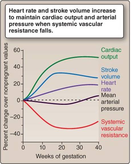
Figure 37.8
Changes in maternal hemodynamic profile during pregnancy.

Figure 37.9
Physiologic anemia of pregnancy.
F. Renal
Glomerular filtration rate rises steadily to ~50% above normal values at 16 weeks’ gestation and remains elevated until parturition. The increase reflects a mother's need to excrete fetal wastes, including urea and nonvolatile acid.
V. FETAL PHYSIOLOGY
Because a fetus receives everything it needs for successful development from the maternal circulation via the placenta, few fetal organ systems are required to support normal growth, although most gain some degree of functionality before birth. The principal exception is the cardiovascular system, which becomes functional very early in pregnancy.
A. Vasculature
During initial development, the embryo relies on simple diffusion to obtain nutrients from fallopian and other maternal secretions. Once the embryo attains a size that exceeds the ability of O2 and other nutrients to reach the innermost cells by diffusion alone, a functional cardiovascular system is required to sustain further growth. A rudimentary single-chambered heart begins pumping blood resembling interstitial fluid during the fourth week after conception. In the adult circulation, the path that blood follows is dictated by a need to pick up O2 from the lungs and nutrients from the GI tract. The placenta provides all of the fetus’ nutritional requirements, and, thus, the vascular circuitry is modified accordingly. There are four adaptations to the adult vascular circuit in the fetus: the placenta, ductus venosus, foramen ovale, and ductus arteriosus (Figure 37.10).
1. Placenta: The fetal placenta functions as fetal lungs, kidneys, GI tract, and liver and, thus, forms a major low-resistance circuit that receives ~40% of fetal CO (Table 37.2).
2. Ductus venosus: Blood coursing from the fetal placenta is shunted past the liver by the ductus venosus. In the adult, the liver filters and processes nutrient-rich blood from the GI system. In the fetus, the GI tract is largely nonfunctional, and, therefore, both GI organs are bypassed. Both receive sufficient blood to meet their nutritional needs via lesser vascular circuits.
3. Foramen ovale: Blood entering the fetal right heart from the inferior vena cava is O2 rich after passing through the placenta (80% saturation). The fetal lungs do not participate in gas exchange, so passage through the pulmonary circulation would serve no purpose. Pulmonary vasculature resistance (PVR) is high also, which makes the lungs difficult to perfuse (see below). Thus, O2-rich blood is shunted past the lungs from the right atrium directly into the left atrium via the foramen ovale.

Figure 37.10
Fetal circulation. LA = left atrium; LV = left ventricle; RA = right atrium; RV = right ventricle.

4. Ductus arteriosus: Blood entering the right heart via the superior vena cava is O2 poor (25% saturation; see below) after having traversed the fetal systemic vascular beds. It is pumped through the right heart and then through the ductus arteriosus to the descending aorta, thereby bypassing the lungs. The foramen ovale and ductus arteriosus together create a vascular circuit in which left and right hearts are arranged in parallel with each other.
B. Vascular resistance
In the adult circulation, SVR > PVR. The adult circulation is dominated by the left heart. In the fetal circulation, PVR > SVR. Fetal lungs are fluid filled, and the air spaces are collapsed. The pulmonary vasculature is tonically constricted as a response to low O2 levels (hypoxic vasoconstriction; see 23·III·E), which makes the pulmonary circuit difficult to perfuse, and, thus, fetal PVR is high. By contrast, the fetal systemic circulation includes the placenta, which is a very low–resistance pathway for blood flow, and, therefore, fetal SVR is low.
C. Oxygen transfer
Maternal uterine blood has an O2 saturation of ~80%–100%. Although the barrier separating fetal and maternal circulations is minimal, the placental route is a relatively inefficient means of gas exchange compared with the lungs, and fetal blood can only achieve PO2 levels of 30–35 mm Hg at best (compare to a PaO2 of 98–100 mm Hg in an adult pulmonary vein). Despite the inherent limitations of the transfer route, fetal blood carries similar amounts of O2 as does an adult circulation. This is made possible by hemoglobin F (HbF), a fetal Hb isoform that has a leftward-shifted O2 dissociation curve (see 23·VI·C·2). HbF's high O2 affinity is well adapted to take up O2 at partial pressures common to the maternal placenta, meaning that blood traveling from the placenta to the fetus in the umbilical veins typically has an O2 saturation of 80%–90% (Figure 37.11). Fetal blood also contains ~20% more Hb than adult blood, which increases overall O2-carrying capacity.
D. Oxygen distribution
Blood traveling from the placenta to the fetus via the umbilical vein is O2 rich. It streams around the liver via the ductus venosus, but then encounters O2-poor blood returning from the lower regions of the body in the inferior vena cava (see Figure 37.11). A filmy membrane ensures that little mixing occurs at the point where the two bloodstreams merge, and the O2-rich stream is preserved all the way to the right atrium (streamline flow; see 19·V·A). Here, the two streams are cleaved by the interatrial septum (crista dividens). The O2-rich portion passes preferentially into the left heart and then into the aorta. The first arteries to branch off the aorta feed the myocardium and the brain, so flow streamlining ensures that these two critical circulations receive highly oxygenated blood.
E. Renal function
Fetal kidneys start producing urine within 9–10 weeks of conception. The ability to concentrate urine is gained around 4 weeks later, but the fetus remains dependent on the placenta for fluid and electrolyte balance throughout gestation. By 18 weeks, the kidneys are producing over 10 mL of urine per hour, and fetal urine has become the primary source of amniotic fluid.

Figure 37.11
O2 distribution by the fetal circulatory system. Circled numbers represent O2 saturations. LA = left atrium; LV = left ventricle; RA = right atrium; RV = right ventricle.
VI. PARTURITION
Human gestation lasts 40 weeks on average. At the end of this time, the fetus is forcibly expelled from the uterus, and the physical link with the mother is broken (parturition). The birthing process requires careful coordination if both mother and newborn are to survive.
A. Staging
Parturition can be divided into three stages of variable duration: dilation, fetal expulsion, and placental.
1. Dilation: Stage one begins with labor and ends when the cervix is dilated fully. The fetus is enclosed within the amniotic sac, but the main barrier preventing it from exiting the uterus is the cervix. During the first stage of parturition, the myometrium begins contracting rhythmically and with increasing intensity. Contraction begins in the uterine fundus and spreads caudally, which pushes the fetus against the cervix and causes the latter to thin and dilate. Stage one usually lasts for ~8–15 hours.
2. Fetal expulsion: The fetus is forcibly expelled from the uterus through the cervix and vaginal canal by frequent and intense waves of contraction. Stage two is complete within 45–100 min (Figure 37.12). The umbilical cord is traditionally clamped shortly after birth, although preterm infants may benefit from delayed clamping and milking blood from the cord toward the newborn to increase the infant's hematocrit.
3. Placental: The uterus continues contracting after the fetus has been expelled, which causes it to shrink in size (involution). Shrinkage shears the placenta from the uterine wall. The placenta and associated membranes are later expelled as afterbirth. Stage three is usually completed within minutes of fetal expulsion.
B. Hormones
Irregular waves of weak uterine contraction occur throughout pregnancy (Braxton Hicks contractions). The reasons why contractions abruptly transition to the forceful contraction of parturition are not understood, although several hormones have been implicated. The fetus stretches the myometrium and increases its overall excitability as it grows and may be a contributing factor. The major hormones driving parturition include estrogen and progesterone, prostaglandins, oxytocin, and cortisol.
1. Estrogen/progesterone ratio: Progesterone suppresses uterine contraction during pregnancy. Estrogens promote excitability by increased expression of Na+ channels, Ca2+ channels, and gap junctions between adjacent smooth muscle cells within the myometrium. The gap junctions allow developing waves of excitation to sweep through the uterine wall, manifesting as a wave of contraction. At parturition, the estrogen/progesterone ratio increases, and the uterus becomes excitable.

Figure 37.12
A newly delivered infant.
2. Prostaglandins: The uterus, placenta, and fetus all produce prostaglandins (PGE2 and PGF2α), which stimulate uterine contractions. Increasing estrogen levels likewise increase prostaglandin production.
3. Oxytocin: Oxytocin is a powerful stimulant of uterine contractions. It is released from the posterior pituitary in response to cervical distension (see 36·VII·B), providing a positive feedback mechanism that couples fetal expulsion with the motive force required for expulsion.
4. Cortisol: The fetal hypothalamic–pituitary–adrenal axis is activated to release cortisol (see 34·V·B). Cortisol increases the estrogen/progesterone ratio.
C. Circulatory transition from fetal to adult
Parturition breaks the link between mother and fetus and forces the fetal vasculature to adopt the serial circulatory pattern that is common to the adult. Transition follows a rapid sequence of coincident events: an SVR increase; lung inflation; decrease in PVR; closure of the ductus arteriosus, foramen ovale, and ductus venosus; and, finally, a shift from right- to left-sided circulatory dominance (Figure 37.13).
1. Systemic vascular resistance: The umbilicus is a highly muscular structure that contracts spontaneously in response to the trauma of birth. Contraction occludes the umbilical arteries and vein and terminates flow to the placenta. Fetal SVR increases when this low-resistance pathway is removed from the systemic vascular circuit.
Remnants of the umbilical arteries and vein can be observed in the adult as the medial umbilical ligaments and ligamentum teres, respectively.
2. Lung inflation: Compression and occlusion of umbilical vessels halts blood flow and deprives the fetus of O2, causing asphyxia. This, together with the sudden cooling experienced by the infant at birth, stimulates respiratory control centers in the fetal brainstem, causing the neonate to gasp and take several breaths. Intraalveolar pressure drops below atmospheric pressure, creating a pressure gradient that drives air inflow, and the lungs inflate.
3. Pulmonary vascular resistance: During development, PVR is high because the lungs are collapsed, and the pulmonary arteries are compressed and constricted in response to low PO2. The first breaths cause alveolar and pulmonary arterial PO2 levels to rise dramatically, promoting vasodilation. Lung inflation also stretches the pulmonary vessels, thinning their walls and increasing their internal diameter. PVR drops dramatically as a result, and there is a coincident increase in pulmonary blood flow.
4. Ductus arteriosus: The fall in PVR and loss of flow from the umbilical vein causes right atrial pressure to fall. SVR simultaneously increases due to loss of the placental circuit, so left ventricular and aortic pressure rises. The sudden inversion of the fetal right-to-left pressure gradient causes a reversal of blood flow in the ductus arteriosus, and it constricts, probably in response to rising PaO2 and falling circulating prostaglandin levels (see Clinical Application 37.2). Complete anatomic closure takes several months, and vestiges of the fetal shunt persist even in the adult as the ligamentum arteriosum.

Figure 37.13
Changes in fetal vascular circuitry during parturition. LAP = left atrial pressure; LVP = left ventricular pressure; PaO2 = partial pressure of oxygen (arterial); PVR = pulmonary vascular resistance; RAP = right atrial pressure; RVP = right ventricular pressure; SVR = systemic vascular resistance.
Clinical Application 37.2: Patent Ductus Arteriosus
Patent ductus arteriosus is a common congenital heart defect, particularly in premature and very low–birth-weight infants among whom the incidence may be as high as 30%. If the ductus arteriosus remains open, high-pressure blood from the systemic circulation shunts into the pulmonary circulation. Depending on severity, the shunt can cause pulmonary hypertension and may result in right-sided heart failure if not addressed. Ductus arteriosus patency is maintained during development in part by high circulating levels of prostaglandin E2, so administering a cyclooxygenase inhibitor, such as indomethacin, is often sufficient to prompt complete closure.1
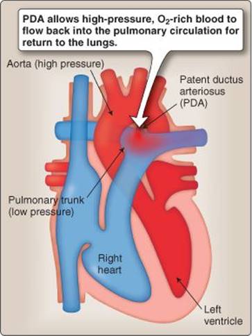
Patent ductus arteriosus (PDA)
5. Foramen ovale: The right–left blood pressure inversion pushes on a valvelike flap that then covers the foramen ovale. Gradually escalating left atrial pressures hold the flap closed to isolate the left and right sides of the heart. In time, the flap fuses with the inter atrial septum to seal off the foramen permanently (seen as the fossa ovale in an adult heart).
Patent foramen ovale is a congenital heart lesion that affects 25%–30% of the general population. Although the pathway between right and left atria remains intact, left atrial pressure is usually higher than right atrial pressure, and, thus, the foramen remains occluded by the one-way valve. Healthy individuals usually remain asymptomatic.
6. Ductus venosus: The ductus venosus closes by a sphincter-like mechanism, persisting at the ligamentum venosum in the adult. The mechanism of closure is unknown.
 1For more information on the actions and uses of cyclooxygenase inhibitors, see LIR Pharmacology, 5e, p. 525.
7. Circulatory dominance: Over ensuing weeks, the left ventricle slowly hypertrophies as a response to a rising SVR. Meanwhile, the right heart pumps against a lower PVR than it did during gestation, so its muscle mass slowly decreases relative to the left heart.
D. Maternal blood loss
A mother typically loses ~500 mL of blood from the placental site during a normal delivery. Although this represents a substantial hemorrhage, the mother has been well prepared for the loss by the massive expansion of blood volume that occurs during the first few weeks of pregnancy. Further loss is prevented by intense uterine contractions, which compress the uterine vasculature and allow hemostasis to occur. The contractions are stimulated by oxytocin during the third stage of parturition.
Chapter Summary
• Pregnancy begins with fertilization. The developing embryo divides rapidly over subsequent days to form a blastocyst and then implants in the maternal uterine wall. Implantation is effected by an outer trophoblast cell layer, which digests and invades the maternal endometrium and develops to create an interface between fetal and maternal circulations (the placenta).
• The placenta exchanges nutrients, hormones, and waste products between fetal and maternal circulations.
• The fetal placenta comprises 60–70 villous trees that serve to increase interface surface area. The maternal placenta comprises 15–20 blood-filled, eroded endometrial sinuses sculpted by fetal trophoblast during placentation. The space between fetal and maternal placenta is filled with ~500 mL of maternal blood. Blood flows in an unregulated manner at relatively high pressure (~70 mm Hg) from eroded spiral arteries and washes over the fetal villous trees.
• The placenta is also an endocrine organ that secretes human chorionic gonadotropin, progesterone, and estrogens.
• Supporting the needs of a developing fetus involves most of a mother's organ systems, including the cardiovascular system, kidneys (increased disposal of waste products), lungs (~30% increase in O2demand), gastrointestinal tract, liver, and skin (thermoregulation).
• Maternal cardiac output increases ~50% during gestation, accomplished through increases in heart rate and stroke volume. Stroke volume increases as a result of fluid retention and increased circulating blood volume.
• Blood volume increases faster than red blood cell production, causing a physiologic anemia of pregnancy. The resulting decrease in blood viscosity reduces shear stress on the heart and vasculature lining.
• The uterus and its contents gain significant weight during pregnancy, which, depending on posture, compresses and impairs blood flow from the mother's lower extremities. The result is pedal edema.
• The fetal circulation includes three shunts that allow blood from the umbilical vein to bypass the liver (ductus venosus) and lungs (foramen ovale and ductus arteriosus) for distribution to the developing organs.
• Fetal blood contains a fetal hemoglobin (HbF) isoform that has a high affinity for O2. HbF helps compensate for the fact that the placenta is a less efficient route for O2 transfer than the lungs and allows fetal blood to carry near-adult levels of O2.
• Parturition is initiated and sustained by changing levels of hormones produced by both the mother and the fetal placenta. Uterine contractions expel the fetus and placenta then compress and collapse the uterine vasculature. Compression limits maternal blood loss during delivery.
• At birth, the neonate's pulmonary vascular resistance decreases, and systemic vascular resistance increases, thereby establishing a left side–dominated circulatory system common to an adult.