
38
I. OVERVIEW
The ability to dissipate and retain heat, combined with the ability to adapt behaviorally to temperature extremes, has allowed humans to occupy most regions of the Earth's surface, including Plateau Station, Antarctica (average temperature = −55°C) and Dallol, Ethiopia (average temperature = 35°C). The body's internal temperature can rise to 39°–40°C without causing irreversible loss of cellular function, but body temperature is typically regulated within a much tighter range (36.5°–37.5°C) as shown in Figure 38.1. Control of internal temperature is one of the body's fundamental homeostatic functions. Internal temperatures are sensed through thermoreceptors located in the brain, spinal cord, and viscera. External temperatures are sensed through cutaneous thermoreceptors. The sympathetic nervous system (SNS), in conjunction with hypothalamic control centers, mediates thermoregulatory responses to environmental stressors. During heat stress (e.g., sitting in a Finnish sauna at 80°–90°C), there is a simultaneous increase in skin blood flow and sweat gland stimulation that mediates evaporative cooling. During cold stress (e.g., as experienced by an officer directing traffic on a cold, wet day), skin blood flow decreases to reduce cutaneous heat loss, while shivering generates heat. The hypothalamus can also actively increase internal temperature as a way of slowing infection by a pathogen. Fever is one of the oldest recognized symptoms of illness.
II. THERMOREGULATION
External and internal temperatures are sensed by thermoreceptors, which relay sensory information to control centers located in the hypothalamus. Thermoregulatory effector organs include the skin, brown adipose tissue, and skeletal musculature.
Internal temperature can be accurately measured with a temperature probe placed in the esophagus or rectum. Oral temperature measurements can also yield good estimates of internal temperature, provided that the patient is breathing nasally, and ventilation is low. Oral temperature measurements are 0.25°–0.5°C lower compared with rectal temperature.

Figure 38.1
Body temperatures.
A. Sensors
The body possesses two diverse groups of thermosensors. Central thermoreceptors monitor internal body temperature, whereas skin thermoreceptors provide information about the external thermal environment.
1. Central: Thermoreceptors that monitor internal temperature are located in the hypothalamus, spinal cord, and viscera, but the sensors that have the greatest influence on thermoregulatory control center output are in the preoptic area of the hypothalamus (Figure 38.2). Warmth-sensitive preoptic neurons are tonically active at normal body temperature. A rise in internal temperature (as reflected by the temperature of blood bathing the preoptic area), increases their firing rate, whereas cooling decreases firing rate.
2. Skin: There are four primary skin thermoreceptor types. Two mediate nociceptive responses to painfully cold or hot stimuli and are discussed elsewhere (see 16·VII·B). The other two, comprising distinct populations of cold and warmth receptors, are involved in thermoregulation.
a. Cold: Cold receptors mediate neutral, cool, and cold sensations (5°–45°C). Cold temperatures are believed to be sensed by TRPM8, one of the transient receptor-potential (TRP) channel family (see 2·VI·D) that mediates a depolarizing receptor potential when active. Afferent firing rate increases as a consequence.
b. Warmth: Heat sensation involves warmth receptors that are activated from 30°–50°C. Heat reception also involves TRP channels (TRPV3 and TRPV4) that are active at neutral and warm temperatures (Figure 38.3).
B. Control center
Body temperature is normally held at 37°C, with a circadian variance of 1°C (i.e., 36.5°–37.5°C). The temperature nadir occurs in the early morning, and the peak occurs in the late afternoon. The internal temperature that the body is trying to maintain is known as the set point. The preoptic area of the hypothalamus contains the thermoregulatory integration and control center. Cooling the preoptic area elicits heating responses and behaviors (e.g., putting on more clothing), whereas heating this area activates cooling responses and behaviors (e.g., seeking shade). If the preoptic area is damaged by ischemia (i.e., stroke), demyelination (e.g., multiple sclerosis), or ablation, internal temperature fluctuates over an exaggerated range, and responses to thermal stress are impaired. Control center output is governed primarily by central thermoreceptors, but the preoptic area integrates signals from many other areas also. These include skin thermoreceptors, the immune system (see Section IV below), and areas of the central nervous system that regulate other systemic variables, such as blood pressure, plasma glucose concentration, and plasma osmolality.
C. Effector pathways
The hypothalamus effects most thermoregulatory responses via the SNS. Sympathetic signals travel via T1–L3 spinal nerves and synapse within sympathetic chain ganglia (see 7·III·B). Efferents project from the ganglia to skin blood vessels and to sweat glands.
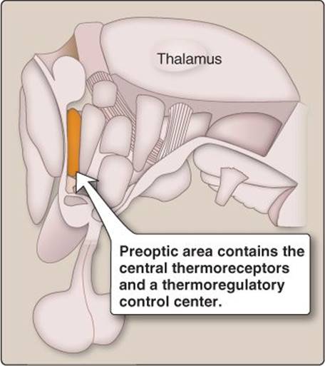
Figure 38.2
Preoptic area of the hypothalamus.
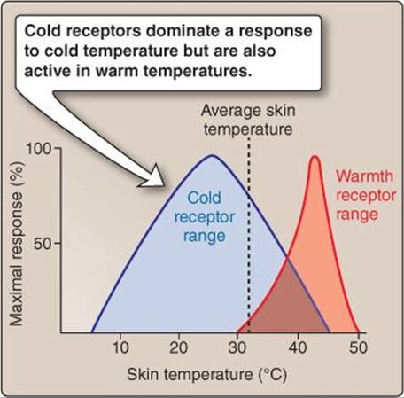
Figure 38.3
Skin thermoreceptor sensitivity.
D. Response
Cold stress initiates pathways that increase tissue insulation and increase metabolic rate via shivering and nonshivering thermogenesis (heat production). Conversely, heat stress reduces tissue insulation and initiates sweating. The primary thermoregulatory effectors are the skin blood vessels, sweat glands, skeletal muscles, and brown adipose tissue (Figure 38.4).
1. Skin blood vessels: Heat loss or gain is most effectively regulated at the level of the skin. To dissipate heat, blood flow is brought in close proximity to the body's surface, whereas to conserve heat, blood flow is shunted away from the body's surface. Glabrous skin contains deep arteriovenous anastomoses that allow blood to bypass surface capillary beds. Hairy (nonglabrous) skin does not have arteriovenous anastomoses but does have deep and superficial capillaries. The most efficient heat transfer with the environment occurs when blood is shunted through these surface capillaries. Postganglionic adrenergic nerves constrict cutaneous arteries, veins, and anastomoses, acting via α-adrenergic receptors. Removing the constrictor influence and then actively dilating vessels increases blood flow through the cutaneous vasculature (Figure 38.5). The vasodilation mechanism is less understood but involves nitric oxide and cholinergic sympathetic nerves. These vasomotor changes allow skin blood flow to change from <6 mL/min to 8 L/min. During heat stress, venous volume increases also to provide additional time for heat transfer. Heart rate and CO increase, and other vascular beds (e.g., renal and splanchnic), vasoconstrict to facilitate the skin blood flow increase.
2. Sweat glands: There are three sweat gland types (see 16·VI·C), but only eccrine sweat glands produce sweat that mediates evaporative cooling during heat stress. Sweating is initiated by SNS cholinergic nerves, but the glands are stimulated by adrenergic compounds (e.g., epinephrine, norepinephrine) also. Sweating can dehydrate the body and cause a hypertonic volume contraction. Even small fluid loss (2% body weight) can decrease work performance and allow internal temperatures to rise during heat stress.
3. Muscles: Shivering is a rapid, cyclical contraction of skeletal muscles that liberates heat but produces minimal force. Muscle contractions always produce large amounts of heat because force production is only 20% efficient. The remaining 80% of expended energy is liberated as heat. Shivering muscles do not perform meaningful work, and, thus, almost all of the energy used is liberated as heat. Shivering is unique in that it is mediated by the somatic motor pathways rather than the SNS, but the response is initiated by the preoptic area.
4. Nonshivering thermogenesis: Nonshivering thermogenesis is a SNS-mediated increase in metabolic rate in muscle and other tissues designed to liberate heat. In brown fat, SNS stimulation activates an uncoupling protein (thermogenin) in the inner mitochondrial membrane (Figure 38.6). Thermogenin is a pore-forming protein that allows H+ to cross the inner mitochondrial membrane without generating adenosine triphosphate. Thus, oxidative phosphorylation becomes uncoupled. Infants rely on brown adipose tissue for heat production, but this pathway is less important in adults.

Figure 38.4
Thermoregulatory response mediators.
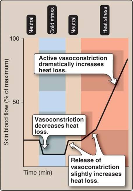
Figure 38.5
Skin blood flow during heat stress.
5. Behavior: Thermoregulatory behaviors can decrease or even eliminate thermal stress. These behaviors are driven by the preoptic area but can be overridden or modified by other areas of the brain. Conscious behavioral responses to cold stress involve increasing insulation (e.g., putting on a coat), increasing physical activity to increase metabolic rate, or seeking an external heat source. Heat stress–related behaviors include drinking fluids to facilitate sweating, removing clothing, seeking shade, or turning on a fan.
III. HEAT PRODUCTION AND TRANSFER
The amount of heat stored within the body reflects a balance between the amount of heat produced and the amount transferred to the external environment. Heat storage can be quantified theoretically using the heat balance equation:
S = (M − Wk) ± (R + K + C) − E
where S is heat storage; M is metabolism; Wk is external work; and R, K, C, and E describe radiative, conductive, convective, and evaporative heat transfer, respectively.
A. Production
Heat is a metabolic byproduct reflecting the inefficiency of the chemical pathways involved. The amount of heat produced at rest is related to basal metabolic rate (BMR), which, in turn, is related to body mass (e.g., two individuals with a mass of 50 kg and 90 kg may be expected to have BMRs of ~1,315 kcal/day and ~2,045 kcal/day, respectively). Any increase in tissue metabolism increases heat production. Food digestion and assimilation increase energy expenditure (the amount of energy used is known as the thermic effect of food), as do spontaneous movements and exercise.
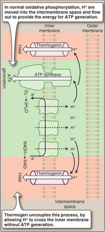
Figure 38.6
Brown adipose tissue. ATP = adenosine triphosphate; NAD = nicotinamide adenine dinucleotide; NADH = NAD hydrogen.
Clinical Application 38.1: Malignant Hyperthermia
Malignant hyperthermia is a syndrome triggered by anesthetics (e.g., halothane) and muscle relaxants (e.g., succinylcholine).1 Metabolic rate increases at a rate that far outpaces heat dissipation, due to excess sarcoplasmic Ca2+, which stimulates exaggerated and prolonged excitation–contraction coupling in skeletal muscle. This heat production mechanism appears to be a genetic abnormality in the Ca2+-release channels (ryanodine receptors) in the sarcoplasmic reticulum (see 12·III·A).
 1For more information on the use of anesthetics, see LIR Pharmacology, 5e, p. 135.
B. Heat transfer
Heat produced by metabolism must be transferred to the external environment, principally via the skin, although a small amount of heat is transferred via the respiratory tract. The term “heat transfer” refers to a mechanism whereby heat is transferred from an area of higher temperature to an area of lower temperature. Human skin temperature is ~32°C in normothermic (i.e., temperatures that support a normal body temperature) environments. Because outside temperatures are usually lower, body heat can be transferred to air or other objects. When the external temperature is greater than skin temperature, the body gains heat. There are four primary mechanisms by which heat is transferred to the environment: radiation, conduction, convection, and evaporation (Figure 38.7).
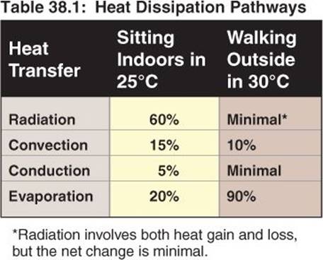
1. Radiation: Radiation refers to the thermal energy that is transferred to objects in the external environment. Heat energy is carried in the infrared spectrum, and the amount transferred depends on the temperature difference and the emissivity (ability to absorb energy) of the object's surface. The majority of heat transfer at rest occurs by radiation (Table 38.1).
2. Conduction: Conduction of thermal energy from one body to another occurs when they are in close physical contact. The high kinetic energy of molecules in a warm region dissipates by collisions with adjacent molecules in a cool region. Solids differ enormously in their ability to conduct heat. Substances with low thermal conductivity are called thermal insulators.
3. Convection: Convection occurs when heat is transferred to the environment by a moving fluid (i.e., air or water). Generally, heating reduces the density of air and water, and gravity creates a “natural” fluid convection current near the skin as the warmer, low-density fluid rises. Forced convection results when an alternative energy source propels the fluid past the skin (e.g., fan, wind, water current). Convective heat loss or gain is proportional to the specific heat of the fluid, the temperature gradient, and the square root of fluid or air velocity.
4. Evaporation: Evaporation dissipates heat by using thermal energy to convert water from a liquid to a gaseous phase, the primary sites of evaporative heat loss being the respiratory tract and the skin. Evaporation is a very effective mode of heat dissipation, such that 1 L of sweat can remove ~580 kcal from the skin surface. The amount of evaporation is dependent on the relative humidity of ambient air: Humid air attenuates and dry air facilitates sweating. During exercise or when ambient air temperature is above skin temperature, sweat evaporation provides the primary and, often, only mode of heat dissipation (see Table 38.1).
IV. CLINICAL ASPECTS
Although internal temperature is normally maintained within a narrow range, the hypothalamus may allow it to increase in an attempt to thwart a pathogen, manifesting as fever. Deviation from normal may also occur when the body's thermoregulatory systems are overwhelmed, resulting in hypothermia or hyperthermia.

Figure 38.7
Heat transfer.
A. Fever
Fever has long been recognized as a symptom of illness and is caused by exogenous and endogenous pyrogens. Exogenous pyrogens include microorganisms, such as Staphylococcus aureus, and their byproducts or toxins. Most often, fever is a response to endogenous pyrogens released during macrophage and monocyte activation,1 which is related to an infection, even though the microorganism is not directly involved. Endogenous pyrogens are interferons and cytokines, including interleukins (e.g., IL-1 and IL-8) and tumor necrosis factor. Although the pathways involved are as yet unelucidated, circulating pyrogens are sensed by the circumventricular organs (see 7·VII·C), which signal their presence to the preoptic hypothalamus via prostaglandin release. Neurons in the preoptic area express a prostaglandin receptor, EP3, that mediates the fever response. When these receptors are stimulated, the hypothalamic set point is reset, and the body begins to regulate internal temperature at a higher value (Figure 38.8). The symptoms associated with a febrile state reflect thermoregulatory effector organs trying to attain the new set point. This elevated temperature is thought to exert both a beneficial effect on the host's immune system and to decrease pathogen growth and proliferation. The febrile state is distinct from the body temperature increase associated with muscle contraction and exercise or ambient heat exposure. In both cases, the body attempts to dissipate heat with the goal of returning internal temperature to 37°C.
The hypothalamic set point can be returned toward 37°C and the symptoms of fever reduced by administering nonsteroidal anti-inflammatory drugs (NSAIDs), such as aspirin, ibuprofen, and acetaminophen.2NSAIDs are cyclooxygenase inhibitors that block prostaglandin synthesis and, thus, have antipyretic effects.
B. Hypothermia and hyperthermia
Hypothermia and hyperthermia are deviations from normal body temperature that occur due to thermoregulatory system failure. These conditions occur most commonly during weather extremes and in patients with an impaired ability to respond to thermal stress such as those with a genetic inability to secrete sweat (congenital anhidrosis).
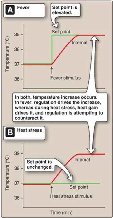
Figure 38.8
Fever versus heat stress.
1 For cellular defense mechanisms and associated cytokines, see LIR Immunology, 2e, p. 49.
1 For more information on the use and actions of NSAIDs, see LIR Pharmacology, 5e, p. 529.
1. Hypothermia: Hypothermia (internal temperature of <35°C) commonly results from immersion in cold water because water transfers heat 25 times faster than does air. Heat loss occurs via normal heat transfer mechanisms, but heat production cannot increase sufficiently to compensate for heat loss. Hypothermia causes symptoms associated with cold-induced decreases in neuronal metabolic rate, including drowsiness, slurred speech, bradycardia, and hypoventilation. Severe hypothermia (internal temperature of <28°C) can cause coma, hypotension, oliguria, and fatal cardiac arrhythmias (ventricular fibrillation). Peripheral tissues can also be injured by the cold. Frostbite is a condition in which fluid in skin and subcutaneous areas crystallizes (freezes), disrupting cell membranes and causing tissue necrosis (Figure 38.9). Necrosed areas often require amputation.
2. Hyperthermia: Precise internal temperature definitions of hyperthermia are not possible without assessing the cause. For example, internal temperatures above 40°C can be achieved during exercise, without developing a heat illness. Heat illnesses form a continuum, from a milder heat exhaustion to the more severe heat stroke. The etiology of heat exhaustion is related to a decrease in circulating blood volume caused by skin vasodilation and a sweating-induced decrease in central venous pressure (CVP). The decrease in CVP can allow blood to pool in the limbs when an individual is in the upright position, causing syncope (fainting). Heat stroke, in the classic sense, refers to failure of the heat dissipation mechanisms due to the continuing increase in internal temperature. Unfortunately, failure of these mechanisms only leads to more rapid gains in temperature. In heat stroke, internal temperatures can climb to over 41°C, which can lead to neural death and organ system failure.
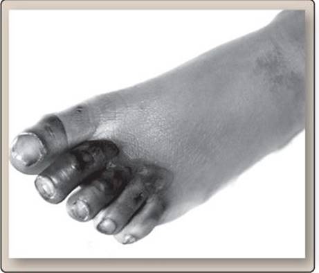
Figure 38.9
Frostbite.
Chapter Summary
• Internal temperature is normally maintained at 37.0°C ± 0.5°C.
• Internal temperature is sensed and controlled by the preoptic area of the hypothalamus. Environmental temperature is sensed by warmth and cold skin thermoreceptors.
• Heat stress induces skin vasodilation and increases cardiac output and sweating to aid in body heat offloading. Cold-seeking behavioral strategies are also stimulated.
• Cold stress induces skin vasoconstriction and increases shivering to decrease heat loss and increase heat production. Heat-seeking behavioral strategies are also stimulated.
• Heat balance is achieved by matching heat production with heat loss. Heat production includes the amount generated by metabolism, the thermic effect of food, spontaneous movements, and exercise. Heat transfer occurs via radiation, convection, conduction, and evaporation.
• Fever is the external manifestation of temperature set point resetting to a higher value. Both exogenous pyrogens (e.g., microbial toxins) and endogenous pyrogens (e.g., interferons, interleukins, and tumor necrosis factor) can increase the set point via prostaglandin production. The body then defends this higher value by normal means, such as shivering to raise temperature and sweating to lower temperature.
• Hypothermia is a low internal temperature and is clinically associated with processes that slow body metabolism. Frostbite results in tissue necrosis from fluid crystallizing within and between cells.
• Hyperthermia is an elevated internal temperature. The most serious form of heat illness is heat stroke, which classically involves complete thermoregulatory system failure.