
4
I. OVERVIEW
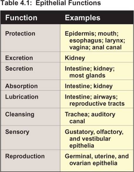
The human body comprises a diverse assemblage of cells that can be placed in one of four groups based on structural and functional similarities. These groups are known as tissues: epithelial tissue, nervous tissue, muscle tissue, and connective tissue. The four tissue types asso ciate with and work in close cooperation with each other. Epithelial tissue comprises sheets of cells that provide barriers between the internal and external environment. Skin (the epidermis) is the most visible example (see Chapter 16), but there are many unseen internal interfaces that are lined with epithelia also (e.g., lungs, gastrointestinal [GI] tract, kidneys, and reproductive organs). Nervous tissue comprises neurons and their support cells (glia) that provide pathways for communication and coordinate tissue function, as will be discussed in greater detail in Unit II. Muscle tissue is specialized for contraction. There are three types of muscle: skeletal muscle (see Chapter 12), cardiac muscle (see Chapter 13), and smooth muscle (see Chapter 14). Connective tissue is a mix of cells, structural fibers, and ground substance that connects and fills the spaces bet ween adjacent cells and gives tissues their strength and form. Bone is a specialized connective tissue that is mineralized to provide strength and to resist compression (see Chapter 15). This chapter considers the structure and varied functions of epithelial tissue (Table 4.1) and connective tissue.
II. EPITHELIA
Epithelia are continuous sheets of cells that line all body surfaces and create barriers that separate the internal and external environments. They help protect us from invasion by microorganisms, and they limit fluid loss from the internal environment: They “keep our insides in.” Epithelia are much more than just barriers, however. Most epithelia have additional specialized secretory or absorptive functions that include sweat formation, food digestion and absorption, and excretion of waste products.
A. Structure
The simplest epithelia comprise a single layer of cells adhered to each other by a variety of junctional complexes that impart mechanical strength and create pathways for communication between adjacent cells (Figure 4.1). The apical surface interfaces with the external environment (or an internal body cavity), whereas the basal surface rests on a basement membrane that provides structural support. The basement membrane comprises two fused layers. The basal lamina is synthesized by the epithelial cells that it supports and is composed of collagen and associated proteins. The inner layer (lamina reticularis, or reticular lamina) is formed by underlying connective tissue. Epithelia are avascular, relying on blood vessels lying close to the basement membrane to deliver O2 and nutrients, but they are innervated.
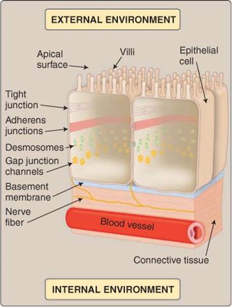
Figure 4.1
Epithelial cell structure.
B. Types
Epithelia are classified based on their morphology, which is usually a reflection of their function. There are three types: simple epithelia, stratified epithelia, and glandular epithelia.
1. Simple: Many epithelia are specialized to facilitate exchange of materials between their apical surface and the vasculature. For example, the pulmonary epithelium facilitates gas exchange bet ween the atmosphere and the pulmonary circulation (see 22·II·C). The renal tubule epithelium transfers fluid between the tubule lumen and blood (see 26·II·C), whereas the epithelium that lines the small intestine transfers materials between the intestinal lumen and the circulation (see 31·II). Exchange and transport functions require that the barrier separating the two compartments be minimal, so all of the above structures are lined with “simple” epithelia. Simple epithelia comprise a single cell layer and can be further subdivided into three groups according to epithelial cell shape. Pulmonary alveoli and blood vessels are lined with simple squamous epithelium. Squamous epithelial cells are extremely thin to maximize diffusional exchange of gases. Many segments of renal tubules and glandular ducts are lined with cube-shaped (simple cuboidal) epithelial cells. Their shape reflects the fact that they actively transport materials and, thus, they must accommodate mitochondria to produce the adenosine triphosphate (ATP) needed to support primary active transporter function. Simple columnar epithelia comprise sheets of cells that are long and narrow to accommodate large numbers of mitochondria and are found in the distal regions of the renal tubule (see 27·IV·A) and in the intestines, for example.
Clinical Application 4.1: Squamous Cell Carcinoma
Squamous cell carcinomas are one of the most common forms of cancer that arise from most epithelia, including the skin, lips, buccal lining, esophagus, lungs, prostate, vagina, cervix, and urinary bladder. Cutaneous squamous cell carcinoma is a prevalent skin cancer that typically occurs in sun- exposed skin areas. Squamous cell malignancies are believed to arise from uncontrolled division of epithelial stem cells rather than squamous epithelial cells. Squamous cell cancers usually remain localized and can be treated by Mohs surgery (a specialized dermatologic surgery for skin malignancies), cryotherapy, or surgical excision.

Squamous cell carcinoma.
2. Stratified: Epithelia that are subject to mechanical abrasion are composed of multiple cell layers (Figure 4.2). The layers are designed to be sacrificed to prevent exposure of the basement membrane and deeper structures. The inner epithelial cell layers are renewed continually and the damaged outer layers sloughed off. Examples include the skin and the lining of the mouth, esophagus, and vagina. The skin suffers constant exposure to mechanical stress associated with contact with and manipulation of external objects, so the outer layers are reinforced with keratin, a resilient structural protein (see 16·III·A). Transitional epithelium (also known as urothelium) is a specialized stratified epithelium that lines the urinary bladder, ureters, and urethra (see 25·VI). A transitional epithelium comprises cells that readily stretch and change shape (from cuboidal to squamous) without tearing to accommodate volume changes within the structures that they line.
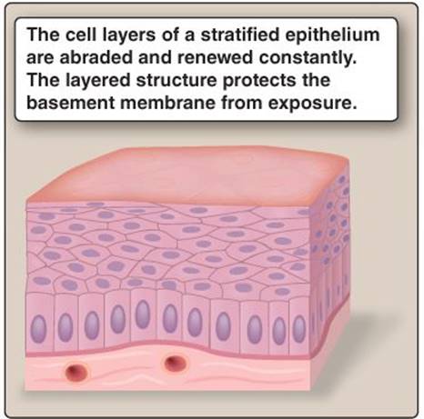
Figure 4.2
Stratified epithelial structure.
3. Glandular: Glandular epithelia produce specialized proteinaceous secretions (Figure 4.3). Glands are formed from columns or tubes of surface epithelial cells that invade the underlying structures to form invaginations. Glandular secretions are then released either via a duct or ductal system onto the epithelial surface (exocrine glands), or across the basement membrane into the bloodstream (endocrine glands). Endocrine glands include the adrenal glands (which secrete epinephrine), the endocrine pancreas (which secretes insulin and glucagon), and reproductive glands, which are considered in Unit VIII. Sweat glands, salivary glands, and mammary glands are all examples of exocrine glands. Exocrine glands are typically composed of two epithelial cell types: serous and mucous.
a. Serous: Serous cells produce a watery secretion containing proteins, typically enzymes. Salivary serous cells produce salivary amylase, gastric chief cells produce pepsinogen (a pepsin precursor) and pancreatic exocrine serous cells produce trypsinogen, chymotrypsinogen, pancreatic lipase, and pancreatic amylase.
b. Mucous: Mucous cells secrete mucus, a slippery glycoprotein (mucin)-rich secretion that lubricates the surface of mucous membranes. Many glands contain a mix of serous and mucous cells that together create an epithelial barrier layer enriched with antibacterial agents such as lactoferrin to help ward off infection (e.g., pancreatic glands) or enriched with HCO3− to neutralize acid (e.g., gastric epithelium).

Figure 4.3
Glandular epithelial structure.

Figure 4.4
Apical surface specializations.
C. Apical specializations
Several epithelia support apical modifications that amplify surface area or serve motile or sensory functions, including villi, cilia (motile and sensory), and stereocilia (Figure 4.4).
1. Villi: Epithelia that are specialized for high-volume fluid uptake or secretion (e.g., epithelia lining the renal proximal tubule and small intestine) are folded extensively to create fingerlike projections (villi) that serve to amplify the surface area available for diffusion and transport (see Figure 4.4A). The epithelial cells that cover villi may also support microvilli, plasma-membrane projections that enhance surface area even further. Villi and microvilli are nonmotile.
2. Motile cilia: The epithelia that line the upper airways, brain ventricles, and fallopian tubes are covered with motile cilia. Cilia are hairlike organelles containing a 9 + 2 arrangement of microtubules that run the length of the organelle (Figure 4.5). Two micro-tubules are located centrally, and nine microtubule doublets run around the ciliary circumference. Adjacent microtubule doublets are associated with dynein(dynein arms are shown in Figure 4.5), which is a molecular motor (an ATPase). When activated, dynein causes adjacent microtubule doublets to slide against each other sequentially around the ciliary circumference, causing the cilium to bend or “beat.” The synchronized beating of many thousands of cilia cause the mucus (e.g., in the airways; see 22·II·A) or cerebrospinal fluid ([CSF] see 6·VII·D) in which they are immersed to move over the epithelial surface (see Figure 4.4B). Respiratory cilia propel mucus and trapped dust, bacteria, and other inhaled particles upward and away from the blood–gas interface. In the brain, ciliary beating helps circulate CSF.
3. Sensory cilia: Epithelial cells lining the renal tubule each sprout a single central cilium that is nonmotile and is believed to monitor flow rates through the tubule. The olfactory epithelium also bears nonmotile cilia whose membranes are dense with odorant receptors (see 10·III·B).
4. Stereocilia: The sensory epithelium that forms the lining of the inner ear expresses mechanosensory stereocilia that transduce sound waves (organ of Corti; see 9·IV·A) and detect head motion (vestibular apparatus; see 9·V·A). Stereocilia are nonmotile epithelial projections more closely related to villi than to true cilia.
D. Basolateral membrane
The membranes of two adjacent epithelial cells come into close apposition just below the apical surface to form tight junctions (zona occludens) as shown in Figure 4.1. Tight junctions comprise continuous structural bands that link adjacent cells together, much as be verage cans are held together by plastic six-pack rings (Figure 4.6). Tight junctions effectively seal the apical surface of an epithelium and create a barrier, which, in some epithelia (e.g., distal segments of the renal tubule), is impermeant to water and solutes. Tight junctions also divide the epithelial plasma membrane into two distinct regions (apical and basal) by preventing lateral movement and mixing of membrane proteins. The membrane located on the basal side of the tight junction includes the lateral and basal membranes, which are contiguous and together form a functional unit known as the basolateral membrane. The basolateral membrane usually contains a different complement of ion channels and transporters from the apical side (e.g., the Na+-K+ATPase is usually restricted to the basolateral membrane) and may be folded to increase the surface area available for transporter proteins (e.g., some portions of the nephron). The basolateral membrane faces the vasculature across an interstitial space.

Figure 4.5
Microtubules within a motile cilium.
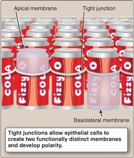
Figure 4.6
Model for an epithelium.
E. Tight junctions
Tight junctions contain numerous different proteins, the principal ones being occludin and claudin. Tight junctions serve several important functions: They form molecular “fences,” they determine tight-junction “leakiness,” and they regulate water and solute flow across epithelia.
1. Fences: Tight junctions prevent apical and basolateral membrane proteins from mixing (a fence function) and, thereby, allow epithelial cells to develop functional polarity (Figure 4.7). The apical membrane becomes specialized for moving material between the external environment and the cell interior, whereas the basolateral membrane moves material between the inside of the cell and the bloodstream (see below).
2. Leakiness: Adjacent cells within an epithelium are separated by a narrow space that creates a physical pathway for transepithelial fluid flow (the paracellular pathway). Tight junctions act as gates that limit paracellular fluid movement and, in so doing, define epithelial leakiness.
a. Leaky epithelia: The tight junctions in a “leaky” epithelium (e.g., renal proximal tubule; see 26·II) are highly permeable and allow solutes and water to pass with relative ease (see Figure 4.7). Leakiness prevents an epithelium from being able to create strong solute concentration gradients between external and internal surfaces, but leaky epithelia are capable of taking up large fluid volumes by paracellular flow.
b. Tight epithelia: Tight junctions in “tight” epithelia effectively bar paracellular flow of water and solutes and allows an epithelium to become highly selective in what it absorbs or secretes (e.g., nephron distal segments; see 27·IV) as shown in Figure 4.8. An epithelium's leakiness is defined by its electrical resistance. Because the tight junctions in tight epithelia restrict passage of ions, they have a high resistance to current flow (>50,000 Ohm), whereas leaky epithelia have low resistance (<10 Ohm).
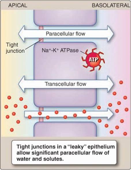
Figure 4.7
Flow across a leaky epithelium. ATP = adenosine triphosphate.
Clinical Application 4.2: Familial Hypomagnesemia with Hypercalciuria and Nephrocalcinosis
Familial hypomagnesemia with hypercalciuria and nephrocalcinosis (FHHNC) is a rare autosomal recessive disorder characterized by an inability to reabsorb Mg2+ from the renal tubule. Plasma Mg2+ levels fall as a consequence (hypomagnesemia). The mutation also impairs Ca2+ reabsorption, which increases urinary excretion rates (hypercalciuria) and the likelihood of kidney stone formation (nephrocalcinosis). Kidney stones form when urinary Ca2+and Mg2+ concentrations are so high that their salts precipitate as crystals, which then aggregate and become lodged within the renal tubule (intrarenal calculi), ureters (ureteral calculi), or bladder. FHHNC is caused by claudin-16 gene mutations (the human genome contains 24 claudin genes). Claudin-16 forms a divalent cation-specific pathway (paracellin-1) for Mg2+ and Ca2+ reabsorption from the thick ascending limb of the loop of Henle. Mutations in claudin-19 can similarly produce renal Mg2+ wasting. Affected individuals typically require magnesium supplements and frequent lithotripsy to mechanically fragment kidney stones and allow them to pass out of the body.

Intrarenal calculi.

Figure 4.8
Tight epithelia. ATP = adenosine triphosphate.
3. Regulation: Although tight junctions in leaky epithelia are highly permeable to water and solutes, they are selective about what they give passage to. Tight-junction permeability can also be regulated to increase or decrease net uptake of water and solutes via the paracellular route. For example, transcellular Na+-glucose transport by intestinal epithelia increases paracellular Na+-glucose transport through changes in tight-junction permeability. The mechanisms involved are not well delineated, but claudins clearly have a central role in determining the size and charge of permeant solutes, and myosin light-chain kinase is involved in regulating junctional permeability.
F. Gap junctions
Gap junctions are the location of gap-junction channels that provide pathways for communication between adjacent cells. They are found in many areas (including muscle and nervous tissue), but are so abundant in some epithelia (e.g., intestinal epithelia) that they are packed into dense crystalline arrays, each containing thousands of individual channels. Gap-junction channels are formed by the association of two connexin hemichannels (connexons) as shown in Figure 4.9.
1. Connexins: Gap-junction channels are formed by six connexin subunits that assemble around a central pore, and each subunit contains four membrane-spanning domains (see Figure 4.9). The human genome contains 21 connexin isoforms that yield channels with distinct gating properties, selectivities, and regulatory mechanisms when expressed. All 21 isoforms are associated with hereditary diseases, which underscores the significance of the gap-junction communication pathway.
Connexin gene mutations produce disorders ranging from idiopathic atrial fibrillation, congenital cataracts, hearing loss, and oculodentodigital dysplasia, to an X-linked form of Charcot- Marie-Tooth disease (CMT). CMT includes a diverse group of demyelinating disorders that primarily affect peripheral nerves, resulting in sensory loss, muscle wasting, and paralysis.
2. Connexons: A gap-junction channel forms when hemichannels from two adjacent cells contact each other end to end, align, and form a tight association (see Figure 4.9). Gap junctions,, have a significant intercellular adhesion function also.
3. Gating: Gap-junction channels are gated by numerous factors, including the potential difference across the junction, membrane-potential changes, Ca2+, pH changes, and by phosphorylation. At rest, when the transjunctional potential is 0 mV, gap-junction channels are usually open.
4. Permeability: The gap-junction channel pore is sufficiently large to allow passage of ions, water, metabolites, second messengers, and even small proteins of up to around 1,000 MW. Gap junctions allow all cells within an epithelium to communicate with each other both electrically and chemically.
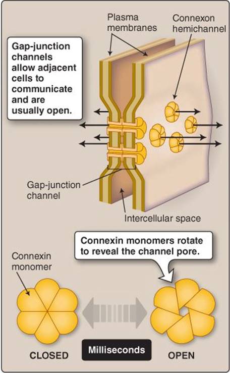
Figure 4.9
Gap-junction channels.
Clinical Application 4.3: Pemphigus Foliaceus
Pemphigus foliaceus is a rare autoimmune disorder that presents as scaly, crusting skin blisters located on the face and scalp, primarily, although the chest and back may become involved in later stages. Affected individuals express antibodies to desmoglein 1, an integral membrane protein that forms a part of the desmosomal complex. Symptoms typically are triggered by drugs (e.g., penicillin) and are caused by desmoglein 1 being targeted and degraded by the immune system. Adjacent skin epithelial cells become detached from one another, and the skin blisters. The blisters ultimately slough off and leave sores. Treatment includes immunosuppressive therapy.

Pemphigus foliaceus.
G. Other junctional structures
Two additional structures provide support to the epithelial sheet: adherens junctions and desmosomes (Figure 4.10).
1. Adherens junctions: All cells in an epithelial sheet are tethered together by bands of protein complexes that lie just below the tight junction known as adherens junctions (zonula adherens; see Figure 4.10). The complexes straddle two adjacent cells and then link to the cell cytoskeleton.
2. Desmosomes: Adjacent cells within an epithelium are also tightly adhered by desmosomes (macula adherens) as shown in Figure 4.10. Desmosomes are small, rounded, membrane specializations that function much like the spot welds used to join metal body panels to an automobile chassis. Protein complexes link the membrane to the cytoskeleton on the intracellular side, whereas adhesion proteins (cadherins) bridge the gap between cells and fuse the two surfaces together. Desmosomes are particularly important for maintaining the integrity of epithelia that are stressed mechanically (e.g., the urinary bladder transitional epithelium).
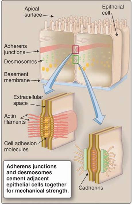
Figure 4.10
Adherens junction and desmosome structures.
III. MOVEMENT ACROSS EPITHELIA
Transepithelial flow of water and solutes occurs via regulated pathways (channels and transporters) and is driven by the same physical forces discussed previously in relation to flow across membranes (i.e., diffusion and carrier-mediated transport; see 1·IV). The principal difference is in the availability of a paracellular route for transepithelial transport.
A. Transcellular transport
Transport epithelia (e.g., intestinal epithelia) are specialized to move large volumes of water and solutes between the outside of the body (e.g., GI tract or renal tubule) and the vasculature by way of the interstitium. Secretory epithelia transfer water and solutes to the outside of the body, whereas absorptive epithelia take up water and solutes from the outside and transfer them to the vasculature. The example below considers the steps involved in glucose uptake by the small intestine (or the renal proximal tubule) as an example, but secretory and absorptive epithelia use the same basic transport principles regardless of body location. The first step involves establishing a Na+-concentration gradient across the surface membrane (Figure 4.11). The steps below correspond to the steps in the figure.
1. Step 1—Create a sodium gradient: Transepithelial transport involves work, the energy for which is supplied by ATP. ATP is used to power primary active transport, which, in virtually all instances, involves the ubiquitous Na+-K+ ATPase located in the basolateral membrane. The Na+-K+ ATPase takes up K+ and expels Na+, thereby creating an inwardly directed Na+-concentration gradient and an outwardly directed K+-concentration gradient.

Figure 4.11
Epithelial transport principles. ATP = adenosine triphosphate.
2. Step 2—Glucose uptake: Gut luminal glucose concentration is usually lower than that in intracellular fluid, meaning that sugar must be transported “uphill” against a concentration gradient. Uptake is powered by the inward Na+-concentration gradient using a SGLT1 Na+-glucose cotransporter (secondary active transport).
3. Step 3—Glucose absorption: Na+-glucose cotransport raises intracellular glucose concentration and creates an outwardly directed concentration gradient that favors glucose movement from the cell to the interstitium. Glucose diffuses across the cell and exits via a GLUT2 transporter in the basolateral membrane. Glucose subsequently diffuses through the interstitium, enters a capillary, and is carried away in the bloodstream.
4. Step 4—Sodium removal: Na+ that crossed the apical membrane during glucose transport is removed from the cell by the basolateral Na+-K+ ATPase.
5. Step 5—Potassium removal: The Na+-K+ exchange during Step 4 above raises intracellular K+ concentrations, but the gradient favoring K+ efflux is already very strong, and excess K+ exits the cell passively via K+channels. K+ channels are usually present in the basolateral membrane but may be located apically also.
B. Water movement
Water cannot be actively transported across epithelia, but a tried-and-true maxim of transport physiology notes that “water follows solutes” (by osmosis). The steps outlined in section (A) caused Na+ and glucose to be translocated from the intestinal lumen to the interstitium, an act that created a transepithelial osmotic gradient that is then used to absorb water. There are two potential routes for water absorption: transcellular and paracellular (Figure 4.12).
1. Transcellular flow: Transcellular water movement only occurs if water is provided with a clear passage through the epithelial cell. In practice, this requires that water channels (aquaporins [AQPs]) be present in both the apical and basolateral membranes. Transport epithelia typically express high AQP levels, which support high volumes of transcellular water uptake (or secretion). The epithelium that lines renal collecting ducts actively regulates its water permeability by modulating apical AQP expression levels (see 27·V·C). When there is a need to reabsorb water from the tubule lumen, AQPs are recruited to the apical membrane from stores located intracellularly in vesicles. When the body contains water in excess of homeostatic requirements, AQPs are removed from the apical membrane, and the epithelium becomes water impermeant.
2. Paracellular flow: Paracellular water flow is also driven by osmotic pressure gradients created by solute transport. The availability of the paracellular route is determined by epithelial leakiness, which is, in turn, determined by tight junctions.

Figure 4.12
Transepithelial water movement. ATP = adenosine triphosphate.
Clinical Application 4.4: Oral Rehydration Therapy
Intestinal epithelia are capable of transporting high volumes of watery fluids. In a healthy person, virtually all of the ~10 L of fluids secreted by intestinal epithelia during the digestive phase are subsequently reabsorbed, so that <200 mL/d is lost from the body in stool. The bacterium Vibrio cholerae secretes a toxin that increases intestinal epithelial Cl− permeability and raises intestinal luminal osmolality.1 Copious amounts of fluid are drawn osmotically across the epithelium as a result. Almost all of the secreted fluid is lost to the external environment, either as a result of vomiting or frequent, watery stools. Death usually occurs as a result of hyponatremia, hypovolemia, and loss of blood pressure. Cholera can be treated and death prevented fairly simply using oral rehydration therapy (ORT). ORT takes advantage of the fact that Na+ and glucose are rapidly absorbed by the intestinal epithelia (via SGLT1), creating an inwardly directed osmotic gradient that drives water reabsorption. Typical home remedies involve giving patients a solution containing 6 tsp sugar and 1/2 tsp salt (NaCl) per liter of water. The advantage of ORT is that it is highly effective, easy to administer, and cheap, which is of particular advantage in developing countries where cholera is endemic and resources are typically limited.

Cholera patients excrete large volumes of watery stool.
C. Solvent drag
Intestinal epithelia secrete and absorb ~10 L of water per day, whereas the renal tubule reabsorbs almost as much on an hourly basis. These secretory and absorptive functions generate high water flow rates, both transcellularly and paracellularly. The resulting water streams carry ions and other small solutes with them, much as a fast-flowing river sweeps along sand and other fine particles. This phenomenon is known as solvent drag and can contribute significantly to transepithelial solute movement. The net result of all this solute and water flow is that the secreted or absorbed fluid usually has a composition that is isosmotic relative to the source (isosmotic flow).
D. Transepithelial voltage effects
Epithelial cells are located at the interface between two compartments that may have very different chemical compositions. The basolateral membrane faces the inside of the body and is bathed in extracellular fluid (ECF) whose chemical composition is well controlled. The apical membrane is bathed in external fluid whose composition may be indeterminate and variable. Charge differences between the two fluids create a transepithelial voltage difference that influences transport (Figure 4.13).

Figure 4.13
Transepithelial voltage difference. Vm = membrane potential.
 1For more information on the pathogenesis and treatment of cholera, see LIR Microbiology, 3e, pp. 122–123.
1. Transport: The distal segment of the renal proximal tubule, for example, is positive charged (~3 mV) with respect to ECF. Although the voltage difference is small, it provides a motive force that drives significant amounts of Na+ out of the tubule and toward the interstitium (see 26·X·B).
2. Local membrane potential effects: Epithelial cells, like all cells in the body, establish a membrane potential (Vm) across their surface membrane, inside negative. Vm is measured with respect to ECF and is uniform throughout the cell. However, the fact that the apical surface is bathed in a medium of different ionic composition can create local differences in Vm. Thus, if Vm is −50 mV and the tubule lumen is +3 mV with respect to ECF, the potential across the apical membrane will be −53 mV relative to the lumen.
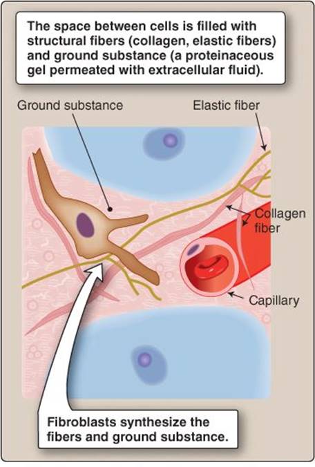
Figure 4.14
Connective tissue.
IV. CONNECTIVE TISSUE
Connective tissue is the most abundant tissue class that can be found in all areas of the body. There are several different connective tissue types, but they all follow a common organizational principle (Figure 4.14). Connective tissues are composed of specialized cells, structural proteins, and a fluid-permeated ground substance.
A. Types
There are three main types of connective tissue: embryonic (not considered further here), specialized connective tissue, and connective tissue proper.
1. Specialized: Specialized connective tissue includes cartilage, bone (see Chapter 15), hematopoietic tissue and blood, lymphatic tissue, and adipose tissue. Cartilage is a flexible connective tissue that cushions bones at sites of articulation and that gives shape to the nose and ears, for example. Lymphatic tissue comprises a system of vessels that drain fluid from the extracellular space (see 19·VII·C). Adipose tissue is composed largely of adipocytes whose primary function is to store energy in the form of triglycerides. Fat conducts heat poorly, so it is layered beneath the skin (subcutaneous fat) to help insulate the body. Adipose tissue deposits can also be associated with internal organs (visceral fat) and in yellow bone marrow.
2. Proper: Connective tissue proper forms the extracellular matrix (ECM) that occupies the interstitial space. Connective tissue proper can be further subdivided into loose connective tissue, a highly pliable form that occupies the space between most cells, dense connective tissue (tendons, ligaments, and fibrous fascia and capsules that enclose muscles and organs), and reticular connective tissue that forms the scaffolding upon which blood vessels, muscle, and the liver is built, for example.
B. Extracellular matrix
The ECM is a mix of cells (fibroblasts), structural proteins (collagen and elastic fibers), ground substance, and ECF. The ECM imparts form and strength to tissues and provides pathways for chemical diffusion and for immune system cell migration (e.g., macrophages).
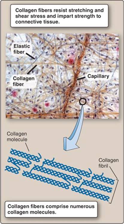
Figure 4.15
Collagen structure.
1. Fibroblasts: Fibroblasts are motile cells that continually synthesize and secrete structural protein precursors and ground substance. They are essential for ECM maintenance and for wound healing.
2. Structural proteins: The ECM is filled with an interconnected structural matrix comprising collagen and elastic fibers.
a. Collagen: Collagen is a tough, fibrous protein that possesses high tensile strength and resistance to shear stress. The body contains 28 different collagen types, but four forms (types I, II, III, and IV) predominate. Type I collagen is abundant in skin and vascular walls and is bundled to form ligaments, tendons, and bone. Type IV organizes into meshlike networks that make up the basal lamina of epithelia, for example. Collagen molecules are composed of three polypeptide chains braided into a triple helix, and then cross-linked extensively for enhanced stress resistance1 (Figure 4.15).
b. Elastic fibers: Elastic fibers are composed of elastin and glycoprotein microfibrils (e.g., fibrillin and fibullin). Elastic fibers stretch like rubber bands when stressed and then recoil and assume their original shape when allowed to relax. Elastic fibers are found in the walls of arteries and veins, which allows them to stretch when intraluminal pressures increase. Elastic fibers also allow lungs to expand during inspiration as well as help reduce stress on teeth during chewing (periodontal fibers). Elastic fibers are synthesized by fibroblasts, which first lay down structural scaffolding made of fibrillin and then deposit tropoelastin monomers onto the scaffolding. Four adjacent elastin monomers are then cross-linked to form an irregular network that comprises the mature elastin molecule (Figure 4.16).

Figure 4.16
Elastin properties.
3. Ground substance: Ground substance is a mix of various proteins (principally proteoglycans) and ECF that creates an amorphous gel filling the spaces between cells and structural fibers. The high water content of the gel facilitates chemical diffusion between cells and the vasculature, yet the structural fibers simultaneously impede movement of invading pathogens. Proteoglycans are formed by attaching numerous glycosaminoglycan (GAG) molecules to a core protein, the final structure resembling a bottlebrush or pipe cleaner (Figure 4.17). GAGs possess a high negative-charge density, which allows them to attract and loosely trap water molecules within the gel. The interstitium contains >10 L of ECF in an average person, representing a substantial buffer volume that helps minimize the impact of changes in total body water on cell and cardiovascular function (see 3·III and 19·VIII·C).

Figure 4.17
Proteoglycan structure.
1For more information on collagen synthesis and assembly, see LIR Biochemistry, 5e, p. 43.
Chapter Summary
• The human body is composed of four tissue types: epithelial tissue, nervous tissue, muscle tissue, and connective tissue. Epithelial tissue comprises sheets of tightly packed cells that line all external and internal body surfaces (e.g., skin and the pulmonary and gastrointestinal linings). Epithelia form barriers that protect the body from invasion by microbes, but many also have specialized transport functions.
• Epithelia are classed morphologically. Simple epithelia are composed of a single cell layer (e.g., pulmonary epithelium). Stratified epithelia (e.g., skin) comprise multiple layers that are sloughed off and renewed. Glandular epithelia (endocrine and exocrine glands) are specialized for secretion.
• Epithelia are polarized with functionally distinct apical and basolateral surfaces. The apical surface faces the external environment, the lumen of a hollow organ, or a body cavity. Apical surfaces may be specialized to include villi, cilia, and stereocilia that amplify surface area, propel mucus layers, or serve a sensory role, respectively.
• The basolateral membrane communicates with the body interior via the interstitium and the vasculature. It rests on a basement membrane that anchors the epithelium to underlying connective tissues.
• Polarization of epithelia is made possible by tight junctions, which are structural bands encircling all cells in an epithelium close to their apical surface. The junctions form tight seals with an important barrier function. The junctions also segregate apical and basolateral membrane proteins, thereby allowing for specialization of membrane function.
• Tight-junction permeability is regulated by claudins, which determine how much water and solutes cross an epithelium via the space between adjacent cells (paracellular flow). The junctions effectively block passage of all water and solutes across a “tight” epithelium. In contrast, “leaky” epithelia secrete and absorb significant amounts of fluid.
• Gap junctions are hexameric channels comprising connexin monomers that connect adjacent cells and allow all cells in an epithelium to communicate chemically and electrically. Adherens junctions and desmosomes are junctional structures that provide strength to an epithelium and help prevent it from tearing when stressed mechanically.
• Many epithelia have secretory and absorptive functions. Transepithelial transport usually occurs transcellularly and paracellularly, although both routes are regulated. The electrochemical and osmotic driving forces for movement of solutes and ions are established by primary and secondary active transporters (e.g., Na+-K+ ATPase and Na+-coupled transport).
• Connective tissue comprises cells; structural fibers; and an amorphous, fluid-permeated ground substance. Specialized connective tissues include cartilage, bone, and adipose tissue. Connective tissue proper forms the extracellular matrix.
• The extracellular matrix fills the space between all cells, providing mechanical strength and support as well as a loose, gel-like medium that facilitates chemical diffusion and cell migration.
• The extracellular matrix (ECM) is synthesized and maintained by fibroblasts. ECM structural proteins include collagen fibers for strength and elastic fibers to allow stretching. Elastic fibers are composed primarily of elastin. Ground substance is a proteinaceous matrix containing large quantities of proteoglycans. Proteoglycans have a high negative-charge density that allows them to attract and immobilize ~10 L of extracellular fluid in an average person.
Study Questions
Choose the ONE best answer.
I.1 A fluid that is composed of 120 mmol/L K+, 12 mmol/L Na+, and 15 mmol/L Cl− but is virtually Ca2+ free (<1 μmol/L) would best approximate which body fluid compartment?
A. Transcellular
B. Plasma
C. Interstitial
D. Intracellular
E. Extracellular
Best answer = D. Intracellular fluid should be recognized by its relatively high K+ concentration, which is due to the Na+K+ ATPase found in the membrane of virtually all cells (1·II). Extracellular fluid (ECF) has a lower K+ and higher Na+, Cl−, and Ca2+ concentration compared with ECF and can be further subdivided into plasma (fluid within the vascular space) and interstitial fluid (fluid outside the vascular space; 3·III·A). Because the barrier between these two ECF compartments does not prevent ion movement, their ionic composition is similar. Transcellular fluid (including cerebrospinal fluid, synovial fluid, and urine) composition is variable depending on the location and, therefore, not the best choice.
I.2 Dye indicators are important physiologic tools used to calculate unknown volumes or concentrations within the body. If a dye is membrane permeable, which of the following changes will most likely increase dye diffusion rate?
A. Lowering dye concentration
B. Increasing membrane surface area
C. Increasing membrane thickness
D. Decreasing fluid temperature
E. Lowering the dye partition coefficient
Best answer = B. Increasing membrane surface area increases the opportunity for dye to cross the membrane, which increases its diffusion rate (1·IV·B). It takes longer for molecules to diffuse across thick membranes than thin ones, and decreasing a fluid's temperature increases its viscosity, which also slows diffusion rate. Concentration gradients provide the driving force for diffusion, so lowering dye concentration flattens the gradient and reduces diffusion rate. A partition coefficient is a measure of dye lipid solubility. Molecules with high lipid solubility diffuse through membranes faster than poorly soluble molecules, so lowering partition coefficient would decrease diffusion rate.
I.3 A 66-year-old male is treated with the loop diuretic furosemide (Na+-K+-2Cl− cotransport inhibitor) to reduce symptoms associated with congestive heart failure. Which of the following best describes this cotransporter's mode of action?
A. It is a primary active transporter.
B. It is electrogenic.
C. A rise in intracellular K+ would decrease transport rate.
D. It transports Na+ and K+ into the cell and 2 Cl− out of the cell.
E. It transports Na+ against its electrochemical gradient.
Best answer = C. Cotransporters, by definition, move two or more ions in the same direction (1·V·C). The Na+-K+-2Cl− cotransporter simultaneously carries two anions and two cations across the plasma membrane and, thus, is not electrogenic. Primary active transporters use adenosine triphosphate to pump ions against their electrochemical gradients. Transporters that move Na+ in one direction while simultaneously bringing other ions back in the opposite direction are exchangers, not cotransporters. The Na+ gradient established by the basolateral Na+ pump (Na+-K+ ATPase) provides the electrochemical driving force for K+ and Cl− uptake, but transport rate is sensitive to transmembrane K+ and Cl− gradients. Increasing intracellular concentrations of either ion will slow net uptake.
I.4 Serum electrolytes levels are ordered on a 12-year-old boy with a gastrointestinal infection, which induced prolonged and severe vomiting episodes. Plasma K+ concentrations were found to be abnormally low (2 mmol/L). Which of the following might be expected to result from mild hypokalemia?
A. Resting potentials would shift positive.
B. K+ equilibrium potential would shift negative.
C. Neuronal action potentials would be inhibited.
D. Na+ channels would inactivate.
E. K+-channel activation would yield K+ influx.
Best answer = B. Hypokalemia, or reduced extracellular K+ concentrations, enhances the electrochemical gradient favoring K+ efflux from cells and causes the K+ equilibrium potential to shift negative (see 2·II·B). Because membrane potential is determined largely by the transmembrane K+ gradient, resting membrane potential (Vm) would shift negative also. A negative shift in Vm means that a stronger depolarization would be necessary to take Vmto the threshold for voltage-gated Na+ channel activation (2·III·B), but, once reached, an action potential would be initiated. K+-channel activation always causes K+ efflux, except in rare instances (e.g., in the inner ear; 9·IV·C).
I.5 A 35-year-old man carries an epilepsy gene. The gene mutation affects the neuronal voltage-dependent Na+ channel, causing it to inactivate more slowly (~50%). How might expression of this epilepsy gene affect nerve function?
A. Resting potential would settle close to 0 mV.
B. Action potentials would no longer overshoot 0 mV.
C. Action potentials would be prolonged.
D. Action potentials would rise very slowly.
E. There would be no action potentials.
Best answer = C. The voltage-dependent Na+ channel is opened by membrane depolarization to yield the upstroke of the neuronal action potential (2·VI·A). An inactivation gate closes shortly after activation, blocking passage of Na+ and allowing membrane potential to return to resting levels. If inactivation were slowed, membrane recovery would be delayed, and the action potential would be prolonged. Resting potential should not be affected by an inactivation defect unless it prevented the channel from closing, causing a sustained Na+ influx. Activation and inactivation are separate processes, and, therefore, the rate at which the action potential rises should be normal.
I.6 An agricultural worker is packing hot chili peppers for transport. He removes his protective mask and becomes incapacitated with a sensation of nasal burning caused by capsaicin from the peppers. What receptor type is capsaicin stimulating?
A. Transient receptor-potential channels
B. Purinergic receptors
C. Ionotropic glutamate receptors
D. Cys-loop family receptors
E. Voltage-gated Na+ channels
Best answer = A. The transient receptor potential channel (TRP) family transduces a variety of sensory stimuli, including heat, cold, and osmolality (2·VI·D). TRPV1 channels are stimulated by capsaicin. Purinergic, glutamate, and cys-loop receptors are activated by specific ligands (i.e., adenosine triphosphate, L-glutamate, and acetylcholine, respectively). Capsaicin is not an agonist for these receptor classes. Voltage-gated Na+ channels are activated primarily by membrane depolarization.
I.7 During a serological analysis, red blood cells (RBCs) were transferred from blood to a solution containing 100 mmol/L CaCl2 and 100 mmol/L urea and then monitored using light microscopy. How would you expect this transfer to affect RBC volume?
A. The solution is isosmotic, so no long-term effect.
B. The solution is isotonic, so no long-term effect.
C. Transient swelling would occur.
D. Swelling to the point of lysis would occur.
E. The cell would shrink by ~50%.
Best answer = B. CaCl2 dissociates into three particles (1 Ca2+ and 2 Cl−) in water. A 100-mmol/L CaCl2solution has an osmolality of 300 mOsm/kg H2O, which approximates that of RBC intracellular fluid (ICF). 100 mmol/L urea brings total osmolality to 400 mOsm/kg H2O (the solution is hyperosmotic), but urea would rapidly enter the cell until intracellular and extracellular fluids equilibrated at ~350 mOsm/kg H2O (3·II·C). The solution is, thus, isotonic. Cell shrinkage would occur if urea was impermeant, but most cells are highly permeable to urea. Cell swelling in this example would only occur if ICF osmolality rose due to active accumulation of one or more of the three solutes.
I.8 Liver damage may result in decreased synthesis of plasma proteins such as albumin. What is the most significant effect of low plasma albumin on osmosis or fluid transport?
A. Interstitial fluid volume increases.
B. Vascular fluid volume increases.
C. Plasma colloid osmotic pressure increases.
D. Plasma osmolality increases.
E. Plasma osmolality decreases.
Best answer = A. Blood contains large amounts of albumin (3.5–5 g/dL) that is trapped in the vascular compartment by virtue of its large size (3·III·B). Its function is to help create an osmotic potential (known as plasma colloid osmotic pressure) that draws extracellular fluid (ECF) into the vasculature. A decrease in plasma albumin concentration would, therefore, allow fluid to leave the vasculature and enter the interstitium. Plasma osmolality does not change significantly with changes in protein concentration. The main determinants of ECF osmolality are ions (e.g., Na+ and Cl−) and other solutes (e.g., glucose and urea, measured as blood urea nitrogen, or “BUN”).
I.9 A 95-year-old man with widely metastatic cancer is receiving morphine to help alleviate pain. Brainstem respiratory center function has been depressed as a result, causing hypoventilation. Which of the following might be expected to result from reducing ventilation?
A. Alkalemia
B. Decreased plasma HCO3− levels
C. Decreased renal HCO3− reabsorption
D. Increased interstitial pH
E. Increased urinary H+ excretion
Best answer = E. Metabolism generates large amounts of volatile acid (H2CO3) that is excreted via the lungs (3·IV·A). Decreasing ventilation allows this acid to accumulate, producing a (respiratory) acidemia. The kidneys help compensate by increasing H+ excretion. Decreasing renal HCO3− reabsorption would exacerbate the acidemia through loss of buffer to urine. Accumulation of volatile acid raises plasma HCO3− levels because H2CO3 dissociates in solution to form HCO3− and H+. H+, along with other small molecules, moves freely between the blood and interstitium. Thus, if blood is acidic, the interstitium will also have a low pH.
I.10 A researcher investigating the properties of intestinal epithelium from a patient with inflammatory bowel disease noted that the diseased areas have a low electrical resistance, whereas the healthy areas have a high resistance. What might be inferred about the properties of the healthy epithelium?
A. It forms weak transepithelial ionic gradients.
B. It is specialized for isosmotic transport.
C. It has a thick basement membrane.
D. The tight junctions are highly impermeable.
E. It lacks a basolateral Na+-K+ ATPase.
Best answer = D. High electrical resistance is characteristic of a “tight” epithelium, a property conferred in part by the impenetrability of the tight junctions between cells to ions and water (4·II·E). Tight epithelia are notable for their ability to establish strong osmotic and ionic concentration gradients. The areas of inflammation have a low electrical resistance, which makes them “leakier” to ions. Leaky epithelia are usually specialized for high-volume isosmotic transport. Basement membranes do not directly contribute to epithelial electrical resistance. All intestinal epithelia express a basolateral Na+-K+ATPase.
I.11 A 52-year-old woman presents with heart palpitations and lightheadedness. An electrocardiogram shows her to be in atrial fibrillation, which has been linked to increased connexin 43 expression. Which of the following best describes connexins normally?
A. They open during membrane depolarization.
B. They are highly ion selective.
C. They mediate Ca2+ influx from the cell exterior.
D. They allow electrical propagation through tissues.
E. They are found only in the heart.
Best answer = D. Connexins form hexameric assemblies (connexons) with a pore at their center (4·II·F). Connexons from two adjacent cells fuse to create a gap-junction channel that provides a pathway for electrical and chemical communication between cells. They are widely distributed. In the heart, they allow waves of contraction to spread across the myocardium (17·III). Gap-junction channels are characterized by their wide, nonselective pores that can allow passage of small peptides. They are usually open at rest and may close upon depolarization. Ca2+ channels mediate Ca2+ influx across surface membranes, not gap-junction channels.
I.12 Hypokalemia is relatively rare in healthy individuals, but which of the following would favor increased K+ up-take by a transport epithelium for transfer to the circulation?
A. Lumen-negative potential difference
B. Increased paracellular water uptake
C. Increased Na+-K+ ATPase activity
D. High interstitial K+ concentrations
E. Apical glucose cotransport
Best answer = B. The paracellular route is a significant pathway for solute and water movement across many epithelia (4·III·B). High paracellular water flow rates generate solvent drag, whereby inorganic ions and other solutes are swept along with water. Because K+ is a cation, a renal or gastrointestinal lumen (for example) that is negatively charged with respect to blood decreases net uptake. The Na+-K+ATPase increases intracellular K+concentrations, which decreases the driving force for apical K+ uptake. High interstitial K+ concentrations also decrease the electrochemical gradient favoring net K+ uptake. Glucose cotransporters generally couple glucose movement with Na+, not K+.