
5
I. OVERVIEW
To a casual observer, a microscopic pond organism such as Paramecium behaves with apparent intent and coordination that suggests the involvement of a sophisticated nervous system. If it bumps into an object, it stops, swims backward, and then moves off in a new direction (Figure 5.1). This simple behavior minimally requires a sensory system to detect touch, an integrator to process information from the sensor, and a motor pathway to effect a response. Paramecium does not possess a nervous system, however. It is a single cell. The human brain contains over a trillion neurons. It has evolved sophisticated structures and networks that allow for self-awareness, creativity, and memory. Yet the human nervous system's basic organizational principles share many similarities with our unicellular cousins. Unicells and neurons both use changes in membrane potential (Vm) to integrate and respond to divergent and, sometimes, conflicting inputs. On an organismal level, humans, like unicells, have sensory systems to inform them about their immediate environment, integrators to process sensory data, and motor systems to effect an appropriate response.
II. NERVOUS SYSTEM
In discussing how the nervous system works, it is useful to define three partially overlapping subdivisions.
• The central nervous system (CNS) includes the neurons of the brain and spinal cord. The CNS is the nervous system's integrative and decision-making arm.
• The peripheral nervous system (PNS) collects sensory information and conveys it to the CNS for processing. It then directs motor commands from the CNS to the appropriate targets. The PNS includes neurons that originate in the cranium and spinal cord and extend beyond the CNS.
• The autonomic nervous system (ANS) is central to many discussions of human physiology because it regulates and coordinates visceral organ function, including the gastrointestinal system, lungs, heart, and vasculature. The distinction between the ANS and the other two divisions is functional rather than anatomic. The ANS can be further subdivided into the sympathetic nervous system and parasympathetic nervous system. Both divisions function largely independently of voluntary control.
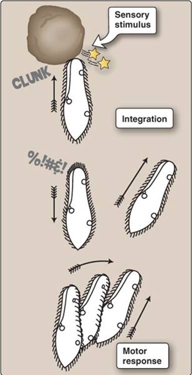
Figure 5.1
Sensory response in Paramecium.
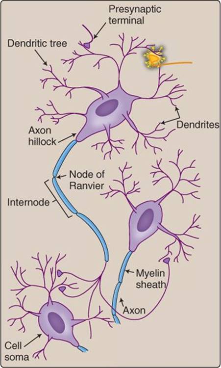
Figure 5.2
Neuronal anatomy.
III. NEURONS
The nervous system comprises a network of neurons. Although their shape may vary according to function and location within the body, the basic principles of neuronal design and operation are universal. Their role is to transmit information as rapidly as possible from one area of the body to the next. In the brain, the distance involved may be a few micrometers, but, in the periphery, it can exceed a meter. Because speed is achieved using electrical signals, a neuron can be thought of as a biologic wire. Unlike a wire, however, a neuron has the ability to integrate incoming signals before transmitting information to a recipient.
A. Anatomy
A neuron can be divided into four anatomically distinct regions: the cell body, dendrites, an axon, and one or more nerve terminals (Figure 5.2).
1. Cell body: The cell body (soma) houses the nucleus and components required for protein synthesis and other normal cellular housekeeping functions.
2. Dendrites: Dendrites are branched projections of the cell body that radiate in multiple directions (“dendrite” is derived from dendros, the Greek word for tree). Some neurons have dense and elaborate dendritic trees, whereas others may be very simple. Dendrites are cellular antennae waiting to receive information from the neural net. Many tens of thousands of nerve terminals may synapse with a single neuron via its dendrites.
3. Axon: An axon is designed to relay information at high speed from one end of the neuron to the other. It arises from a swelling of the soma called an axon hillock. An axon is long and thin like a wire. It is often wrapped with an insulating material (myelin) that enhances signal-transmission rate (see below). Myelination begins some distance distal to the axon hillock, leaving a short initial segment that is unmyelinated. The axoplasm (axonal cytoplasm) is filled with parallel arrays of microtubules and microfilaments. They are partly structural, but they also act like railway tracks in a mineshaft. “Ore carts” (vesicles) filled with neurotransmitters and other materials attach to the tracks and then motor along at relatively high speed (~2 μm/s) from one end of the cell to the other. Movement away from the cell body toward the nerve terminal (anterograde transport) is powered by kinesin. The return trip (retrograde transport) relies on a different molecular motor (dynein).
4. Nerve terminal: The nerve terminal is specialized to convert an electrical signal (an action potential) into a chemical signal for dispatch to one or more recipients. The junction between the terminal and its target is called a synapse. The presynaptic and postsynaptic cell membranes are separated by a ~30–50 nm synaptic cleft. Facing the terminal across the cleft may be any of a number of different postsynaptic effector cells, including myocytes, secretory cells, or even a dendrite extending from the cell body of another neuron.
Clinical Application 5.1: Polio
Retrograde transport is believed to be the mechanism by which polio and many other viruses enter the central nervous system from the periphery.1 Poliovirus is an enterovirus distributed by fecal–oral contact that causes paralytic poliomyelitis. After infecting the host, the virus enters and spreads through the nervous system via nerve terminals. After fusing with the surface membrane and entering the axoplasm, the viral capsid (a protein shell) attaches to the retrograde transport machinery and motors to the cell body. Here, it proliferates and, ultimately, destroys the neuron. The result is a flaccid paralysis of the musculature, classically affecting the lower limbs, but it can also cause fatal paralysis of the respiratory musculature. Polio has been largely eradicated in North America and Europe but is endemic in many other regions of the world. Defects in axonal transport are also believed to have a role in precipitating the neuronal death that accompanies Alzheimer disease, Huntington disease, Parkinson disease, and several other adult-onset neurodegenerative diseases.

Poliovirus
B. Excitability
The speed with which neural nets process and output data is limited by the rate at which signals are transmitted from one component to the next. Extracting maximal speed from a neuron is achieved by using a fast action potential, by optimizing axonal geometry, and by insulating the axons.
1. Action potentials: Axons that convey signals over long distances typically display action potentials that have a very simple form and function as binary digits on the neural information net. Neuronal action potentials are mediated primarily by voltage-gated Na+ channels, which are very fast activating (Figure 5.3). When Na+ channels open, Na+ flows into the neuron down its electrochemical gradient, and Vmdepolarizes rapidly toward the equilibrium potential for Na+ (see 2·II·B). It is the rapidity of Na+-channel opening (“gating kinetics”) that allows electrical signals to propagate at high speeds down an axon's length. Membrane repolarization occurs largely as a result of Na+-channel inactivation. Voltage-dependent K+ channels activate during a spike also, but their numbers are small and, thus, their contribution to membrane repolarization is limited.
 1For a discussion of enteroviruses, see LIR Microbiology, 2e, pp. 283–286.

Figure 5.3
Time course of ion channel events during a neuronal action potential.
2. Axon diameter: The rate at which electrical signals travel down an axon increases with axonal diameter (Figure 5.4). This is because internal resistance, which is inversely proportional to diameter, determines how far passive current can reach down the axon's length before the signal decays and needs amplifying by an active current (i.e., a Na+-channel–mediated current). The amplification step is slow compared with transmission of passive current, so wide axons transmit information over long distances much faster than do thin ones.
3. Insulation: The passive currents that flow during excitation dissipate with distance because the membrane contains K+ “leak” channels that lose current to the extracellular medium (see Figure 2.11). Leak channels are always open. Conduction velocity is improved significantly by insulating the axon with myelin to prevent such leak (see Figure 5.4B). Myelin is formed by glial cells and comprises concentric layers of sphingomyelinrich membrane (see Section V below). Insulation increases conduction velocity up to 250-fold.
4. Saltatory conduction: An axon's myelin sheath is not continuous. Every 1–2 mm is a 2–3-μm segment of exposed axonal membrane known as a node of Ranvier. Nodes are tightly packed with Na+channels, whereas the internodal regions (the areas lying hidden beneath the myelin sheath) have virtually no channels. In practice, this means that an action potential leapfrogs from one node to the next down the length of the axon, a behavior known as nodal, or saltatory, conduction (see Figure 5.4C).
C. Classification
CNS neurons are a diverse group of cells, and there are many ways of classifying them. Morphologically, they can be grouped on the basis of the number of neurites (processes, such as axons and dendrites) extending from the cell body.
1. Pseudounipolar: Pseudounipolar neurons are usually sensory. The cell body gives rise to a single process (the axon) that then splits into two branches. One branch returns sensory information from the periphery (the peripheral branch), whereas the other branch projects and conveys this information to the CNS (central branch).
2. Bipolar: Bipolar neurons are usually specialized sensory neurons. Bipolar neurons can be found in the retina (see 8·VII·A) and olfactory epithelium (see 10·III·B), for example. Their cell body gives rise to two processes. One conveys sensory information from the periphery, and the other (the axon) travels to the CNS.
3. Multipolar: Multipolar neurons have a cell body that gives rise to a single axon and numerous dendritic branches. Most CNS neurons are multipolar. They can be further subcharacterized based on the size and complexity of their dendritic tree.
D. Neurons as integrators
The unicell mentioned in the introduction is capable of integrating multiple sensory signals (e.g., mechanical, chemical, thermal) through changes in Vm. For example, a noxious signal that depolarizes Vm and increases the tendency to turn might be ignored if an attractant signal indicating nearby food hyperpolarizes the membrane and negates or overrides noxious signal input. A paramecium is not capable of conscious thought, yet it makes a decision that affects behavior based on the summed effect of multiple stimuli on Vm. The dendritic trees of higher cortical neurons receive tens of thousands of competing inputs. The likelihood that neuronal output (spiking) will be modified on the basis of these signals is similarly determined by their net effect on Vm.
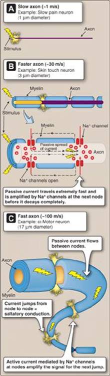
Figure 5.4
Myelin and diameter effects on axonal conduction velocity.
1. Incoming signals: Neurons hand off information to each other via dendrites. When a presynaptic neuron fires, it releases transmitter into the synaptic cleft. If the neuron is excitatory, transmitter binding to the postsynaptic dendritic membrane causes a transient depolarization known as an excitatory postsynaptic potential (EPSP) as shown in Figure 5.5A. Inhibitory neurons release transmitters that cause transient hyperpolarizations known as inhibitory postsynaptic potentials (IPSPs). EPSP and IPSP amplitudes are graded with incoming signal(s) strength.
2. Filtering: Much of the information being received by neurons at their dendrites represents sensory “noise.” Isolating the strongest and most relevant signals is accomplished using a noise filter that takes advantage of a dendrite's natural electrical properties. Postsynaptic potentials (PSPs) are passive responses that degrade rapidly as they travel toward the cell body (see Figure 2.11). Degradation is enhanced by a dendrite's inherent electrical leakiness and its lack of myelin. In practice, this means that a small PSP may never reach the cell body. PSPs generated by strong presynaptic activity activate voltage-gated ion currents along the length of the dendrite (see Figure 2.12). These enhance the signals and, thereby, increase their likelihood of reaching the cell body.
3. Integration: Signal integration also begins at the dendritic level. PSPs may meet and combine with PSPs arriving from other synapses as they travel toward the soma. This phenomenon is known as summation and is reminiscent of the way in which waves (e.g., sound waves and ripples spreading across a pond surface) interfere constructively and destructively. There are two types of summation: spatialand temporal.
a. Spatial summation: If EPSPs from two different dendrites collide, they combine to create a larger EPSP (see Figure 5.5B). This is known as spatial summation and applies to IPSPs also. EPSPs and IPSPs can also summate to yield an attenuated membrane response (see Figure 5.5C).
b. Temporal summation: Two EPSPs (or IPSPs) traveling along a dendrite in rapid succession can also combine to produce a single, larger event. This is known as temporal summation (see Figure 5.5D).
4. Output: The net effect of multiple PSPs on Vm determines the likelihood and intensity of neuronal output. If a depolarization is sufficiently strong, it may elicit a train of spikes. Spikes arise from the initial segment (also known as the spike initiation zone) and travel down the length of the axon toward the presynaptic terminal.

Figure 5.5
Summation. EPSP = excitatory postsynaptic potential; IPSP = inhibitory postsynaptic potential; Vm = membrane potential.
5. Encoding: Action potentials are all-or-nothing events, so neurons must pass on information about signal strength using digital encoding. Weak stimuli may yield one or two spikes. Strong stimuli elicit spike trains (volleys) that travel in rapid succession down the axon's length. There is tremendous variability in the size, shape, and frequency of spikes generated by different neurons. As a general rule, the number of spikes in a volley reflects incoming stimulus strength (Figure 5.6).
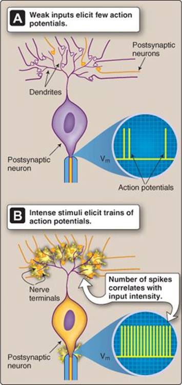
Figure 5.6
Digital encoding by neurons. Vm = membrane potential.
IV. NEUROTRANSMISSION
Neurons communicate with each other at synapses, specialized regions where two cells come into close apposition with each other. Communication typically occurs chemically via neurotransmitter release and is unidirectional. Although an inherently slow form of communication, placing a neurotransmitter receptor in the signaling pathway allows for a great diversity of responsiveness and unlimited opportunities for regulation.
A. Neurotransmitters
There are two main classes of neurotransmitter: small-molecule transmitters and peptides. A third, lesser group includes gases and other unconventional transmitters such as adenosine triphosphate (Table 5.1). Many tens of neuroactive peptides have been described also, many of which are coreleased along with a small-molecule transmitter. Most neural interactions involve just a handful of molecules, whose synthetic pathways are summarized in Figure 5.7.
B. Synaptic vesicles
Neurotransmitters are released into the synaptic cleft from synaptic vesicles. The vesicles are synthesized in the presynaptic cell body and then shuttled by fast axonal transport to the nerve terminal. Here, they are filled with locally produced neurotransmitter for storage and eventual release. Mature vesicles then dock at specialized release sites on the presynaptic membrane and remain there awaiting the arrival of an action potential.

Peptide transmitters are synthesized and prepackaged in vesicles within the cell body rather than in the nerve terminal.
C. Release
Neurotransmitter release occurs when an action potential arrives at the nerve terminal and opens voltage-dependent Ca2+ channels in the nerve terminal membrane (Figure 5.8). Ca2+ influx raises local Ca2+concentrations and initiates a Ca2+-dependent secretory event. The details are complex and not fully resolved. The Ca2+ signal is sensed by a vesicle-associated Ca2+-binding protein called synaptotagmin, which activates a SNARE-protein complex that includes synaptobrevin, syntaxin, and SNAP-25. The vesicle then fuses with the presynaptic membrane at an active zone, and the contents are emptied into the synaptic cleft. Each vesicle releases a single quantum of neurotransmitter (“quantal signaling”).

Figure 5.7
Common small-molecule neurotransmitters and their synthesis pathways. GABA = γ-aminobutyric acid.
Botulinum toxin and tetanus toxin, two of the most lethal known neurotoxins, both paralyze their victims by targeting the synaptic terminal and disrupting neurotransmitter release. Both toxins degrade SNAPS and SNARES by virtue of their intrinsic protease activity.

Figure 5.8
Synaptic vesicle release.
D. Receptors
Once released, a neurotransmitter diffuses across the narrow synaptic cleft and binds to a specific neurotransmitter receptor expressed on the postsynaptic membrane. The receptors are associated with numerous proteins that anchor them and that regulate their activity and expression levels, manifesting as a postsynaptic density in micrographs (see Figure 5.8). Receptors can be classed in at least two ways.
1. Ionotropic versus metabotropic: Ionotropic receptors are ion channels that mediate ion fluxes when active (Figure 5.9). The nicotinic acetylcholine receptor (AChR) is an ionotropic receptor that mediates Na+ influx, for example.
Metabotropic receptors couple to an intracellular signaling pathway and are usually associated with a G protein. Examples include the muscarinic AChR.
2. Excitatory versus inhibitory: Excitatory receptors (e.g., the NMDA receptor) cause membrane depolarization and increased firing rates when occupied. Conversely, inhibitory receptors (e.g., the glycine receptor) hyperpolarize the membrane and decrease spike frequency.
Properties of the main neurotransmitter receptor types are summarized in Table 5.2.
E. Signal termination
Signal termination can occur at the receptor level through receptor internalization or desensitization, but, more usually, signaling ends when the transmitter is removed from the synaptic cleft. A neurotransmitter typically suffers one of three fates: degradation, recycling, or diffusion out of the cleft (“spillover”; Table 5.3).
1. Degradation: The synaptic cleft usually contains high levels of enzymes that limit signaling by degrading neurotransmitter. For example, cholinergic synapses contain acetylcholinesterase, which degrades ACh.
2. Recycling: Many nerves and their support cells (see below) actively take up transmitter from the synaptic cleft and recycle it by repackaging it in synaptic vesicles.
3. Diffusion: Transmitter can also diffuse out of the synaptic cleft to affect neighboring neurons. During intense neuronal activity, significant amounts of transmitter may appear in the circulation, ultimately being degraded by systemic enzymes or being excreted by the kidneys.

Figure 5.9
Ionotropic versus metabotropic receptors.
V. NEUROGLIA
Glia (or neuroglia) are inexcitable cells that support many aspects of neuronal function. In addition to forming and maintaining myelin, they control local ion concentrations, help recycle neurotransmitters, and provide neurons with nutrients. They are found throughout the PNS and CNS (Table 5.4), where neurons and glia are present in equal numbers.

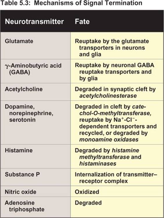


Figure 5.10
Myelin sheath formation.
A. Myelination
Myelin is formed by Schwann cells (in the PNS) and oligodendrocytes (in the CNS). Oligodendrocytes may simultaneously myelinate the axons of multiple neurons, but Schwann cells remain dedicated to a single axon. Glia form myelin by extending processes that rotate around an axon over 100 times (Figure 5.10). Cytoplasm is squeezed out from between the membrane layers as the myelin builds, so that the lipid layers become highly compacted. Glial cells remain viable after the layering is complete, with the nucleus and residual cytoplasm occupying the outermost layer.
B. Potassium homeostasis
Renormalization of Vm after neuronal excitation involves K+ release (see Figure 5.3). During intense neuronal activity, extracellular K+ concentration can rise significantly as a result of this release. Because Vm is dependent on the transmembrane K+ gradient (see 2·II·C), K+ buildup can be detrimental to neuronal function. Astrocytes (the predominant glial cell type in the CNS) are inexcitable, but they do possess K+ channels and K+ transporters that allow them to siphon K+ away from active neurons and redistribute it over nonactive regions of the CNS (Figure 5.11). “Spatial buffering” takes advantage of the fact that adjacent astrocytes are tightly coupled via gap junctions (see 4·II·F) that provide pathways for K+ to flow down their concentration gradient from the active zone to a remote site.
Clinical Application 5.2: Multiple Sclerosis
Myelin is essential for normal neural communication, so diseases that affect myelin or the cells that produce it have devastating physiologic effects. Multiple sclerosis (MS) is a demyelinating disease of central nervous system neurons, and the cause is unknown. Symptoms arise when autoreactive immune cells produce antibodies against one or more sheath components. The myelin swells and degrades, and axonal conduction is interrupted, producing pathologic changes that can be visualized using computed tomography (commonly known as a CT scan). Patients can present with a variety of neurologic symptoms, including tremors, visual disturbances, autonomic dysfunction, weakness, and fatigue. MS may be relapsing in nature, characterized by acute onset of clinical symptoms, followed by a period of remission with full or partial recovery of function. There is no known treatment for MS, however, and the disease usually progresses over a period of 10 to 20 years.

Periventricular frontal lobe demyelination.
C. Neurotransmitter uptake and recycling
Glutamate, γ-aminobutyric acid (GABA), and glycine are three of the most widely used neurotransmitters in the CNS. Many neurons employing these transmitters spike at such high frequencies that their ability to control synaptic transmitter levels and prevent spillover into adjacent regions is challenged. Intense activity also strains the transmitter synthesis pathways. Glial cells assist with both issues. They extend foot processes that surround the synapse and rapidly take up transmitter from the cleft using high-affinity transport systems (Figure 5.12). Glutamate is subsequently converted to glutamine by glutamine synthetase and returned to the presynaptic terminal for conversion back to glutamate by glutaminase. Glutamine is also distributed to inhibitory neurons for GABA synthesis. GABAergic neurons have very limited glutamine reserves and are dependent on glia to supply the substrates necessary for continued signaling.
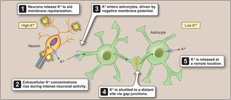
Figure 5.11
Spatial buffering.

Figure 5.12
Neurotransmitter recycling by astrocytes.
D. Nutrient supply
Neurons are highly dependent on O2 and glucose for continued activity, the nervous system accounting for 20% of total body usage. Glia play a unique role in ensuring that these needs are met (see also 21·II for a discussion of the role of glial cells in maintaining the blood-brain barrier).
1. Lactate shuttle: Astrocytes ferry glucose from the vasculature to neurons. They extend foot processes that surround cerebral capillaries and absorb glucose from blood using transporters. Glucose then diffuses through the glial network via gap junctions. Some glucose is converted to glycogen, and the rest is metabolized to lactic acid. Lactate is then excreted into the extracellular fluid for uptake by surrounding neurons, a process known as the lactate shuttle.
2. Storage: Neurons have few energy reserves. They rely on astrocytes to maintain a steady lactate supply in the face of changes in neuronal activity or waning blood glucose levels. Astrocytes contain extensive glycogen stores and the necessary pathways to convert them to lactate when needed.
VI. NERVES
The terms neuron and nerve are often confused. A neuron is a single excitable cell. A nerve is a bundle of nerve fibers (axons and their supporting cells) that runs through the periphery like a modern telecommunications cable.
A. Conduction velocity
The characteristics of the individual fibers that make up a nerve vary considerably. Some may be thin, unmyelinated, and slow to conduct. Others are thick, myelinated, and conduct impulses at high speed. Thick fibers take up more space than thin ones and are expensive to sustain metabolically. They are used only where speed of communication is paramount. In practice, that means the fastest fibers are used for motor reflexes (see Figure 5.4 and Table 11.1).
B. Assembly
Individual nerve fibers are wrapped loosely in connective tissue (the endoneurium), and then several are bundled together to form a fascicle (Figure 5.13). A fascicle is wrapped in yet more connective tissue (the perineurium), and, finally, several fascicles are gathered together with blood vessels to form a nerve. The nerve is heavily protected with a dense layer of connective tissue (the epineurium). Peripheral nerves are subjected to considerable mechanical stress associated with locomotion, so the multiple reinforcing layers are essential for protection.

Figure 5.13
Peripheral nerve anatomy.
C. Ganglia
The cell bodies of axons that make up a nerve are clustered in swellings called ganglia. Ganglia are the sites of information relay between neurons. Ganglia may also contain intrinsic circuits that allow for reflex arcs and signal processing.
D. Types
Nerves can be classified according to the type of information that they carry. Afferent nerves contain fibers relaying sensory information from the various body regions to the CNS. Efferent nerves originate in the CNS and contain somatic motor neurons that innervate skeletal muscles and ANS visceral motor neurons. Some peripheral nerves contain a mix of both sensory and motor fibers (mixed nerves). This can result in reflex arcs that travel within the same nerve. The vagus nerve, for example, informs the CNS when food is entering the stomach. The CNS responds with a motor command that makes the stomach relax, and it travels via an efferent fiber contained within the vagus nerve (see 30·IV·A). The arc is called a vagovagal reflex.
Chapter Summary
• The nervous system comprises the central nervous system (CNS) and the peripheral nervous system (PNS). The autonomic nervous system (ANS) is a functional subdivision of the CNS and PNS. The CNS includes brainand spinal cord neurons. The PNS includes nerves that carry sensory and motor information to the periphery. The ANS monitors and controls internal organ function.
• Neurons are excitable cells that communicate with each other and with target organs using action potentials. Electrical signals are relayed from one cell to the next at a synapse using chemical neurotransmitters.
• Neurotransmitters are a diverse group that includes gases and polypeptides. The central nervous system (CNS) uses small molecules such as amino acids (glutamate, aspartate, glycine, and γ-aminobutyric acid) as transmitters. Acetylcholine is the neurotransmitter used at the neuromuscular junction and by some neurons of the autonomic nervous system (ANS). The ANS and CNS also use monoamines(dopamine and norepinephrine) as transmitters.
• A transmitter may excite or inhibit the postsynaptic cell, depending on the nature of the receptor on the postsynaptic membrane. Excitatory transmitters cause postsynaptic cell depolarization (an excitatory postsynaptic potential), whereas inhibitory transmitters cause hyperpolarization and reduced excitability (inhibitory postsynaptic potential).
• Central nervous system neurons may receive thousands of synaptic inputs via their dendritic trees. The electrical properties of neuronal dendrites ensure that weak inputs do not propagate. Stronger inputs may summate to push membrane potential beyond the threshold for excitation, causing the neuron to fire an action potential.
• Neuronal function is supported by glial cells. Glia may regulate extracellular ion concentrations, aid in uptake and recycling of neurotransmitters, supply nutrients, and encase the axons in myelin.
• Myelin is an insulating material laid down by Schwann cells (peripheral nervous system) and oligodendrocytes (central nervous system). Myelin increases the speed at which electrical signals propagate down an axon. Myelin is formed from compacted layers of glial surface membrane. Myelinated neurons are used in reflex arcs where response timing is critical. Motor neurons also have axons that are wider than normal to further increase signal propagation rates.
• Nerves are bundles of axons and their supporting cells. Peripheral nerves may convey afferent signals from sensory cells to the central nervous system (CNS), efferent signals from the CNS to effector cells, or a signal mixture.