
6
I. OVERVIEW
The central nervous system (CNS) comprises the spinal cord and brain (Figure 6.1). The spinal cord is a thick communications tract that relays sensory and motor signals between the peripheral nervous system (PNS) and the brain. The cord also contains intrinsic circuits that support certain muscle reflexes. The brain is a highly sophisticated data processor containing neural circuits that analyze sensory data and then execute appropriate responses via the spinal cord and PNS efferents. Large portions of the brain are devoted to associative functions that integrate information from the various senses and allow us to assign meaning to sounds, associate smells with specific memories, and recognize objects and faces, for example. Associative regions also provide for abstract thinking, language skills, social interactions, and learning and memory. The human body has bilateral symmetry, and the structures of the spinal cord and brain are, for the most part, mirrored about a midline. Sensory and motor information generally crosses the midline at some point in its journey between the brain and the periphery. In practice, this means that the left side of the brain controls the right side of the body and vice versa. For the purposes of discussion, the brain can be divided into four principal areas: the brainstem, cerebellum, diencephalon, and the cerebral hemispheres(triencephalon). A full discussion of CNS function is beyond the scope of this book, which focuses on the sensory and motor aspects of CNS function. For more information on higher brain functions, see LIR Neuroscience.
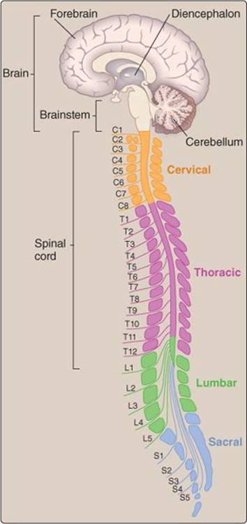
Figure 6.1
Central nervous system. C1–C8, T1–T12, L1–L5, and S1–S5 are spinal nerves.
II. SPINAL CORD
The spinal cord is housed within the vertebral canal. It extends from the foramen magnum at the base of the skull caudally to the second lumbar vertebra.
A. Segments
The vertebral column consists of a series of stacked vertebrae divided anatomically into five regions: cervical, thoracic, lumbar, sacral, and coccygeal. The cervical, thoracic, and lumbar vertebrae are separated by intervertebral disks that allow the bones to articulate, but the sacral and coccygeal vertebrae are fused to form the sacrum and coccyx, respectively. The spinal cord can be divided into 31 named segments. Thirty-one pairs of spinal nerves (one on each side of the body) emerge from corresponding segments (see Figure 6.1). Although the spinal cord terminates before it reaches the sacrum, spinal nerves continue caudally within the vertebral canal until they reach an appropriate exit level.
Rostral and caudal are anatomical terms meaning “beak” (or mouth) and “tail,” respectively. They are commonly used to indicate direction of information flow in the CNS.
B. Nerves
Spinal nerves are a component of the PNS. The nerves contain sensory afferent and motor efferent fibers (spinal nerves are sometimes called mixed spinal nerves for this reason) that generally serve tissues on the same level as the nerves. Thus, nerves emerging from the cervical region (C2) control head and neck movements, whereas sacral nerves (S2 and S3) project to the bladder and large intestine.
1. Sensory: Somatic and autonomic sensory fibers travel to the spinal cord via peripheral nerves (Figure 6.2). They relay sensations of pain, temperature, and touch from the skin; proprioceptive signals from muscle and joint receptors; and sensory signals from numerous visceral receptors. Multiple peripheral nerves come together to form the posterior root of a spinal nerve and enter the vertebral canal via an intervertebral foramen. The cell bodies of these nerves cluster within a prominent spinal ganglion located within the foramen. The posterior root then divides into a number of rootlets and joins the spinal cord. Sensory nerves travel rostrally to synapse within nuclei en route to the brain. Branches of sensory afferents may also synapse directly with motor neurons or on interneurons that synapse with motor neurons, which makes local spinal cord–mediated reflexes possible (see 11·III).
2. Motor: Motor efferents from the brain travel caudally and synapse with peripheral motor nerves within the spinal cord. These nerves include both somatic and autonomic motor efferents. They leave the spinal cord via anterior rootlets, which join to form an anterior root and then travel out to the periphery alongside sensory fibers in spinal nerves.
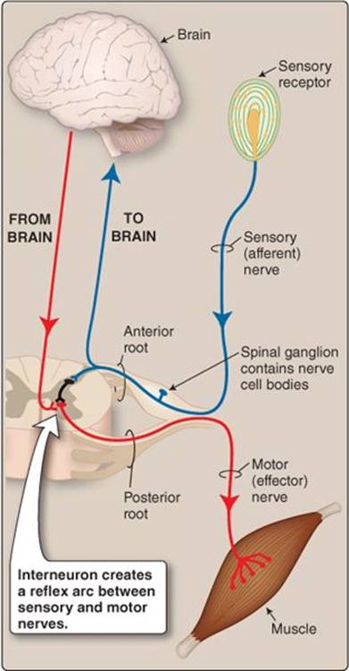
Figure 6.2
Sensory and motor pathways.
C. Tracts
The spinal cord's interior is roughly organized into a butterfly-shaped central area of gray matter surrounded by white matter (Figure 6.3). The white matter contains bundles of nerve fibers with common origins and destinations that relay information between the PNS and the brain. Sensory nerve fibers from the periphery travel rostrally to the brain in discrete ascending tracts. Descending tracts carry bundles of motor efferents from the CNS en route to the periphery.
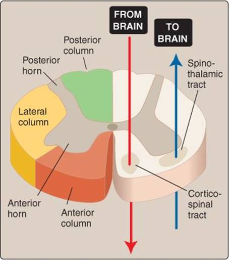
Figure 6.3
Spinal cord organization.
The tracts are named according to their origin and destination. For example, the spinothalamic tract carries pain fibers from the spine upward to the thalamus. The corticospinal tract carries motor fibers from the cortex downward to the spine. The tracts (also known as fasciculi) are grouped in posterior, lateral, and anterior columns (also known as funiculi). The “wings” of the gray butterflies are divided into posterior and anterior horns and act as synaptic relay stations for information flow between neurons. They contain neuronal cell bodies, which may be clustered in functionally related groups, or nuclei. The gray matter on either side of the cord is connected by commissures containing bundles of fibers that allow for information flow across the midline.
CNS tissue typically appears white or gray in color. White matter is largely composed of myelinated nerve axons (it gets its white color from myelin). Gray matter is composed of cell bodies, dendrites, and unmyelinated axons.
III. BRAINSTEM
All sensory and motor information flowing to and from the brain passes through the brainstem (Figure 6.4). The brainstem contains several important nuclei that act as relay stations for information flow between brain and periphery. Many of the 12 cranial nerves (CNs) originate from nuclei within the brainstem also (Figure 6.5). The CNs provide sensory and motor innervation to the head and neck and include nerves that mediate vision, hearing, smell, and taste, among many other functions. Intrinsic circuits within the brainstem create control centers that allow for reflex responses to sensory data. The location and functions of these centers are discussed in more detail in Chapter 7. The brainstem can be subdivided anatomically into three areas:
• Medulla: The medulla contains autonomic nuclei involved in the control of respiration and blood pressure and in coordination of swallowing, vomiting, coughing, and sneezing reflexes.
• Pons: The pons helps control respiration.
• Midbrain: The midbrain contains areas involved in controlling eye movements.
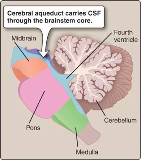
Figure 6.4
Brainstem organization. CSF = cerebrospinal fluid.
CN I and CN II do not originate in the brainstem. CN I, the olfactory nerve, is a sensory nerve that relays information from the olfactory epithelium in the roof of the nasal cavities directly to the olfactory bulb. CN II, the optic nerve, enters the brain at the level of the diencephalon.
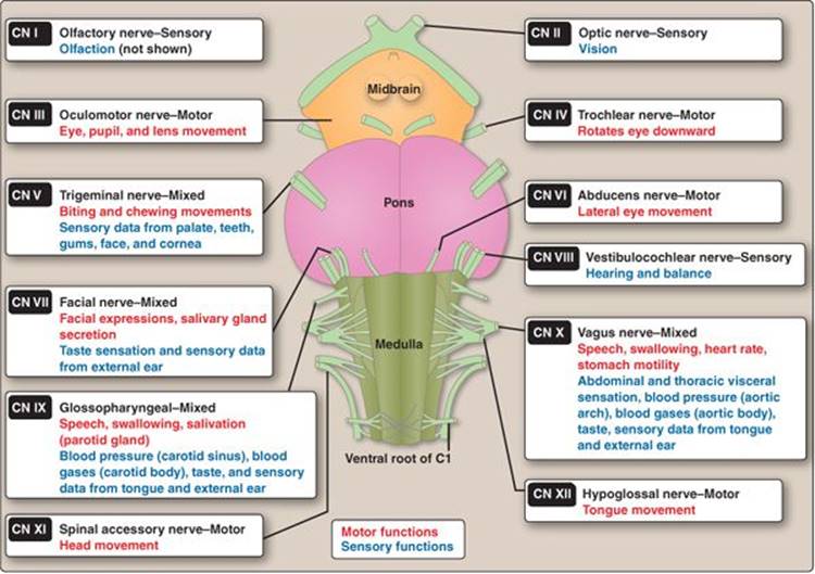
Figure 6.5
Cranial nerve (CN) functions. C1 = first cervical vertebra.
IV. CEREBELLUM
The cerebellum fine-tunes motor control and facilitates smooth execution of learned motor sequences (see 11·IV·C). Cerebellar function requires massive integrative and computational capabilities, which is why this small area contains more neurons than the rest of the brain combined, even though it accounts for only ~10% of total brain mass! The cerebellum is attached to the brainstem by three peduncles that contain thick afferent and efferent nerve fiber bundles. The cerebellum receives sensory data from muscles, tendons, joints, skin, and the visual and vestibular systems and inputs from all regions of the CNS involved in motor control. It also sends signals back to most of these areas and modifies their output (Figure 6.6). Integration of sensory data with motor commands is achieved using feedback and feedforward circuits that include the Purkinje cell, a neuronal type renowned for its immense dendritic tree. The dendrites are sites of information flow from hundreds of thousands of presynaptic neurons. The cerebellar circuits allow movements to be finessed with reference to incoming sensory data, even as they are being executed.
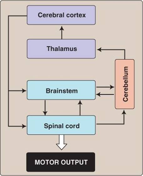
Figure 6.6
Functional relationships among central nervous system components.
Cerebellar injury does not cause paralysis, but it does have profound motor effects (ataxia, or an inability to coordinate muscle activity). Patients with cerebellar damage walk with a staggering gait that mimics alcohol intoxication. They may also have slurred speech and difficulties with swallowing and eye movement.
V. DIENCEPHALON
The diencephalon and telencephalon together make up the forebrain. The diencephalon contains two major structures: the thalamus and hypothalamus (Figure 6.7).
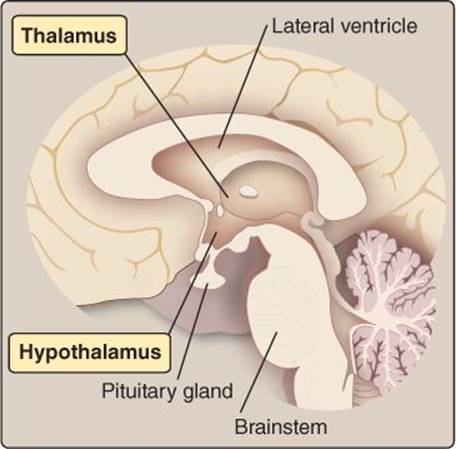
Figure 6.7
Thalamus and hypothalamus location.
A. Thalamus
Sensory information from the periphery passes through the thalamus for processing before reaching a conscious level. Output from the olfactory system is the single exception, insofar as it bypasses the thalamus and feeds raw olfactory data to the cortex directly. The thalamus also controls sleep and wakefulness and is required for consciousness. Damage to the thalamus can result in deep coma. The thalamus is also involved in motor control and has areas that project to the cortical motor regions.
B. Hypothalamus
The hypothalamus is a major autonomic nervous system control center that is discussed in detail in Chapter 7. Its functions include control of body temperature, food intake, thirst and water balance, and blood pressure, and it also controls aggression and rage. The hypothalamus exerts control through direct neural connections to autonomic centers in the brainstem, but it also controls the endocrine system. Endocrine control occurs directly through hormonal synthesis and release (oxytocin and antidiuretic hormone) and indirectly by secreting hormones that affect release of pituitary hormones.
VI. TELENCEPHALON
The telencephalon, or cerebrum, is the seat of human intellect. It is organized into two cerebral hemispheres comprising the basal ganglia and the cerebral cortex.
A. Basal ganglia
The basal ganglia are a group of functionally related nuclei (Figure 6.8) that work closely with the cerebral cortex and thalamus to effect motor control. Their function is discussed at length in Chapter 11. Major structures within the basal ganglia include the caudate nucleus and putamen (together forming the striatum) and the globus pallidus.
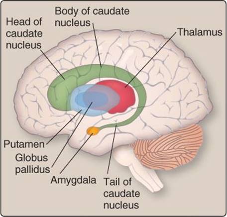
Figure 6.8
Basal ganglia.
B. Cerebral cortex
The cerebral cortex is involved in conscious thought, awareness, language, and learning and memory.
1. Anatomy: The cortex comprises a sheet of neural tissue organized in six layers that is folded to accommodate the 15 to 20 billion (1.5–2.0 × 10¹⁰) neurons contained within. The folds (gyri) are separated by sulci (grooves). Deep fissures separate the cortex into four lobes: frontal, parietal, occipital, and temporal (Figure 6.9). The lobes contain discrete areas that can be distinguished on a cytoarchitectural basis and that correlate with regions of specialized function.
2. Function: The cortex can be functionally divided into three general areas that stretch across both hemispheres: sensory, motor, and associative.
a. Sensory: Sensory regions process information from the sensory organs (see Chapters 8–10). Primary sensory regions receive and process information directly from the thalamus. Spatial information is preserved as data flows from the senses to the sensory areas and then accurately maps onto the cortex (topographic mapping). Thus, the pattern of light falling on the retina is faithfully replicated in the pattern of excitation within the primary visual cortex.
b. Motor: Motor areas are involved with planning and executing motor commands. Primary motor areas execute movements. Axons from these areas project to the spinal cord, where they synapse with and excite motor neurons. Supplementary motor areas are involved with planning and fine control of such movements (see Chapter 11).
c. Associative: The majority of cortical neurons are involved in associative functions. Each cortical sensory region feeds information to a corresponding association area. Here, patterns of color, light, and shade are recognized as a human face, for example, or a series of notes can be recognized as coming from a songbird. Other associative areas integrate sensory information from other parts of the brain to allow for higher mental functions. These include abstract thinking, acquisition of language, musical and mathematical skills, and the ability to engage in social interactions.
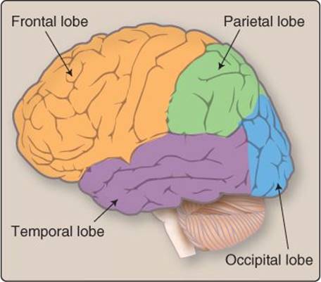
Figure 6.9
Cortical lobes.
VII. CEREBROSPINAL FLUID
Because the CNS has a central role in all aspects of life, its neurons are provided with multiple layers of protection and support.
A. Protective layers
The role of the blood–brain barrier in protecting CNS neurons against bloodborne chemicals is discussed in Chapter 21 (see 21·II·B). The CNS is also enclosed within five protective layers, including three membranes (the meninges), a layer of cerebrospinal fluid (CSF), and an outer layer of bone (Figure 6.10). The meninges comprise the pia mater, the arachnoid mater, and the dura mater.
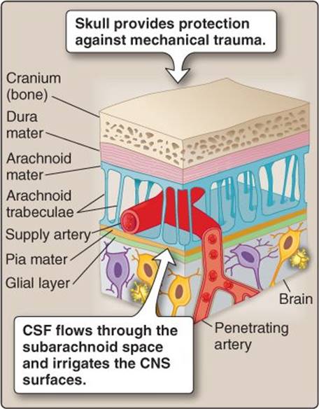
Figure 6.10
Layers that protect the brain and provide a pathway for cerebrospinal fluid (CSF) flow. CNS = central nervous system.
Clinical Application 6.1: Bacterial Meningitis
Bacterial meningitis is a life-threatening disease caused by bacterial infection of the cerebrospinal fluid (CSF) and meningeal inflammation.1 It is a leading cause of infectious death worldwide. The most common community-acquired causes of meningitis are Streptococcus pneumoniae (~70%) and Neisseria meningitiditis (12%), whereas hospital-acquired cases are usually caused by Staphylococcus. Infection is caused by bacteria crossing the blood–brain barrier and establishing colonies in the CSF, affecting both the brain and spinal cord. Symptoms are usually rapid in onset and include a triad comprising a severe headache, nuchal (neck) rigidity, and a change in mental status. Most patients also present with a high fever. Nuchal rigidity is caused by pain and muscle spasm when attempting to flex or turn the head, reflecting meningeal inflammation in the cervical region. Immediate treatment to reduce swelling and address the infection usually leads to full recovery.
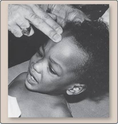
Patient lifts her shoulders rather than flexing her neck when her head is elevated (nuchal rigidity).
1. Pia mater: The entire surface of the brain and spinal cord is tightly adhered to a thin, fibrous membrane called the pia mater (Latin for “dutiful mother”). The pia's cerebral portion is held in place by a continuous layer of astrocyte foot processes.
2. Arachnoid mater: The arachnoid mater comprises a layered epithelial membrane that is loosely connected to the pia by trabeculae, small structural supports that give the arachnoid mater a cobwebbed appearance. The trabeculae create a subarachnoid space through which CSF flows unhindered over the brain's surface. The layer of CSF has multiple functions (see below), including cushioning the brain against trauma.
3. Dura mater: The “tough mother” is a thick, leathery membrane comprising two layers. An inner, meningeal layer is firmly attached to the arachnoid mater and covers the entire surface of both brain and spinal cord. The cranium is lined by a periosteal layer. The two layers separate in places to create an intracranial venous sinus that drains blood and CSF from the brain and channels it to the circulation.
4. Bone: The brain is protected by the cranium. The spinal cord lies within the vertebral canal, protected by the vertebral column.
B. Functions
CSF is a highly purified, sterile, colorless fluid nearly devoid of proteins that surrounds and bathes the tissues of the CNS. It has four main functions: providing buoyancy, absorbing shock, permitting limited intracranial volume changes, and maintaining homeostasis.
 1For a more complete discussion of bacterial meningitis, see LIR Microbiology, 2e, p. 376.
1. Buoyancy: The brain's high lipid content gives it a relatively high specific gravity compared with CSF (1.036 versus 1.004). In practice, this means that the brain floats in CSF. The advantage is that flotation distributes brain mass evenly and helps prevent cerebral tissues from being compressed by gravity against the skull. Compression must be avoided because it impedes blood flow through the cerebral vasculature and causes ischemia.
2. Shock absorption: CSF surrounds the brain on all sides and envelops it in a liquid cushion. Cushioning reduces the chance of mechanical trauma to the brain when the skull is impacted or impacts an object at speed.
3. Volume changes: During periods of intense activity, neurons and glia tend to swell due to accumulation of metabolites and other osmotically active materials. CSF allows water to shift from CSF to cells without causing any gross change in CNS volume. Because the CNS is constrained by bone on all sides, volume changes can potentially compress the cerebral vasculature and cause ischemia (see 21·II·D).
4. Homeostasis: Cell membrane potential (Vm) and neuronal excitability is highly sensitive to changes in extracellular K+ concentration. Plasma K+ concentrations can rise by >40% even under normal conditions (normal plasma K+ concentration = 3.5–5.0 mmol/L), changes that are unacceptable for a Vm-dependent organ such as the CNS. CSF K+ concentrations are tightly maintained at a relatively low level (2.8–3.2 mmol/L), thereby insulating neurons from large swings in plasma concentration. CSF is also free of potentially neuroactive compounds (such as glutamate and glycine) that constantly circulate in blood. CSF thus provides the CNS with a stable, rarified extracellular environment that is renewed constantly to prevent buildup of neuronal waste products, transmitters, and ions.
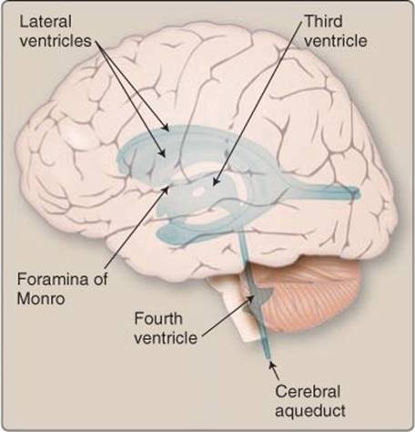
Figure 6.11
Location of the cerebrospinal fluid-filled ventricles and cerebral aqueduct.
C. Choroid plexus
CSF is formed by the choroid plexus, a specialized epithelium that lines four fluid-filled ventricles within the brain's core (Figure 6.11).
1. Ventricles: The brain contains four ventricles: two lateral ventricles and a third and fourth ventricle. They are all connected by foramina that allow CSF to flow caudally to the spinal cord and through its central canal.
a. Lateral: The two lateral ventricles are the largest of the four. They are symmetrical C shapes and are located at the center of the two cerebral hemispheres. They connect with the third ventricle via two interventricular channels called the foramina of Monro.
b. Third: The third ventricle lies on the midline at the level of the thalamus and hypothalamus. It connects with the fourth ventricle via the cerebral aqueduct (of Sylvius).
c. Fourth: The fourth ventricle is located within the brainstem. The caudal end communicates with the spinal cord's central canal. The ventricle also provides a pathway for CSF to flow into the subarachnoid space via three openings. The foramen of Magendie is located at the midline. Two foramina of Luschka are located laterally.

Figure 6.12
Choroid plexuses.
2. Location: The choroid plexuses are localized to specific regions of the ventricles (Figure 6.12). They line the floor of the lateral ventricles and continue through the interventricular channels to line the roof of the third ventricle. In the fourth ventricle, choroid plexus occupies a small portion of the roof.
3. Structure: The ventricles and the spinal cord's central canal are lined with ependymal epithelium. In the region of choroid plexus, the ependyma gives way to a ciliated choroid epithelium, which is responsible for secreting CSF. Choroid epithelial cells contain large numbers of mitochondria, and their apical surfaces are amplified by microvilli, features that are characteristic of an epithelium specialized for high-capacity ion and water transport. The epithelium rests on a basal lamina, which separates it from the vasculature below, and adjacent cells are all coupled by tight junctions. Choroid epithelial activity is supported by a vascular plexus comprising a dense network of arteries, capillaries, and veins. The capillaries are large and leaky, and their walls contain fenestrations to facilitate fluid filtration from blood.
D. Formation
About 30% of total CSF production can be attributed to secretion by the brain parenchyma. The remaining 70% is produced by the choroid plexus.
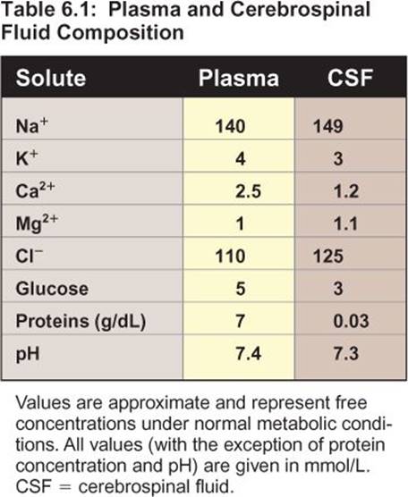
1. Composition: The basal side of the choroid epithelium is bathed in plasma filtrate, but tight junctions between adjacent epithelial cells create an effective barrier to exchange of ions and other solutes between blood and CSF. The differences between CSF and plasma are notable in several respects (Table 6.1):
• CSF contains minimal protein or other large molecules. The lack of protein makes the CSF reliant on HCO3– for pH buffering.
• HCO3– levels are higher to help buffer acids produced by the CNS.
• Na+ and Cl– levels are higher, which compensates osmotically for the lack of protein.
• K+ concentrations are lower.
The necessary gradients for CSF formation are established at the apical (luminal) surface of the choroid epithelium (Figure 6.13).
2. Sodium secretion: The choroid epithelium is highly unusual in that the Na+-K+ ATPase is located on the apical membrane rather than the basolateral membrane. The pump drives Na+ into the CSF.
3. Potassium absorption: The Na+-K+ ATPase simultaneously removes K+ from the CSF. More K+ may be absorbed by an apical Na+-K+-2Cl– cotransporter, using the energy of the Na+ gradient that favors Na+ entry into the epithelial cell.

Figure 6.13
Cerebrospinal fluid (CSF) formation. AQP = aquaporin; CA = carbonic anhydrase; ECF = extracellular fluid.
4. Bicarbonate secretion: HCO3– is generated by carbonic anhydrase activity. For every HCO3– molecule generated, a H+ is liberated also. This is released to the vasculature via a Na+-H+ exchanger in the basolateral membrane. HCO3– is likely secreted into the ventricle via anion (Cl–) channels and Na+-HCO3– cotransporters.
5. Chloride secretion: Cl– is concentrated within the cells by anion exchangers in the basolateral membrane and then flows across the apical membrane via Cl– channels.
6. Water secretion: Water follows an osmotic gradient generated by secretion of Na+, HCO3–, and Cl–. Aquaporins provide a pathway for movement.
E. Flow
CSF is produced at prodigious rates (~500 mL/day), flushing the ventricles and CNS surfaces once every 7–8 hrs. High flow rates ensure that byproducts of neuronal activity (inorganic ions, acids, and transmitters spilling over from synapses) are removed in a timely manner before they can build to levels that might interfere with CNS function.
The choroid plexuses have a mass of only ~2 g. Their ability to generate so much CSF is made possible both by a blood flow that is higher than that of virtually any other tissue (and 10 times that supplying neurons) and by the enhanced surface area for secretion created by the villi and microvilli.
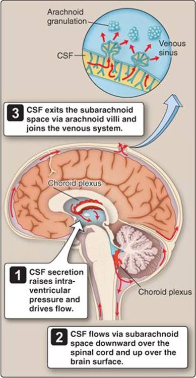
Figure 6.14
Pathways for cerebrospinal fluid (CSF) flow over the surfaces of the central nervous system (CNS).
1. Pathways: CSF secretion by the choroid plexus increases the pressure within the ventricles by a few millimeters of H2O, sufficient to drive CSF flow through the ventricles, through the foramina in the fourth ventricle and into the subarachnoid space (Figure 6.14). CSF then percolates through the space and flows over the CNS surfaces, eventually joining venous blood contained within the intracranial sinus. CSF enters the sinuses via arachnoid villi, which may be organized into large clumps called arachnoid granulations. CSF is transported across the villi via giant vesicles, creating a one-way valve that prevents backflow from sinus to subarachnoid space if CSF pressure drops.
2. Exchange between extracellular fluids: CSF and brain ECF are separated in the ventricles by ependymal cells and in other regions by the pia and supporting layer of astrocytic foot processes. Although the pia and astrocytes layers are continuous, the junctions between adjacent cells are leaky and they allow free exchange of materials between CSF and ECF. This allows neuronal and glial waste products to diffuse out of the ECF and be carried away by the CSF.
Clinical Application 6.2: Lumbar Puncture
Cerebrospinal fluid (CSF) pressure is normally within a range of 60–200 mm H2O (~4.5–14.7 mm Hg), but can rise dramatically when the subarachnoid villi become clogged with bacteria or blood cells. Lumbar puncture offers an opportunity both to measure CSF pressure and retrieve fluid samples to test for the presence of white or red blood cells, which might indicate bacterial meningitis or subarachnoid hemorrhage, respectively. Lumbar puncture involves inserting a long, thin (spinal) needle through the dura mater into the subarachnoid space. Fluid is withdrawn from the subarachnoid space in the lumbar region, which is below where the spinal cord terminates. Up to 40 mL of CSF can be withdrawn safely for cytologic analysis and culturing.
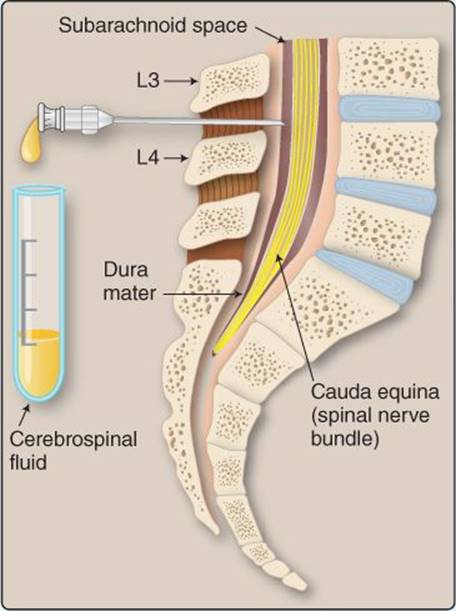
Lumbar puncture.
Chapter Summary
• The central nervous system comprises the spinal cord and brain. The spinal cord contains bundles of nerve fibers organized into tracts that relay information between the peripheral nervous system(PNS) and the brain. Ascending tracts relay sensory information from the PNS to the brain, whereas descending tracts convey motor commands to the PNS. The spinal cord also contains intrinsic circuits that facilitate local reflex arcs that do not require input from the brain.
• Peripheral nervous system neurons enter and leave the spinal cord via 31 pairs of spinal nerves. Posterior roots of these nerves contain sensory afferent fibers, whereas anterior roots contain motor efferents.
• All information flowing between the central and peripheral nervous systems must pass through the brainstem, which contains the medulla, pons, and midbrain. These areas contain autonomic nucleiinvolved in control of respiration, blood pressure, and upper gastrointestinal tract reflexes. The brainstem is associated with 10 cranial nerves that innervate the head and neck.
• The cerebellum facilitates fine motor control. It integrates sensory information from muscles, joints, and the visual and vestibular systems, and fine-tunes motor commands in anticipation of and during movements.
• The diencephalon comprises the thalamus and hypothalamus. The thalamus processes sensory information, whereas the hypothalamus is an autonomic nervous system control center.
• The telencephalon comprises the basal ganglia, which are involved in motor control, and the cerebral cortex. The cerebral cortex contains sensory, motor, and associative areas and is the seat of higher functioning.
• The central nervous system is protected by five layers, comprising the pia mater, a layer of cerebrospinal fluid, arachnoid mater, dura mater, and bone.
• Cerebrospinal fluid (CSF) is a colorless, protein-free fluid produced by the choroid plexus, a secretory epithelium located within the brain ventricles. CSF flows through the ventricles under pressure and then over the surface of both the brain and spinal cord. It drains into venous sinuses located within the dura mater.
• Cerebrospinal fluid (CSF) also acts as a liquid cushion that protects the brain from mechanical trauma and helps distribute its weight evenly within the cranium. CSF is produced at high rates, flushing the ventricles and central nervous system surfaces and carrying away accumulated waste products.