
Mandana Khalili, MD, MAS,&Blaire Burman, MD
Although many different pathogenic agents and processes can affect the liver (Table 14-1), they are generally manifested in individual patients in a limited number of ways that can be assessed by evaluation of some key parameters. Liver disease can be acute or chronic, focal or diffuse, mild or severe, and reversible or irreversible. Most cases of acute liver disease (eg, caused by viral hepatitis) are so mild that they never come to medical attention. Transient symptoms of fatigue, loss of appetite, and nausea are often ascribed to other causes (eg, flu), and minor biochemical abnormalities referable to the liver that would be identified in blood studies are not discovered. The patient recovers without any lasting medical consequences. In other cases of acute liver injury, symptoms and signs are severe enough to call for medical attention. The entire range of liver functions may be affected or only a few, as is the case with liver injury resulting from certain drugs that cause isolated impairment of the liver’s role in bile formation (cholestasis). Occasionally, viral, drug-induced, and other acute liver injury occurs in an overwhelming manner, resulting in massive liver cell death and progressive multiorgan failure. This syndrome of acute liver failure (also referred to as fulminant hepatic failure) carries a high mortality rate; however, in recent years, use of emergency liver transplantation has significantly improved survival.
TABLE 14-1 Liver diseases.

Liver injury may continue beyond the initial acute episode or may be recurrent (chronic hepatitis). In some cases of chronic hepatitis, liver function remains stable or the disease process ultimately resolves altogether. In other cases, there is progressive and irreversible deterioration of liver function.
Cirrhosis is ultimately the consequence of progressive liver injury. Cirrhosis can occur in a subset of cases of chronic hepatitis that do not resolve spontaneously or after repeated episodes of acute liver injury, as in the case of chronic alcoholism. In cirrhosis, the liver becomes hard, shrunken, and nodular and displays impaired function and diminished reserve because of a decreased amount of functioning liver tissue. More importantly, the physics of blood flow is altered such that the pressure in the portal vein is elevated. As a result, the blood is diverted around the liver rather than filtered through the liver. This phenomenon, termed portal-to-systemic (or portosystemic) shunting, has profound effects on the function of various organ systems and sets the stage for certain devastating complications of liver disease that are described later.
Although liver disease resulting from many different causes may present in common ways, the reverse is also true (ie, liver disease from specific causes may have distinctly different presentations in different patients). For example, consider two patients with acute viral hepatitis: One may present with yellow eyes and skin—a manifestation of impaired liver function—complaining of nothing more than itching, fatigue, and loss of appetite, whereas the other may be brought to the emergency room moribund, with massive gastrointestinal (GI) bleeding and encephalopathy. Such variations in the severity of liver disease are probably due to genetic, immunologic, and environmental (including perhaps nutritional) factors that are currently poorly understood.
The consequences of liver disease can be either reversible or irreversible. Those arising directly from acute damage to the functional cells of the liver, most notably hepatocytes, without destruction of the liver’s capacity for regeneration, are generally reversible. Like many organs of the body, the liver normally has both a huge reserve capacity for the various biochemical reactions it carries out and the ability to regenerate fully differentiated cells and thereby recover completely from acute injury. Thus, only in the most fulminant cases or in end-stage disease are there insufficient residual hepatocytes to maintain minimal essential liver functions. More commonly, patients display transient signs of liver cell necrosis and disordered function followed by full recovery. The symptoms and signs of this sort of acute liver injury can best be understood as an impairment of normal biochemical functions of the liver.
Other consequences of liver disease are irreversible, typically seen in the patient with cirrhosis. These are best understood as a result of portosystemic shunting of blood flow. They include a heightened sensitivity to noxious substances absorbed from the GI tract (encephalopathy), an increased risk of massive GI bleeding (development of varices and coagulopathy), and malabsorption of fat in the stool (as a result of decreased bile flow). Nevertheless, some of these consequences are treatable. Commonly, patients with cirrhosis present with superimposed acute liver injury (eg, caused by an alcoholic binge or other drug exposure). Because they have a decreased hepatocyte mass and much less functional reserve, they are more sensitive to acute liver injury than patients with a normal liver.
CHECKPOINT
1. What parameters must you consider in assessing a patient with liver disease?
2. What factors may determine the difference in severity of liver disease between two patients with acute hepatitis resulting from the same cause?
3. In what ways is the patient with underlying cirrhosis who presents with acute hepatitis likely to be different from the patient with a previously normal liver and acute hepatitis?
STRUCTURE & FUNCTION OF THE LIVER
ANATOMY, HISTOLOGY, & CELL BIOLOGY
The liver is located in the right upper quadrant of the abdomen in the peritoneal space just below the right side of the diaphragm and under the rib cage (Figure 14-1). It is anatomically separated into two predominant lobes, a right and a left lobe. The right lobe has two lesser segments, the posterior caudate lobe and the inferior quadrate lobe. The liver can also be differentiated functionally via the portal blood flow into four sectors, which are further subdivided into eight segments. The liver weighs approximately 1400 g in the adult and is covered by a fibrous capsule. It receives nearly 25% of the cardiac output, approximately 1500 mL of blood flow per minute, via two sources: venous flow from the portal vein, which is crucial to performance of the liver’s roles in bodily functions, and arterial flow from the hepatic artery, which is important for liver oxygenation and which supplies the biliary system via the cystic artery. These vessels converge within the liver, and the combined blood flow exits via the so-called central veins (also called terminal veins or hepatic venules) that drain into the hepatic vein and ultimately the inferior vena cava.

FIGURE 14-1 Location of the liver. (Redrawn, with permission, from Wolf DC. Evaluation of the size, shape and consistency of the liver. In: Walker HK et al, eds. Clinical Methods, 3rd ed. Butterworth, 1990.)
The portal vein carries venous blood from the small intestine, rich in freshly absorbed nutrients—as well as drugs and poisons—directly to the liver. Also flowing into the portal vein before its entry into the liver is the pancreatic venous drainage, rich in pancreatic hormones (insulin, glucagon, somatostatin, and pancreatic polypeptide). The portal vein forms a specialized capillary bed that allows individual hepatocytes to be bathed directly in portal blood. In part because of this system of blood supply, the liver is a prime site for metastatic spread of cancer, especially from the GI tract, breast, and lung.
Concepts of Liver Organization
The substance (parenchyma) of the liver is organized into plates of hepatocytes lying in a cage of supporting cells termed reticuloendothelial cells (Figure 14-2A). The plates of hepatocytes are generally only one cell thick, and individual plates are separated from each other by vascular spaces called sinusoids. It is in these sinusoids that blood from the hepatic artery is mixed with blood from the portal vein on the way to the central vein. The reticuloendothelial cell meshwork in which the hepatocytes reside includes diverse cell types, most importantly the endothelial cells that make up the walls of the sinusoids; specialized macrophages, termed Kupffer cells, which are anchored in the sinusoidal space; and stellate cells or lipocytes, fat-storing cells involved in vitamin A metabolism, which lie between the hepatocytes and the endothelial cells. Approximately 30% of all cells in the liver are reticuloendothelial cells, and about 33% of these are Kupffer cells. Yet, because reticuloendothelial cells are smaller than hepatocytes, the reticuloendothelial system accounts for only 2–10% of the total protein in the liver. The reticuloendothelial cells are much more than just a cage for hepatocytes. They perform specific functions, including phagocytosis and secretion of cytokines, and communicate with each other as well as with hepatocytes. Their dysfunction contributes to both hepatocyte necrosis in acute liver disease and to hepatic fibrosis in chronic liver disease.

FIGURE 14-2 A: Detailed structure of the liver lobule. (Redrawn, with permission, from Chandrasoma P et al. Concise Pathology, 3rd ed. Originally published by Appleton & Lange. Copyright © 1998 by The McGraw-Hill Companies, Inc.) B: Relationship of lobule to acinus. (CV, central vein; PS, portal space [or triad].) (Redrawn, with permission, from Leeson CR. Histology, 2nd ed. Saunders, 1970.) C:Hepatic acinus. (HV, hepatic venule.) (Reproduced, with permission, from Gumucio JJ. Hepatic transport. In: Kelley WN (ed): Textbook of Medicine. Lippincott, 1989.)
A. Lobules
Under the microscope at low-power magnification, liver architecture has been traditionally described in terms of the lobule (Figure 14-2B). Neat arrays of hepatocyte plates are organized around individual central veins to form hexagons with portal triads or tracts (sheathlike structures containing a portal venule, hepatic arteriole, and bile canaliculus) at their corners. The hepatocytes adjacent to the portal triad are termed the limiting plate. Disruption of the limiting plate is a significant diagnostic marker of some forms of immune-mediated liver disease. This may be seen in liver biopsies from patients with liver disease of unknown cause.
B. Functional Zonation
Physiologically, it is more useful to think of liver architecture in terms of the portal-to-central direction of blood flow: Blood entering the sinusoids from a terminal portal venule or hepatic arteriole flows past hepatocytes closest to those vessels first (termed zone 1 hepatocytes) and then percolates past zone 2 hepatocytes (so called because they are not the first hepatocytes reached by blood entering the hepatic parenchyma). The last hepatocytes reached by the blood before it enters the central vein are termed zone 3 hepatocytes. Thus, the microscopic organization of the liver can be viewed in terms of functional zones. A liver acinus is defined as the unit of liver tissue centered around the portal venule and hepatic arteriole whose hepatocytes can be imagined to form concentric rings of cells in the order in which they come into contact with portal blood, first to last (Figure 14-2C). Hepatocytes at either extreme of the acinus (zones 1 and 3) appear to differ in both enzymatic activity and physiologic functions. Zone 1 hepatocytes, exposed to the highest oxygen concentrations, are particularly active in gluconeogenesis and oxidative energy metabolism. They are also the major site of urea synthesis (because freely diffusible substances such as ammonia absorbed from protein breakdown in the gut are largely extracted in zone 1). Conversely, zone 3 hepatocytes are more active in glycolysis and lipogenesis (processes requiring less oxygen). Zone 2 hepatocytes display attributes of both zone 1 and zone 3 cells.
C. Receptor-Mediated Uptake
Functional zonation applies only to processes driven by the presence of diffusible substances. The liver, however, is also involved in many pathways participating in receptor-mediated uptake and active transport of substances that are unable to diffuse freely into cells. These substances enter whichever hepatocytes have the appropriate transporters regardless of their zone. Similarly, substances that are tightly bound to carrier proteins for which the liver does nothave receptors are cleared equally poorly by hepatocytes in all three zones.
Hepatocytes: Polarized Cells with Segregation of Functions
All surfaces of a hepatocyte are not the same. Hepatocytes have three sides. One side, the apical surface, forms the wall of the bile canaliculus. The second side, the basolateral surface, is in contact with the bloodstream via the sinusoids. The last side, the lateral domain, is bordered by the two other surfaces. Very different activities go forward at these regions of the hepatocyte plasma membrane; tight junctionsbetween hepatocytes serve to maintain segregation of apical and basolateral plasma membrane domains. Processes related to bile transport and excretion act at the apical plasma membrane (Figure 14-3A). Uptake from and secretion into the bloodstream are activities that occur across the basolateral membrane (Figure 14-3B).
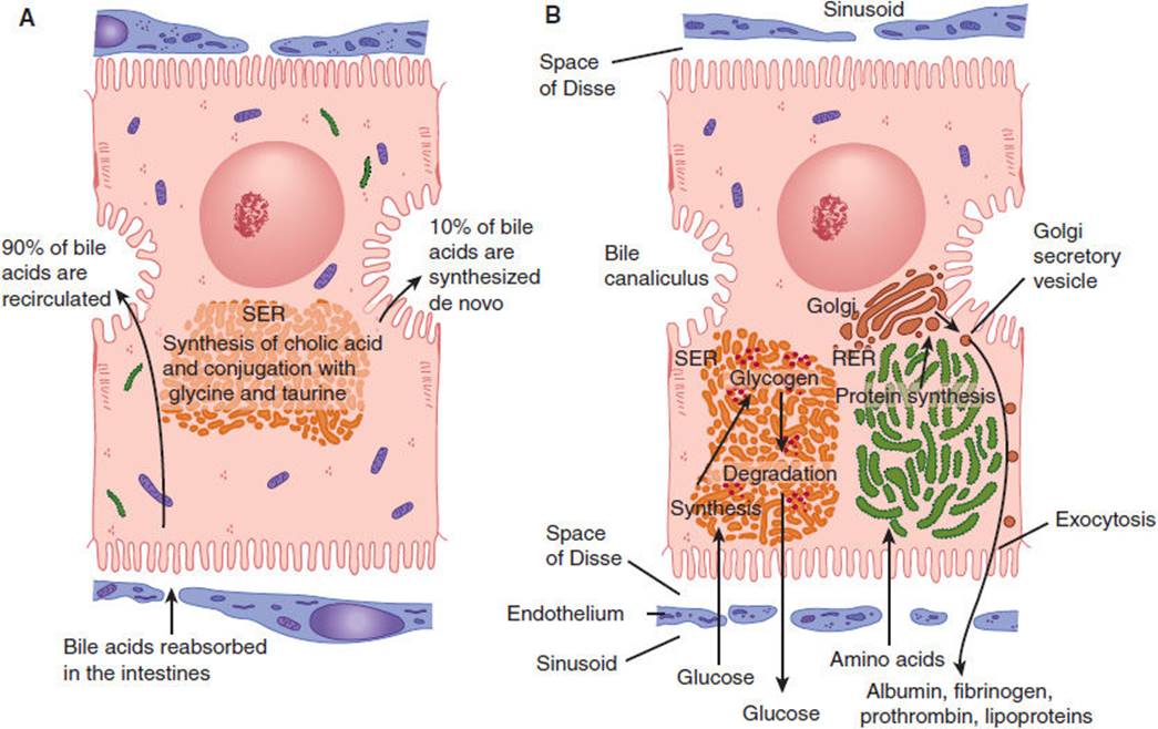
FIGURE 14-3 A: Mechanism of secretion of bile acids. About 90% of these compounds derive from bile acids absorbed in the intestinal epithelium and recirculated to the liver. The remainder are synthesized in the liver by conjugating cholic acid with the amino acids glycine and taurine. This process occurs in the smooth endoplasmic reticulum (SER). B: Protein synthesis and carbohydrate storage in the liver. Protein synthesis occurs in the rough endoplasmic reticulum, which explains why liver cell lesions or starvation leads to a decrease in the amounts of albumin, fibrinogen, and prothrombin in a patient’s blood. In several diseases, glycogen degradation is depressed, with abnormal intracellular accumulation of this compound. (SER, smooth endoplasmic reticulum; RER, rough endoplasmic reticulum.) (Redrawn, with permission, from Junqueira LC et al. Basic Histology, 10th ed. McGraw-Hill, 2003.)
Effects of Hepatocyte Dysfunction
In view of this organization, it is perhaps not surprising that hepatocyte dysfunction can sometimes involve disruption of bile flow (cholestasis) with relative preservation of other functions. There is, however, no clear line between the consequences of disturbed apical and basolateral functions: Cholestasis, although initially a disorder of apical bile flow, is ultimately manifested at the basolateral surface. This is because it is at the basolateral surface that bilirubin and other substances to be excreted across the apical plasma membrane into the bile must first be taken up from the bloodstream. Similarly, disruption of energy metabolism or protein synthesis, although initially impinging on the secretory and metabolic processes of the hepatocyte, will ultimately affect the bile transport machinery in the apical plasma membrane as well.
Capacity for Regeneration
Although the normal liver contains very few cells in mitosis, when hepatocytes are lost, poorly understood mechanisms stimulate proliferation of the remaining hepatocytes. This is why in most cases of fulminant hepatic failure with massive hepatocellular death, if the patient survives the acute period of hepatic dysfunction (usually with medical therapy in the hospital), recovery will be complete. Similarly, surgical resection of liver tissue is followed by proliferation of the remaining hepatocytes (hyperplasia). Numerous growth factors (eg, HGF, TGF-α) and cytokines (eg, TNF, IL-1, IL-6) are involved in positioning the liver on a continuum between cell proliferation and cell death.
CHECKPOINT
4. From which vascular beds do the hepatic central veins derive their blood flow?
5. Why is the liver a major site for metastasis of malignant neoplasms from other parts of the body?
6. What cell types make up the liver, and what are their distinguishing characteristics?
7. What is the difference between the lobule concept and the acinus concept of liver subarchitecture?
8. What are some physiologic consequences of functional zonation in the liver?
9. What activities are found in zone 1 hepatocytes? In zone 3 hepatocytes?
10. What structures normally maintain the separation of apical and basolateral plasma membrane domains of the hepatocyte?
11. What happens to the remaining hepatocytes when part of the liver is surgically resected?
LIVER BLOOD FLOW & ITS CELLULAR BASIS
The portal blood flow, being venous in nature, is normally under low hydrostatic pressure (about 10 mm Hg). Accordingly, there must be little resistance to its flow within the liver, allowing the blood to percolate through the sinusoids and achieve maximal contact—for exchange of substances—with hepatocytes. Two unique features—fenestrations in the endothelial cells and lack of a typical basement membrane between endothelial cells and hepatocytes—aid in making the liver a low-pressure circuit for the flow of portal blood. These features are altered in cirrhosis, resulting in increased portal pressure and profound changes in liver blood flow, with devastating clinical consequences.
Fenestrations are spaces between the endothelial cells that make up the walls of the portal capillary system, which allow plasma and its proteins, but not red blood cells, free and direct access to the surface of the hepatocytes. This feature is crucial to the liver’s function of uptake from and secretion into the bloodstream. This feature also contributes to the efficiency of the liver as a filter of portal blood. Most of the capillary beds in the body lack such fenestrations.
PHYSIOLOGY
The diverse functions of the liver are listed as four broad categories in Table 14-2. Although there is considerable overlap between them, systematic consideration of each category is a useful way of approaching the patient with liver disease.
TABLE 14-2 Functions of the normal liver.

Energy Generation & Substrate Interconversion
Much of the body’s carbohydrate, lipid, and protein is synthesized, metabolized, and interconverted in the liver; products are removed from or released into the bloodstream in response to the energy and substrate needs of the body.
A. Carbohydrate Metabolism
After a meal, the liver achieves net glucose consumption (eg, for glycogen synthesis and generation of metabolic intermediates via glycolysis and the tricarboxylic acid cycle). This occurs as a result of a confluence of several effects. First, the levels of substrates such as glucose increase. Second, the levels of hormones that affect the amount and activity of metabolic enzymes change. Thus, when blood glucose levels increase, the ratio of insulin to glucagon in the bloodstream also increases. The net effect is increased glucose utilization by the liver. In times of fasting (low blood glucose) or stress (when higher blood glucose is needed), hormone and substrate levels in the bloodstream drive metabolic pathways of the liver responsible for net glucose production (eg, the pathways of glycogenolysis and gluconeogenesis). As a result, blood glucose levels are raised to, or maintained in, the normal range in spite of wide and sudden changes in the rate of glucose input (eg, ingestion and absorption) and output (eg, utilization by tissues) from the bloodstream (Figure 14-4).
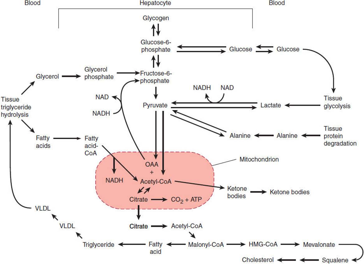
FIGURE 14-4 Pathways of hepatic carbohydrate and lipid metabolism. (ATP, adenosine triphosphate; CoA, co-enzyme A; HMG, hepatic 3-methylglutaryl; OAA, oxaloacetic acid; VLDL, very low-density lipoproteins.) (Redrawn from Schwartz CC. Hepatic metabolism. In: Kelley WN, ed. Textbook of Medicine. Lippincott, 1989.)
B. Protein Metabolism
Related to its important role in protein metabolism, the liver is a major site for processes of oxidative deamination and transamination (Figure 14-5). These reactions allow amino groups to be shuffled among molecules in order to generate substrates for both carbohydrate metabolism and amino acid synthesis. Likewise, the urea cycle allows nitrogen to be excreted in the form of urea, which is much less toxic than free amino groups in the form of ammonium ions. Impairment of this function in liver disease is discussed in greater detail later.

FIGURE 14-5 Urea cycle. Dashed lines signify pathways whose extent of involvement varies from patient to patient depending on genetic, dietary, and other factors. (Redrawn, with permission, from Powers-Lee SG et al. Urea synthesis and ammonia metabolism. In: Arias IM et al, eds. The Liver: Biology and Pathology, 3rd ed. Raven Press, 1994.)
C. Lipid Metabolism
The liver is the center of lipid metabolism. It manufactures nearly 80% of the cholesterol synthesized in the body from acetyl-CoA via a pathway that connects metabolism of carbohydrates with that of lipids (Figure 14-4). Moreover, the liver can synthesize, store, and export triglycerides (Figure 14-4). The liver is also the site of keto acid production via the pathway of fatty acid oxidation that connects lipid catabolism with activity of the tricarboxylic acid cycle.
In the process of controlling the body’s level of cholesterol and triglycerides, the liver assembles, secretes, and takes up various lipoprotein particles (Figure 14-6). Dietary fat is first absorbed into the small intestine then packaged into chylomicrons. Following removal of triglycerides, the chylomicron remnant is taken up by the liver via low-density lipoprotein (LDL) receptor–mediated endocytosis. To distribute lipids systemically, very-low-density lipoproteins (VLDLs) are secreted by the liver and transport triglycerides and cholesterol to adipose tissue for storage or to other tissues for immediate use. As triglycerides are removed, the structure of VLDL particles is modified by loss of lipid and protein components rendering intermediate-density lipoprotein (IDL) and further downstream LDL. LDL particles are then returned to the liver via the LDL receptor. On the other hand, high-density lipoproteins (HDLs), a lipoprotein synthesized and secreted from the liver, scavenge excess cholesterol and triglycerides from other tissues and from the bloodstream, returning them to the liver where they are excreted. Thus, secretion of HDL and removal of LDL are both mechanisms by which cholesterol in excess of that needed by various tissues is removed from the circulation (Figures 14–6B and 14–6C).
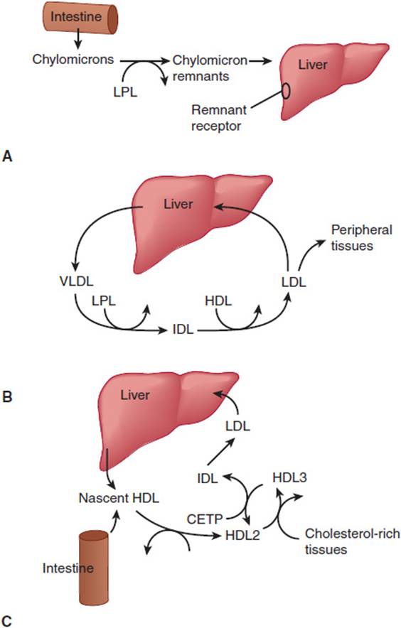
FIGURE 14-6 Lipoprotein metabolism in the liver. A: Exogenous fat transport pathway. B: Endogenous fat transport pathway. C: Pathway of reverse cholesterol transport. In each of these three pathways, lipoprotein particles are used to solubilize cholesteryl esters (and triglyceride), either for the purpose of import from the GI tract (A), distribution to various tissues (B), or transport to the liver for excretion in bile (C). During their circulation, specific lipoprotein particles are transformed by addition and removal of apoproteins and by the action of enzymes in plasma or in tissues (eg, LPL, lipoprotein lipase; CETP, cholesteryl ester transfer protein). Intermediate-density lipoproteins (IDL) are intermediates in the conversion of VLDL to LDL. (HDL, high-density lipoproteins; LDL, low-density lipoproteins.) (Redrawn from Breslow JL. Genetic basis of lipoprotein disorders. J Clin Invest. 1989;84:373.)
Synthesis & Secretion of Plasma Proteins
The liver manufactures and secretes many of the proteins found in plasma, including albumin, several of the clotting factors, a number of binding proteins, and even certain hormones and hormone precursors. By virtue of the actions of these proteins, the liver has important roles in maintaining plasma oncotic pressure (serum albumin), coagulation (clotting factor synthesis and modification), blood pressure (angiotensinogen), growth (insulin-like growth factor-1), and metabolism (steroid and thyroid hormone–binding proteins).
Solubilizing, Transport, & Storage Functions
The liver plays an important role in solubilizing, transporting, and storing a variety of very different substances that would otherwise be difficult for the tissues to obtain or to move in and out of cells. Specific cells in the liver perform these functions by manufacturing specialized proteins that serve as receptors, binding proteins, or enzymes.
A. Enterohepatic Circulation of Bile Acids
Bile is a detergent-like substance synthesized by the liver that permits a variety of otherwise insoluble substances to be dissolved in an aqueous environment for transport into or out of the body. Bile acids are a major component of bile and are recycled via the so-called enterohepatic circulation between the liver and the intestines. After synthesis and active transport from hepatocyte cytoplasm into the bile canaliculus (across the apical plasma membrane of the hepatocyte), bile is collected in the biliary tract (and sometimes stored in the gallbladder) and excreted via the common bile duct into the duodenum. While still in the cytoplasm of the hepatocyte, many bile acids are conjugated to sugars, which increases their water solubility. Once in the duodenum, bile acids serve to solubilize lipids, facilitating digestion and absorption of fats. In the terminal ileum, both conjugated and deconjugated bile acids are taken up and transported from enterocytes to portal blood flow. Portal blood returns them to the liver, where specialized bile acid transporters (predominantly sodium taurocholate cotransporter, or Ntcp) return them to the hepatocyte cytosol via the basolateral plasma membrane facing the space of Disse. There they are subject to reconjugation and secretion across the apical membrane along with other components (eg, pigment, cholesterol) to form new bile. Thereafter, they engage in another cycle of enterohepatic transport.
B. Drug Metabolism and Excretion
Most of the enzymes that catalyze metabolic processes necessary for the detoxification and excretion of drugs and other substances are located in the smooth endoplasmic reticulum of hepatocytes. These pathways are used not only for metabolism of exogenous drugs but also for many endogenous substances that would otherwise be difficult for cells to excrete (eg, bilirubin and cholesterol). In most cases, this metabolism involves the conversion of hydrophobic (lipophilic) substances (which are difficult to excrete from cells because they tend to partition into cellular membranes) into more hydrophilic (polar) substances. This process involves catalysis of covalent modifications to make the substance more charged, so that it will partition more readily into an aqueous medium or at least be solubilized sufficiently in bile. As a result of these processes, collectively termed biotransformations, some substances that would otherwise be retained in cellular membranes can be excreted directly in the urine or transported into the bile for excretion in feces.
C. Phases of Biotransformation
Biotransformation generally occurs in two phases. Phase I reactions involve oxidation reductions in which an oxygen-containing functional group is added to the substance to be excreted. While oxidation itself does not necessarily have a major effect on water solubility, it usually introduces into the drug a reactive “handle” that makes possible other reactions that do render the modified substance water soluble. These phase II reactions usually involve covalent attachment of the drug to a water-soluble carrier molecule such as the sugar glucuronic acid or the peptide glutathione. Unfortunately, by making substances more chemically reactive, phase I oxidation reactions often convert mildly toxic drugs into more toxic reactive intermediates. If conjugation by phase II enzymes is impaired for some other reason, the reactive intermediate can sometimes react with and damage other cellular structures. This feature of drug detoxification has important clinical implications.
D. Role of Apolipoprotein in Solubilization and Transport of Lipids
The detoxification and bile transport pathways allow hepatocytes to convert a wide range of hydrophobic low-molecular-weight substances (eg, drugs and bilirubin) into a more hydrophilic and hence water-soluble form in which they can be excreted (eg, in bile or by the kidney). However, these are not the only solubilization challenges facing the body. The body also needs a mechanism that makes lipids available to various tissues (eg, to synthesize membranes) and one that removes any excess lipid the tissues do not use. For these processes to occur, lipid must be solubilized in a dispersed form that can be carried through the bloodstream. For this purpose, hepatocytes synthesize a class of specialized apolipoproteins. Apolipoproteins assemble into a variety of lipoprotein particles that transport lipids to and from various tissues by receptor-mediated endocytosis (see prior discussion of lipid metabolism).
E. Role in Production of Binding Proteins
Various cells in the liver synthesize proteins that bind certain substances very tightly (eg, some vitamins, minerals, and hormones). In some cases, this allows their transport in the bloodstream, where they would otherwise not be soluble (eg, steroids bound to steroid-binding globulin, which is synthesized and secreted by hepatocytes). In other cases, binding proteins made by the liver (eg, thyroid hormone–binding globulin) allow transport of specific substances (eg, thyroxine) in a form not fully accessible to tissues. In this way, the effective concentration of the substance is limited to its free concentration at equilibrium, and the tightly bound fraction forms a reservoir of the substance that is made available slowly as the free fraction is metabolized, thereby prolonging its half-life.
In some cases, binding proteins allow the liver to accumulate specific substances in relatively high concentrations and store them in a nontoxic form. Consider iron, for example, an essential nutrient. Free iron can be quite toxic to cells both directly as an oxidant and indirectly as an essential nutrient needed by infectious agents. Control of body iron occurs at the level of the enterocyte in the duodenum (see Chapter 13). Thus, the primary defect in the iron overload disorder hemochromatosis probably involves the enterocyte. Nevertheless, the liver has the responsibility of making a variety of proteins crucial for the binding and metabolism of iron. Through the actions of these proteins, the body gets the iron it needs without allowing excess free iron to cause damage or support pathogens.
Transferrin is an iron-binding protein synthesized and secreted into the bloodstream by the liver. On binding of free iron at normal pH, transferrin undergoes a conformational change that gives it high affinity for a specific membrane receptor of the hepatocyte (transferrin receptor). On receptor binding, the transferrin–transferrin receptor complex is internalized into the endocytic pathway, a progressively more acidic environment. There, at low pH, iron no longer remains bound to transferrin. However, conformational changes that occur at low pH allow transferrin to maintain high-affinity binding to its receptor even in the absence of bound iron. Thus, when the receptor recycles back to the surface, it brings the “empty” transferrin with it. On presentation to the pH 7.4 environment of the bloodstream, transferrin lacking bound iron is released from the receptor, and the cycle can start over again. In this way, transferrin and its receptor keep the bloodstream free of unbound iron. Meanwhile, the free iron released from transferrin in the acidic environment of the endosome is transported into the cytoplasm of the hepatocyte, where it binds to ferritin, a cytoplasmic iron storage protein. This provides a reservoir that can be mobilized in response to the body’s needs but makes iron inaccessible to pathogens and keeps it from causing direct toxic effects. Similar dynamics of plasma-binding proteins, receptors, or cytosolic storage proteins occur for many other substances, including fat-soluble vitamins and steroid hormones.
Whereas most solubilization functions are performed in hepatocytes, some of the binding and storage functions involve accessory cells. Thus, vitamin A storage occurs in fat droplets seen in the lipocytes of the reticuloendothelial system. Lipocytes have been implicated in the pathogenesis of chronic liver injury and cirrhosis. Injury to other cells releases cytokines, which activate the lipocytes. The lipocytes respond by proliferating and by synthesizing collagen and other basement membrane components, leading to an increase in the extracellular matrix and contributing to hepatic fibrosis.
Protective & Clearance Functions
Many of the functions of the liver already discussed (eg, drug detoxification and excretion of excess cholesterol by conversion to and solubilization in bile) can also be considered protective. Nevertheless, it is useful to conceptualize the protective function as a separate category because of its clinical importance in ameliorating the consequences of liver disease.
A. Phagocytic and Endocytic Functions of Kupffer Cells
The liver helps remove bacteria and antigens that breach the defenses of the gut to enter the portal blood and also participates in clearing the circulation of endogenously generated cellular debris. It appears that specialized receptors on the Kupffer cell surface bind to glycoproteins (via carbohydrate receptors), to material coated with immunoglobulin (via the Fc receptor), or to complement (via the C3 receptor), thus allowing damaged plasma proteins, activated clotting factors, immune complexes, senescent blood cells, and so on to be recognized and removed.
B. Endocytic Functions of Hepatocytes
Hepatocytes have a number of specific receptors for damaged plasma proteins distinct from the receptors present on Kupffer cells (eg, the asialoglycoprotein receptor that specifically binds glycoproteins whose terminal sialic acid sugar residues have been removed). The precise physiologic significance of this metabolic action remains unclear.
C. Ammonia Metabolism
Ammonia generated from deamination of amino acids is metabolized within hepatocytes into the much less toxic substance urea. Loss of this function results in altered mental status, a common manifestation of severe or end-stage liver disease.
D. Hepatocyte Synthesis of Glutathione
Glutathione is the major intracellular (cytoplasmic) reducing reagent and thus is crucial for preventing oxidative damage to cellular proteins. This molecule is a nonribosomally synthesized tripeptide (γ-glutamyl-cystinyl-glycine) that is also a substrate for many phase II drug detoxification conjugation reactions. The liver may also export glutathione for use by other tissues.
Some additional indirect liver functions (eg, its role in maintaining normal sodium and water balance) are inferred from the derangements observed in patients with liver disease, as discussed in the following section.
Tests to Assess Liver Function
A number of blood tests are commonly used to assess liver injury. Serum aspartate aminotransferase (AST) and alanine aminotransferase (ALT) are measurements of levels of enzymes normally situated within hepatocytes. Their presence in the serum is thus actually a sign of liver cell necrosis rather than an indication of liver function.
To assess liver function more directly, a number of other tests can be used. The levels of albumin, clotting factors, and bilirubin can be measured in blood samples. Each of these tests has advantages and disadvantages, and no one of them serves as an ideal sole indicator of liver function. For example, albumin has a relatively long half-life (18–20 days); its synthesis can be stimulated in excess of need, and it can be lost via the kidneys in renal disease. Furthermore, about two thirds of body albumin is located in the extravascular, extracellular space, so changes in fluid distribution can alter serum albumin concentration. Likewise, the simplest measure of clotting factor levels, the prothrombin time (PT), is a relatively insensitive measure because it does not become abnormal until more than 80% of hepatic synthetic capacity is lost. Furthermore, vitamin K deficiency, occurring in patients with nutritional deprivation, chronic cholestasis, or fat malabsorption, can prolong the PT. Serum bilirubin is a good measure of cholestasis, and determination of conjugated (direct) versus unconjugated (indirect) bilirubin provides a good assessment of whether cholestasis is intrinsic to the liver or due solely to obstruction (eg, by a stone in the common bile duct). Furthermore, cholestasis, even when caused by liver disease, often does not reflect the degree to which other hepatic functions are lost, and unconjugated hyperbilirubinemia can occur for other reasons (eg, hemolysis).
For these reasons, an accurate assessment of liver function requires several blood tests (eg, AST, ALT, albumin, PT, bilirubin) as well as clinical assessment of the patient.
The two most common schemes for grading liver function is the modified Child-Turcotte-Pugh (CTP) score (Table 14-3) and the Model for End-Stage Liver Disease (MELD score = 3.78[Ln serum bilirubin (mg/dL)] + 11.2[Ln INR] + 9.57[Ln serum creatinine (mg/dL)] + 6.43). The CTP score predicts 1- and 2-year survival, and the MELD score predicts short-term (3-month) survival. In the United States, the MELD score is currently used to prioritize patients for donor allocation and liver transplantation.
TABLE 14-3 Grading liver function using the modified Child-Turcotte-Pugh score.

CHECKPOINT
12. What are the roles of the liver in carbohydrate, protein, and lipid metabolism?
13. What are two physiologic mechanisms by which the body transports cholesterol?
14. Explain phase I and phase II reactions in drug detoxification.
15. Name and explain four clearance or protective functions of the liver.
16. What specializations allow the liver normally to be a low-pressure conduit for blood flow?
OVERVIEW OF LIVER DISEASE
TYPES OF LIVER DYSFUNCTION
Most of the clinical consequences of liver disease can be understood either as a failure of one of the liver’s four broad functions (summarized in Table 14-2) or as a consequence of portal hypertension, the altered hepatic blood flow of cirrhosis.
Hepatocyte Dysfunction
One mechanism of liver disease, particularly in acute liver injury, is dysfunction of the individual hepatocytes that make up the liver parenchyma. The pathway and extent of hepatocellular dysfunction determine the specific manifestations of liver disease. The outcomes to be anticipated when normal hepatic functions fail are described later.
Portal Hypertension
Some consequences of liver disease, particularly of cirrhosis, are best understood in terms of what we know about hepatic blood flow. Of greatest clinical importance are the existence under normal circumstances of a low-pressure portal venous capillary bed throughout the liver parenchyma and the functional zonation of portal blood flow.
When pathologic processes (eg, fibrosis) result in elevation of the normally low intrahepatic venous pressure, blood backs up and a substantial fraction of it finds alternative routes back to the systemic circulation, bypassing the liver. Thus, blood from the GI tract is, in effect, filtered less efficiently by the liver before entering the systemic circulation. The consequences of this portal-to-systemic shunting are loss of the protective and clearance functions of the liver, functional abnormalities in renal salt and water homeostasis, and a greatly increased risk of GI hemorrhage from the development of engorged blood vessels carrying venous blood bypassing the liver (eg, esophageal, gastric, umbilical varices, etc).
Even in the absence of any intrinsic parenchymal liver disease, portal-to-systemic shunting of blood can produce or contribute to encephalopathy (altered mental status resulting from failure to clear poisons absorbed from the GI tract), GI bleeding (resulting from esophageal varices), and malabsorption of fats and fat-soluble vitamins (caused by loss of enterohepatic recirculation of bile), with associated coagulopathy. In Table 14-4, the syndromes observed in liver disease are categorized as being a consequence of hepatocyte dysfunction, portal-to-systemic shunting, or both.
TABLE 14-4 Pathophysiology of syndromes of aberrant function in liver disease.

Pathophysiology of Functional Zonation
The fact that hepatocytes in the different zones of the acinus “see” blood in a particular sequence has great pathophysiologic significance. Because zone 1 hepatocytes see blood that has just left the portal venule or hepatic arteriole, they have access to the highest concentrations of various substances, both good (eg, oxygen and nutrients) and bad (eg, drugs and toxins absorbed from the GI tract). Zone 2 hepatocytes receive blood containing less of these substances, and zone 3 hepatocytes are bathed in blood largely depleted of them. However, zone 3 hepatocytes see the highest concentrations of products (eg, drug metabolites) released into the bloodstream by hepatocytes of zones 1 and 2. Thus, direct poisons have their most severe impact on zone 1 hepatocytes, whereas poisons that are generated as a result of hepatic metabolism cause more damage to those of zone 3. Similarly, because sinusoidal blood around zone 3 has the lowest oxygen concentration, hepatocytes of this zone are at greatest risk of injury under conditions of hypoxia.
MANIFESTATIONS OF LIVER DYSFUNCTION
Whether as a result of hepatocyte dysfunction or portal-to-systemic shunting, failure of normal hepatic function underlies the clinical manifestations of liver disease. An understanding of these mechanisms offers insight into the probable causes of illness in a patient with acute or chronic liver disease.
Diminished Energy Generation & Substrate Interconversion
A first category of altered liver function involves the intermediary metabolism of carbohydrates, fats, and proteins.
A. Carbohydrate Metabolism
Severe liver disease can result in either hypoglycemia or hyperglycemia. Hypoglycemia results largely from a decrease in functional hepatocyte mass, whereas hyperglycemia is a result of portal-to-systemic shunting, which decreases the efficiency of postprandial extraction of glucose from portal blood by hepatocytes, thus elevating systemic blood glucose concentration.
B. Lipid Metabolism
Disturbance of lipid metabolism in the liver can result in syndromes of fat accumulation within the liver early in the course of liver injury. Perhaps this is because the complex steps in assembly of lipoprotein particles for export of cholesterol and triglycerides from the liver are more sensitive to disruption than the pathways of lipid synthesis. Such disruption results in a buildup of fat that cannot be exported in the form of VLDL.
In certain chronic liver diseases such as primary biliary cirrhosis, bile flow decreases as a result of destruction of bile ducts. The decrease in bile flow results in decreased lipid clearance via bile, with consequent hyperlipidemia. These patients often develop subcutaneous accumulations of cholesterol termed xanthomas.
C. Protein Metabolism
Any disturbance of protein metabolism in the liver can result in a syndrome of altered mental status and confusion known as hepatic encephalopathy. As with carbohydrate metabolism, altered protein metabolism can result from either hepatocyte failure or portal-to-systemic shunting, with the net effect of elevation of blood concentrations of centrally acting toxins, including ammonia generated by amino acid metabolism.
Loss of Solubilization & Storage Functions
A. Disordered Bile Secretion
The clinical significance of bile synthesis can be seen in the prominence of cholestasis—failure to secrete bile—in many forms of liver disease. Cholestasis can occur as a result of extrahepatic obstruction (eg, from a gallstone in the common bile duct) or selective dysfunction of the bile synthetic and secretory machinery within the hepatocytes themselves (eg, from a reaction to certain drugs). The mechanisms responsible for cholestatic drug reactions are not well understood. Regardless of the mechanism, however, the clinical consequences of severe cholestasis may be profound: A failure to secrete bile results in a failure to solubilize substances such as dietary lipids and fat-soluble vitamins, resulting in malabsorption and deficiency states, respectively. Retained bile salts are also cytotoxic, but in the setting of cholestasis hepatocytes adapt to decrease uptake of bile salts by downregulating Na+-bile acid cotransporter while maintaining bile salt excretion. As a result, hepatic necrosis is minimized in predominantly cholestatic syndromes, with the typical laboratory findings of minimally elevated levels of AST and ALT in the presence of marked jaundice and high levels of bilirubin. However, prolonged exposure to bile salts in chronic cholestatic diseases such as primary biliary cirrhosis leads to portal tract cytotoxic injury and inflammation, leading eventually to fibrosis and cirrhosis.
The solubilization function of bile works both to excrete and to absorb substances. Thus, in cholestasis, endogenous substances that are normally excreted via the biliary tract can accumulate to high levels. One such substance is bilirubin, a product of heme degradation (Figure 14-7). The buildup of bilirubin results in jaundice (icterus), which is a yellow discoloration of the sclera and skin. In the adult, the most significant feature of jaundice is that it serves as a readily monitored index of cholestasis, which may occur alone or with other abnormalities in hepatocyte function (ie, as part of the presentation of acute hepatitis). In the neonate, however, elevated bilirubin concentrations can be toxic to the developing nervous system, producing a syndrome termed kernicterus.

FIGURE 14-7 The secretion of bilirubin. This water-insoluble compound is derived from the metabolism of hemoglobin in macrophages of the mononuclear phagocyte system. Glucuronyl transferase activity in the hepatocytes causes bilirubin to be conjugated with glucuronide in the smooth endoplasmic reticulum, forming a water-soluble compound. Accumulation of bilirubin and bilirubin glucuronide in the tissues produces jaundice. Several defective processes in the hepatocytes can cause diseases that produce jaundice: a defect in the capacity of the cell to trap and absorb bilirubin (rectangle 1), the inability of the cell to conjugate bilirubin because of a deficiency in glucuronyl transferase (rectangle 2), or problems in the transfer and excretion of bilirubin glucuronide into the biliary canaliculi (rectangle 3). One of the most frequent causes of jaundice, however—unrelated to hepatocyte activity—is the obstruction of bile flow as a result of gallstones or tumors of the pancreas. This causes jaundice primarily as a result of accumulation of bilirubin glucuronide in the tissues. (Redrawn, with permission, from Junqueira LC et al, eds. Basic Histology, 10th ed. McGraw-Hill, 2003.)
Similarly, cholesterol is normally excreted either by conversion into bile acids or by forming complexes, termed micelles, with preexisting (recycled) bile acids. In cholestasis, the resultant buildup of bile acids can lead to their deposition in the skin. This is believed to cause intense itching, or pruritus. Data suggest that, in at least some patients, cholestasis results in altered levels of endogenous opioids. Instead of skin deposition of bile acids altered endogenous opioid-mediated neurotransmission may be responsible for pruritus. Disorders of bile production are a basis for the formation of cholesterol gallstones. Nevertheless, as mentioned, other hepatocyte functions are often relatively well preserved in the face of significant cholestasis. The syndromes that produce jaundice are summarized in Table 14-5.
TABLE 14-5 Laboratory findings in the diff erential diagnosis of jaundice.

Hemolysis causes an unconjugated hyperbilirubinemia because the hepatic capacity to take up and conjugate bilirubin is exceeded. Gilbert syndrome reflects a genetic defect in bilirubin conjugation. Thus, the findings in blood and urine are different from what is observed in hemolytic jaundice even though the pathway of bilirubin metabolism is backed up at a similar initial point. Extrahepatic biliary tract obstruction presents the other extreme, in which the actual pathway of bile formation is entirely intact, at least initially. In obstruction, the bilirubin level in the urine is high because the backed-up metabolite is conjugated and hence much more water soluble than unconjugated bilirubin, which accumulates in hemolysis. Most forms of jaundice that result from liver dysfunction caused by hepatocellular damage reflect variable degrees of overlap between unconjugated and conjugated hyperbilirubinemia.
B. Impaired Drug Detoxification
Two features of the mechanisms of drug detoxification are of particular clinical importance. One is the phenomenon of enzyme induction. It is observed that the presence in the bloodstream of any of the large class of drugs inactivated by phase I enzymes increases the amount and activity of these enzymes in the liver. This property of enzyme induction makes physiologic sense (as a response to the body’s need for increased biotransformation) but can have undesired effects as well: A patient who chronically consumes large amounts of a substance that is metabolized by phase I enzymes (eg, ethanol) will induce high levels of these enzymes and thus speed up the metabolism of other substances metabolized by the same detoxifying enzymes (eg, antiseizure or anticoagulant medications, resulting in subtherapeutic blood levels of the drugs).
A second clinically important phenomenon in drug metabolism is that phase I reactions often convert relatively benign compounds into more reactive and hence more toxic ones. Normally, this heightened reactivity of phase I reaction products serves to facilitate phase II reactions, making detoxification more efficient. However, under certain conditions when phase II reactions are impaired (eg, during glutathione deficiency from inadequate nutrition), continued phase I enzyme activity can cause increased liver injury. This is because the products of many phase I reactions, in the absence of glutathione, react with and damage cellular components. Such damage rapidly kills the hepatocyte.
Thus, the combined effects of certain common conditions can make the individual abnormally sensitive to the toxic effects of drugs. For example, the combination of induced phase I activity (eg, caused by alcoholism) with low phase II activity (eg, caused by low glutathione levels from nutritional deprivation) can result in heightened generation of reactive intermediates with an inadequate capacity to conjugate and detoxify them. A classic example of this phenomenon is acetaminophen toxicity. As little as 2.5 g of acetaminophen can produce significant liver damage in such susceptible individuals, whereas normal individuals have the capacity to detoxify 10 g/d or more. Table 14-6lists common drugs and chemicals that cause morphologically distinctive changes in the liver.
TABLE 14-6 Principal alterations of hepatic morphology produced by some commonly used drugs and chemicals.


C. Lipoprotein Dynamics and Dyslipidemias
The liver’s role in lipid metabolism is illustrated by the genetic defect causing familial hypercholesterolemia. Lack of a functional LDL receptor in such cases renders the liver unable to clear LDL cholesterol from the bloodstream, resulting in markedly elevated serum cholesterol and accelerated atherosclerosis and coronary artery disease. Heterozygotes with one normal LDL receptor allele can be treated with drugs (eg, HMG-CoA reductase inhibitors) that inhibit endogenous cholesterol synthesis and thus upregulate LDL receptor levels. However, there is no effective drug therapy for homozygotes because they have no normal LDL receptors. Hepatic transplantation is effective therapy for homozygous familial hypercholesterolemia because it provides a genetically different liver with normal LDL receptors.
In acquired liver diseases, the serum cholesterol is elevated in biliary tract obstruction as a result of blockage of cholesterol excretion in bile, and it is diminished in severe alcoholic cirrhosis, in which fat malabsorption prevents cholesterol intake.
D. Altered Hepatic Binding and Storage Functions
Liver disease influences the liver’s ability to store various substances. As a result, patients with liver disease are at high risk for certain deficiency states such as folic acid and vitamin B12 deficiency. Because these vitamins are needed for DNA synthesis, their deficiency results in macrocytic anemia (low red blood cell count with large red cells reflecting abnormal nuclear maturation), a common finding in patients with liver disease.
Diminished Synthesis & Secretion of Plasma Proteins
The clinical significance of liver protein synthesis and secretion derives from the wide range of functions carried out by these proteins. For example, because albumin is the major contributor to plasma oncotic pressure, hypoalbuminemia as a consequence of liver disease or nutritional deficiency presents with marked edema formation. Other important proteins synthesized and secreted by the liver include clotting factors and hormone-binding proteins.
Loss of Protective & Clearance Functions
A crucial protective function of the liver is its role as a filter of blood from the GI tract, by which various substances are removed from portal blood before it reenters the systemic circulation.
A. Clearance of Bacteria and Endotoxins
Clearance of bacteria by Kupffer cells of the liver is the final line of defense in keeping gut-derived bacteria and their endotoxins out of the systemic circulation. Loss of this capacity in liver disease as a result of portal-to-systemic shunting may help to explain why, in patients with severe liver disease, infections can rapidly become systemic and result in sepsis and the effects of endotoxins.
B. Altered Metabolism of Ammonia
Impairment of the liver’s ability to detoxify ammonia to urea leads to hepatic encephalopathy that manifests as an altered mental status. While several overlapping mechanisms have been implicated in the pathogenesis of hepatic encephalopathy, ammonia is the most well-characterized neurotoxin that precipitates encephalopathy. Ammonia is primarily produced from deamination of glutamine by glutaminase in the enterocytes of the small bowel and colon but is also produced from hydrolysis of urea via bacterial catabolism of nitrogenous sources including dietary protein and urea. The intact liver clears nearly all of the ammonia that arrives via the portal vein by converting it to glutamine, which prevents entry of ammonia into the systemic circulation. Increased ammonia in the bloodstream can be the direct consequence of impaired liver function (either acute hepatocellular dysfunction or progressive chronic disease) and/or significant shunting of blood around the liver (portal-to-systemic shunting) with consequent bypass of its clearance mechanisms.
Precipitants of increased ammonia levels in the bloodstream and consequent altered mental status include the following: 1) increased protein intake (hydrolysis of urea via bacterial catabolism of nitrogenous sources); 2) GI bleeding (increased levels of ammonia and other nitrogenous substances are produced by breakdown of blood proteins by GI tract microbes); and 3) the systemic inflammatory response to infection (stimulation of proinflammatory cytokine release and endogenous protein catabolism, leading to elevated ammonia production). Thus, once increased protein intake has been excluded, the development of encephalopathy in a patient with chronic liver disease calls for investigation of possible acute GI bleeding as well as search for a potentially catastrophic infection. Pending the outcome of diagnostic studies (eg, serial hemoglobin and hematocrit measurements and cultures of blood, urine, and ascitic fluid), therapy is aimed at diminishing the absorption of ammonia and other noxious substances from the GI tract. When the patient is given the nonabsorbable carbohydrate lactulose, its metabolism by microbes creates an acidic environment. Ammonia is trapped as charged NH4+ species in the gut lumen and excreted by the resultant osmotic diarrhea. In this way, the toxin is prevented from ever entering the portal circulation, and the patient’s mental status gradually improves. Lactulose also selects for a gut bacterial flora that produces less ammonia. Antibiotics, and in particular rifaximin, have been used in conjunction with lactulose for treatment of hepatic encephalopathy. Antibiotics are thought to act by decreasing the intestinal production and absorption of ammonia via modulation of the intestinal microbiota and prevention of the bacterial translocation across the gut mucosal surface.
Furthermore, the resulting elevations in blood ammonia and other nitrogen-containing compounds can upregulate peripheral receptors for endogenous benzodiazepine-like products. These effects may also contribute to altered systemic hemodynamics in liver disease.
C. Altered Hormone Clearance in Liver Disease
Normally, the liver removes from the bloodstream the fraction of steroid hormones not bound to steroid hormone-binding globulin. On uptake by hepatocytes, these steroids are oxidized, conjugated, and excreted into bile, where a fraction undergoes enterohepatic circulation. In liver disease accompanied by significant portal-to-systemic shunting, steroid hormone clearance is diminished, extraction of the circulated enterohepatic fraction is impaired, and enzymatic conversion of androgens to estrogens (peripheral aromatization) is increased. The net effect is an elevation of blood estrogens, which in turn alters hepatocyte protein synthesis and secretion together with activation of the metabolizing P450 enzymes. Synthesis of some hepatic proteins increases, whereas synthesis of others is diminished. Enzymatic P450 activity increases as the liver attempts to partially compensate for the higher blood estrogen levels by increased metabolism. Thus, male patients with liver disease display both gonadal and pituitary suppression as well as feminization.
Sodium & Water Balance
Patients with liver disease often display renal abnormalities and complications, most commonly sodium retention and difficulty excreting water. An intrinsic renal lesion is apparently not involved, because the kidneys of patients with liver disease typically function normally when transplanted into patients whose liver is normal. Instead, renal abnormalities associated with liver disease are functional, occurring because liver disease induces altered intravascular pressures and perhaps because of elevated nitric oxide levels or loss of as yet poorly understood factors secreted from the liver or the endothelium. By whatever homeostatic mechanisms, intravascular volume is perceived as being inadequate when it is actually only maldistributed. Renal mechanisms of salt and water retention are then stimulated to correct what has been sensed as volume depletion. Some of the factors influencing renal sodium retention in liver disease are summarized in Table 14-7. Patients with severe liver disease are at risk for renal failure related to these hemodynamic alterations.
TABLE 14-7 Factors influencing renal sodium retention in liver disease.

CHECKPOINT
17. Under what circumstances is hypoglycemia seen in liver disease?
18. Name three clinical consequences of cholestasis.
19. The development of hepatic encephalopathy in a patient with chronic liver disease should lead you to investigate what possible precipitating factors?
20. By what mechanisms can coagulation defects be a consequence of liver disease?
21. What is an explanation for hypogonadism in male patients with liver disease?
PATHOPHYSIOLOGY OF SELECTED LIVER DISEASES
ACUTE HEPATITIS
Acute hepatitis is an inflammatory process causing liver cell death either by necrosis or by triggering apoptosis (programmed cell death). A wide range of clinical entities can cause global hepatocyte injury of sudden onset. Worldwide, acute hepatitis is most commonly caused by infection with one of several types of viruses. Although these viral agents can be distinguished by serologic laboratory tests based on their antigenic properties, all produce clinically similar illnesses. Other less common infectious agents can result in liver injury (Table 14-1). Acute hepatitis is also sometimes caused by exposure to drugs (eg, isoniazid) or poisons (eg, ethanol).
Clinical Presentation
The severity of illness in acute hepatitis ranges from asymptomatic and clinically unapparent to fulminant and potentially fatal. The clinical presentation of acute hepatitis can also be quite variable. Some patients are relatively asymptomatic, with abnormalities noted only by laboratory studies. Others may have a range of symptoms and signs, including anorexia, fatigue, weight loss, nausea, vomiting, right upper quadrant abdominal pain, jaundice, fever, splenomegaly, and ascites. The extent of hepatic dysfunction can also vary tremendously, correlating roughly with the severity of liver injury. The relative extent of cholestasis versus hepatocyte necrosis is also highly variable. The potential interrelationship of acute hepatitis, chronic hepatitis, and cirrhosis is illustrated in Figure 14-8.
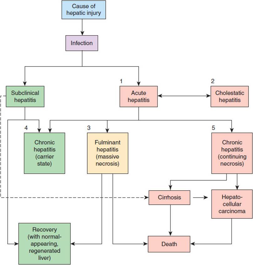
FIGURE 14-8 Clinical syndromes associated with hepatitis: acute hepatitis (1), which is sometimes associated with intrahepatic cholestasis (2). Fulminant hepatitis (3) is associated with massive necrosis and has a high mortality rate. Chronic viral hepatitis may lead to a carrier state without (4) or with (5) continuing hepatocyte necrosis. Chronic hepatitis associated with continuing necrosis often progresses to cirrhosis, whereas that associated simply with a carrier state does not. (Redrawn, with permission, from Chandrasoma P et al, eds. Concise Pathology, 3rd ed. Originally published by Appleton & Lange. Copyright © 1998 by The McGraw-Hill Companies, Inc.)
Etiology
A. Viral Hepatitis
Acute hepatitis is commonly caused by one of five major viruses: hepatitis A virus (HAV), hepatitis B virus (HBV), hepatitis C virus (HCV), hepatitis D virus (HDV), and hepatitis E virus (HEV). Table 14-8summarizes important characteristics of these viral agents. Other viral agents that can cause acute hepatitis, though less commonly, include the Epstein-Barr virus (cause of infectious mononucleosis), cytomegalovirus, varicella virus, measles virus, herpes simplex virus, rubella virus, and yellow fever virus. A newly discovered DNA virus, SEN virus, may be associated with transfusion-associated acute hepatitis not attributable to other viruses. HAV, a small RNA virus, causes liver disease both by direct killing of hepatocytes and by stimulating the host’s immune response to infected hepatocytes. It is spread by the fecal-oral route from infected individuals. Although most cases are mild, hepatitis A occasionally causes fulminant liver failure and massive hepatocellular necrosis, resulting in death. Regardless of the severity, patients who recover do so completely, show no evidence of residual liver disease, and have antibodies that protect them from reinfection.
TABLE 14-8 Characteristics of various types of viral hepatitis.

HBV is a DNA virus that is transmitted by sexual contact or by contact with infected blood or other bodily fluids. Perinatal and early childhood transmission is the most common mode of HBV acquisition worldwide, whereas sexual transmission is most common among adults in the United States. This virus does not kill the cells it infects. Rather, the infected hepatocytes die almost exclusively as a consequence of attack by the immune system after recognition of viral antigens on the hepatocyte surface. Although most cases of hepatitis B infection are asymptomatic or produce only mild disease, an excessive immune response can result in acute liver injury and even liver failure. In a minority of those infected as adults but a majority of individuals infected at birth, the immune response is inadequate to clear the virus and chronic hepatitis B develops. The incidence of infection has significantly declined in the era of HBV vaccination, although prevalence remains high, in part because of immigration of infected patients from endemic countries. Although the true burden of chronic hepatitis B infection in the United States is unknown, it is estimated that 1.25 million Americans are infected with HBV, with a likely higher prevalence among those who are foreign-born. Additionally, complications of HBV-induced liver disease result in 3000–5000 deaths each year in the United States.
HCV is a RNA virus, also transmitted by blood and body fluids, causes a form of hepatitis similar to HBV infection but with a far greater proportion of cases (60–85%) progressing to chronic hepatitis. Acute infection can be characterized by mild to moderate illness but is usually asymptomatic. However, chronic HCV can lead to life-threatening complications including cirrhosis and hepatocellular carcinoma (HCC), usually after decades of infection. It is estimated that between 2.7 and 3.9 million Americans are infected with HCV, many unaware of their infection, and the rate of attributable mortality is rising at 12,000 deaths each year. The Centers for Disease Control and Prevention (CDC) estimates that persons born during 1945–1965 account for approximately three fourths of all HCV infections in the United States. End-stage liver disease due to HCV is the most common indication for liver transplantation in the United States.
HDV, also known as delta agent, is a defective RNA virus that requires helper functions of HBV to cause infection. Thus, individuals who are chronically infected with HBV are at high risk for HDV infection, whereas those who have been vaccinated against HBV are at no risk. HDV infection occurs either as coinfection with HBV or superinfection in the setting of chronic HBV. HDV infection causes a much more severe form of hepatitis both in terms of the proportion of fulminant cases and in the percentage of cases that progress to chronic hepatitis. In North America, HDV coinfection primarily occurs in high-risk groups such as injection drug users and hemophiliacs, and in up to 9% of those high-risk patients who have chronic HBV infection. In the United States, the prevalence of HDV coinfection in the general HBV-infected population is not well known.
HEV is an unclassified RNA virus, and like HAV, it is spread via the fecal-oral route. The clinical disease is generally benign and self-limited, resembling hepatitis A, but HEV infection may result in acute liver failure in pregnant women. More recently, it has been recognized that HEV is an underdiagnosed cause of many cases of “idiopathic” hepatitis in nonpregnant patients, cases of presumed drug-induced liver injury, and even chronic hepatitis in immune-compromised hosts. HEV remains an under-recognized clinical entity because there is no laboratory test for HEV viral load in routine clinical practice.
B. Toxic Hepatitis
Most cases of drug-induced liver injury present as acute hepatitis, although some present as cholestasis or other patterns (Table 14-6). The incidence of drug-induced hepatitis has been rising. Although fewer than 10% of drug-induced liver injury cases progress to acute liver failure, acetaminophen is now the most common cause of acute liver failure in the United States and the United Kingdom. Hepatic toxins can be further subdivided into those for which hepatic toxicity is predictable and dose dependent for most individuals (eg, acetaminophen) and those that cause unpredictable (idiosyncratic) reactions without relationship to dose. The pathogenesis of drug-induced liver injury is not well understood; Table 14-9 and Figure 14-9 summarize speculations on the mechanisms of idiosyncratic and dose-related drug-induced liver injury. Idiosyncratic reactions to drugs may be due to genetic predisposition in susceptible individuals to certain pathways of drug metabolism that generate toxic intermediates. Prominent examples of drugs causing acute liver failure that have been withdrawn from the U.S. market include bromfenac, a nonsteroidal anti-inflammatory drug (NSAID), and troglitazone sulfate, a thiazolidinedione used as an insulin-sensitizing agent in diabetes mellitus. Other thiazolidinediones such as rosiglitazone and pioglitazone do not seem to have the same complication, although routine testing of transaminases has been recommended for those taking the drugs. HMG-CoA reductase inhibitors (eg, “statins”) are associated with elevated levels of transaminases in less than 3% of individuals but very rarely result in clinical acute liver failure.
TABLE 14-9 Idiosyncratic drug reactions and the cells that are affected.
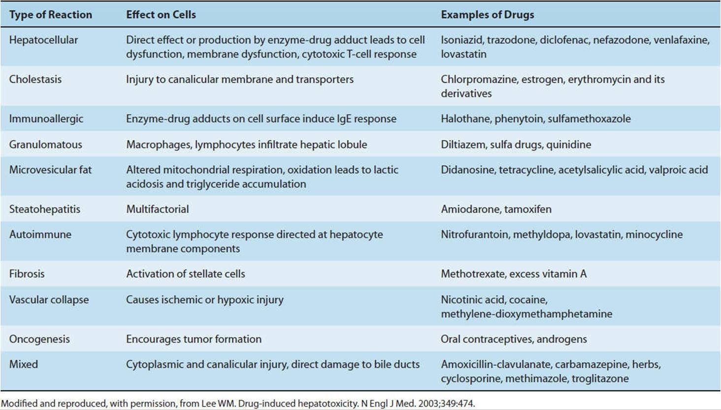
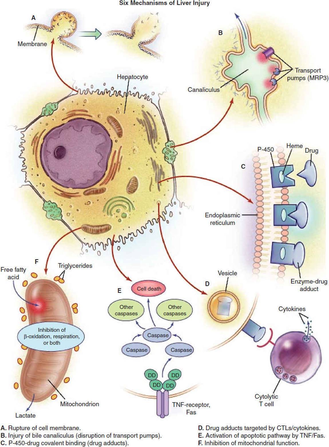
FIGURE 14-9 Potential mechanisms of drug-induced liver injury. The normal hepatocyte may be affected adversely by drugs through (A) disruption of intracellular calcium homeostasis that leads to the disassembly of actin fibrils at the surface of the hepatocyte, resulting in blebbing of the cell membrane, rupture, and cell lysis; (B) disruption of actin filaments next to the canaliculus (the specialized portion of the cell responsible for bile excretion), leading to loss of villous processes and the interruption of transport pumps such as multidrug resistance-associated protein 3 (MRP3), which in turn prevents the excretion of bilirubin and other organic compounds; (C) covalent binding of heme-containing cytochrome P450–metabolizing enzymes to the drug, thus creating nonfunctioning adducts; (D) migration of these enzyme-drug adducts to the cell surface in vesicles to serve as target immunogens for cytolytic attack by T cells, stimulating an immune response involving cytolytic T cells and cytokines; (E) activation of apoptotic pathways by tumor necrosis factor (TNF) receptor or Fas (DD denotes death domain), triggering the cascade of intercellular caspases, resulting in programmed cell death; or (F) inhibition of mitochondrial function by a dual effect on both β-oxidation and the respiratory-chain enzymes, leading to failure of free fatty acid metabolism, a lack of aerobic respiration, and accumulation of lactate and reactive oxygen species (which may disrupt mitochondrial DNA). Toxic metabolites excreted in bile may damage bile-duct epithelium (not shown). CTLs, cytolytic T lymphocytes. (Reproduced, with permission, from Lee WM. Drug-induced hepatotoxicity. N Engl J Med. 2003;349:474.)
The time course of acute hepatitis is highly variable. In hepatitis A jaundice is typically seen 4–8 weeks after exposure, whereas in hepatitis B jaundice occurs usually from 8–20 weeks after exposure (Figure 14-10). Drug- and toxin-induced hepatitis typically occurs at any time during or shortly after exposure and resolves with discontinuance of the offending agent. This is usually the case for both idiosyncratic and dose-dependent reactions.

FIGURE 14-10 (A) Serum antibody and antigen levels in hepatitis A and hepatitis B. (AST, aspartate aminotransferase, a marker for hepatocellular injury and necrosis; IgM anti-HAV, early antibody response to hepatitis A infection; IgG anti-HAV, late antibody response to hepatitis A infection; HBsAg, hepatitis B surface antigen, a marker of active viral gene expression; HBeAg, hepatitis B early antigen, a marker of infectivity.) Antibodies to the surface or early antigens (anti-HBs or anti-HBe) indicate immunity. (Redrawn, with permission, from Chandrasoma P et al, eds. Concise Pathology, 3rd ed. Originally published by Appleton & Lange. Copyright © 1998 by The McGraw-Hill Companies, Inc.) (B) Course of acute, resolving HCV infection. (ALT, Alanine aminotransferase; HCV RNA, hepatitis C viral load; anti-HCV, HCV antibody.) (Redrawn, with permission, from Hoofnagle JH. Course and outcome of hepatitis C. Hepatology. 2002 Nov;36(5 Suppl 1):S21–9.)
Acute hepatitis typically resolves in 3–6 months. Hepatic injury continuing for more than 6 months is arbitrarily defined as chronic hepatitis and suggests, in the absence of continued exposure to a noxious agent, that immune or other mechanisms are at work.
Pathogenesis
A. Viral Hepatitis
The viral agents responsible for acute hepatitis first infect the hepatocyte. During the incubation period, intense viral replication in the liver cell leads to the appearance of viral components (first antigens, later antibodies) in urine, stool, and body fluids. Liver cell death and an associated inflammatory response then ensue, followed by changes in laboratory tests of liver function and the appearance of various symptoms and signs of liver disease.
1. Liver damage—Host immunologic response plays an important though incompletely understood role in the pathogenesis of liver damage. In hepatitis B, for example, the virus is probably not directly cytopathic. Indeed, there are asymptomatic HBV carriers who have normal liver function and histologic features. Instead, the host’s cellular immune response has an important role in causing liver cell injury. Patients with defects in cell-mediated immunity are more likely to remain chronically infected with HBV than to clear the infection. Histologic specimens from patients with HBV-related liver injury demonstrate lymphocytes next to necrotic liver cells. It is thought that cytolytic T lymphocytes become sensitized to recognize hepatitis B viral antigens (eg, small quantities of hepatitis B surface antigen [HBsAg]) and host antigens on the surfaces of HBV-infected liver cells.
2. Extrahepatic manifestations—Immune factors may also be important in the pathogenesis of the extrahepatic manifestations of acute viral hepatitis. For example, in hepatitis B, a serum sickness-like prodrome characterized by fever, urticarial rash and angioedema, and arthralgias and arthritis appears to be related to immune complex–mediated tissue damage. During the early prodrome, circulating immune complexes are composed of HBsAg in high titer in association with small quantities of anti-HBs. These circulating immune complexes are deposited in blood vessel walls, leading to activation of the complement cascade. In patients with arthritis, serum complement levels are depressed, and complement can be detected in circulating immune complexes containing HBsAg, anti-HBs, immunoglobulin (Ig) G, IgM, IgA, and fibrin.
Cryoglobulinemia is a common finding in chronic hepatitis C infection. Diabetes mellitus also occurs frequently in patients with HCV infection and is now considered an extrahepatic manifestation of HCV. Although the mechanism of diabetes in HCV is not fully understood, it is thought to be predominantly related to an increase in insulin resistance. Insulin resistance also appears to improve following HCV therapy.
Immune factors are thought to be important in the pathogenesis of some clinical manifestations in patients who become chronic HBsAg carriers after acute hepatitis. For example, in patients developing glomerulonephritis with nephrotic syndrome, histopathologic investigation demonstrates deposition of HBsAg, immunoglobulin, and complement in the glomerular basement membrane. In patients developing polyarteritis nodosa, similar deposits have been demonstrated in affected small and medium-sized arteries.
Other more rare extrahepatic manifestations include papular acrodermatitis, and Guillain-Barré syndrome for HBV, and idiopathic thrombocytopenic purpura, lichen planus, Sjögren syndrome, lymphoproliferative diseases, membranoproliferative glomerulonephritis, and porphyria cutanea tarda for HCV.
B. Alcoholic Hepatitis
Ethanol has both direct and indirect toxic effects on the liver as well as effects on many other organ systems of the body. Its direct effects may result from increasing the fluidity of biologic membranes and thereby disrupting cellular functions. Its indirect effects on the liver are in part a consequence of its metabolism. Ethanol is sequentially oxidized to acetaldehyde and then to acetate, with the generation of NADH and adenosine triphosphate (ATP). As a result of the high ratio of reduced to oxidized NAD that is generated, the pathways of fatty acid oxidation and gluconeogenesis are inhibited, whereas fatty acid synthesis is promoted. Ethanol can also quantitatively and qualitatively alter the pattern of gene expression in various tissues but especially in the liver, resulting in impaired homeostasis and greater sensitivity to other toxins. These and other biochemical mechanisms may contribute to the common observation of fat accumulation in the liver of alcoholics and the tendency of hypoglycemia to develop in alcoholics whose liver glycogen has been depleted by fasting. Ethanol metabolism also affects the liver by generating acetaldehyde, which reacts with primary amino groups to inactivate enzymes, resulting in direct toxicity to the hepatocyte in which it is generated. Furthermore, proteins so modified may activate the immune system against antigens that were previously tolerated as “self.”
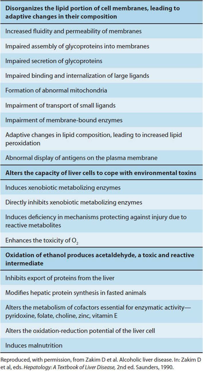
There is considerable variation among individuals in the amount of ethanol required to cause acute liver injury. Whether nutritional, genetic, or other factors are responsible for these differences has not been determined. The mechanisms thought to be responsible for ethanol-induced liver injury are listed in Table 14-10.
TABLE 14-10 Mechanisms of hepatocyte injury by ethanol.
Pathology
In uncomplicated acute hepatitis, the typical histologic findings consist of (1) focal liver cell degeneration and necrosis, with cell dropout, ballooning, and acidophilic degeneration (shrunken cells with eosinophilic cytoplasm and pyknotic nuclei); (2) inflammation of portal areas, with infiltration by mononuclear cells (small lymphocytes, plasma cells, eosinophils); (3) prominence of Kupffer cells and bile ducts; and (4) cholestasis (arrested bile flow) with bile plugs. Characteristically, although the regular pattern of the cords of hepatocytes is disrupted, the reticulin framework is preserved. This reticular framework provides scaffolding for liver cells when they regenerate.
Recovery from acute hepatitis from any cause is characterized histologically by regeneration of hepatocytes, with numerous mitotic figures and multinucleated cells, and by a largely complete restoration of normal lobular architecture.
Less commonly in acute hepatitis (1–5% of patients), there will be a more severe histologic lesion called bridging hepatic necrosis (also called subacute, submassive, or confluent necrosis). Bridging is said to occur between lobules because necrosis involves contiguous groups of hepatocytes, resulting in large areas of hepatic cell loss and collapse of the reticulin framework. Necrotic zones (“bridges”) consisting of condensed reticulin, inflammatory debris, and degenerating liver cells link adjacent portal or central areas or may involve entire lobules.
Rarely, in massive hepatic necrosis or fulminant hepatitis (<1% of patients), the liver becomes small, shrunken, and soft (acute yellow atrophy). Histologic examination reveals massive hepatocyte necrosis in most of the lobules, leading to extensive collapse and condensation of the reticulin framework and portal structures (bile ducts and vessels).
The pathology of alcoholic hepatitis is different from that of viral hepatitis in some ways. The specific pathologic features of alcoholic hepatitis include accumulation of Mallory hyalin and infiltration of polymorphonuclear leukocytes.
Clinical Manifestations
A. Viral Hepatitis
Acute viral hepatitis usually is manifested in three phases: the prodrome, the icteric phase, and the convalescent phase.
1. Prodrome—The prodrome, typically lasting 3 or 4 days, is characterized by three sets of symptoms and signs: (1) nonspecific constitutional symptoms and signs: malaise, fatigue, and mild fever; (2) GI symptoms and signs: anorexia, nausea, vomiting, altered senses of olfaction and taste (loss of taste for coffee or cigarettes), and right upper quadrant abdominal discomfort (reflecting the enlarged liver); and (3) extrahepatic symptoms and signs: headache, photophobia, cough, coryza, myalgias, urticarial skin rash, arthralgias or arthritis (10–15% of patients with HBV), and, rarely, hematuria and proteinuria.
2. Icteric phase—The icteric phase typically lasts for 1–4 weeks. The constitutional symptoms usually improve, although mild weight loss may occur. Pruritus occurs if cholestasis is severe. Right upper quadrant abdominal pain as a result of the enlarged and tender liver, which was present in the prodromal phase, continues. Splenomegaly is noted in 10–20% of patients.
Jaundice may be observed as a yellowing of the scleras, skin, or mucous membranes. Jaundice is generally not appreciated on physical examination before the serum bilirubin rises above 2.5 mg/dL (41.75 μmol/L). Direct hyperbilirubinemia is elevation of the level of conjugated bilirubin in the bloodstream. Its occurrence indicates unimpaired ability of hepatocytes to conjugate bilirubin but a defect in the excretion of bilirubin into the bile as a result of intrahepatic cholestasis or posthepatic obstructive biliary tract disease, with overflow of conjugated bilirubin out of hepatocytes and into the bloodstream.
Changes in stool color (lightening) and urine color (darkening) often precede clinically evident jaundice. This reflects loss of bilirubin metabolites from the stool as a consequence of disrupted bile flow. Water-soluble (conjugated) bilirubin metabolites are excreted in the urine, whereas water-insoluble metabolites accumulate in tissues, giving rise to jaundice. Note that in most cases of acute viral hepatitis the degree of liver impairment is sufficiently mild that jaundice does not develop.
Ecchymoses suggest coagulopathy, which may be due to loss of vitamin K absorptive capacity from the intestine (caused by cholestasis) or decreased coagulation factor synthesis. Rarely, loss of clearance of activated clotting factors triggers disseminated intravascular coagulation. Coagulopathy in which the prothrombin time can be corrected by vitamin K injections but not by oral vitamin K suggests cholestatic disease, because vitamin K uptake from the gut is dependent on bile flow. If the prothrombin time cannot be corrected with either oral or parenteral vitamin K, inability to synthesize clotting factor polypeptides (eg, as a result of massive hepatocellular dysfunction) should be suspected. Correction of prothrombin time with oral vitamin K alone suggests a nutritional deficiency rather than liver disease as the basis for the coagulopathy.
Tests for serum levels of various enzymes normally localized primarily within hepatocytes provide an indication of the extent of liver cell necrosis. For unclear reasons, perhaps related to liver cell polarity, certain forms of liver disease typically result in disproportionate elevations in some parameters. Thus, in alcoholic hepatitis but not in viral hepatitis, AST is often disproportionately elevated relative to ALT (AST:ALT ratio >2.0). One hypothesis is that this is due to pyridoxine deficiency in alcoholics. Likewise, in cholestasis, alkaline phosphatase is commonly disproportionately elevated relative to AST or ALT.
Measurement of antigen and antibody titers is a convenient way to assess whether an episode of acute hepatitis is due to viral infection. Moreover, because IgM antibodies are produced early after exposure to antigens (ie, soon after onset of illness), the presence of IgM antibodies to either HAV or to core antigen of HBV (HBcAg) is strong evidence that an episode of acute hepatitis is due to the corresponding viral infection. Several months after onset of illness, IgM antibody titers wane and are replaced by antibodies of the IgG class, indicating immunity to recurrence of infection by the same virus. Presence of hepatitis B “e” antigen (HBeAg) correlates well with a high degree of infectivity (Table 14-11). However, more sensitive DNA tests have shown low levels of viral DNA in the blood of many who are HBeAg negative and who are thus still infectious.
TABLE 14-11 Commonly encountered serologic patterns in hepatitis B infection.

Subtle or profound mental status changes are seen in fulminant hepatic necrosis. Encephalopathy is believed to be related in part to failure of detoxification of ammonia, which normally occurs through the urea cycle. Other products such as γ-aminobutyric acid (GABA) may not be metabolized. Although ammonia is a neurotoxin, it remains unclear whether it is the major agent of CNS dysfunction or whether elevated blood levels of GABA (or other compounds) may act synergistically to alter mental status because of its role as a major inhibitory neurotransmitter. In addition to encephalopathic changes caused by accumulation of toxins, acute hepatic failure is associated with encephalopathy from cerebral edema caused by increased intracranial pressure, perhaps related to alterations in the blood-brain barrier.
Renal dysfunction may complicate fulminant hepatic failure. Affected patients may develop prerenal azotemia when the glomerular filtration rate falls secondary to intravascular volume depletion. A state of intravascular volume depletion can be induced by the combination of decreased oral intake, vomiting, and formation of ascites. If uncorrected, this process can lead to acute tubular necrosis and acute kidney injury. Other causes of renal dysfunction in fulminant hepatic failure include toxins (eg, acetaminophen or Amanita poisoning) or hepatorenal syndrome. Serum creatinine is a more accurate measure than blood urea nitrogen of renal impairment in fulminant hepatic failure resulting from decreased hepatic urea production. Other complications of fulminant hepatic failure include cardiovascular dysfunction as a result of systemic vasodilation and hypotension, pulmonary edema, coagulopathy, sepsis, and hypoglycemia.
3. Convalescent phase—The convalescent phase is characterized by complete disappearance of constitutional symptoms but persistent abnormalities in liver function tests. Symptoms and signs gradually improve.
CHECKPOINT
22. Describe the range of clinical presentations of acute hepatitis.
23. Which viruses can cause hepatitis?
24. What are some extrahepatic manifestations of viral hepatitis?
25. What is the basis for the extrahepatic manifestations of viral hepatitis?
CHRONIC HEPATITIS
Chronic hepatitis is a category of disorders characterized by the combination of liver cell necrosis and inflammation of varying severity persisting for more than 6 months. It may be due to viral infection; drugs and toxins; genetic, metabolic, or autoimmune factors; or unknown causes. The severity ranges from an asymptomatic stable illness characterized only by laboratory test abnormalities to a severe, gradually progressive illness culminating in cirrhosis, liver failure, and death. Based on clinical, laboratory, and biopsy findings, chronic hepatitis is best assessed with regard to (1) distribution and severity of inflammation, (2) degree of fibrosis, and (3) etiology, which has important prognostic implications. A simplified scoring system for assessment of liver biopsies for chronic hepatitis is presented in Table 14-12.
TABLE 14-12 Three simple systems for histologic grading and staging of chronic hepatitis.

Clinical Presentation
Patients may present with fatigue, malaise, low-grade fever, anorexia, weight loss, mild intermittent jaundice, and mild hepatosplenomegaly. Others are initially asymptomatic and present late in the course of the disease with complications of cirrhosis, including variceal bleeding, coagulopathy, encephalopathy, jaundice, and ascites. In contrast to chronic persistent hepatitis, some patients with chronic active hepatitis, particularly those without serologic evidence of antecedent HBV infection, present with extrahepatic symptoms such as skin rash, diarrhea, arthritis, and various autoimmune disorders (Table 14-13).
TABLE 14-13 Extrahepatic manifestations of chronic viral hepatitis.

Etiology
Either type of chronic hepatitis can be caused by infection with several hepatitis viruses (eg, hepatitis B with or without hepatitis D superinfection and hepatitis C); a variety of drugs and poisons (eg, ethanol, isoniazid, acetaminophen), often in amounts insufficient to cause symptomatic acute hepatitis; genetic and metabolic disorders (eg, α1-antitrypsin deficiency, Wilson disease); or immune-mediated injury of unknown origin. Table 14-1summarizes known causes of chronic hepatitis. Less than 5% of otherwise healthy adults with acute hepatitis B remain chronically infected with HBV; the risk is higher in those who are immunocompromised or of young age (ranging from 90% in newborns of HBeAg-positive mothers to 25–30% in infants and children younger than 5 years). Among those chronically infected, about two thirds develop mild chronic hepatitis and one third develop severe chronic hepatitis (see later discussion). Those with HDV coinfection progress to chronic hepatitis at higher rates than seen with isolated HBV infection. HDV superinfection is also associated with a high incidence of acute liver failure. Finally, 60–85% of individuals exposed to acute hepatitis C develop chronic hepatitis, and rates are not significantly affected by age, mode of acquisition, or coinfections.
Pathogenesis
Many cases of chronic hepatitis are thought to represent an immune-mediated attack on the liver occurring as a result of persistence of certain hepatitis viruses or after prolonged exposure to certain drugs or noxious substances (Table 14-14). In some, no mechanism has been recognized. Evidence that the disorder is immune mediated is that liver biopsies reveal inflammation (infiltration of lymphocytes) in characteristic regions of the liver architecture (eg, portal versus lobular). Furthermore, a variety of autoimmune disorders occur with high frequency in patients with chronic hepatitis (Table 14-13).
TABLE 14-14 Drugs implicated in the etiology of chronic hepatitis.

A. Chronic Viral Hepatitis
Viral hepatitis is the most common cause of chronic liver disease in the United States. In approximately 5% of adult cases of HBV infection and 60–85% of hepatitis C infections, the immune response is inadequate to clear the liver of virus, resulting in persistent infection. The individual becomes a chronic carrier, intermittently producing the virus and hence remaining infectious to others. Biochemically, these patients are often found to have viral DNA integrated into their genomes in a manner that results in abnormal expression of certain viral proteins with or without production of intact virus. Viral antigens expressed on the hepatocyte cell surface are associated with class I HLA determinants, thus eliciting lymphocyte cytotoxicity and resulting in hepatitis. The severity of chronic hepatitis is largely dependent on the activity of viral replication and the response by the host’s immune system.
Chronic hepatitis B infection predisposes the patient to the development of hepatocellular carcinoma (HCC). Although in the setting of HBV infection most cases of HCC occur in the presence of cirrhosis, 10–30% of cases occur in the absence of cirrhosis or advanced fibrosis. It remains unclear whether hepatitis B infection is the initiator or simply a promoter in the process of tumorigenesis. In hepatitis C infection, HCC develops exclusively in the setting of cirrhosis.
B. Alcoholic Chronic Hepatitis
Chronic liver disease in response to some poisons or toxins may represent triggering of an underlying genetic predisposition to immune attack on the liver. In alcoholic hepatitis, however, repeated episodes of acute injury ultimately cause necrosis, fibrosis, and regeneration, leading eventually to cirrhosis (Figure 14-11). As in other forms of liver disease, there is considerable variation in the extent of symptoms before development of cirrhosis.

FIGURE 14-11 Changes in the hepatic subendothelial space during fibrosing liver injury. Cellular and matrix alterations in the space of Disse are critical events in the pathogenesis of hepatic fibrosis. The activation of lipocytes, characterized by proliferation and increased fibrogenesis, is associated with the replacement of the normal low-density matrix with a high-density matrix. These alterations are likely to underlie, at least in part, the loss of both endothelial fenestrations (pores) and hepatocytic microvilli typical of chronic liver injury. (Redrawn, with permission, from Bissell DM. The cellular basis of hepatic fibrosis. N Engl J Med. 1993;328:1828.)
C. Nonalcoholic Fatty Liver Disease
In light of increasing obesity in the United States, there has been a significant increase in the prevalence of nonalcoholic fatty liver disease (NAFLD), a form of chronic liver disease that is associated with the metabolic syndrome. Nonalcoholic fatty liver disease (NAFLD) refers to the presence of hepatic steatosis, with or without inflammation and fibrosis when no other causes for secondary hepatic fat accumulation (eg, heavy alcohol consumption) are present. NAFLD is an umbrella term for a spectrum of liver disease severity, ranging from nonalcoholic fatty liver (NAFL), where inflammation is minimal, to nonalcoholic steatohepatitis (NASH), where active inflammation poses a risk for fibrosis and progression to cirrhosis. On biopsy, the inflammation associated with NASH may be histologically indistinguishable from alcoholic steatohepatitis.
NAFLD is prevalent worldwide and is the most common liver disease in Western industrialized countries. In the United States, the estimated prevalence of NAFLD ranges from 10% to 46%, and the prevalence of NASH is from 3% to 5%, with variation by age, gender, and ethnicity. NAFLD is strongly associated with metabolic risk factors such as obesity, dyslipidemia, insulin resistance, and type 2 diabetes mellitus, and the rising incidence of NAFLD parallels rising rates of obesity worldwide.
The pathogenesis of NAFLD has not been fully elucidated, but the most widely supported theory implicates insulin resistance as the key mechanism leading to hepatic steatosis and steatohepatitis. Additional oxidative injury may also play a role. In general, patients with simple steatosis have low risk of histologic progression, but patients with NASH can progress to cirrhosis and end-stage liver disease and are at risk for HCC. Long-term studies of patients with NAFL and NASH have revealed that such patients have increased overall mortality and that the most common cause of death in these patients is cardiovascular disease. Moreover, patients with NASH (but not NAFL) have increased rates of liver-related mortality. Management of NAFLD is centered around risk factor modification and treatment of metabolic comorbidities. Vitamin E is the only agent that has been shown to improve liver histology in a subgroup of adults who are nondiabetic with biopsy-proven NASH.
D. Idiopathic Chronic Hepatitis
Some patients develop chronic hepatitis in the absence of evidence of preceding viral hepatitis or exposure to noxious agents (Figure 14-12). These patients typically have serologic evidence of disordered immunoregulation, manifested as hyperglobulinemia and circulating autoantibodies. Nearly 75% of these patients are women, and many have other autoimmune disorders. A genetic predisposition is strongly suggested. Most patients with autoimmune hepatitis show histologic improvement in liver biopsies after treatment with systemic corticosteroids. The clinical response, however, can be variable. Primary biliary cirrhosis and autoimmune cholangitis represent cholestatic forms of an autoimmune-mediated liver disease.
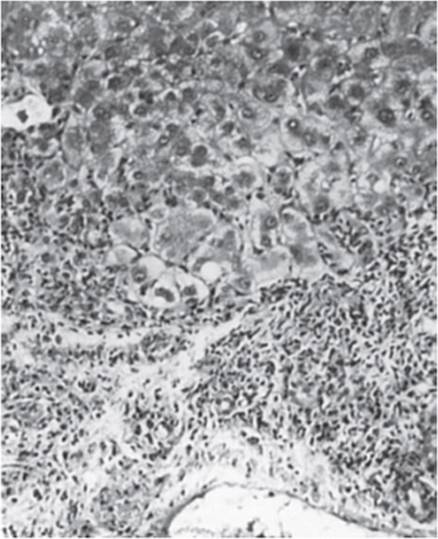
FIGURE 14-12 Chronic hepatitis, showing marked lymphocytic infiltration and fibrosis of the portal areas. The lymphocytes extend into the peripheral part of the lobule through the limiting plate. There is ongoing necrosis of hepatocytes in the peripheral part of the lobule (piecemeal necrosis). (Reproduced, with permission, from Chandrasoma P et al, eds. Concise Pathology, 3rd ed. Originally published by Appleton & Lange. Copyright © 1998 by The McGraw-Hill Companies, Inc.)
Pathology
All forms of chronic hepatitis share the common histopathologic features of (1) inflammatory infiltration of hepatic portal areas with mononuclear cells, especially lymphocytes and plasma cells, and (2) necrosis of hepatocytes within the parenchyma or immediately adjacent to portal areas (periportal hepatitis, or “piecemeal necrosis”).
In mild chronic hepatitis, the overall architecture of the liver is preserved. Histologically, the liver reveals a characteristic lymphocyte and plasma cell infiltrate confined to the portal triad without disruption of the limiting plate and no evidence of active hepatocyte necrosis. There is little or no fibrosis, and what there is generally is restricted to the portal area; there is no sign of cirrhosis. A “cobblestone” appearance of liver cells is seen, indicating regeneration of hepatocytes.
In more severe cases of chronic hepatitis, the portal areas are expanded and densely infiltrated by lymphocytes, histiocytes, and plasma cells. There is necrosis of hepatocytes at the periphery of the lobule, with erosion of the limiting plate surrounding the portal triads (piecemeal necrosis; Figure 14-12). More severe cases also show evidence of necrosis and fibrosis between portal triads. There is disruption of normal liver architecture by bands of scar tissue and inflammatory cells that link portal areas to one another and to central areas (bridging necrosis). These connective tissue bridges are evidence of remodeling of hepatic architecture, a crucial step in the development of cirrhosis. Fibrosis may extend from the portal areas into the lobules, isolating hepatocytes into clusters and enveloping bile ducts. Regeneration of hepatocytes is seen with mitotic figures, multinucleated cells, rosette formation, and regenerative pseudolobules. Progression to cirrhosis is signaled by extensive fibrosis, loss of zonal architecture, and regenerating nodules.
Clinical Manifestations
Some patients with mild chronic hepatitis are entirely asymptomatic and identified only in the course of routine blood testing; others have an insidious onset of nonspecific symptoms such as anorexia, malaise, and fatigue or hepatic symptoms such as right upper quadrant abdominal discomfort or pain. Fatigue in chronic hepatitis may be related to a change in the hypothalamic-adrenal neuroendocrine axis brought about by altered endogenous opioidergic neurotransmission. Jaundice, if present, is usually mild. There may be mild tender hepatomegaly and occasional splenomegaly. Palmar erythema and spider telangiectases are seen in severe cases. Other extrahepatic manifestations are unusual. By definition, signs of cirrhosis and portal hypertension (eg, ascites, collateral circulation, and encephalopathy) are absent. Laboratory studies show mild to moderate increases in serum aminotransferase, bilirubin, and globulin levels. Serum albumin and the prothrombin time are normal until late in the progression of liver disease.
The clinical manifestations of chronic hepatitis probably reflect the role of a systemic genetically controlled immune disorder in the pathogenesis of severe disease. Acne, hirsutism, and amenorrhea may occur as a reflection of the hormonal effects of chronic liver disease. Laboratory studies in patients with severe chronic hepatitis are invariably abnormal to various degrees. However, these abnormalities do not correlate with clinical severity. Thus, the serum bilirubin, alkaline phosphatase, and globulin levels may be normal and aminotransferase levels only mildly elevated at the same time that a liver biopsy reveals severe chronic hepatitis. However, an elevated prothrombin time usually reflects severe disease.
The natural history and treatment of chronic hepatitis varies depending on its cause. The complications of severe chronic hepatitis are those of progression to cirrhosis: variceal bleeding, encephalopathy, coagulopathy, hypersplenism, and ascites. These are largely due to portosystemic shunting rather than diminished hepatocyte reserve (see later discussion).
CHECKPOINT
26. What are the categories of chronic hepatitis based on histologic findings on liver biopsy?
27. What are the causes of chronic hepatitis?
28. What are the consequences of chronic hepatitis?
CIRRHOSIS
Clinical Presentation
Cirrhosis is an irreversible distortion of normal liver architecture characterized by hepatic injury, fibrosis, and nodular regeneration. The clinical presentations of cirrhosis are a consequence of both progressive hepatocellular dysfunction and portal hypertension (Figure 14-13). As with other presentations of liver disease, not all patients with cirrhosis develop life-threatening complications. Indeed, in nearly 40% of cases of cirrhosis, it is diagnosed at autopsy in patients who did not manifest obvious signs of end-stage liver disease.

FIGURE 14-13 Clinical effects of cirrhosis of the liver. (Redrawn, with permission, from Chandrasoma P et al, eds. Concise Pathology, 3rd ed. Originally published by Appleton & Lange. Copyright © 1998 by The McGraw-Hill Companies, Inc.)
Etiology
The causes of cirrhosis are listed in Table 14-1. The initial injury can be due to a wide range of processes. A crucial feature is that the liver injury is not acute and self-limited but rather chronic and progressive. In the United States, alcohol abuse is the most common cause of cirrhosis. In other countries, infectious agents (particularly HBV and HCV) are the most common causes. Other causes include chronic biliary obstruction, drugs, genetic and metabolic disorders, chronic heart failure, and primary (autoimmune) biliary cirrhosis.
Pathogenesis
Increased or altered synthesis of collagen and other connective tissue or basement membrane components of the extracellular matrix is implicated in the development of hepatic fibrosis and thus in the pathogenesis of cirrhosis. The role of the extracellular matrix in cellular function is an important area of research, and studies suggest that it is involved in modulating the activities of the cells with which it is in contact. Thus, fibrosis may affect not only the mechanics of blood flow through the liver but also the functions of the cells themselves.
Hepatic fibrosis occurs in three situations: (1) secondary to inflammation and subsequent activation of immune responses, (2) as part of the process of wound healing, and (3) in response to agents that induce primary fibrogenesis. HBV and Schistosoma species are good examples of agents that lead to hepatic fibrosis by stimulating an immune response. Agents such as carbon tetrachloride that attack and kill hepatocytes directly can produce fibrosis as part of wound healing. In both immune responses and wound healing, the fibrosis is triggered indirectly by the effects of cytokines released from invading inflammatory cells. Finally, certain agents such as ethanol and iron may cause primary fibrogenesis by directly increasing collagen gene transcription and thus increasing also the amount of connective tissue secreted by cells.
The actual culprit in all of these mechanisms of increased fibrogenesis may be the fat-storing cells (stellate cells) of the hepatic reticuloendothelial system. In response to cytokines, they differentiate from quiescent stellate cells in which vitamin A is stored into myofibroblasts, which lose their vitamin A storage capacity and become actively engaged in extracellular matrix production. In addition to the stellate cells, fibrogenic cells are also derived from portal fibroblasts, circulating fibrocytes, bone marrow, and epithelial-mesenchymal cell transition. It appears that hepatic fibrosis occurs in two stages (Figure 14-14). The first stage is characterized by a change in extracellular matrix composition from non–cross-linked, non–fibril-forming collagen to collagen that is more dense and subject to cross-link formation. At this stage, liver injury is still reversible. The second stage involves formation of subendothelial collagen cross-links, proliferation of myoepithelial cells, and distortion of hepatic architecture with the appearance of regenerating nodules. Cirrhosis remains a dynamic state in which certain interventions, even at these advanced stages, may yield benefits such as regression of scar tissue and improvements in clinical outcomes.

FIGURE 14-14 Pathways of hepatic stellate cell activation. Features of stellate cell activation can be distinguished between those that stimulate initiation and those that contribute to perpetuation. Initiation is provoked by soluble stimuli that include oxidant stress signals (reactive oxygen intermediates), apoptotic bodies, lipopolysaccharide (LPS), and paracrine stimuli from neighboring cell types including hepatic macrophages (Kupffer cells), sinusoidal endothelium, and hepatocytes. Perpetuation follows, characterized by a number of specific phenotypic changes including proliferation, contractility, fibrogenesis, altered matrix degradation, chemotaxis, and inflammatory signaling. (FGF, fibroblast growth factor; ET-1, endothelin-1; NK, natural killer; NO, nitric oxide; MT, membrane type.) (Reproduced, with permission, from Friedman SL. Molecular regulation of hepatic fibrosis, an integrated cellular response to tissue injury. J Biol Chem. 2000 Jan 28:275(4):2247–50.)
The manner in which alcohol causes chronic liver disease and cirrhosis is not well understood. However, chronic alcohol abuse is associated with impaired protein synthesis and secretion, mitochondrial injury, lipid peroxidation, formation of acetaldehyde and its interaction with cellular proteins and membrane lipids, cellular hypoxia, and both cell-mediated and antibody-mediated cytotoxicity. The relative importance of each of these factors in producing cell injury is unknown. Genetic, nutritional, and environmental factors (including simultaneous exposure to other hepatotoxins) also influence the development of liver disease in chronic alcoholics. Finally, acute liver injury (eg, from exposure to alcohol or other toxins) from which a person with a normal liver would fully recover may be sufficient to produce irreversible decompensation (eg, hepatorenal syndrome) in a patient with underlying hepatic cirrhosis.
Pathology
The liver may be large or small, but it always has a firm and often nodular consistency. Although several noninvasive methods for staging the extent of fibrosis exist, including use of serum biomarkers and imaging techniques to measure liver stiffness (eg, elastography), these methods are accurate for severe (fibrosis stage F3) or minimal (F1) fibrosis, but not intermediate stages in between. Liver biopsy remains the only method for definitively diagnosing significant fibrosis (F ≥ 2) and cirrhosis (F4). Histologically, all forms of cirrhosis are characterized by three findings: (1) marked distortion of hepatic architecture, (2) scarring as a result of increased deposition of fibrous tissue and collagen, and (3) regenerative nodules surrounded by scar tissue. When the nodules are small (<3 mm) and uniform in size, the process is termed micronodular cirrhosis. In macronodular cirrhosis, the nodules are more than 3 mm and variable in size. Cirrhosis from alcohol abuse is usually micronodular but can be macronodular or both micronodular and macronodular. Scarring may be most severe in central regions, or dense bands of connective tissue may join portal and central areas.
More specific histopathologic findings may help to establish the cause of cirrhosis. For example, invasion and destruction of bile ducts by granulomas suggests primary (autoimmune) biliary cirrhosis; extensive iron deposition in hepatocytes and bile ducts suggests hemochromatosis; and alcoholic hyalin and infiltration with polymorphonuclear cells suggest alcoholic cirrhosis.
Clinical Manifestations
The clinical manifestations of progressive hepatocellular dysfunction in cirrhosis are similar to those of acute or chronic hepatitis and include constitutional symptoms and signs: fatigue, loss of vigor, and weight loss; GI symptoms and signs: nausea, vomiting, jaundice, and tender hepatomegaly; and extrahepatic symptoms and signs: palmar erythema, spider angiomas, muscle wasting, parotid and lacrimal gland enlargement, gynecomastia and testicular atrophy in men, menstrual irregularities in women, and coagulopathy.
Clinical manifestations of portal hypertension include ascites, portosystemic shunting, encephalopathy, splenomegaly, and esophageal and gastric varices with intermittent hemorrhage (Table 14-15).
TABLE 14-15 Manifestations of cirrhosis.

A. Portal Hypertension
Portal hypertension is defined by a portal venous pressure gradient greater than 5 mm Hg. Portal hypertension is due to a rise in intrahepatic vascular resistance. The cirrhotic liver loses the physiologic characteristic of a low-pressure circuit for blood flow seen in the normal liver. The increased blood pressure within the sinusoids is transmitted back to the portal vein. Because the portal vein lacks valves, this elevated pressure is transmitted back to other vascular beds, resulting in splenomegaly, portal-to-systemic shunting, and many of the complications of cirrhosis discussed later.
B. Ascites
Abdominal ascites refers to the presence of excess fluid within the peritoneal cavity. Patients with ascites develop physical examination findings of increasing abdominal girth, a fluid wave, a ballotable liver, and shifting dullness. Ascites can develop in patients with conditions other than liver disease, including protein-calorie malnutrition (from hypoalbuminemia) and cancer (from lymphatic obstruction). In patients with liver disease, ascites is due to portal hypertension and can be confirmed by the presence of a serum-to-ascites albumin gradient (SAAG) of 1.1 g/dL (11 g/L) or more. Calculating the SAAG involves measuring on the same day the albumin concentration in serum and ascitic fluid (via abdominal paracentesis) and subtracting the ascitic fluid value from the serum value. Ascites can develop in approximately 50% of patients with compensated cirrhosis over a 10-year follow-up period, and it is associated with significant morbidity and mortality.
It is useful to recognize that liver disease with ascites formation occurs in a wide clinical spectrum. At one end is fully compensated portal hypertension with no ascites present because the volume of ascites generated is less than the approximately 800–1200 mL/d capacity of the peritoneal lymphatic drainage. At the other extreme is the typically fatal hepatorenal syndrome, in which patients with liver disease, usually with massive ascites, succumb to rapidly progressing acute kidney injury. The hepatorenal syndrome seems to be precipitated by intense and inappropriate renal vasoconstriction and is characterized by extreme sodium retention typical of prerenal azotemia but in the absence of true volume depletion (see Chapter 16). Nonetheless, the presence of clinically apparent ascites in a patient with liver disease is associated with poor long-term survival. Over the years, various mechanisms have been proposed to explain ascites formation. No single hypothesis of pathogenesis easily explains all findings at all points in time during the natural history of portal hypertension. Portal hypertension and inappropriate renal retention of sodium are important elements of all theories. The end result of ascites occurs when excess peritoneal fluid exceeds the capacity of lymphatic drainage, leading to increased hydrostatic pressure. The fluid can then be seen to visibly weep from the lymphatics and pool in the abdominal cavity as ascites.
The underfill/vasodilatation hypothesis proposes that the primary event in ascites formation is vascular, with reduced effective circulating volume leading to the activation of the renin-angiotensin system and subsequent renal sodium retention. The classic underfill hypothesis postulates that elevated hepatic sinusoidal pressure leads to sequestration of blood in the splanchnic venous bed. This results in underfilling of the central vein with diversion of intravascular volume to the hepatic lymphatics, which, like the central vein, drain the space of Disse. The peripheral arterial vasodilatation or splanchnic vasodilatation hypothesis adds the idea that, with portal-to-systemic shunting, vasodilatory products (eg, nitric oxide) that are normally cleared by the liver are instead delivered to the systemic circulation, where they cause peripheral arteriolar vasodilation, particularly in the splanchnic arterial bed. The resultant reduced arterial vascular resistance (Figure 14-15) is associated with decreased central filling pressures, decreased renal arterial perfusion, reflex renal arterial vasoconstriction, and increased renal tubular sodium resorption. Retention of sodium expands the intravascular volume, which exacerbates portal venous hypertension. The imbalance between hydrostatic versus oncotic pressure in the portal vein results in ascites formation. Although the splanchnic vasodilatation hypothesis accounts for many of the findings in ascites formation, the use of transhepatic intrajugular portal-to-systemic shunting (TIPS) as a means of decompressing the portal vein in patients with ascites provides a counterargument. As a result of the procedure, peripheral arteriolar vasodilation appears to increase (perhaps as a result of shunting of vasodilators such as nitric oxide that are normally cleared by the liver), yet ascites is generally dramatically improved.
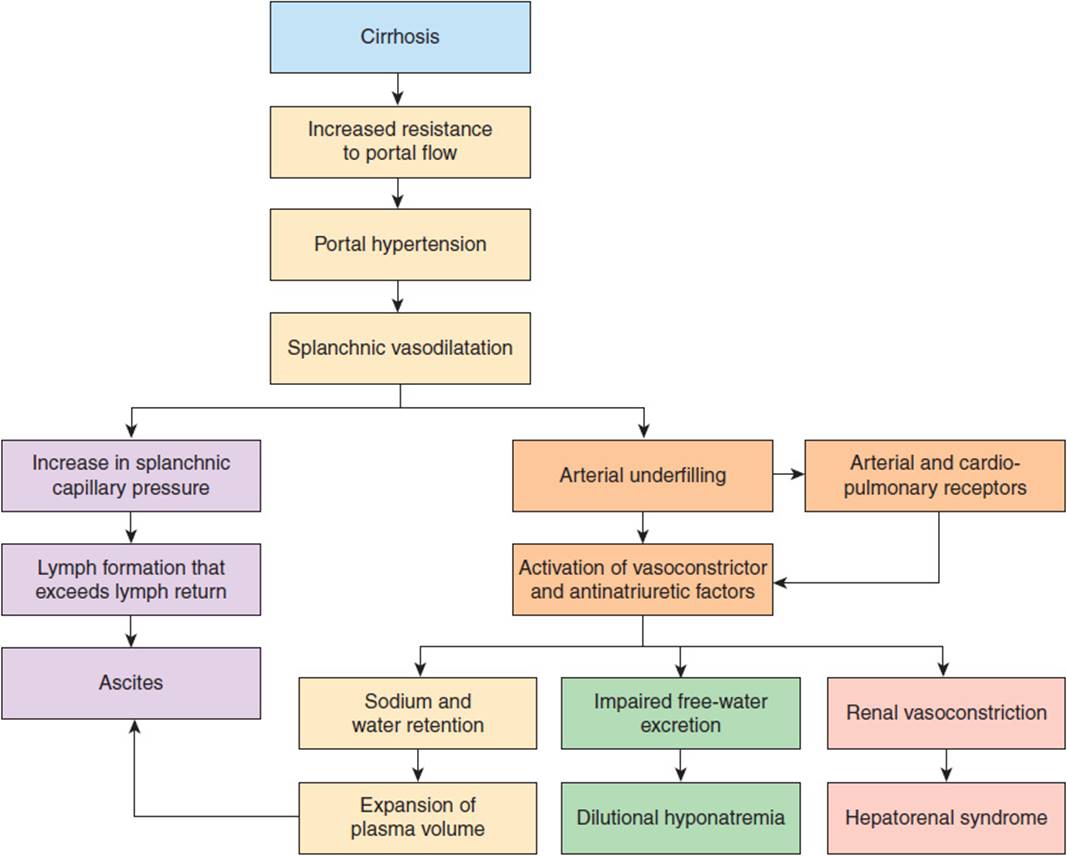
FIGURE 14-15 Proposed mechanism for ascites formation in cirrhosis through the splanchnic vasodilation hypothesis. This hypothesis incorporates elements of the underfill and vasodilation theories. (Redrawn, with permission, from Gines P et al. Management of cirrhosis and ascites. N Engl J Med. 2004;350:1646.)
Those who support the overflow hypothesis have proposed that the primary event in the development of ascites is inappropriate renal sodium retention. In this view, ascites is the consequence of overflow of fluid from the intravascular volume-expanded portal system into the peritoneal cavity. But what triggers the inappropriate renal sodium retention? One possibility is that there may exist a hepatorenal reflex by which elevated sinusoidal pressure triggers increased sympathetic tone or endothelin-1 secretion. Either of these pathways could cause an inappropriate degree of renal vasoconstriction, a decrease in glomerular filtration rate, and, by tubuloglomerular feedback (see Chapter 16), sodium retention. Note that endothelin-1 is both a renal vasoconstrictor and a stimulant of epinephrine secretion, which in turn stimulates more endothelin-1 secretion. Alternatively, it is possible that an as yet unidentified product from the diseased liver interferes with atrial natriuretic peptide (ANP) action at the kidney or is in some other way responsible for an inappropriate increase in renal sodium retention. Supporters of the overflow hypothesis point to the fact that many cirrhotic patients have sodium handling defects in the absence of ascites and do not have a measurable increase in renin-angiotensin activity. However, studies have shown that the renal sodium retention in these patients can be reversed by the use of an angiotensin II receptor antagonist.
Most likely, multiple mechanisms contribute to the development of ascites and to its perpetuation, worsening, or improvement in diverse clinical situations. Regardless of the initial events, once fully established, many if not all of the mechanisms described in Figure 14-15 are likely to contribute to ascites formation.
C. Hepatorenal Syndrome
Hepatorenal syndrome refers to a distinct form of kidney injury resulting from renal vasoconstriction that develops in response to the systemic and splanchnic arterial vasodilation in patients with advanced liver disease. The incidence of hepatorenal syndrome in patients who develop decompensated liver disease is 18% within 1 year of diagnosis and up to 40% at 5 years. This disorder generally occurs in patients with cirrhosis and ascites and is characterized by a progressive rise in serum creatinine (>1.5 mg/dL) with no improvement after 48 hours of withholding diuretics and volume expansion with albumin, in the absence of shock, ingestion of nephrotoxic agents, or underlying parenchymal kidney disease. The urine produced is notable for an extremely low sodium content (<10 mmol/L) and an absence of casts, resembling the findings in prerenal azotemia. Yet when central venous pressures are measured, the patient does not show intravascular volume depletion and the disorder does not respond to hydration with normal saline. The renal abnormalities of the hepatorenal syndrome appear to be functional because no pathologic changes are identifiable in the kidney. In addition, when a kidney is transplanted from a patient dying of hepatorenal syndrome, it functions well in a recipient without liver disease. While diagnostic criteria for hepatorenal syndrome have been developed and recently modified, diagnosing and differentiating hepatorenal syndrome from other causes of acute kidney injury in cirrhotic patients can be a difficult task. Other than hepatorenal syndrome, acute tubular necrosis and other causes of prerenal azotemia are the most common diagnoses in this setting.
The hepatorenal syndrome can be classified into two types, each having different clinical and prognostic characteristics. Type 1 hepatorenal syndrome is rapidly progressive with a doubling of the serum creatinine concentration to a level greater than 2.5 mg/dL in less than 2 weeks. It is associated with multiorgan failure. By contrast, type 2 hepatorenal syndrome is characterized by less severe renal insufficiency, is more slowly progressive, and generally occurs in the setting of ascites resistant to diuretics. The onset of hepatorenal syndrome can be insidious or dramatic and can be precipitated by an acute event, such as infection (notably spontaneous bacterial peritonitis) or hypovolemia from GI bleeding or overdiuresis. Prognosis after development of hepatorenal syndrome is dismal (overall survival of 50% at 1 year). The untreated survival is in the range of weeks for type 1 hepatorenal syndrome and 4–6 months for type 2 hepatorenal syndrome.
The pathophysiology of hepatorenal syndrome is related to the distinct hemodynamic and circulatory changes that occur in patients with severe hepatic dysfunction. Portal hypertension triggers arterial vasodilation in the splanchnic circulation and subsequent reduction in systemic vascular resistance, which can no longer be compensated for by an augmented cardiac output. Increased production or activity of vasodilators within the splanchnic circulation, particularly nitric oxide, carbon monoxide, and endogenous cannabinoids, leads to such arterial vasodilation. In advanced cirrhosis, arterial pressure must be maintained through the activation of vasoconstrictor systems, including the renin-angiotensin system and the sympathetic nervous system, as well as excess secretion of antidiuretic hormone (arginine vasopressin). These compensatory mechanisms help maintain effective arterial blood volume and relatively normal arterial pressure but lead to intrarenal vasoconstriction and hypoperfusion, which impairs renal function. By the same mechanisms, affected patients may develop further retention of sodium and free water, worsening edema and ascites.
The best approach to the management of the hepatorenal syndrome, based on knowledge of its pathogenesis, is the administration of vasoconstrictor drugs. Use of the vasopressin analog, terlipressin, together with albumin, can be considered as initial therapy for hepatorenal syndrome. Terlipressin is effective in approximately 40–50% of patients with type 1 hepatorenal syndrome; data on use of vasoconstrictors in type 2 hepatorenal syndrome is limited. Renal replacement therapy in the form of hemodialysis or continuous venovenous hemofiltration has been used, particularly in patients awaiting transplantation or in those with acute, potentially reversible hepatorenal syndrome. There is no evidence, however, that renal replacement therapy improves the prognosis of patients with cirrhosis who are not candidates for a liver transplant. Liver transplantation remains the optimal treatment for patients with hepatorenal syndrome.
D. Hypoalbuminemia and Peripheral Edema
Progressive worsening of hepatocellular function in cirrhosis can result in a fall in the concentration of albumin and other serum proteins synthesized by the liver. As the concentration of these plasma proteins decreases, the plasma oncotic pressure is lowered, thereby tilting the balance of hemodynamic forces toward the development of both peripheral edema and ascites.
These hemodynamic changes further contribute to an avid sodium-retaining state despite total body water and sodium overload seen by urinalysis in the cirrhotic patient. Serum sodium may be low as a result of superimposed water retention caused by antidiuretic hormone release triggered by volume stimuli. There are typically no obvious clinical manifestations until the serum sodium concentration falls below 120 mEq/L, at which point neurologic symptoms can occur. Attempts to raise the serum sodium, including fluid restriction and administration of vasopressin receptor antagonists (eg, tolvaptan and conivaptan) are generally not recommended due to adverse effects and lack of clear benefit. Hyponatremia is simply a late manifestation of end-stage liver disease and a strong predictor of mortality in patients with cirrhosis.
A low serum potassium and metabolic alkalosis may be observed as a consequence of elevated aldosterone levels responding to renin release (and angiotensin II release) by the kidneys, which sense afferent intravascular depletion.
E. Spontaneous Bacterial Peritonitis
Spontaneous bacterial peritonitis is defined as infection of ascitic fluid in the absence of an intra-abdominal event (such as a bowel perforation or another surgically treatable source) that would account for the entry of pathogenic organisms into the peritoneal space. This complication carries a high mortality rate and is predictive of poor overall prognosis. The presence of infection is confirmed by an elevated ascitic fluid absolute polymorphonuclear leukocyte count of 250 cells/μL or more and definitively by a positive ascitic fluid bacterial culture. Symptoms and signs include fever, hypotension, abdominal pain or tenderness, decreased or absent bowel sounds, and abrupt onset of hepatic encephalopathy in a patient with ascites. Alternatively, patients with spontaneous bacterial peritonitis can have subtle or no symptoms, and hence, a high index of suspicion may be required for timely diagnosis.
Patients with advanced liver disease with large-volume ascites or very low ascitic fluid protein levels, prior history of spontaneous bacterial peritonitis, and episodes of upper GI bleeding are at increased risk for this complication. Ascitic fluid is an excellent culture medium for a variety of pathogens, including Enterobacteriaceae (chiefly Escherichia coli), group D streptococci (enterococci), Streptococcus pneumoniae, and viridans streptococci. The greater risk in patients with low ascitic fluid protein levels may be due to a low level of opsonic activity in the fluid.
While the exact pathogenesis of spontaneous bacterial peritonitis is not known, cirrhosis predisposes to the development of GI bacterial overgrowth and increased intestinal permeability. Peritonitis may occur because of bacterial seeding of the ascitic fluid via the blood or lymph or by bacteria traversing the gut wall. Enteric organisms may also enter the portal venous blood via the portosystemic collaterals, bypassing the reticuloendothelial system of the liver.
F. Gastroesophageal Varices and Bleeding
As blood flow through the liver is progressively impeded, hepatic portal venous pressure rises. In response to the elevated portal venous pressure, there is a decrease in blood vessel wall thickness and enlargement of blood vessels that anastomose with the portal vein, such as those on the surface of the bowel and lower esophagus. These enlarged vessels are termed gastroesophageal varices. They eventually develop in approximately 50% of patients with cirrhosis, generally when the portal hypertensive gradient exceeds 12 mm Hg. Physical examination may reveal enlargement of hemorrhoidal and periumbilical vessels. Gastroesophageal varices are of more significance clinically, however, because of their tendency to rupture. Variceal hemorrhage occurs in 25–40% of patients with cirrhosis and is a leading cause of morbidity and mortality in these individuals. Each episode of active variceal bleeding is associated with a 30% mortality risk, and survivors have a 70% risk of recurrent bleeding within 1 year. GI bleeding from varices and other sources (eg, duodenal ulcer, gastritis) in patients with cirrhosis is often exacerbated by concomitant coagulopathy (see later discussion).
G. Hepatic Encephalopathy
Hepatic encephalopathy presents as a range of reversible neuropsychiatric abnormalities that occur as a consequence of advanced decompensated liver disease or portal-to-systemic shunting (see Table 14-16for common precipitants). Neuropsychiatric symptoms can be episodic or persistent. Changes in the sleep pattern starting with hypersomnia and progressing to reversal of the sleep-wake cycle are often an early sign. Cognitive changes range from mild confusion, apathy, and agitation, to marked confusion, obtundation, and even coma. More advanced neurologic features include tremor, bradykinesia, asterixis(flapping motions of outstretched, dorsiflexed hands), hyperactive deep tendon reflexes, and less commonly, transient decerebrate posturing and flaccidity. Cerebral edema, which is an important accompanying feature in patients with encephalopathy in acute liver disease, is not seen in cirrhotic patients with encephalopathy. Subtle changes of hepatic encephalopathy are present in up to 15% of patients with advanced liver disease and may only be detectable by a number of specialized measures, such as psychometric testing. Such patients have sometimes been referred to as having subclinical or minimal hepatic encephalopathy.
TABLE 14-16 Common precipitants of hepatic encephalopathy.
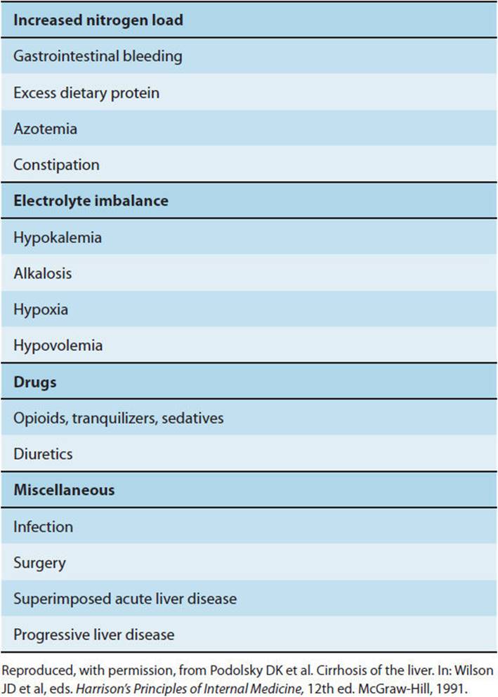
Hepatic encephalopathy is diagnosed by history and clinical features in the appropriate context and after exclusion of other causes of altered mental status. Common precipitants of encephalopathy are onset of GI bleeding, increased dietary protein intake, and an increased catabolic rate resulting from infection (including spontaneous bacterial peritonitis). Similarly, because of compromised first-pass clearance of ingested drugs, affected patients are exquisitely sensitive to sedatives and other drugs normally metabolized in the liver. Other causes include electrolyte imbalance as a result of diuretics, vomiting, alcohol ingestion or withdrawal, or procedures such as TIPS. TIPS also exacerbates hepatic encephalopathy given direct bypass of portal venous blood flow into systemic circulation via the hepatic vein, while bypassing the hepatic parenchyma.
The pathogenesis of hepatic encephalopathy is likely multifactorial and complex. One proposed mechanism is related to toxins in the gut such as ammonia, derived from metabolic degradation of urea or protein; glutamine, derived from degradation of ammonia; or mercaptans, derived from degradation of sulfur-containing compounds; and manganese. Because of anatomic or functional portal-systemic shunts, these toxins bypass the liver’s detoxification processes and produce alterations in mental status. Exposure to these toxins can cause astrocyte swelling and structural changes in neurons. In addition, high ammonia levels can result in abnormal cerebral blood flow and glucose metabolism. Increased levels of ammonia, glutamine, and mercaptans can be found in the blood and cerebrospinal fluid. There is also an increase in cerebral manganese deposition in patients with cirrhosis. However, blood ammonia and spinal fluid glutamine levels correlate poorly with the presence and severity of encephalopathy. In addition, the role of manganese in hepatic encephalopathy remains unclear.
Alternatively, there may be impairment of the normal blood-brain barrier, rendering the CNS susceptible to various noxious agents. Increased levels of other substances, including metabolic products such as short-chain fatty acids and endogenous benzodiazepine-like metabolites, have also been found in the blood. Importantly, some patients show improvement in encephalopathy when treated with flumazenil, a benzodiazepine receptor antagonist.
Another proposed mechanism postulates a role for GABA, the principal inhibitory neurotransmitter of the brain. GABA is produced in the gut, and increased levels are found in the blood of patients with liver failure. More recently, cerebral and systematic inflammation has been implicated in the pathogenesis of hepatic encephalopathy. Although the exact mechanisms are not known, possibilities include cytokine-mediated changes in blood-brain barrier permeability, potential changes in glutamate uptake by astrocytes, and changes in expression of GABA receptors.
Once the diagnosis is made, it is helpful to grade the severity of hepatic encephalopathy. Stages I through IV are based on degree of behavioral changes, intellectual dysfunction, and alterations in consciousness. Therapy includes management of potential precipitants and is directed at reduction of intestinal ammonia production or increasing the removal of ammonia from the circulation. Nonabsorbable synthetic disaccharides (eg, lactulose) are catabolized by colonic bacteria to short-chain fatty acids, which lower luminal pH. This change in pH favors the formation of ammonium (NH4+), which reduces absorption of ammonia (NH3) into the circulation. Disaccharides such as lactulose are thus the mainstay of therapy. As discussed above in the section Altered Metabolism of Ammonia, the antibiotic rifaximin has been used in conjunction with lactulose for treatment of hepatic encephalopathy.
H. Coagulopathy
Factors contributing to coagulopathy in cirrhosis include loss of hepatic synthesis of clotting factors, some of which have a half-life of just a few hours. Under these circumstances, a minor or self-limited source of bleeding can become massive.
Hepatocytes are also functionally involved in the maintenance of a normal coagulation cascade through the absorption of vitamin K (a fat-soluble vitamin whose absorption is dependent on bile flow), which is necessary for the activation of some clotting factors (II, VII, IX, X). An ominous sign of the severity of liver disease is the development of a coagulopathy that does not respond to parenteral vitamin K, suggesting deficient clotting factor synthesis rather than impaired absorption of vitamin K because of fat malabsorption. Finally, loss of the liver’s capacity to remove activated clotting factors and fibrin degradation products may play a role in the increased susceptibility to disseminated intravascular coagulation, a syndrome of coagulation factor consumption that results in uncontrolled simultaneous clotting and bleeding.
I. Splenomegaly and Hypersplenism
Enlargement of the spleen is a consequence of elevated portal venous pressure and consequent engorgement of the organ. Thrombocytopenia and hemolytic anemia occur because of sequestering of formed elements of the blood in the spleen, from which they are normally cleared as they age and are damaged.
J. Hepatocellular Carcinoma
The 5-year cumulative risk of HCC in patients with cirrhosis ranges from 5% to 30% depending on the patient’s sex, ethnicity, liver disease cause, and stage of cirrhosis. In the United States, the incidence of HCC has been rising over the past few decades, with over 20,000 new cases diagnosed each year, attributable to increased prevalence of NAFLD, HCV cirrhosis, and chronic HBV infections due to immigration from high prevalence regions. Several etiologic factors have been identified in the development of this tumor, although cirrhosis is present in the vast majority (80–90%) of patients who develop HCC.
The risk of developing HCC is increased 100-fold in those with chronic hepatitis B infection, and worldwide, HBV accounts for over 50% of all HCC cases and nearly all childhood cases. While HCC can occur in the absence of cirrhosis, over 70% of HBV-related cases occur in those with advanced fibrosis or cirrhosis. Risk factors for HCC in this population include male sex, older age or longer duration of infection, coinfection (HCV, HDV, HIV), mycotoxin aflatoxin exposure, genotype C, and in particular, high levels of viral replication, as evidenced by persistent elevation of HBV viral load.
In patients with chronic hepatitis C, the risk of developing HCC is increased 15- to 20-fold, with risk limited to those with advanced fibrosis and cirrhosis. It has been projected that the incidence of HCV-related HCC cases in the United States will continue to rise over the next several decades. Risk factors for HCC development include male sex, older age and duration of chronic HCV infection, coinfections (HBV, HIV), heavy alcohol use, obesity, and metabolic factors.
Chronic hepatitis B and C account for 60–70% of all HCC cases in the United States. While any cause of cirrhosis can lead to HCC, alcoholic cirrhosis and nonalcoholic steatohepatitis account for most of the remaining U.S. cases. Obesity and the metabolic syndrome are increasingly recognized as risk factors for liver cancer.
K. Pulmonary Complications
Up to one third of patients with decompensated cirrhosis have problems associated with oxygenation and may present with shortness of breath. There are three main pulmonary complications of cirrhosis to consider: hepatopulmonary syndrome, portopulmonary syndrome, and hepatic hydrothorax. In addition, mild hypoxemia can be caused by massive ascites, with resulting diaphragmatic elevation and ventilation/perfusion mismatch.
The hepatopulmonary syndrome consists of the triad of advanced liver failure, hypoxemia, and intrapulmonary vascular dilation and shunting. The cause of pulmonary precapillary and capillary vasodilatation is unknown, but substances such as nitric oxide, endothelin, and arachidonic acid are thought to be involved. As a result of ventilation-perfusion mismatch, patients often present with platypnea, dyspnea that worsens in the upright position secondary to preferential perfusion of dilated vessels in the lung bases. Classically, contrast-enhanced echocardiography is used for definitive diagnosis and can reveal opacification of the left heart chambers within three to six cardiac cycles if a right-to-left intrapulmonary shunt is present. Liver transplantation leads to resolution of the hepatopulmonary syndrome. However, development of severe pulmonary hypertension in patients with advanced liver failure may be a contraindication to liver transplantation.
Portopulmonary hypertension refers to the development of pulmonary hypertension in patients with advanced liver disease and advanced portal hypertension. Patients can present with hypoxia, dyspnea on exertion, fatigue, and even signs of right heart failure. Patients have evidence of elevated pulmonary vascular resistance and a transpulmonary gradient in the setting of pulmonary arterial vasoconstriction. Targeted therapy (eg, epoprostenol, vasodilators) and management of right heart failure can delay progression, but prognosis is poor. Liver transplantation is associated with high operative risk when pulmonary hypertension becomes severe.
Individuals with cirrhosis and ascites can develop hepatic hydrothorax and present with shortness of breath, cough, or chest discomfort. In this condition, fluid accumulates in the pleural space due to small defects in the diaphragm, most commonly on the right side. Negative intrathoracic pressure generated during inspiration favors the passage of fluid from the intra-abdominal cavity to the pleural space. Diagnostic thoracentesis should be performed to exclude alternative causes of pleural effusion, particularly infection. Treatment aims to prevent or reduce fluid accumulation with diuretics, low sodium diet, and occasionally therapeutic thoracentesis (or paracentesis to decrease pressure from tense ascites) for highly symptomatic patients refractory to or intolerant of conservative measures. TIPS may benefit selected patients (eg, Child-Pugh class A or B with no encephalopathy) who are requiring repeated thoracenteses. If they are otherwise suitable candidates, patients with cirrhosis and persistent hepatic hydrothorax should be referred for liver transplantation.
L. Miscellaneous Manifestations
Other findings on physical examination of patients with cirrhosis include spider angiomas (prominent blood vessels with a central arteriole and small vessels radiating from it seen in the skin, particularly on the face and upper trunk), Dupuytren contractures (fibrosis of the palmar fascia), testicular atrophy, gynecomastia (enlargement of breast tissue in men), palmar erythema, lacrimal and parotid gland enlargement, and diminished axillary and pubic hair (Figure 14-13). These findings are largely a consequence of estrogen excess resulting from decreased clearance of endogenous estrogens by the diseased liver combined with decreased hepatic synthesis of steroid hormone–binding globulin. Both of these mechanisms result in tissues receiving higher than normal concentrations of estrogens. In addition, a longer half-life of androgens may allow a greater degree of peripheral aromatization (conversion to estrogens by, eg, adipose tissue, hair follicles), further increasing estrogen-like effects in patients with cirrhosis. Xanthomas of the eyelids and extensor surfaces of tendons of the wrists and ankles can occur with chronic cholestasis such as occurs in primary biliary cirrhosis. Finally, profound muscle wasting and cachexia in cirrhosis probably reflect diminution of the liver’s synthesis of carbohydrate, lipid, and amino acids.
CHECKPOINT
29. What are the defining features of cirrhosis?
30. What are the three categories of hepatic fibrosis? Name one agent causing each.
31. What are the two postulated stages in the development of cirrhosis?
32. What are some ways alcohol may injure the liver?
33. What are the major clinical manifestations of cirrhosis?
34. For each major clinical manifestation of cirrhosis, suggest a reasonable hypothesis to account for its pathogenesis.
CASE STUDIES
Yeong Kwok, MD
(See Chapter 25, p. 725 for Answers)
CASE 71
A 28-year-old man, recently emigrating from the Philippines, was noted to have a positive tuberculin skin test result in the clinic. His chest radiograph showed no active tuberculosis, and he denied any symptoms of this infection, including weight loss, cough, or night sweats. To prevent future disease, daily dosing with isoniazid was recommended for the next 9 months. Two weeks after initiating therapy, the patient reported progressive fatigue, intermittent bouts of nausea, and abdominal pain. He also noticed darkening of his urine and light-colored stools. His sister noted a gradual yellowing of his eyes and skin. Blood tests showed a marked increase in serum bilirubin and aminotransferases. The isoniazid was discontinued, and his symptoms subsided with normalizing of his liver enzymes.
Questions
A. Describe the subtypes of toxic hepatitis.
B. What typical histologic findings are noted during uncomplicated acute hepatitis?
C. What is the pathogenesis of clinical jaundice seen in this patient?
CASE 72
A 44-year-old man is concerned about abnormal liver tests drawn for his preemployment physical 6 months ago. His serum aminotransferase levels were two times normal at that time and remain unchanged after repeat testing. On further questioning, he denies regular alcohol use but states that he used to inject heroin. Currently, he reports some fatigue but says he feels well otherwise. His primary care physician orders serologic testing, which reveals HBsAg-positive, anti-HBs-negative, and anti-HBc-positive IgG. Anti-HDV and anti-HCV test results are both negative.
Questions
A. Based on these antigen and antibody test results, what is the patient’s diagnosis?
B. What percentage of patients with acute hepatitis B remain chronically infected with HBV? Of those patients, how many develop chronic active disease? What are the significant complications of chronic active infection?
C. What is the significance of hepatitis D superinfection?
D. What evidence exists supporting immune-mediated damage in chronic active hepatitis?
CASE 73
A 63-year-old man with a long history of alcohol use presents to his new primary care physician with a 6-month history of increasing abdominal girth. He has also noted easy bruisability and worsening fatigue. He denies any history of GI bleeding. He continues to drink three or four cocktails a night but says he is trying to cut down. Physical examination reveals a cachectic man who appears older than his stated age. Blood pressure is 108/70 mm Hg. His scleras are anicteric. His neck veins are flat, and chest examination demonstrates gynecomastia and multiple spider angiomas. Abdominal examination is significant for a protuberant abdomen with a detectable fluid wave, shifting dullness, and an enlarged spleen. The liver edge is difficult to appreciate. He has trace pitting pedal edema. Laboratory evaluation shows anemia, mild thrombocytopenia, and an elevated prothrombin time. Abdominal ultrasonogram confirms a shrunken, heterogeneous liver consistent with cirrhosis, significant ascites, and splenomegaly.
Questions
A. Describe possible mechanisms for alcohol-induced cirrhosis.
B. What is the proposed mechanism of portal hypertension, and how does it affect ascites formation?
C. Significant hematologic abnormalities exist. How might they be explained?
REFERENCES
General
Bass NM et al. Rifaximin treatment in hepatic encephalopathy. N Engl J Med. 2010 Mar 25;362(12):1071–81. [PMID: 20335583]
Bohinc BN et al. Mechanisms of disease progression in NASH: new paradigms. Clin Liver Dis. 2012 Aug;16(3):549–65. [PMID: 22824480]
Boyer TD et al, eds. Zakim and Boyer’s Hepatology: A Textbook of Liver Disease, 6th ed. WB Saunders, 2011.
Cauli O et al. Glutamatergic and gabaergic neurotransmission and neuronal circuits in hepatic encephalopathy. Metab Brain Dis. 2009 Mar;24(1):69–80. [PMID: 19085094]
Dooley JS et al. Sherlock’s Diseases of the Liver and Biliary System, 12th ed. Wiley-Blackwell, 2011.
Gao B et al. Alcoholic liver disease: pathogenesis and new therapeutic targets. Gastroenterology. 2011 Nov;141(5):1572–85. [PMID: 21920463]
Jones EA et al. Theories of the pathogenesis of hepatic encephalopathy. Clin Liver Dis. 2012 Feb;16(1):7–26. [PMID: 22321462]
Krawczyk M et al. Nonalcoholic fatty liver disease. Best Pract Res Clin Gastroenterol. 2010 Oct;24(5):695–708. [PMID: 20955971]
Rose CF. Ammonia-lowering strategies for the treatment of hepatic encephalopathy. Clin Pharmacol Ther. 2012 Sep;92(3):321–31. [PMID: 22871998]
Acute Hepatitis
Bernal W et al. Acute liver failure. Lancet. 2010 Jul 17;376(9736):190–201. [PMID: 20638564]
Davern TJ. Drug-induced liver disease. Clin Liver Dis. 2012 May;16(2):231–45. [PMID: 22541696]
Dienstag JL. Hepatitis B virus infection. N Engl J Med. 2008 Oct 2;359(14):1486–500. Erratum in: N Engl J Med. 2010 Jul 15;363(3):298. [PMID: 18832247]
Ghany MG et al. American Association for the Study of Liver Diseases. Diagnosis, management, and treatment of hepatitis C: an update. Hepatology. 2009 Apr;49(4):1335–74. [PMID: 19330875]
Gossard AA et al. Autoimmune hepatitis: a review. J Gastroenterol. 2012 May;47(5):498–503. [PMID: 22526272]
Hoofnagle JH et al. Hepatitis E. N Engl J Med. 2012 Sep 27;367(13):1237–44. [PMID: 23013075]
Kowdley KV et al. Prevalence of chronic hepatitis B among foreign-born persons living in the United States by country of origin. Hepatology. 2012 Aug;56(2):422–33. [PMID: 22105832]
Maheshwari A et al. Management of acute hepatitis C. Clin Liver Dis. 2010 Feb;14(1):169–76. [PMID: 20123448]
Mufti AR et al. Liver disease in pregnancy. Clin Liver Dis. 2012 May;16(2):247–69. [PMID: 22541697]
Peppa D et al. Pathogenesis of hepatitis B virus infection and potential for new therapies. Br J Hosp Med (Lond). 2012 Oct;73(10):581–4. [PMID: 23124289]
Smith BD et al. Centers for Disease Control and Prevention. Recommendations for the identification of chronic hepatitis C virus infection among persons born during 1945–1965. MMWR Recomm Rep. 2012 Aug 17;61(RR-4):1–32. Erratum in: MMWR Recomm Rep. 2012 Nov 2;61(43):886. [PMID: 22895429]
Chronic Hepatitis
Bals R. Alpha-1-antitrypsin deficiency. Best Pract Res Clin Gastroenterol. 2010 Oct;24(5):629–33. [PMID: 20955965]
Bertoletti A et al. The host-pathogen interaction during HBV infection: immunological controversies. Antivir Ther. 2010;15(Suppl 3): 15–24. [PMID: 21041900]
Boyer TD et al, eds. Zakim and Boyer’s Hepatology: A Textbook of Liver Disease, 6th ed. WB Saunders, 2011.
Brandman D et al. Impact of insulin resistance on HCV treatment response and impact of HCV treatment on insulin sensitivity using direct measurements of insulin action. Diabetes Care. 2012 May;35(5):1090–4. [PMID: 22399695]
Chalasani N et al. The diagnosis and management of non-alcoholic fatty liver disease: practice guideline by the American Gastroenterological Association, American Association for the Study of Liver Diseases, and American College of Gastroenterology. Gastroenterology. 2012 Jun;142(7):1592–609. Erratum in: Gastroenterology. 2012 Aug;143(2):503. [PMID: 22656328]
Jacobson IM et al. Manifestations of chronic hepatitis C virus infection beyond the liver. Clin Gastroenterol Hepatol. 2010 Dec;8(12):1017–29. [PMID: 20870037]
Jou JH et al. In the clinic. Hepatitis C. Ann Intern Med. 2012 Dec 4;157(11):ITC6-1–ITC6-16. [PMID: 23208180]
Sanyal AJ et al. NASH CRN. Pioglitazone, vitamin E, or placebo for nonalcoholic steatohepatitis. N Engl J Med. 2010 May 6;362(18):1675–85. [PMID: 20427778]
Yang JD et al. Cirrhosis is present in most patients with hepatitis B and hepatocellular carcinoma. Clin Gastroenterol Hepatol. 2011 Jan;9(1):64–70. [PMID: 20831903]
Cirrhosis
Abraldes JG et al. Diagnosing and monitoring cirrhosis: liver biopsy, hepatic venous pressure gradient and elastography. Gastroenterol Hepatol. 2012 Aug–Sep;35(7):488–95. [PMID: 22560536]
Ascha MS et al. The incidence and risk factors of hepatocellular carcinoma in patients with nonalcoholic steatohepatitis. Hepatology. 2010 Jun;51(6):1972–8. [PMID: 20209604]
Bals R. Alpha-1-antitrypsin deficiency. Best Pract Res Clin Gastroenterol. 2010 Oct;24(5):629–33. [PMID: 20955965]
Baranova A et al. Non-invasive markers for hepatic fibrosis. BMC Gastroenterol. 2011 Aug 17;11:91. [PMID: 21849046]
El-Serag HB. Hepatocellular carcinoma. N Engl J Med. 2011 Sep 22;365(12):1118–27. [PMID: 21992124]
Garcia-Tsao G et al. Management of varices and variceal hemorrhage in cirrhosis. N Engl J Med. 2010 Mar 4;362(9):823–32. Erratum in: N Engl J Med. 2011 Feb 3;364(5):490. [PMID: 20200386]
Ginès P et al. Management of critically-ill cirrhotic patients. J Hepatol. 2012;56(Suppl 1):S13–24. [PMID: 22300462]
Ginès P et al. Renal failure in cirrhosis. N Engl J Med. 2009 Sep 24;361(13):1279–90. [PMID: 19776409]
Ilan Y. Leaky gut and the liver: a role for bacterial translocation in nonalcoholic steatohepatitis. World J Gastroenterol. 2012 Jun 7;18(21):2609–18. [PMID: 22690069]
Invernizzi P et al. Update on primary biliary cirrhosis. Dig Liver Dis. 2010 Jun;42(6):401–8. [PMID: 20359968]
Krok KL et al. Hepatic hydrothorax. Semin Respir Crit Care Med. 2012 Feb;33(1):3–10. [PMID: 22447255]
Pietrangelo A. Hereditary hemochromatosis: pathogenesis, diagnosis, and treatment. Gastroenterology. 2010 Aug;139(2):393–408. [PMID: 20542038]
Pinzani M et al. Liver cirrhosis. Best Pract Res Clin Gastroenterol. 2011 Apr;25(2):281–90. [PMID: 21497745]
Rahimi RS et al. Complications of cirrhosis. Curr Opin Gastroenterol. 2012 May;28(3):223–9. [PMID: 22343347]
Riggio O et al. A simplified psychometric evaluation for the diagnosis of minimal hepatic encephalopathy. Clin Gastroenterol Hepatol. 2011 Jul;9(7):613–6. [PMID: 21440091]
Rodríguez-Roisin R et al. Hepatopulmonary syndrome—a liver-induced lung vascular disorder. N Engl J Med. 2008 May 29;358(22):2378–87. [PMID: 18509123]
Rosen HR. Clinical practice. Chronic hepatitis C infection. N Engl J Med. 2011 Jun 23;364(25):2429–38. [PMID: 21696309]
Runyon BA; AASLD Practice Guidelines Committee. Management of adult patients with ascites due to cirrhosis: an update. Hepatology. 2009 Jun;49(6):2087–107. [PMID: 19475696]
Sigal SH. Hyponatremia in cirrhosis. J Hosp Med. 2012 Apr;7(Suppl 4): S14–7. [PMID: 22489081]
Silverman EK et al. Clinical practice. Alpha1-antitrypsin deficiency. N Engl J Med. 2009 Jun 25;360(26):2749–57. [PMID: 19553648]
Tsouka A et al. Complications of chronic liver disease. Clin Res Hepatol Gastroenterol. 2012 Jun;36(3):262–7. [PMID: 22521556]
Wadei HM. Hepatorenal syndrome: a critical update. Semin Respir Crit Care Med. 2012 Feb;33(1):55–69. [PMID: 22447261]
Zhang J et al. Hepatopulmonary syndrome: update on pathogenesis and clinical features. Nat Rev Gastroenterol Hepatol. 2012 Sep;9(9):539–49. [PMID: 22751459]