
Jason C. Mills, MD, PhD, AGAF, &
Thaddeus S. Stappenbeck, MD, PhD
Gastrointestinal (GI) diseases most often present with one or more of four common classes of symptoms and signs: (1) abdominal or chest pain; (2) altered ingestion of food (eg, resulting from nausea, vomiting, dysphagia [difficulty swallowing], odynophagia [painful swallowing], or anorexia [lack of appetite]); (3) altered bowel movements (ie, diarrhea or constipation); and (4) GI tract bleeding, either occurring without warning or preceded by one or more of the foregoing (Table 13-1). However, not all cases of a particular GI disease present in the same way. For example, peptic ulcer disease, although typically accompanied by abdominal pain, may be painless.
TABLE 13-1 Common presentations of GI disease.

GI disease may be limited to the GI tract (eg, reflux esophagitis, peptic ulcer, diverticular disease), be a manifestation of a systemic disorder (eg, inflammatory bowel disease), or present as a systemic disease resulting from a primary GI pathologic process (eg, vitamin deficiencies resulting from malabsorption). Because different parts of the GI tract are specialized for certain functions, the most prominent causes, consequences, and manifestations of disease differ from one anatomic site to another.
Acutely, GI disease can be complicated by dehydration, sepsis, or bleeding or by their consequences, such as shock. Dehydration can occur as a consequence of even subtle alterations in fluid input or outflow because the volume of fluid traversing the GI tract daily is enormous (see later discussion). Sepsis can result from disruption of the barrier function against pathogens in the environment, including bacteria resident in the colon. The tendency for bleeding is a reflection of the tremendous vascularity of the GI tract and the difficulty of applying pressure at the site of bleeding.
Chronically, GI disease can be complicated by malnutrition and deficiency states. These occur because many primary GI diseases result in malabsorption (failure to absorb one or more necessary nutrients in ingested food).
GI tract disease can present as partial or complete obstruction (blockage of movement of contents down the GI tract) caused by adhesions and stenosis resulting from proliferation of connective tissue in response to inflammation. The symptoms and signs of obstruction can range from mild nausea, abdominal pain, and anorexia to projectile vomiting and rebound tenderness. In severe cases, obstruction can result in perforation, infarction and bleeding, hypotension, shock, sepsis, and death. The severity of symptoms depends on the extent of obstruction, the degree to which the obstruction compromises blood flow to the affected region, and the stage in the natural history of the process at which the patient presents for medical attention.
CHECKPOINT
1. What are the cardinal symptoms and signs of GI disease?
2. What are some acute systemic complications of primary GI disease?
3. What additional systemic manifestations can occur as a result of chronic GI disease?
STRUCTURE, FUNCTION, & CONTROL OF THE GI TRACT
STRUCTURE OF THE GI TRACT
The GI tract is one of the most complex and important organ systems. It comprises the alimentary canal, a hollow structure extending from the mouth to the anus, and associated glandular organs (salivary glands, pancreas, gallbladder, and liver) that empty their contents into the canal (Figure 13-1). The GI tract, which is 7–9 m in the adult, includes the mouth, esophagus (23–25 cm), stomach, small intestine (duodenum, jejunum, ileum; 6–7 m), large intestine (cecum and colon; 1.0–1.5 m), rectum, and anus. The GI tract is connected to the salivary glands, the pancreas, and the gallbladder, the sources of exocrinesecretions that play an essential role in digestion.
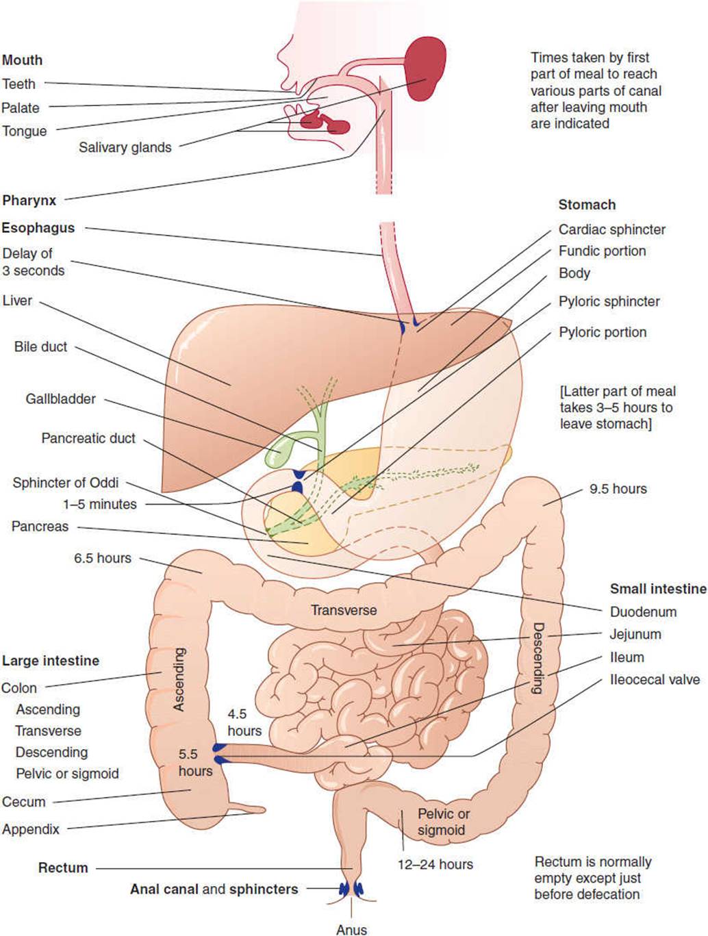
FIGURE 13-1 Progress of food along the alimentary canal. Food undergoes mechanical as well as chemical changes to render it suitable for absorption and assimilation. (Redrawn, with permission, from Mackenna BR et al, eds. Illustrated Physiology, 6th ed. Churchill Livingstone, 1997.)
The wall of the GI tract is composed of four main layers. From the lumen outward, these include the mucosa, submucosa, muscularis externa, and serosa (Figure 13-2). The precise structure of some of these layers, most notably the mucosa, varies from one region of the GI tract to the next. The mucosa has three components: specialized epithelial cells that line the lumen; the underlying lamina propria, a layer of connective tissue that contains small blood and lymphatic vessels, immune cells and nerve fibers; and the muscularis mucosa, a thin layer of muscle cells. The muscularis mucosa is an important boundary in determining whether cancer of the GI tract is still localized to its site of origin or is likely to have metastasized (ie, spread to distant regions of the body). The submucosa is a layer of loose connective tissue directly beneath the mucosa containing larger blood and lymphatic vessels and a nerve plexus of the intrinsic or enteric nervous system, termed the submucosal nerve (Meissner) plexus. This nerve plexus is particularly important for control of secretion in the GI tract. In some areas, the submucosa also contains glands and lymphoid tissue. The muscularis externa is composed of an inner circular and an outer longitudinal layer of smooth muscle and is responsible for motility of the GI tract. Between these muscle layers lies the myenteric nerve (Auerbach) plexus, a division of the enteric nervous system that regulates motility. The serosa is an outer sheath of squamous mesothelial cells and connective tissues, where larger nerves and blood vessels travel in a bed of connective and adipose tissue.
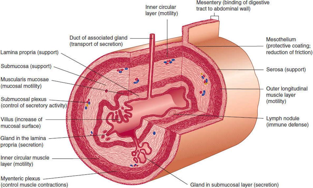
FIGURE 13-2 Schematic structure of a portion of the digestive tract with various possible components. (Redrawn, with permission, from Bevelander G. Outline of Histology, 7th ed. Mosby, 1971.)
FUNCTIONS OF THE GI TRACT
The overall function of the GI tract is to take in nutrients and process them to a form that can be used by the body and to eliminate wastes. The major physiological processes that occur in the GI tract are digestion, secretion, motility, and absorption.
A. Digestion
Food is taken into the mouth as large particles containing macromolecules that are not immediately absorbable into the body. Digestion is the process that converts nutrients in food to products that can be absorbed by cells of the mucosa. Digestion includes physical processes (eg, chewing, GI contractions) that break up the food, mix it with digestive secretions, and propel it along the alimentary canal, and chemical processes (eg, digestive enzymes) that degrade food components (proteins, fats, polysaccharides) to products that can be absorbed (amino acids, fatty acids, monosaccharides). Digestive enzymes arise from exocrine glands (salivary gland, pancreas, gallbladder, and liver) and from cells and glands in the mucosa or are found on the apical surface of certain epithelial cells.
B. Secretion
During the process of digestion, large volumes of fluid are secreted into the lumen of the GI tract. Secretions arise from exocrine glands (salivary glands, pancreas, gallbladder) and from epithelial cells lining the GI lumen (or glands that connect to the lumen). The daily fluid load in the GI tract is approximately 2 L of oral intake and 7 L of secretions (1.5 L saliva, 2.5 L gastric juice, 0.5 L bile, 1.5 L pancreatic juice, and 1 L intestinal secretions). From this total of 9 L, approximately 100 mL ends up in stool daily; the balance is recycled (Figure 13-3).

FIGURE 13-3 Approximate flow rates per day and ionic constituents of fluid passing through different levels of the intestine. (Redrawn, with permission, from Fine KD et al. Diarrhea. In: Sleisenger MH et al, eds. Gastrointestinal Disease, 5th ed. Saunders, 1993.)
C. Motility
Secretions and luminal contents are moved from mouth to anus and mixed by a process termed motility, because of the coordinated contractions of smooth muscle. Smooth muscle cells have a resting membrane potential (small excess of negative charge) in their interior as a result of the activity of pumps in the plasma membrane. When a cell is depolarized, this potential difference is transiently abolished, generating a signal that (1) causes contraction of actin and myosin filaments and (2) is propagated to neighboring cells, resulting in the coordinated response of muscle contraction. Depolarization of a cell can occur spontaneously or in response to a neural or hormonal stimulus depending on the specific characteristics of different cells. GI smooth muscle displays differences in contractile properties in different regions of the tract. “Slow-wave” oscillating depolarizations occur in some areas and rapid “spike” depolarizations in other areas. Each type occurs with a characteristic intrinsic frequency, but each can also be triggered by specific stimuli such as stretch, neuronal input, or hormones. Short bursts of spikes cause phasic motor activity; longer bursts cause tonic muscle contraction. Tonic contraction occurs at sphincters (“gates” that allow further movement down the GI tract only during relaxation). Phasic electrical activity occurs at the intervening regions of the GI tract (between sphincters).
D. Absorption
The products of digestion (amino acids, small peptides, monosaccharides, fatty acids) are taken into the body by the process of absorption. Absorbed molecules can pass across (transcellular route) or between (paracellular route) the epithelial cells lining the intestine to enter the blood or lymphatic systems. In general, this transport can occur by either a passive, energy-independent mechanism that occurs down an electrochemical gradient (of charge or concentration), or by an active, energy-requiring process that occurs against an electrochemical gradient. Passive transport can occur by simple diffusion(random molecular motion) of uncharged molecules that readily pass the lipid layer plasma membrane. In this manner, short-chain fatty acids are absorbed in the small intestine. Charged molecules that cannot cross the plasma membrane diffuse through specialized channels (transmembrane proteins) within the apical and basolateral membrane of epithelial cells. For instance, water is absorbed by diffusion through aquaporins (proteins that form water channels) in the small intestine. Some molecules that are absorbed by diffusion bind to transporter proteins in the plasma membrane that facilitate their transfer into the cell (facilitated diffusion). For example, fructose is absorbed into epithelial cells of the small intestine by facilitated diffusion through the apical membrane GLUT-5 transporter.
Active transport requires metabolic energy. There are two classes of active transport. In primary active transport, the transport molecule itself hydrolyzes adenosine triphosphate (ATP). An example of primary active transport is the Na-K ATPase found in the basolateral membrane of intestinal epithelial cells, which expels three Na+ ions from cells in exchange for two K+ ions that are pumped into the cell. This unequal transport of ions generates a transmembrane potential (negative inside; ie, transport is electrogenic). In secondary active transport, the transporter itself does not hydrolyze ATP, but transport depends on an electrochemical gradient that has been established by primary active transport. The Na-K ATPase maintains a low intracellular Na+ concentration and an inside negative potential in epithelial cells, thereby providing the electrochemical gradient for secondary active transport of many absorbed molecules. For example, glucose is absorbed against a concentration gradient across the apical membrane of epithelial cells in the small intestine by secondary active transport with Na+ ions by the SGLT1 transporter. Two Na+ ions are transported down their electrochemical gradient (generated by the Na-K ATPase), dragging with them one glucose molecule. For large molecules such as proteins, transport occurs by pinching off from, and fusion of membrane vesicles with, the plasma membrane. These processes are termed endocytosis (uptake into epithelial cells) and exocytosis (export out of epithelial cells).
In addition to the major roles of the GI tract that are related to digestion and absorption, the digestive tract has other functions that are essential for maintenance of health and homeostasis.
E. Defense
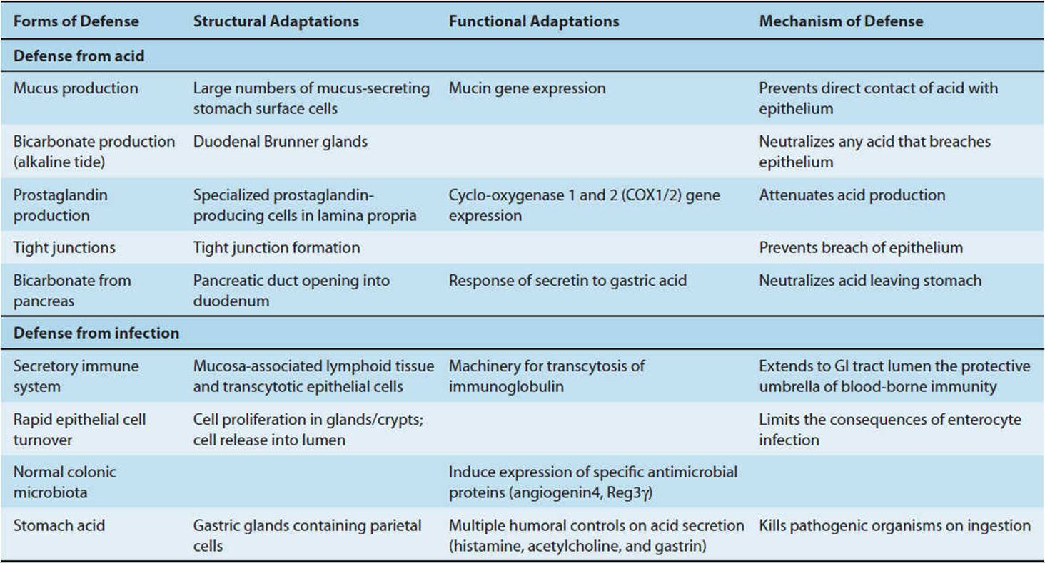
The mucosa of the GI tract is the largest surface of the body that is exposed to the environment, and the gut, like the skin, must protect the body from the external environment. Defense involves protection against ingested toxins, bacteria, and viruses, as well as the bacteria and toxins that normally exist in the large intestine (Table 13-2). The magnitude of the problem is illustrated by the observation that there are more bacterial cells in the human colon than cells in the entire body. Defense involves two mechanisms.
TABLE 13-2 Mechanisms of defense of the GI tract (and features of structure and function involved).
1. Adaptive immune defense—The mucosal immune system or gut-associated lymphoid tissue (GALT) surveys the contents of the intestinal lumen through a variety of mechanisms that utilize cells of both myeloid and lymphoid lineages. Myeloid-derived cells (specific populations of dendritic cells and macrophages) extend processes through the intestinal epithelial barrier that sense the luminal environment. Aggregates of lymphoid cells include Peyer patches (larger aggregates in the distal small intestine) and isolated lymphoid follicles located throughout the intestine. These lymphoid aggregates also play important roles in immune surveillance (Figure 13-4). GALT protects against pathogenic bacteria, viruses, and toxins and enables tolerance to potentially immunogenic dietary substances and bacteria.
2. Innate immune defense—These mechanisms include secretion of fluid (eg, abundant acid secreted by the stomach), electrolytes, and mucus, as well as the tight junctions between epithelial cells. The secretions neutralize and flush away potentially damaging bacteria and macromolecules, and the tight junctions of the gut epithelium prevent their ingress into tissues.

FIGURE 13-4 Systemic and local features of gut immunology. (Redrawn, with permission, from Kagnoff M. Immunology and disease of the gastrointestinal tract. In: Sleisenger MH et al, eds. Gastrointestinal Disease, 6th ed. Saunders, 1998.)
In the intestine, mucus is secreted by specialized goblet cells. The mucus forms a protective layer over the epithelial cells. A group of antimicrobial peptides are secreted into the intestinal lumen. The specialized cells in the small intestine that perform this function are the Paneth cells that produce and secrete lysozyme and alpha-defensins that contribute to defense and healing. Alpha-defensins have broad-spectrum activity and are thought to create holes in bacterial cell walls and prevent them from colonizing the small intestine. Trefoil peptides are secreted into the lumen of the GI tract with mucus. Among their many effects, they promote healing of mucosal lesions.
F. Regulation of Fluid & Electrolyte Balance
The small intestine receives 8–9 L of fluid with electrolytes per day and secretes a further 1 L and electrolytes per day. Most of the fluid is absorbed. Thus, secretion and absorption must be regulated to maintain balance. Increased secretion or diminished absorption causes diarrhea, which can be fatal because of fluid and electrolytes loss.
G. Excretion
Undigested food products, bacteria, and certain heavy metals (eg, copper and iron excreted in bile) are excreted in feces.
CHECKPOINT
4. What are the major functions of the GI tract?
5. Describe the four major layers of a cross-section through the GI tract.
6. What volumes of fluid are transferred into and out of the GI tract each day?
7. Describe the general mechanism of electrolyte transport across epithelial cells.
8. Describe the defense mechanism of the GI tract.
MECHANISMS OF REGULATION OF THE GI TRACT
The processes of motility, secretion, digestion, and absorption are under close physiologic regulation by nerves, hormones, and paracrine substance (Figure 13-5).

FIGURE 13-5 Neural, endocrine, and paracrine mechanisms of control in the GI tract.
A. Neural Control
There are two components of GI innervation.
1. Intrinsic innervation by the enteric nervous system—The enteric nervous system is the third division of the autonomic nervous system (Figure 13-6). An enteric neuron has its cell body within the wall of the GI tract and is thus intrinsic to the gut. The enteric nervous system comprises a series of ganglionated nerve plexuses that extend from the esophagus to the rectum, which are organized into two principal components: 1) the myenteric, or Auerbach, plexus, which is sandwiched between the layers of the muscularis externa; and 2) the submucosal or Meissner plexus, which lies in the submucosa. The enteric nervous system is extensive, containing as many neurons as are present in the spinal cord. It contains sensory or afferent neurons (sometimes called intrinsic primary afferent neurons [IPANs]) that sense the environment (eg, intestinal pH, osmolality, wall stretch), interneurons (the connectors), and secretomotor or efferent neurons that control many cell types to stimulate or inhibit motility, secretion, absorption, and immune function of the GI tract. In this manner, the enteric nervous system can regulate the GI tract in a reflex manner without input from the CNS. For this reason, it is often called the “little brain.” Enteric neurons use many neurotransmitters, most notably neuropeptides.
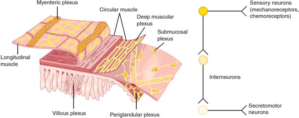
FIGURE 13-6 The enteric nervous system. Left: Enteric nervous system of the small intestine shows that enteric neurons are organized in two nerve plexuses, the submucosal plexus and myenteric plexus, with other plexuses including the deep muscular, periglandular, and villous plexus. (Redrawn with permission from Costa M et al. Histochemistry of the enteric nervous system. In: Johnson LR, ed. Physiology of the Gastrointestinal Tract, 2nd ed. Raven Press, 1987.) Right: The enteric nervous system includes sensory neurons, interneurons, and motor neurons. Complete reflex arcs exist within the enteric nervous system.
The degree to which the CNS regulates the enteric nervous system varies with region. The characteristic functions of structures derived from the embryonic foregut (eg, esophageal peristalsis, relaxation of the lower esophageal sphincter, gastric accommodation and peristalsis, pyloric sphincter function) are more dependent on CNS control. However, functions of structures derived from the embryonic midgut and hindgut (eg, intestinal peristalsis and mucosal secretion) can continue without input from the CNS.
The clinical importance of the enteric nervous system is seen in clinical syndromes in which its function is lost, which can occur at several levels. In esophageal achalasia, for example, as a result of enteric nervous system defects, the body of the esophagus is quiet and the lower sphincter is tonically contracted, making ingestion of food difficult or impossible. More distally, failure of the enteric nerves to migrate into the colon during development (in Hirschsprung disease) or loss of enteric nervous system function (in pseudo-obstruction of the small bowel) have severe clinical consequences, including abdominal pain, distension, and a risk of catastrophic intestinal perforation.
2. Extrinsic innervation by parasympathetic and sympathetic nerves—Extrinsic neurons that innervate the GI tract have cell bodies outside of the gut wall and allow a bidirectional communication between the brain and the gut (the brain-gut axis) (Figure 13-7). This communication can regulate the function of the enteric nervous system or directly control the activity of other cell types.
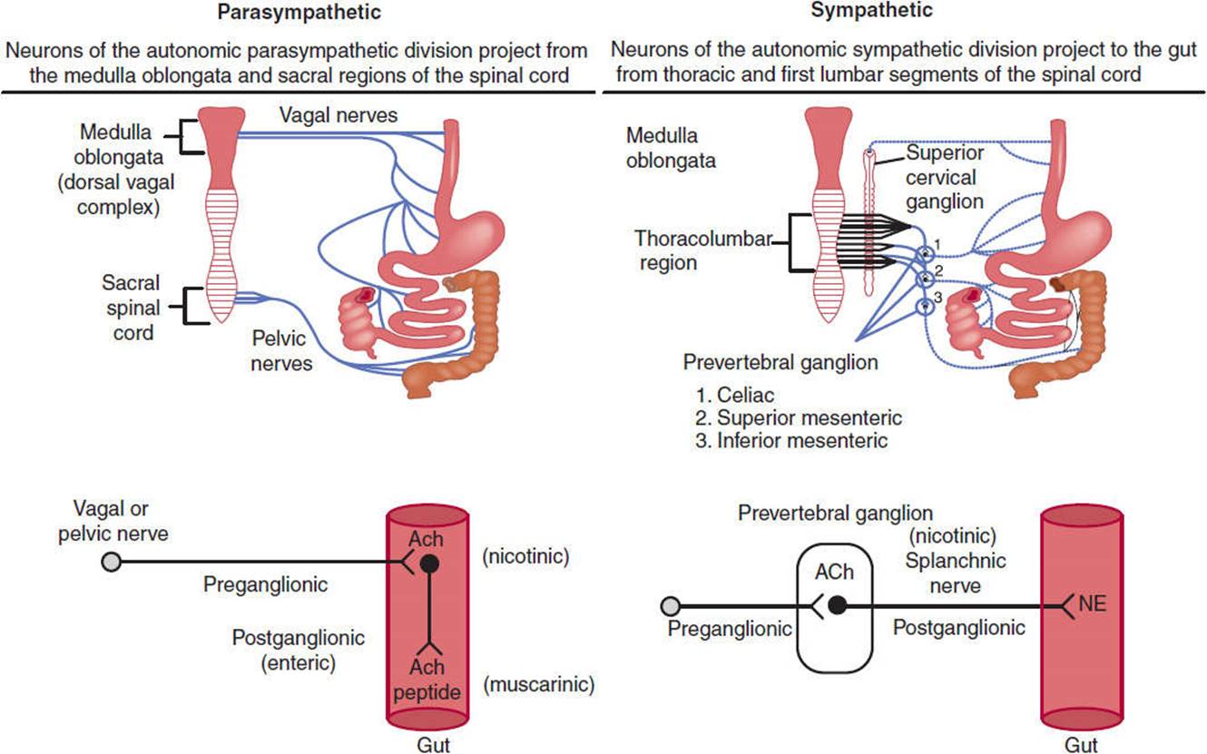
FIGURE 13-7 The extrinsic innervation of the GI tract by the parasympathetic and sympathetic nerves. Preganglionic parasympathetic nerves from the medulla and the sacral spinal cord project fibers in the vagal and pelvic nerves, respectively, to the wall of the GI tract and innervate enteric neurons that serve as postganglionic parasympathetic nerves. Preganglionic sympathetic nerves project fibers from the thoracolumbar regions of the spinal cord to the prevertebral ganglia, where they innervate postganglionic sympathetic nerves that project to the GI tract. Both the parasympathetic and sympathetic preganglionic nerves release acetylcholine (ACh), which activates nicotinic receptors on postganglionic nerves. Postganglionic parasympathetic nerves release acetylcholine and peptides, whereas postganglionic sympathetic nerves release norepinephrine (NE).
In parasympathetic innervation, the vagus nerve (cranial nerve X) innervates the esophagus, stomach, gallbladder, pancreas, and the first part of intestine, cecum, and proximal colon. The pelvic nerve from the sacral spinal cord innervates the distal colon and the rectum. Preganglionic cell bodies in the medulla (vagus) or sacral spinal cord (pelvic nerve) project fibers to some enteric neurons in the gut wall, which are thus in a sense postganglionic parasympathetic nerves. The preganglionic nerves use acetylcholine as a neurotransmitter, which activates nicotinic receptors on enteric neurons. The postganglionic enteric nerves use acetylcholine (acting on muscarinic receptors) and neuropeptides as neurotransmitters. Parasympathetic stimulation can stimulate and inhibit GI functions.
In sympathetic innervation, preganglionic sympathetic nerves arise from cell bodies in the thoracic spinal cord and project fibers to prevertebral ganglia (celiac, cranial, and caudal mesenteric ganglion). They release acetylcholine as a neurotransmitter that interacts with nicotinic receptors on the postganglionic nerves. Postganglionic fibers innervate some enteric neurons or directly innervate effector cells in the GI tract, such as vascular smooth muscle cells. Norepinephrine is the major postganglionic neurotransmitter. Sympathetic innervation is often inhibitory to GI functions.
Regarding extrinsic sensory nerves, parasympathetic and sympathetic nerve tracts also carry sensory fibers from the gut to cell bodies that are located in nodose ganglia and the dorsal root ganglia, respectively. Cell bodies in the nodose and dorsal root ganglia then project fibers to the brain stem (from nodose ganglia) or spinal cord (from dorsal route ganglia). Sensory nerve fibers in the wall of the GI tract detect mucosal pH and osmolality and can respond to amino acids or glucose, temperature, tension, and touch. In this manner, the extrinsic sensory nerves sense changes in the environment of the intestine and trigger central reflexes that initiate secretomotor changes to maintain normal homeostasis. Extrinsic sensory nerves also contribute to GI inflammation and pain. Sensory nerve endings in the wall of the gut detect noxious chemical and mechanical stimuli, including acid, inflammatory agents, and distension. These stimuli trigger the release of the neuropeptides, substance P, and calcitonin gene-related peptide, from the endings of sensory nerves within the gut wall, where they induce extravasation of plasma proteins and infiltration of granulocytes and arteriolar vasodilatation to cause neurogenic inflammation. The same stimuli induce release of neuropeptides from the central projections of these neurons, where they participate in pain transmission. Additional research is required to define the mechanisms of neurogenic inflammation and GI pain.
B. Hormonal Control
Hormones are blood-borne messengers released from endocrine cells or glands into the circulation, which carries them to distant target cells (Figure 13-5). This mechanism of endocrine regulation was discovered in the GI tract in 1902, when Bayliss and Starling discovered the hormone secretin in the small intestine and showed that it stimulates secretion from the exocrine pancreas. Since then, a large number of hormones have been identified in all regions of the GI tract. In this respect, the GI tract is the largest endocrine organ.
GI hormones have several characteristics in common. They are secreted from endocrine cells that are scattered throughout the mucosa of the stomach and intestine rather than being concentrated in specialized glands. This diffuse distribution made purification a truly Herculean task: Many hundreds of kilograms of intestine were required to isolate a few milligrams of pure hormone. GI hormones are invariably peptides, and many of these peptides are present not only in endocrine cells but also in nerves of the enteric system and CNS (Table 13-3). Thus, they have dual functions as hormones and neurotransmitters. After feeding, there are elevated levels of many GI hormones in the circulation. When administered to reproduce postprandial plasma concentrations, these hormones have multiple biological effects, ranging from the stimulation of gastric acid secretion to the suppression of appetite. The physiologic role of some GI hormones has been clearly established by demonstration that antagonists of hormone receptors block certain physiologic processes. However, in many cases, such antagonists are not available, and the physiologic relevance of hormones that cannot be antagonized remains to be determined.
TABLE 13-3 Secretory products of the GI tract.

C. Paracrine Control
Many substances that are used for intercellular signaling are rapidly removed from the extracellular fluid by uptake into nearby cells or by enzymatic degradation. Such substances have a short half-life in the extracellular fluid and are consequently only capable of regulating neighboring cells. Paracrine substances are released from nonneuronal sensory cells and neurons and regulate the function of neighboring cells rather than influencing distant organs by passage through the circulation (Figure 13-5 and Table 13-3). Examples include histamine and somatostatin, which are released from cells in the stomach to control acid secretion, and serotonin (5-hydroxytryptamine [5-HT]), which is released in the small intestine to control activity of the vagus nerve.
CHECKPOINT
9. What are the three general mechanisms of control observed in the GI tract?
10. What are the two components of the enteric nervous system?
11. What are the three general types of enteric neuron?
12. Describe the parasympathetic and sympathetic innervation of the GI tract.
13. What is the relationship between the enteric and central nervous systems?
GI Smooth Muscle
A. Structure of GI Smooth Muscle
The two principal muscle layers that control motility of the GI tract are the inner circular layer and the outer longitudinal layer of the muscularis externa. They vary in thickness in different regions of the GI tract. For example, the muscles are thickened in the gastric antrum, where strong contractions break up food before it can enter the small intestine, and muscle layers are thickened to form sphincters. Most of the GI muscle is smooth muscle, except the pharynx, parts of the esophagus, and the external anal sphincter, which are made up of striated (skeletal) muscle. GI smooth muscle is similar to smooth muscle in other organs: Fusiform cells are packed together in bundles by connective tissue sheaths. Gap junctions between cells allow signals to readily pass from cell to cell so that the contraction of bundles occurs synchronously. Interstitial cells of Cajal form an extensive network of stellate cells in the muscle layers of the stomach and intestine that are intimately associated with smooth muscle cells and enteric neurons (Figure 13-8). They may have two functions. First, they transmit information from enteric neurons to the smooth muscle cells. Second, they are the pacemaker cells, which have the capacity to generate the basic electrical rhythm or slow waves that are a consistent feature of GI smooth muscle. Animals lacking interstitial cells of Cajal show markedly abnormal GI motility, including defective gastric emptying and intestinal stasis or ileus. Defects in interstitial cells of Cajal may be associated with motility disturbances in patients, and this is an area of active investigation.
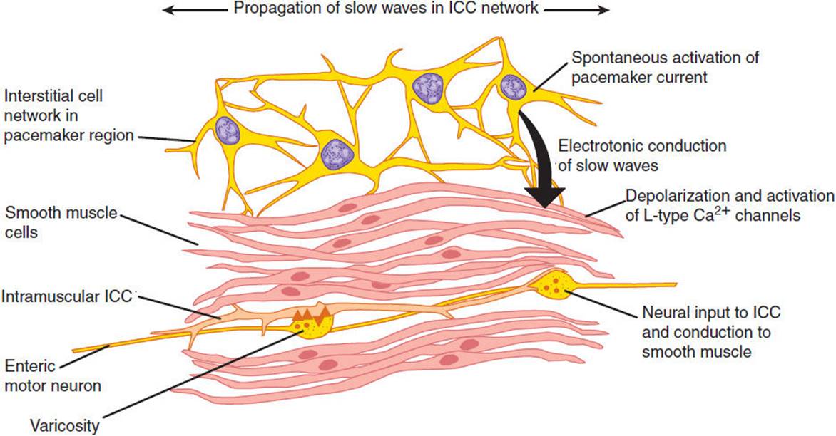
FIGURE 13-8 Diagrammatic view of interstitial cells of Cajal (ICC) in the intestine, showing their interaction with enteric nerves and smooth muscle cells.
B. Electrophysiology of GI Smooth Muscle
GI smooth muscle cells have a resting membrane potential of −40 to −80 mV as a result of the relative conductances of K+, Na+, and Cl− ions. An electrogenic Na+-K+ ATPase contributes significantly to the resting membrane potential. Less is known about the electrophysiological properties of interstitial cells of Cajal, in part because of difficulties in isolating these cells for study. The resting membrane potential of smooth muscle cells varies characteristically with time and is called a slow wave or basic electrical rhythm. Slow waves occur at 3–5/min in the stomach and at 12–20/min in the intestine. Interstitial cells of Cajal set the frequency of the slow waves, and slow waves are transmitted between cells through gap junctions. Nerves and hormones modulate the amplitude of slow waves. Depending on the amplitude of the slow waves and the excitability of the smooth muscle, slow waves can give rise to action potentials. If the slow-wave depolarization reaches a threshold, a train of action potentials will fire. Action potentials depolarize the membrane of the smooth muscle cells and induce an influx of Ca2+ ions into the cytoplasm through voltage-sensitive Ca2+ channels in the plasma membrane and from intracellular stores, causing contraction. What causes an action potential to occur? The presence of neurotransmitters or hormones that are released close to the smooth muscle cells alters the resting membrane potentials of the cells, which makes the oscillations in membrane potential (the slow waves) more or less likely to reach threshold and initiate an action potential. However, because inhibitory motor neurons of the GI tract are highly active and can prevent generation of action potentials, not all slow waves result in active contractions. Action potentials and contractions can only occur when these inhibitory motor neurons are switched off by input from interneurons. Thus, the tonic inhibition serves to contrail the inherent excitability of the pacemaker cells.
C. Mechanical Properties of GI Smooth Muscle
Several characteristic patterns of contraction can be observed in GI smooth muscle. Tonic contractions are best represented by sphincters that act as one-way valves to prevent retrograde movement of material from distal to more proximal regions and thus to facilitate flow in an aboral direction. The proximal parts of the stomach and the gallbladder also exhibit tonic contractions. Peristaltic contractionsare moving waves of contraction that propel digesta along the GI tract. Peristalsis involves neurally mediated contraction of smooth muscle on the oral side of a bolus of digesta and a neurally mediated relaxation of muscle on the anal side of the digesta. Peristalsis occurs in the pharynx, esophagus, gastric antrum, and small and large intestine. Segmental contractions produce narrow contracted segments between relaxed segments. These movements allow mixing of the luminal contents with GI tract secretions and increase exposure to mucosal surfaces where absorption occurs. Segmentation occurs in the stomach and intestine. Pathologic patterns of motility include spasms, which are very strong and often painful contractions that occur continuously in a dysregulated manner, and ileus, where there is a markedly decreased or absent contractile activity. Ileus often results from irritation of the peritoneum involved in surgery, peritonitis, and pancreatitis. Further research is required to understand the mechanisms of these abnormal contractions, which may lead to improved therapies.
CHECKPOINT
14. What are the positive and negative regulators of smooth muscle cell action potentials?
15. What are the functions of interstitial cells of Cajal?
16. What are the general types of contractions observed in the GI tract after feeding?
OROPHARYNX & ESOPHAGUS
Anatomy & Histology
The oropharynx provides entry to the GI tract during swallowing and to the respiratory tract during breathing. It includes the vocal cords, which separate the two tracts and provide the structural basis for speech. Much of the oropharynx is lined with a respiratory-type ciliated pseudocolumnar epithelium.
The esophagus is a hollow tube (25–30 cm long, 2–3 cm wide). The wall of the esophagus consists of a stratified squamous epithelial cell layer, an inner layer of circular muscle, a myenteric nerve plexus, and an outer layer of longitudinal muscle. The first third of esophagus is composed of striated muscle, the middle third is mixed striated and smooth muscle, and the lower third is purely smooth muscle. The esophagus is delimited by an upper esophageal sphincter (a distinct thickening of striated circular muscle) and a lower esophageal sphincter (a tonically contracted 3–4 cm ring of smooth muscle). The two sphincters generate small luminal zones of high pressure, whereas the rest of the esophageal lumen is at a pressure equal to the surrounding body cavities. Between swallows, the two sphincters are closed, preventing entry of air and gastric acid into the esophagus. Regulation of the lower esophageal sphincter is especially important because it controls the passage of digesta into the stomach and prevents the reflux of gastric contents into the esophagus, where they can damage the mucosa. Between swallows, the lower esophageal sphincter is contracted, in large part by vagal cholinergic mechanisms. During swallowing, vagal inhibitory fibers allow the lower esophageal sphincter to relax, possibly because of release of inhibitory neurotransmitters from enteric nerves, including nitric oxide and vasoactive intestinal peptide (VIP).
Swallowing Reflex
Swallowing begins as a voluntary process that rapidly becomes an involuntary reflex mechanism. During the voluntary oral phase, the tongue pushes a bolus of food to the back of the mouth and into the oropharynx. From there on, the process is involuntary. In the pharyngeal phase, the food bolus stimulates touch receptors in the pharynx. Sensory signals pass by the glossopharyngeal, vagal, and trigeminal nerves to the swallowing center in the medulla and pons. Motor impulses pass through cranial nerves to control an involuntary process that directs food into the esophagus and away from the airway. Breathing is interrupted and the soft palate is elevated, closing the pharyngeal opening of the nasopharynx and preventing food from entering the internal openings of the nostrils. The tongue is pressed against the hard palate, closing the oral opening of the pharynx. The glottis is pulled under the epiglottis, which blocks the laryngeal opening. Cartilages around the larynx are pulled together, further restricting food from entering the respiratory tract. When all openings to the pharynx are closed, a wave of muscular contraction pushes the bolus of food toward the opening of the esophagus. As the food reaches the esophagus, the upper esophageal sphincter relaxes to accept the material and then closes after the bolus has moved through. The esophageal phase of swallowing begins when the bolus passes through the upper esophageal sphincter. Vagal stretch receptors in the wall of the esophagus detect distension by the bolus and induce a vagovagal reflex, during which vagal motor nerves induce a wave of contraction that spreads along the esophagus at 3–5 cm/s. This is termed primary peristalsis (Figure 13-9). As the wave of primary peristalsis reaches the lower esophageal sphincter, the sphincter relaxes to allow the bolus to enter the stomach. Distension of the esophagus by the bolus can initiate another wave of contraction called secondary peristalsis. Often repetitive waves of secondary peristalsis are required to clear the esophagus of food. Various hormones and neurotransmitters, foods, and drugs can affect the tone of the lower esophageal sphincter pressure.
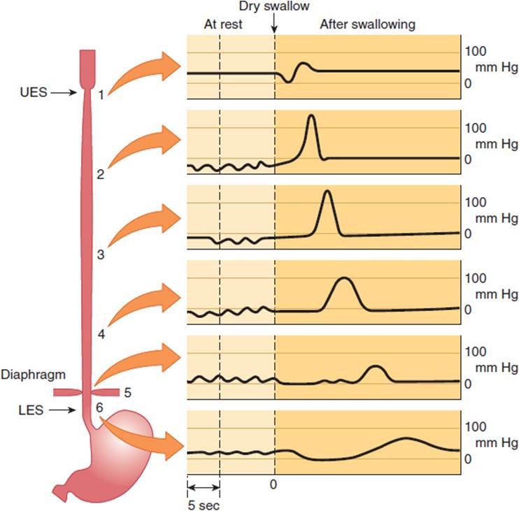
FIGURE 13-9 Primary peristalsis of the esophagus. The tracings show pressures in the indicated regions of the esophagus at rest and at various times after swallowing. UES, upper esophageal sphincter; LES, lower esophageal sphincter. (Redrawn from data in Conklin JL et al. Motor functions of the pharynx and esophagus. In: Johnson LR, ed. Physiology of the Gastrointestinal Tract, 3rd ed. Lippincott-Raven, 1994.)
The importance of oropharyngeal motility and its control is seen in patients who have had strokes or are demented. Inability to swallow properly often makes them unable to manage their own oral secretions, resulting in aspiration of oral contents into the lungs with development of pneumonia. This is a common cause of death in individuals with these kinds of CNS disorders. Disordered lower esophageal sphincter tone can cause gastroesophageal reflux disease (GERD), presenting as heartburn and potentially increasing risk for adenocarcinoma of the esophagus.
CHECKPOINT
17. What is the histologic difference between the proximal one-third and the distal two-thirds of the esophagus?
18. What are the functions of the upper and lower esophageal sphincters, and how are they regulated?
19. Describe the three phases of the swallowing reflex.
STOMACH
Anatomy & Histology
The stomach is a complex glandular organ that is guarded by two sphincters: the lower esophageal sphincter and the pyloric sphincter (Figure 13-10). The mucosa is formed by a single layer of epithelial cells that line the lumen of the stomach and descend into funnel-shaped invaginations that are broad at the top near the surface and then narrow as they descend deeper. The broad upper portion is called the pit zone. The middle portion, where the invaginations narrow into glands, is the neck, and the deepest zone is the base. The stomach can be divided anatomically into several regions on the basis of structure and function. The cardia is a small region just distal to the lower esophageal sphincter where the gastric glands are almost entirely composed of mucus-secreting cells. The corpus, or body, is the major part of the stomach and includes the fundus, which is the portion of the body superior to the insertion of the esophagus. Gastric glands in the corpus contain parietal cells (mostly in the neck zone), which secrete hydrochloric acid and intrinsic factor, and chief cells (mostly in the base zone), which secrete pepsinogen. The corpus is the principal site of gastric digestion. The pyloric antrum is the distal region of the stomach that secretes the hormone gastrin from G cells. Glands of the antrum, like those of the cardia, secrete mostly mucus. The antrum, a highly muscular portion of the stomach, grinds food and regulates gastric emptying.
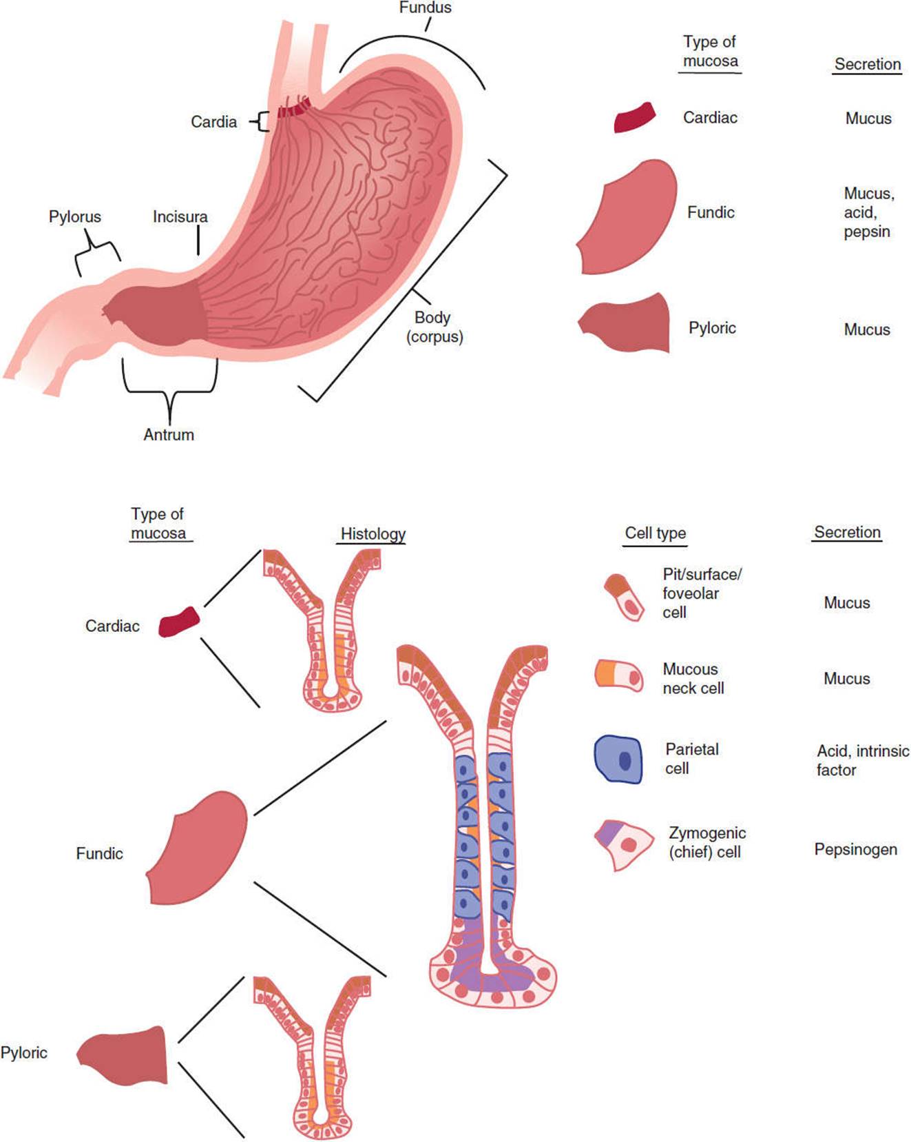
FIGURE 13-10 Anatomy and histology of the stomach. (Redrawn, with permission, from Boron WF et al, eds. Medical Physiology. Saunders, 2009.)
Gastric Acid Secretion
A number of products are secreted from the stomach. Of these, hydrochloric acid is perhaps the most important from a pathophysiologic standpoint. Secretion of acid by the parietal cells of the gastric glands occurs in a basal diurnal pattern but can be stimulated by such diverse factors as the thought of food, distension of the stomach, and protein ingestion.
A. Molecular Mechanisms of HCl Secretion
The mechanisms by which parietal cells secrete HCl into the stomach have been intensively studied because of the importance of acid secretion to digestion and in disease states. Parietal cells are roughly pyramidal in shape. Their membranes express a H+-K+ ATPase, a primary active transporter that is responsible for the secretion of HCl. Parietal cells undergo a remarkable change in appearance when stimulated to secrete HCl (Figure 13-11). In the unstimulated state, they harbor an intracellular tubulovesicular network studded with H+-K+ ATPase molecules. On activation, the tubulovesicular membranes fuse with the plasma membrane to form a canalicular membrane with microvilli. The result is an increase in the area of the apical membrane by 50- to 100-fold that allows markedly increased secretion of HCl by the H+-K+ ATPase pumps directly into the glandular lumen, which in turn squirts the acid into the lumen of the stomach.
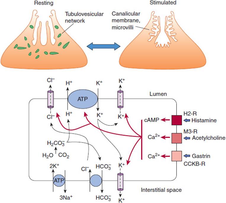
FIGURE 13-11 Acid secretion by parietal cells. Top: Upon stimulation, tubulovesicular network in the parietal cell fuses to form an extensive canalicular membrane with microvilli, which increases the surface area. Bottom: The mechanisms of HCl secretion by parietal cells, stimulated by histamine, acetylcholine, and gastrin, are demonstrated. For abbreviations, see legend for Figure 13-12.
The H+-K+ ATPase is a heterodimer of an α-subunit (the catalytically active unit) and a β-subunit (involved in fixing the intracellular location). The H+-K+ ATPase pumps H+ ions from the cell across the apical membrane in exchange for K+ ions (Figure 13-11). This is an example of primary active transport that is driven by ATP, which pumps H+ ions against an enormous concentration gradient (1 million:1). Tight junctions between cells prevent the reentry of H+ ions into the mucosa. The K+ ions that have entered the cells then recycle to the lumen or enter interstitial fluid by K+ channels. To maintain electroneutrality, Cl− ions are secreted passively across the apical membrane into the lumen through Cl− channels, forming HCl. The secreted H+ ions are provided by H2O and CO2, which form H2CO3. Carbonic anhydrase generates H+ ions for secretion and HCO3− ions, which enter the interstitial fluid by exchange for Cl− ions. Cl− ions enter against their electrochemical gradient, driven by efflux of HCO3−down an electrochemical gradient. The secretion of HCO3− into the blood forms the “alkaline tide,” which can lead to alkalosis when H+ ion secretion is excessive. Water movement maintains the osmotic balance in all regions.
An understanding of the mechanisms of HCl secretion by parietal cells permitted the development of proton pump inhibitors (PPIs), a class of drugs that inhibit the H+-K+ ATPase. Drugs such as omeprazole, a benzimidazole, are inactive at neutral pH levels but, when acidified (in the stomach), bind to sulfhydryl groups of cysteine residues on the external surface of the H+-K+ ATPase, irreversibly inhibiting activity and blocking hypersecretion of gastric acid. Other experimental drugs, termed acid pump antagonists, competitively interfere with K+ ion binding to block acid secretion. These drugs can be used to inhibit the hypersecretion of gastric acid, which causes GERD.
B. Stimulants and Inhibitors of HCl Secretion
The three main stimulants of H+ ion secretion are acetylcholine, gastrin, and histamine, all of which stimulate HCl secretion and induce characteristic shape changes of the stimulated parietal cell. Acetylcholine is released from vagal postganglionic or enteric neurons during feeding. It binds to muscarinic M3-type muscarinic receptors on parietal cells to stimulate H+ ion secretion. Gastrin is a peptide hormone of 17 or 34 amino acids that is secreted from G cells in the gastric antrum during feeding. Gastrin binds to cholecystokinin (CCK) type B receptors on parietal cells, which also stimulates H+ ion secretion.
Both acetylcholine and gastrin receptors activate the same signal transduction pathways: activation of phospholipase Cβ, leading to generation of inositol trisphosphate that mobilizes Ca2+ from intracellular stores, and diacylglycerol, which activates protein kinase C. Because both acetylcholine and gastrin act through similar intracellular pathways, the combined effects of gastrin and acetylcholine are additive.
Histamine is a paracrine substance secreted by enterochromaffin-like (ECL) and mast cells in the corpus mucosa during feeding. Histamine binds to H2 receptors on parietal cells to activate adenylyl cyclase and increase cAMP. The cAMP activates protein kinase A to stimulate H+ ion secretion. The combination of histamine and acetylcholine or gastrin can increase the rate of acid production by up to 10-fold over basal levels, a much greater effect than simple addition of the effects of the agonists would predict. This effect is known as potentiation. Potentiation requires that two different signal molecules bind to receptors that act through different intracellular mechanisms. Increased intracellular Ca2+ and cAMP activate K+ channels on the apical membrane of parietal cells, thereby promoting K+ ion efflux from the cell. This hyperpolarizes the cell (more negative inside) to promote Cl− ion secretion across the apical membrane. Ca2+ and cAMP also increase insertion of Cl− channels and H+-K+ ATPase into the apical membrane. The combined effects are to stimulate HCl secretion.
Gastrin also regulates growth of the gastric epithelium. Excess gastrin produced by certain tumors causes hyperproliferation of gastric glands and parietal cells and excess secretion of gastric acid. The excess acid in the small intestine can lead to ulceration of the mucosa, steatorrhea as a result of inactivation of pancreatic lipases (which are inhibited by low pH), and diarrhea. This condition is termed Zollinger-Ellison syndrome. Excessive administration of PPIs can result in prolonged high luminal pH, which stimulates hypersecretion of gastrin and increased mucosal growth. Termination of drug treatment then results in an acid production rebound because of the increased content of parietal cells and gastrin-secreting G cells.
In addition to the direct mechanisms by which acetylcholine, gastrin, and histamine stimulate HCl secretion from parietal cells, acetylcholine and gastrin also indirectly stimulate secretion by acting on enterochromaffin-like cells to promote the release of histamine, which in turn stimulates parietal cells. The importance of histamine to H+ ion secretion is illustrated by studies with histamine H2 receptor antagonists, such as cimetidine. These drugs not only inhibit histamine-stimulated H+ ion secretion but also block the effects of acetylcholine and gastrin. By preventing such potentiation, these agents can be used to effectively treat the hypersecretion of gastric acid.
Somatostatin, a peptide of 14 or 28 amino acids, is an important inhibitor of gastric acid secretion. Somatostatin directly inhibits proton secretion by activating receptors on parietal cells, which couple to produce inhibition of cAMP. Somatostatin also inhibits gastrin and histamine secretion, which indirectly inhibits proton secretion. Somatostatin is secreted by D cells in the gastric antrum and corpus. D cells in the gastric antrum have direct contact with the stomach lumen (open endocrine cells), allowing them to sense the luminal contents. Protons in the antrum stimulate somatostatin secretion, which acts as a paracrine agent to inhibit gastrin secretion from neighboring G cells and to thereby indirectly reduce gastric acid secretion. This is an example of negative-feedback regulation. D cells in the corpus do not have contact with the lumen (closed cells) and thus cannot sense luminal protons. Instead, multiple neurohumoral factors (eg, noradrenalin, CCK, VIP) increase release of corpus somatostatin, which in turn inhibits acid production indirectly by decreasing histamine release from ECL cells and directly by inhibiting parietal cells. Vagal ACh and the TH1 cytokine interferon-γ inhibit somatostatin release and promote acid secretion. Recent studies indicate that the endocrine cells in the stomach sense luminal nutrient and acid via primary cilia on their apical surfaces.
C. Integrated Regulation of Gastric Acid Secretion
Secretion of gastric acid between meals is low. Three phases of acid secretion occur during feeding (Figure 13-12). The cephalic phase (∼30% of response) of secretion is initiated by the sight, smell, taste, and swallowing of food. These stimuli activate the dorsal motor nucleus of the vagal nerve in the medulla and result in vagal discharge and parasympathetic motor nerves. Stimulation has several consequences. In the corpus, postganglionic nerves release acetylcholine, which directly activates parietal cells by M3 receptors. Acetylcholine also induces histamine release from enterochromaffin cells, which stimulates H+ ion secretion by parietal cells. In the antrum, vagal stimulation induces release of the peptide, gastrin-releasing peptide, from postganglionic fibers, which stimulates gastrin release and thus indirectly stimulates secretion of H+ ion secretion. Acetylcholine also inhibits somatostatin release from D cells in the corpus and pylorus to stimulate secretion of H+ ions.
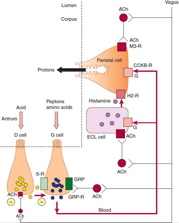
FIGURE 13-12 Regulation of gastric acid secretion by nerves and hormones. During the cephalic phase of digestion, vagal cholinergic nerves directly stimulate parietal cells and induce release of histamine from ECL cells, which also stimulate parietal cells. Vagal fibers also release gastrin-releasing peptide (GRP) in the antrum to induce gastrin secretion, which is carried in the bloodstream to induce release of histamine and stimulate parietal cells. During the gastric phase of digestion, food in the stomach triggers vagovagal reflexes and also stimulates gastrin secretion. Acidification of the gastric antrum stimulates the release of somatostatin, which inhibits gastrin release and thus acid secretion; vagal ACh inhibits somatostatin release. (ACh, acetylcholine; G, gastrin; S, somatostatin; M3-R, muscarinic 3 receptor; H2-R, histamine 2 receptor; CCKB-R, cholecystokinin B receptor; ECL, enterochromaffin-like; GRP-R, GRP receptor; GRP, gastrin-releasing peptide.)
The gastric phase (∼70% of response) of secretion is induced by stimuli within the stomach. Vagal sensory nerves detect gastric distension with food and trigger a vagovagal reflex during which vagal motor nerves release acetylcholine in the stomach to promote acid secretion. Partially digested proteins and amino acids stimulate gastrin release from G cells in the pylorus. G cells, like D cells, are open-type endocrine cells that directly sense the contents of the stomach. Gastrin then further stimulates acid secretion. Acidification of the pylorus stimulates somatostatin release, which inhibits acid secretion by a negative-feedback loop as described.
During the intestinal phase, the products of protein digestion, on entering the small intestine, can stimulate gastrin release from G cells in the duodenum. Many substances, most notably fat and acid, stimulate the secretion of hormones from the small intestine that inhibit gastric acid secretion. Examples include secretin and cholecystokinin.
Helicobacter pylori is a bacterium that lives in the mucous layer of the stomach where the enzyme urease is active, converting urea to CO2 and ammonia. Ammonia buffers luminal acid and protects the organism. H pylori also secretes proteins, such as CagA and VacA, that modulate immune responses and directly alter mucosal cell signaling pathways. More than half of the world’s population is infected with H pylori. In most cases, the infection, though chronic, is mild and does not cause symptoms. In some individuals, however, the infection leads to increased acid secretion and symptomatic inflammation that causes ulceration of the stomach or duodenum. Almost all duodenal peptic (i.e., acid-associated) ulcers and about half of gastric peptic ulcers have H pylori infection as a root cause; the remaining gastric ulcers are caused by medications (such as aspirin and nonsteroidal anti-inflammatory drugs). In some patients, chronic H pylori infection can lead to death (atrophy) of parietal cells, chronic inflammation, and altered mucosal differentiation patterns (metaplasias) that increase the risk of progression to gastric cancer. In certain geographical regions (eg, East Asia and parts of Central and South America), due to environmental and/or lifestyle factors that have not yet been elucidated, risk for progression to gastric cancer is much higher than in other regions (eg, United States and Canada).
Other Gastric Secretions
Chief cells in the glands of the gastric corpus secrete pepsinogen, an inactive precursor (zymogen) of the active protease, pepsin. Acetylcholine is the main stimulant of pepsinogen secretion, although other factors (eg, gastrin) also stimulate secretion. Once released into the lumen of the stomach, gastric acid and preexisting pepsin convert pepsinogen to pepsin. Pepsin has a pH optimum of 3.0 and is thus active in the stomach. It is an endopeptidase that begins the degradation of dietary proteins to peptides. However, pepsin accounts for only 10% of the total protein digestion.
Mucins are high-molecular-weight glycoproteins secreted by mucous cells of gastric glands in the corpus and antrum. The peptide backbone of mucins is densely populated with carbohydrate side chains enriched with sulfate groups. Mucins combine with phospholipids, bicarbonate, and water to form the mucus gel layer that adheres to the surface of epithelial cells of the stomach. This layer forms physical protection for epithelial cells from damage by contractile grinding of food as well as noxious substances such as acid, pepsin, and bile acids. Acetylcholine and mucosal irritation stimulate secretion of mucin.
Epithelial cells of the corpus and antrum secrete HCO3− ions. Although the secretion of HCO3− is minor compared with H+ ion secretion, HCO3− plays a major role in epithelial defense. HCO3− ions are trapped in the mucous gel to form an “unstirred layer” in proximity to the epithelium, where the pH is 7.0 compared with 1.0–3.0 in the lumen. Acetylcholine and intraluminal acid stimulate HCO3− secretion.
Intrinsic factor is a glycoprotein secreted by parietal cells that is required for vitamin B12 absorption. Vitamin B12 (cobalamin) is not made in mammalian cells, and the only source is the diet: meat, fish, dairy products, but not vegetables or fruit. In the stomach, acid and pepsin release B12 from dietary carrier proteins. The acid environment permits binding of B12 to haptocorrin (R factor), a glycoprotein produced by salivary glands and gastric glands. The B12-haptocorrin complex enters the duodenum, where pancreatic proteases digest the haptocorrin. Free intrinsic factor also enters the duodenum. Intrinsic factor combines with B12 in the less acidic environment of the small intestine, forming a degradation-resistant complex for transport to the ileum. Specific receptors on epithelial cells lining the ileum bind the vitamin B12–intrinsic factor complex, which is taken into cells by endocytosis. The absorbed complex dissociates within the epithelial cells, and then vitamin B12 binds to transcobalamin II, a protein required for exocytosis and transport to the liver. In autoimmune gastritis, parietal cells are destroyed, leading to loss of intrinsic factor secretion, which can result in vitamin B12 deficiency and pernicious anemia. This anemia is caused by impaired synthesis of purines and thymine for which vitamin B12 is required. The only reliable therapy is regular intramuscular injections of vitamin B12.
GASTRIC MOTILITY
A. Patterns of Gastric Motility
In terms of motility, the proximal and distal regions of the stomach are distinct. The gastric corpus is a reservoir for gastric digestion. During each swallow, stretch of the esophagus induces a vagovagal reflex that causes the gastric corpus to relax in preparation to receive the food, a phenomenon known as receptive relaxation. When food enters the stomach, it relaxes further to accommodate a meal of 1.5 L without any increase in pressure, a phenomenon called accommodation, which involves vagovagal and local enteric reflexes. Thus, the stomach is a reservoir for ingested food. The antrum of the stomach is highly muscular, and here contractions serve to break food to smaller pieces and thereby facilitate digestion. The pyloric sphincter controls the rate at which the antral contractions propel partially digested food, or chyme, into the duodenum. During fasting, the antrum is relatively quiescent, with occasional forceful contractions that occur every 75–90 min. These intense contractions, of 5- to 10-min duration, are part of a general wave of contractions that sweep the entire length of the GI tract during fasting: the migrating myoelectric complex. Feeding disrupts the migrating myoelectric complex, and now the antrum contracts frequently at a rate of about three contractions per minute. These slow waves of peristaltic contraction originate from spontaneously active interstitial cells of Cajal in the pacemaker zone in the middle of the body of the stomach, and they sweep toward the antrum. When the membrane potential of muscle cells depolarizes to reach threshold, action potentials fire. Contractions occur during the plateau phase of the action potential. Gastrin and acetylcholine stimulate contraction by increasing the magnitude and duration of the action potentials.
B. Gastric Emptying
Immediately after a meal, the stomach may contain up to 1 L of material, which empties slowly into the small intestine. Regulation of gastric emptying occurs by alterations in motility of the proximal and distal stomach, pylorus, and duodenum. Gastric emptying is brought about by an increase in tone (intraluminal pressure) in the proximal stomach, increase in strength of antral contractions, opening of the pylorus, and inhibition of duodenal segmental contractions.
The rate of gastric emptying depends on the chemical and physical composition of chyme that enters the duodenum through the stimulation of both neural and hormonal pathways. Solids and liquids empty at different rates: Liquids empty rapidly, and solids empty only after a lag phase. Acid, fat, and hyperosmolar solutions entering the duodenum slow gastric emptying through stimulation of neuronal and hormonal mechanisms. Sensory neurons in the duodenum, both vagal and spinal, respond to nutrients, H+ ions, and hyperosmolar content of chyme. Vagal motor nerves decrease antral contractions, contract the pylorus, and decrease proximal gastric motility. This results in intestinal feedback inhibition (slowing) of gastric emptying. The main vagal mediator that stimulates contraction is acetylcholine. VIP and nitric oxide are neuronal mediators that inhibit contraction. Many hormones that are released by endocrine cells in the small intestine have been implicated in the feedback inhibition of gastric emptying. Secretin, the release of which is stimulated by acid, inhibits antral contractions and stimulates contraction of the pyloric sphincter to slow emptying. Cholecystokinin, the release of which is stimulated by fat, acts on receptors on vagal sensory nerves to produce a vagovagal reflex that decreases gastric emptying.
The importance of nervous system control over gastric motility is reflected in the high incidence of the dumping syndrome (nausea, bloating, flushing, and explosive diarrhea) that occurs as a consequence of stomach dysmotility in some patients who have undergone surgical procedures such as partial gastrectomy or nonselective vagotomy.
CHECKPOINT
20. Describe the cell types found in the mucosa of the gastric corpus and antrum, and indicate the products of each cell type.
21. What are the roles of the proximal and distal stomach?
22. Describe the ionic basis of secretion of HCl from the gastric parietal cells.
23. Name a neurotransmitter, hormone, and paracrine agent that stimulates acid secretion from parietal cells.
24. Name a peptide that inhibits acid secretion from the parietal cells.
25. Describe the mechanisms of the cephalic, gastric, and intestinal phases of gastric acid secretion.
26. Name two types of drugs with distinct mechanisms of action that can be used to treat hypersecretion of gastric acid.
27. What is the role of the parietal cell in absorption of vitamin B12?
28. Describe two processes by which the gastric mucosa is protected from acid in the lumen.
29. What are the patterns of motility in the corpus and the antrum?
30. How does the composition of the digesta in the lumen of the small intestine affect the rate of gastric emptying?
GALLBLADDER
Anatomy & Histology
The gallbladder is a muscular sac with a resting volume of about 50 mL that lies on the inferior surface of the liver. It is connected to the hepatic biliary system by the cystic duct, which leads to the common bile duct whose opening into the proximal duodenum is controlled by the sphincter of Oddi. The common bile duct and the pancreatic duct usually join just proximal to this sphincter.
Physiology
A. Bile Secretion
Bile, which is produced by the liver, flows down the hepatic duct and into the gallbladder through the cystic duct. It is stored there until stimulation of gallbladder contraction expels the contents of the gallbladder back through the cystic duct into the common bile duct and through the sphincter of Oddi into the duodenum. Stimuli for gallbladder contraction and sphincter of Oddi relaxation necessary for proper bile flow include both hormones and neural inputs. Fat in the intestine stimulates secretion of the hormone CCK from I-cells. CCK causes contraction of the gallbladder and relaxation of the sphincter of Oddi. Depending on how long it remains in the gallbladder, bile becomes concentrated. Bile composition is further modified by mucin production under the control of prostaglandins and by saturation of bile cholesterol controlled in part by estrogens. The most prominent disorders of the gallbladder involve gallstone formation (see later discussion).
SMALL INTESTINE
Anatomy & Histology
Three regions can be distinguished along the approximately 6–7 m length of the small intestine. The pyloric sphincter marks the beginning of the duodenum, which is largely retroperitoneal and fixed in its location and is 20–25 cm in length. Because of this sphincter, stomach contents normally enter the duodenum in small spurts containing tiny suspended particles. In the duodenum, gastric contents are mixed with the secretions of the common bile duct and pancreatic duct. Beyond the duodenum, the small intestine is mobile and suspended in the peritoneal cavity by a mesentery. The proximal two fifths is called the jejunum. The distal three fifths is called the ileum, which ends at the ileocecal valve at the start of the large intestine.
The most striking gross structural features of the small intestine are the numerous villi (projections of the mucosa into the lumen of the intestine that measure approximately 1 mm in height) (Figure 13-13). Each villus contains a single terminal branch of the arterial, venous, and lymphatic trees. Villi increase the absorptive capacity 5-fold and allow efficient transfer to the circulatory system of substances absorbed from the gut lumen by enterocytes (surface epithelial cells). By electron microscopy, each enterocyte contains 3000–5000 microvilli, plasma membrane evaginations on the apical side of the cell that further increase the absorptive surface area by 200-fold. Many digestive enzymes expressed by intestinal epithelial cells are located at the tips of these microvilli. As a group, these densely packed microvilli make up a “brush border” facing the intestinal lumen.
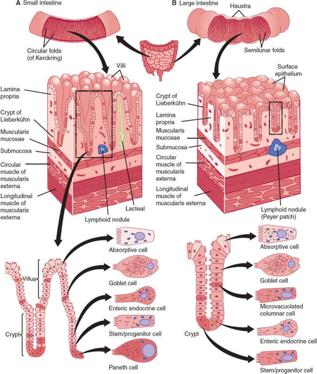
FIGURE 13-13 Anatomy and histology of the small and large intestine. (Redrawn, with permission, from Boron WF et al, eds. Medical Physiology. Saunders, 2003.)
Invaginations of the intestinal epithelium into the wall surround the villi and are called the crypts of Lieberkühn. These structures are the location of epithelial intestinal stem cells and their proliferative daughters that together constantly produce new differentiated epithelial cells that form the epithelial lining of the intestine. Each small intestinal crypt contains tetrapotential stem cells at or near the crypt base that produces the four mature epithelial cell types: absorptive enterocytes, mucus-secreting goblet cells; hormone-secreting enteroendocrine cells, and antimicrobial peptides and growth factor–secreting Paneth cells. Enterocytes, goblet, and enteroendocrine cells migrate out of crypts and onto adjacent villi. These cells then die by apoptosis at the tips of villi and are extruded into the lumen of the intestine; the average life span is about 4–6 days. On the other hand, Paneth cells are much longer lived (∼60 days) and they migrate to the crypt base where they are in close contact with epithelial stem cells.
It has been increasingly appreciated that the external and internal surfaces of the human body are inhabited by commensal microbes that are not mere passengers but that actually perform critical functions. In the adult small intestine, a large and diverse population of commensal microbes inhabit the lumen. Most of these microbes are bacteria and the major phyla represented are Bacteroides and Firmicutes. Most of these microbes are anaerobes (they are able to live in the absence of oxygen). The density increases dramatically in the lumen of the small intestine (from hundreds per milliliter in the duodenum to trillions per milliliter in the colon). These bacteria function to aid in the digestion of complex carbohydrates. Based on studies in germ-free organisms, it is estimated that these intestinal microbiota increase our ability to extract nutrients from food by as much as 30%. The microbiota also play key roles in training the mucosal immune system and development of blood vessels in the intestine. The microbial populations that are most closely associated with the mucosa appear to be quite distinct from those associated with the lumen and include many members of the Lachnospiraceae and Ruminococcaceae families. Functioning cooperatively, these microbes form a critical barrier to pathogens. Furthermore, bacteria are not the only commensal microorganisms in the intestine. We now appreciate that Archaea (single-cell microorganisms without nuclei), fungi, and viruses are normally present in the lumen of the intestine. In most cases, their roles are still undefined.
Digestion & Absorption in the Small Intestine
The small intestine is the main site of digestion and the principal site for nutrient absorption. Thus, it is appropriate to review all steps of digestion in the GI tract and then to consider the mechanisms by which these nutrients are absorbed.
A. Carbohydrates
Carbohydrates, which are mainly present in the diet as polysaccharides and disaccharides, must be digested to monosaccharides for absorption. Intestinal microbes (in particular, Bacteroides spp) contain a large repertoire of glycoside hydrolases that aid in the breakdown of complex plant polysaccharides. This is an important beneficial function of commensal intestinal microbes. Alpha-amylases in salivary and pancreatic secretions cleave interior α-1,4 glucose linkages in large polymers of starch to form fragments (disaccharides, trisaccharides, and oligosaccharides). Oligosaccharidases and disaccharidases in the brush border of enterocytes digest small fragments to the monosaccharides, glucose, galactose, and fructose. Glucose and galactose, along with two Na+ ions, are absorbed across the apical membrane of enterocytes by the same transporter, SGLT1. Passive uptake of water also occurs, maintaining osmolality on both sides of the cell membrane. The extrusion of Na+ out of the basolateral membrane by the Na+-K+ ATPase provides an electrochemical Na+ gradient that drives the absorption of glucose and galactose against their concentration gradients. Fructose is absorbed into the cell by facilitated diffusion through the apical membrane by a different transporter, GLUT-5. All three hexoses leave the cell by facilitated diffusion through a common transporter, GLUT-2, located in the basolateral membrane.
Lactose intolerance is the most common problem of carbohydrate digestion. It results mainly from the reduction of lactase activity in adults. Lactase is expressed normally at high levels in the jejunum of neonatal and infant humans. In many parts of the world, lactase levels are gradually reduced after weaning. However, lactase levels do not decrease significantly in populations where milk products are an important part of the adult diet. Lactase activity is rate limiting for lactose digestion in most adults throughout other regions of the world. If lactase is deficient, nondigested lactose is not absorbed. The nonabsorbed lactose retains water in the lumen to maintain the osmolality of chyme equivalent to that of plasma. This retention of fluid causes abdominal pain (cramps), nausea, and diarrhea. Bacterial fermentation of lactose in the distal small intestine and colon further exacerbates these symptoms.
Mutations of the gene encoding SGLT1 impair glucose and galactose absorption in some patients. Affected individuals develop diarrhea when they consume sugars that are normally absorbed by SGLT1, because of defects in absorption of Na+, monosaccharides, and water. In contrast, fructose, which is absorbed by GLUT-5, does not cause diarrhea.
B. Proteins
Proteins entering the intestine derive from the diet and also from cells shed from the mucosa. Protein digestion begins in the stomach by the action of pepsin, but most protein digestion occurs in the lumen of the duodenum and the jejunum by the action of pancreatic proteases (trypsin, chymotrypsin, carboxypeptidases), yielding small oligopeptides and free amino acids. Peptidases on the surfaces of intestinal epithelial cells are required for the digestion of larger oligopeptides to yield smaller peptides and additional amino acids. Dipeptides and tripeptides are absorbed into enterocytes by secondary active cotransport with H+ ions by the oligopeptide cotransporter, PepT1. The H+ ions in the lumen are provided by a Na+-K+ transporter in the apical membrane. Amino acid uptake from the lumen occurs through several different transporters. Each transporter is specific for various side chain groups: acidic, basic, neutral, and imino. Uptake of most amino acids into enterocytes is coupled to cotransport with Na+ ions that is driven by the Na+-K+ ATPase in the basolateral membrane. Absorbed dipeptides and tripeptides are hydrolyzed to amino acids within the enterocytes by independent cytosolic peptidases. Amino acids exit the cell through the basolateral membrane by cation-independent amino acid transporters. Infants can absorb proteins by endocytosis, providing a mechanism for transfer of immunoglobulins, and thus passive immunity, from mother to child.
C. Lipids
Triglycerides constitute about 90% of dietary lipid; cholesterol, phospholipids, sphingolipids, fatty acids, and fat-soluble vitamins make up the balance. Dietary lipids are first emulsified by mechanical digestion (chewing, antral contractions, segmentation), which produces fine droplets that are suspended in aqueous fluid. Digestion of lipids begins in the stomach by the combined action of swallowed lingual lipase from salivary glands, and gastric lipasesecreted by gastric gland chief cells in the fundus. These lipases convert triglycerides to fatty acids and diglycerides. Most lipid digestion occurs in the duodenum and jejunum. Lipids in the lumen form micelles as a result of the emulsifying properties of bile salts, phospholipids, and mixing contractions of the stomach and intestine. The most important enzyme in lipid digestion is pancreatic lipase. Lipase is secreted as an active enzyme, but full activity requires an alkaline pH and binding to a cofactor called colipase. Procolipase is also secreted in pancreatic juice and is converted to colipase by trypsin in the intestinal lumen. Lipase is only active at the oil-water interface of the triglyceride droplets. Colipase promotes binding of lipase to the surface of micelles and thereby facilitates digestion. Lipase cleaves the fatty acid ester linkages at the 1 and 3 positions of the glycerol backbone of triglycerides to yield free fatty acids and a 2-monoglyceride.
The major barrier to lipid absorption is an unstirred layer on the surface of the enterocytes that is not readily mixed with the bulk fluid in the intestinal lumen because of the highly convoluted surface of the epithelium. The short- and medium-chain fatty acids that are water soluble and the long-chain fatty acids, monoglycerides, lysophospholipids, and cholesterol in the micelles diffuse through the unstirred layer to the surface of the enterocytes. Proton secretion creates an acidic microenvironment at the surface of enterocytes and promotes the protonation of fatty acids. Protonated fatty acids, monoglycerides, lysophospholipids, and cholesterol leave the micelles. Being uncharged (protonated) and thus lipid soluble, they readily diffuse into the cell. Fatty acids of less than 10 carbon atoms in length can pass through cells and enter the blood directly. Uptake of long-chain fatty acids (and some phospholipids) appears to be mediated by a specialized fatty acid transporter protein (microvillous membrane fatty acid–binding protein). Within the enterocyte, long-chain fatty acids bind to fatty acid–binding proteins that transport the newly absorbed long-chain fatty acids to the smooth endoplasmic reticulum for reassembly into triglycerides with absorbed 2-monoglycerides. The triglycerides, cholesterol esters, and phospholipids are combined with specific proteins in the Golgi apparatus of enterocytes and assembled into chylomicrons, which are exported from the basolateral membrane of the cell. They enter the lymphatic system through the large intraendothelial channels and subsequently are delivered to the bloodstream. During a relatively brief circulation, they are partially lipolyzed by cell-surface lipases and acquire more protein components. The liver is the main destination for chylomicron remnants. Note that chylomicrons serve as the primary transporters of fat-soluble vitamins in the circulation.
D. Fluid and Electrolytes
The small intestine is the major site of water absorption. Water moves into and out of the lumen of the intestine to keep its contents iso-osmotic with plasma. Water transport in either direction is thus passive, being secondary and proportional to the movement of ions (especially Na+ and Cl− ions) and nutrients. In the small intestine, water absorption is greatest in mature epithelial cells at villous tips. Water secretion is greatest in immature cells at villous crypts. Most passage of water (and ions) occurs by transcellular transport through aquaporins, a family of water channels. There is also some paracellular transport of water and ions. Epithelial cells lining the GI tract are interconnected by tight junctions. Junctions are somewhat leaky, allowing some water and small ions to move between the lumen and the mucosa via paracellular transport. The resistance of tight junctions is an important determinant of the relative degree that transcellular transport occurs, and this resistance varies throughout the intestines. Tight junctions are most leaky in the duodenum and jejunum, becoming progressively less leaky (tighter) in the ileum and colon. Larger ions and organic solutes are more restricted in their movement across tight junctions.
The jejunum is the main site of absorption of Na+ ions. Na+ absorption is mainly transcellular, either by cotransport with nutrients (sugars, amino acids) or by Na+-K+ exchange. There is also parallel Na+and Cl− absorption by a paracellular route. HCO3− ions are secreted in the proximal duodenum, but in the jejunum HCO3− and Cl− ions are absorbed in large amounts. In the ileum, HCO3− is secreted and Cl−is absorbed. K+ ion absorption from the lumen of the small intestine occurs mainly by passive paracellular transport. The Na+-coupled glucose transporter (SGLT1) in the apical membrane of the small intestine takes up two Na+ ions with each glucose molecule. This property is central to the development of effective therapeutic oral rehydration solutions that contain glucose, Na+, Cl−, and HCO3− to enhance water and electrolyte uptake during severe diarrhea (eg, cholera).
The absorption of electrolytes and water is regulated by hormones and neurotransmitters. For example, angiotensin II and aldosterone, which are generated and released during dehydration, promote absorption of NaCl in the intestine.
Secretion in the Small Intestine
The cells of the crypts of Lieberkühn are important sites of electrolyte and water secretion. The Na+-K+ ATPase in the basolateral membrane of epithelial cells provides the electrochemical gradients for secondary active transport and diffusion of other ions. An Na-K-2Cl− transporter in the basolateral membrane mediates the uptake of Na+, Cl−, and K+ ions into the cell (Figure 13-14). This is an example of secondary active transport: with the entry of Na+ ions, an electrochemical gradient drives the uptake of K+ and Cl− ions against electrochemical gradients. Excess K+ ions leave the cell by basolateral K+channels that can be regulated by Ca2+ and cAMP. Cl− ions diffuse across the apical membrane of the enterocytes and into the intestinal lumen through a Cl− channel that is regulated by cAMP. This electrogenic secretion of Cl− ions provides a small negative charge to the lumen relative to the interstitial fluid, which drives the secretion of Na+ ions by a paracellular route. Water follows by transcellular and paracellular routes to maintain iso-osmolality with plasma. The net result is thus the secretion of NaCl and water.

FIGURE 13-14 Mechanisms of fluid and electrolyte secretion by epithelial cells of the intestinal crypts. Top: Ionic basis of secretion of Cl− and Na+ ions. Bottom: Regulation of fluid and electrolyte secretion by submucosal neurons and mast cells of the lamina propria. Activated mast cells release histamine, which either directly acts on epithelial cells or acts on submucosal neurons to stimulate release of acetylcholine, which then acts on epithelial cells.
Fluid and electrolyte secretion flushes bacterial products and toxins away from the surface of the epithelium and thus plays a role in mucosal defense. Numerous substances, termed secretagogues, stimulate fluid and electrolyte secretion in both health and diseases (Figure 13-14). Neurotransmitter secretagogues from the submucosal plexus include VIP and acetylcholine. Paracrine secretagogues include bradykinin, serotonin, histamine, and prostaglandins. Some products from immune cells indirectly stimulate secretion by acting on submucosal neurons to induce release of acetylcholine or VIP, which then act on enterocytes to stimulate secretion. Luminal secretagogues include bacterial toxins. A toxin from cholera modifies G proteins and thereby permanently activates adenylyl cyclase and increases intracellular levels of cAMP. Strong activation of the apical Cl− channels of crypt cells results in massive secretion of Cl− ions and, in consequence, of water and Na+ ions. Patients with cholera may excrete 20 L of diarrhea per day, leading to rapid dehydration and death. An inexpensive and effective treatment is oral rehydration with glucose-containing solutions. The glucose drives the sodium-glucose cotransporter to transport both molecules into enterocytes, and with them chloride and water, thereby offsetting the fluid efflux mediated by the bacterial toxin. Because these cotransporters are lacking in the colon, its maximum absorptive capacity (5 L/d) is considerably less than that of the small intestine (12 L/d).
One type of Cl− ion channel in the apical membrane is encoded by the gene for cystic fibrosis and is termed the cystic fibrosis conductance regulator, or CFTR. The CFTR is expressed in many epithelial cells throughout the body. Mutations in the channel result in improper folding and premature degradation of the channel protein. The secretion of Cl− ions and, in consequence, of Na+ ions and water is diminished. In the airway, this results in production of thick secretions that impair ventilation.
Motility of the Small Intestine
A. Electrical Activity of Small Intestinal Muscle
In the human duodenum, slow waves occur at a frequency of 11–13/min. The slow-wave frequency declines to the ileum. The slow waves may or may not be associated with action potentials. In the intestine, slow waves alone do not cause contractions. However, when action potentials fire, they give rise to strong but highly localized contractions, the magnitude of which depends on the frequency of the action potentials. The slow waves are entirely intrinsic: They are generated within the intestine and probably depend on the unstable membrane potentials of the interstitial cells of Cajal. The frequency with which action potentials fire depends on the excitability of the muscle cells, which is influenced by circulating hormones, extrinsic nerves, and the enteric nervous system.
B. Mechanical Activity of Small Intestinal Muscle
During periods of fasting, the intestine is quiescent. However, every 90–120 min, there are bursts of action potentials in the muscle that induce waves of contraction lasting about 5 min. These migrating myoelectric complexes take 90 min to traverse the small intestine. By the time the migrating myoelectric complex reaches the ileum, another begins in the stomach. These waves of contraction clear the small intestine of its contents, acting as a “housekeeper” to keep the lumen relatively clean, thereby minimizing bacterial overgrowth (Figure 13-15). The migrating myoelectric complex is associated with cycling levels of motilin, a 22-amino acid peptide hormone secreted by endocrine cells in the duodenum. Motilin may act on the enteric nervous system to regulate the migrating myoelectric complex. Its release appears to be under neural control, although luminal contents can also stimulate motilin release. The effect of motilin is to stimulate contraction of gastric and intestinal smooth muscle during the interdigestive period between meals.

FIGURE 13-15 Mechanical activity of the small intestine during fasting and after feeding. The recordings are of intraluminal pressures measured at indicated regions of the intestine from a conscious dog. The migrating myoelectric complexes in the fasting state are disrupted by feeding, which induces segmentation and peristaltic contractions. (Redrawn, with permission, from Boron WF et al, eds. Medical Physiology. Saunders, 2003.)
During feeding, the migrating myoelectric complexes cease, probably because of the action of the vagus and gut hormones such as gastrin and cholecystokinin (Figure 13-15). The migrating myoelectric complexes are replaced by phasic contractions that are brief (a few seconds at each site) and restricted to short lengths of intestine (a few centimeters). Phasic contractions serve both to mix and propel food through the small intestine. Rhythmic segmented contractions provide the major local mixing activity in the small intestine. In this process, a short segment contracts while adjacent segments are relaxed. Then, the contracted segment relaxes while previously relaxed adjacent segments contract. As these contractions alternate, chyme is forced in both directions, mixed with cell secretions, and brought into contact with cells lining the lumen. Short waves of peristalsis propel chyme distally, mixing chyme in successive segments and propelling it through the intestine.
C. Peristaltic Reflex
Localized chemical or mechanical stimulation of the small intestine results in a contraction on the oral side of the stimulus and relaxation on the anal side. These responses are controlled by the enteric nervous system. Sensory neurons that respond to chemicals (eg, acids) or mechanical stimuli (stroking the mucosa or stretch of the muscle with a bolus of digesta) activate excitatory ascending interneurons, which then innervate excitatory motor neurons (Figure 13-16). These neurons release excitatory neurotransmitters, acetylcholine, and the neuropeptide substance P, which activates receptors on circular muscle cells to trigger contraction. The sensory neurons also excite descending interneurons that innervate inhibitory motor neurons. They, in turn, release inhibitory neurotransmitters, VIP, and nitric oxide, which relax circular muscle.
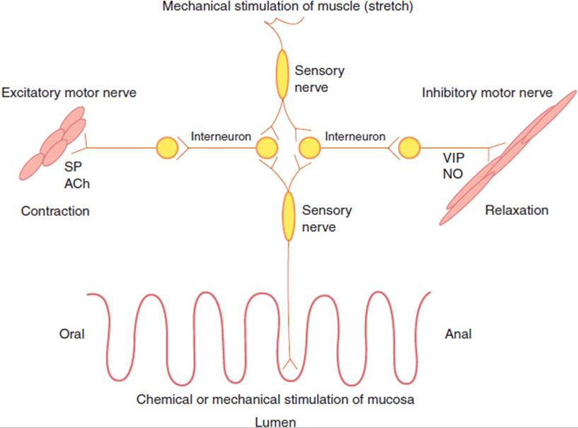
FIGURE 13-16 The peristaltic reflex of the small intestine. Enteric sensory nerves detect chemical or mechanical stimulation of the mucosa or stretch of the muscle layer. Signals are transmitted in an oral or anal direction by interneurons. Excitatory motor nerves release acetylcholine (ACh) and substance P (SP), which cause muscle contraction on the oral side of the stimulus. Inhibitory motor nerves release vasoactive intestinal peptide (VIP) and nitric oxide (NO), which cause muscle relaxation on the anal side of the stimulus.
Opiate drugs such as morphine, which are highly effective for relief of chronic pain (eg, cancer pain), have the detrimental side effect of inhibiting motility of the small intestine. Opioids act on enteric nerves to inhibit secretion of excitatory neurotransmitters to thereby inhibit peristalsis. The inhibition of motility slows down intestinal transit, allowing for a more complete absorption, so the volume entering the colon is diminished and constipation results.
CHECKPOINT
31. Describe the hormonal reflex by which fat in the intestine stimulates the secretion of bile.
32. Describe the mechanism by which glucose is absorbed across the apical and basolateral membranes of an enterocyte.
33. What is the mechanism of absorption of tripeptides across an intestinal epithelial cell?
34. What is the role of bile in lipid absorption in the intestine?
35. List three general mechanisms of absorption of Na+ ions in the small intestine.
36. Describe the mechanism of fluid and electrolyte secretion in the crypts of Lieberkühn.
37. Name two neurotransmitters that are secretagogues.
38. How do certain bacterial toxins stimulate fluid and electrolyte secretion in the crypts of Lieberkühn?
39. Describe the pattern of intestinal motility during fasting and after feeding.
40. Name one hormone that maintains the fasting pattern of motility and one that induces the fed pattern of motility in the small intestine.
41. Name the neurotransmitters that mediate the ascending and descending limbs of the peristaltic reflex.
COLON
Anatomy & Histology
The adult colon is 1.0–1.5 m in length. Its various segments (cecum, ascending, transverse, descending, sigmoid colon, and rectum) are involved in absorption of water and electrolytes, secretion of mucus, and formation, propulsion, and storage of unabsorbed material (feces). The colon is also the home of the majority of the intestinal microbes.
The surface of the colon consists of a columnar epithelium with no villi and few folds except in the distal rectum (Figure 13-13). The epithelial cells include absorptive cells and contain microvilli on their surface as well as mucus-secreting goblet cells. Colonic crypts contain goblet cells, endocrine cells, absorptive cells, and epithelial stem cells. As in the small intestine, the stem cells and their daughter progenitor cells serve to replenish the differentiated cells of the epithelium that continually turnover throughout life.
Digestion & Absorption of the Colon
Digestion in the colon occurs as a consequence of the action of the colonic microbiota. Short-chain fatty acids released by microbial action on dietary fiber are an important source of energy for the colon. More importantly, these short-chain fatty acids promote survival of healthy colonic epithelium while inducing apoptosis (programmed cell death) in epithelial cells that are progressing toward malignant transformation.
Absorption of fluid and electrolytes has been well studied and is a major function of the colon. Up to 5 L of water can be absorbed per day across the colonic epithelium. Furthermore, the colonic epithelium can also take up sodium against a considerable concentration gradient. Aldosterone, a hormone involved in fluid and electrolyte homeostasis, increases colonic sodium conductance in response to volume depletion, thus playing an important role in maintaining fluid and electrolyte balance.
Secretion of the Colon
Major secretory products of the colon are the mucin proteins produced and secreted by goblet cells that reside in the epithelial layer. Mucins are very high molecular weight proteins (mostly due to the extensive glycosylation). In the lumen of the colon, they are hydrated and form a layer overlying the epithelial cells. They serve to lubricate and prevent the opposing sides of the intestinal tube from sticking together (thus collapsing the tube). In addition, mucins participate in innate immunity. As antimicrobial peptides bound by immunoglobulins that are secreted into the lumen, mucins form a barrier to intestinal microbes and pathogens.
Motility of the Colon
Unlike the stomach and small intestine, the colon is rarely inactive, although its activity is less easily characterized than that of the stomach, which has the pattern known as receptive relaxation, or than that of the small intestine, which displays the pattern known as the migrating motor complex and segmental to-and-fro action. Some patterns are discernible, however, such as the gastrocolic reflex (colonic mass peristalsis after a meal). Disorders of colonic motility are common complications of autonomic neuropathy in patients with diabetes mellitus and can cause severe GI complaints. Continence of stool requires contraction of the puborectalis muscle and the anal sphincter. Defecation involves relaxation of the puborectalis by the sacral parasympathetic nerves, resulting in straightening of the anorectal angle. Rectal distension results in reflex sympathetic-mediated internal and external sphincter relaxation.
CHECKPOINT
42. How does colonic motility differ from that in the small intestine?
43. What is the major secretory product of the colon?
44. What volume of water is the colon capable of absorbing per day?
OVERVIEW OF GI DISORDERS
DISORDERS OF MOTILITY
Disorders of motility affect all major regions of the GI tract. Because GI tract motility is a complex result of smooth muscle contraction under neural and hormonal control, abnormal motility of the GI tract can occur through damage to GI smooth muscle or to the neural and hormonal mechanisms by which it is controlled, or both. An example of muscle damage leading to abnormal motility is seen in esophageal stricture as a result of caustic ingestions or acid reflux. Abnormal neural control of motility is seen in esophageal achalasia. Esophageal motility disorders are typically characterized by dysphagia and odynophagia. An example of a neural defect that affects motility is Hirschsprung disease. These patients are typically younger than 2 years and either present after birth with the inability to pass meconium or later develop severe constipation. The structural defect is a lack of myenteric neurons in the distal colon due to a congenital defect whereby the migration of neural crest precursor cells does not occur properly.
Motility disorders of the stomach include gastroparesis, a complication of diabetes mellitus, and dysmotility as a consequence of stomach surgery, from either resection of part of the stomach or vagotomy. Vagotomy entails surgical transection of the vagus nerve trunks, which prevents vagus-stimulated acid secretion and regulation of gastric motility. Before the availability of histamine H2 receptor antagonists and PPIs, selective vagotomy of the stomach was used as a treatment for the hypersecretion of gastric acid. Vagotomy is still sometimes performed as treatment for Zollinger-Ellison syndrome (ie, acid hypersecretion and severe peptic ulcer disease caused by a gastrin-secreting tumor). In hypertrophic pyloric stenosis, food cannot pass freely out of the stomach due to spasmodic narrowing of the pyloric outlet caused either by congenital narrowing or by hypertrophy of the pyloric musculature. It is more common in boys and presents shortly after birth with nonbilious vomiting. It is readily managed surgically.
The symptoms and signs of motility disorders in the stomach depend on their cause. Because vagotomy cuts fibers influencing the enteric nervous system as well as the intended fibers that influence acid secretion, a classic complication of vagotomy is disordered gastric motility. This may present clinically as either partial outlet obstruction or as too-rapid emptying of gastric contents into the duodenum, with resulting fluid shifts and vasomotor symptoms (“dumping syndrome”). Sometimes, however, patients may develop symptoms of stomach distension, nausea, early satiety, and vomiting suggestive of partial gastric outlet obstruction. To ameliorate the latter symptoms, pyloroplasty (severing the fibers of the pyloric sphincter) is done to render the sphincter less competent, so that food can pass more easily into the duodenum. Intrinsic neuropathy (eg, in diabetes mellitus) results in delayed gastric emptying, nausea, vomiting, and constipation rather than the classic dumping syndrome. The pathophysiologic basis for these differences is not known.
In the small intestine and colon, disordered motility occurs in irritable bowel syndrome. Irritable bowel syndrome is characterized by recurrent episodes of abdominal pain, bloating, and diarrhea alternating with constipation in the absence of detectable organic disease or structural abnormalities. The cause of this condition is still unclear.
DISORDERS OF SECRETION
Clinically recognized disorders of secretion involve the production of acid, intrinsic factor, or mucus by the stomach, digestive enzymes and bicarbonate by the pancreas, bile by the liver, and water and electrolytes by the small intestine in response to secretagogues.
Either elevated gastric acid secretion or diminished mucosal defense can predispose to development of peptic ulcers. Ulcers represent discrete regions of erosion through the entire mucosa. Acid-induced damage may occur in the form of an ulcer either in the stomach (gastric ulcer) or in the first part of the small intestine (duodenal ulcer). Acid-induced injury may also occur in the form of more diffuse and less clearly demarcated inflammation anywhere along the GI tract from the lower esophagus through the duodenum. It appears that elevated acid secretion, almost always in the setting of H pylori infection, is relatively more important in the development of duodenal ulcer, whereas diminished mucosal defense (eg, from diminished mucus secretion in some cases) is a more crucial factor in development of gastric ulcer, with H pylori underlying only about half the cases. Disorders of secretion involving the liver and pancreas are discussed in Chapters 14 and 15, respectively. Diarrhea, the major secretory disorder of the small intestine, is discussed later.
DISORDERS OF DIGESTION & ABSORPTION
Physiologically significant digestion and absorption can occur throughout the GI tract. Indeed, the effectiveness of sublingual nitroglycerin therapy for patients with angina is a testimonial to the efficacy of sublingual absorption. Nevertheless, the clinically prominent disorders of digestion and absorption focus on the small intestine and colon and the accessory organs (pancreas and liver) whose secretions (digestive enzymes, bicarbonate, and bile) are necessary for digestion and absorption in the small intestine.
GI MANIFESTATIONS OF SYSTEMIC DISEASE
A wide range of systemic conditions and diseases may produce symptoms and signs in the GI tract. These include endocrine disorders that alter control of GI tract functions or that predispose to pancreatitis or peptic ulcer disease; complications of diabetes mellitus, including autonomic neuropathy and ketoacidosis; pregnancy; deficiency disorders, including deficiency of zinc, niacin, and iron; and neoplastic, rheumatologic, and other syndromes (Table 13-4).
TABLE 13-4 GI manifestations of systemic diseases and their pathophysiologic mechanisms.
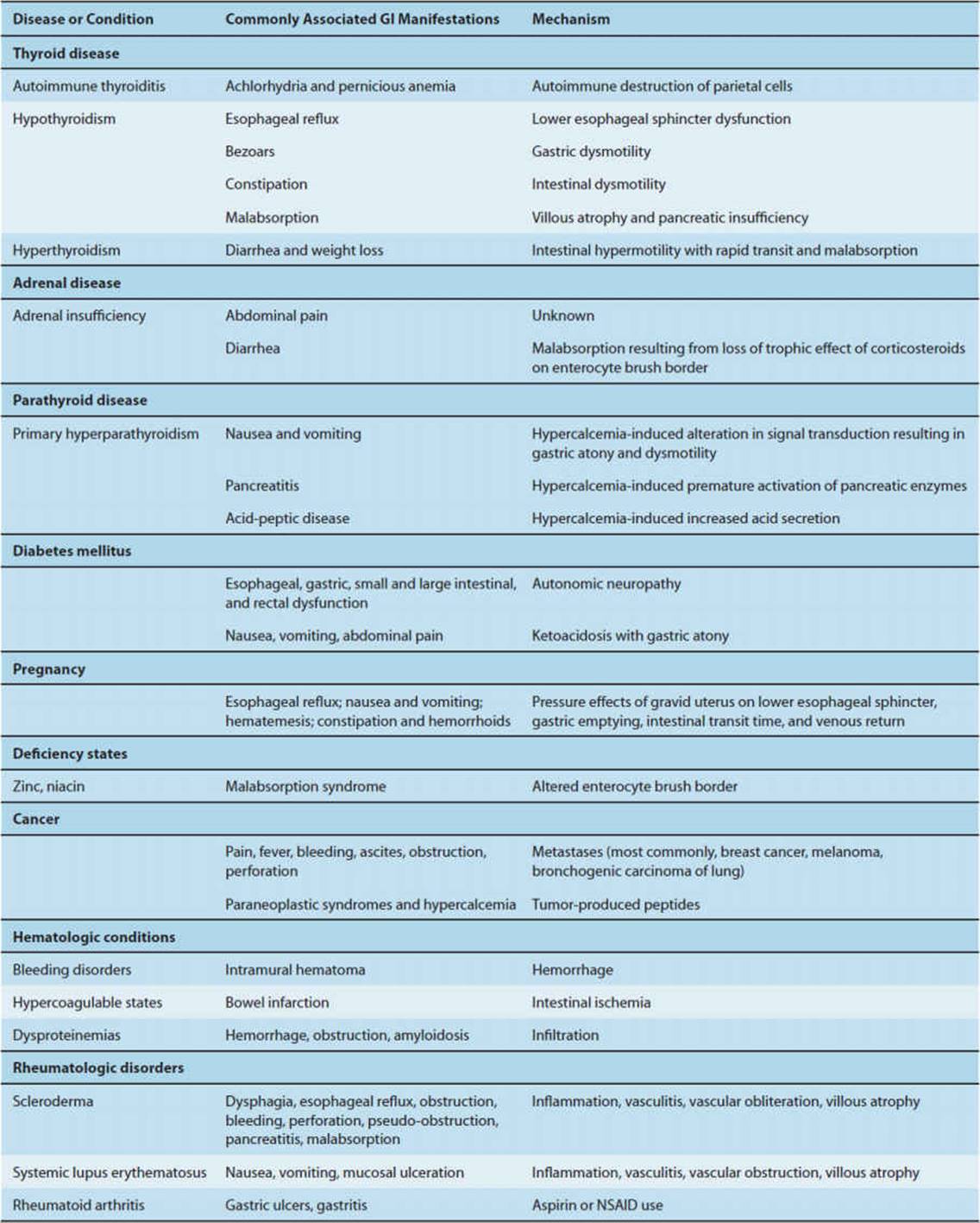
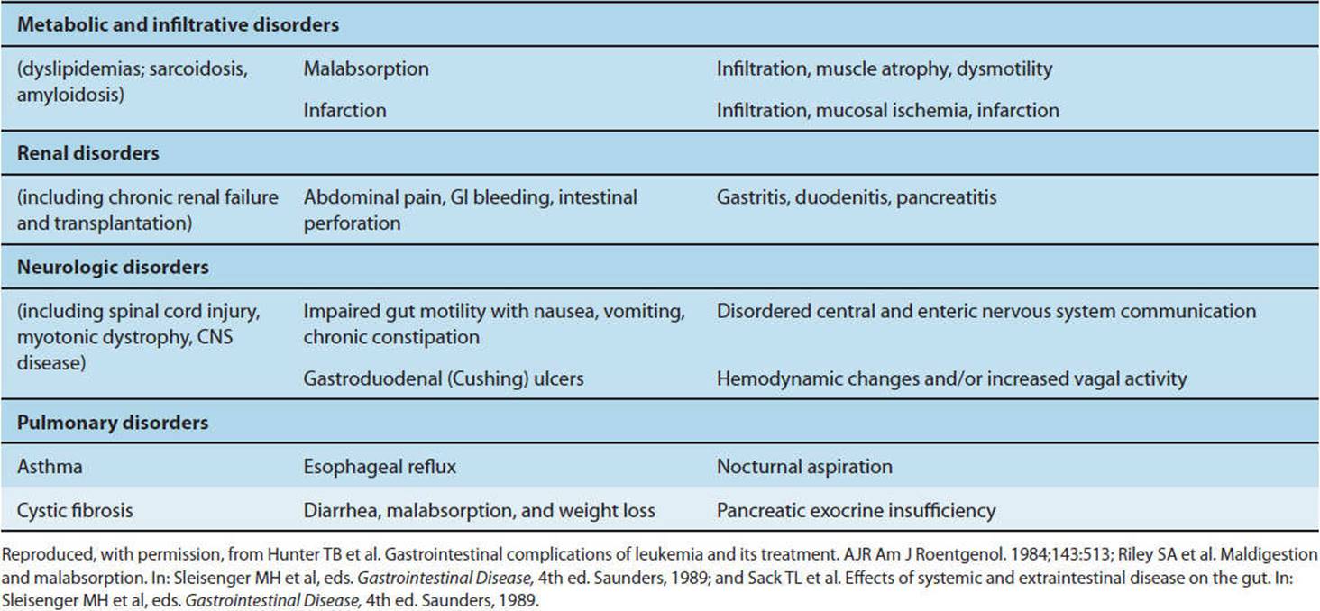
CHECKPOINT
45. What are some common symptoms of esophageal dysmotility?
46. Why does vagotomy often create motor disorders in the stomach?
PATHOPHYSIOLOGY OF DISORDERS OF THE ESOPHAGUS
The major disorders of the esophagus are related to motor functions: Disordered peristalsis and increased lower esophageal sphincter tone are seen in esophageal achalasia, whereas inappropriate lower esophageal sphincter relaxation results in reflux esophagitis.
ESOPHAGEAL ACHALASIA
Clinical Presentation
Esophageal achalasia is a motor disorder in which the lower esophageal sphincter fails to relax properly. As a result, a functional obstruction (ie, obstruction from abnormal function in the absence of a visible mass or lesion) is created that is manifested as dysphagia (inability to swallow), regurgitation, and chest pain. It is a progressive disease in which severe radiographic distortion of the esophagus develops.
Etiology
The underlying cause of esophageal achalasia, which occurs with an incidence of 0.5–1.0 per 100,000 population per year, is unknown. Degeneration of the myenteric plexus and loss of inhibitory neurons that release VIP and nitric oxide, which dilate the lower esophageal sphincter, may contribute. Esophageal involvement in Chagas disease, resulting from damage of the neural plexuses of the esophagus by the parasite Trypanosoma cruzi, bears a striking resemblance to esophageal achalasia. A number of other disorders, including malignancies, may present with manometric pressure characteristics or radiographic features similar to those observed in idiopathic esophageal achalasia.
Pathology & Pathogenesis
Although achalasia is manifested as a motor disorder of esophageal smooth muscle, it is actually due to defective innervation of smooth muscle in the esophageal body and lower esophageal sphincter. Lower esophageal sphincter tone is normally characterized by tonic contraction with intermittent relaxation resulting from a neural reflex arc (see earlier discussion). In achalasia, it is even more tightly contracted and does not relax properly in response to swallowing because of partial loss of neurons in the wall of the esophagus. Thus, achalasia can be thought of as a disorder caused by defective inhibitory pathways of the esophageal enteric nervous system. Interestingly, injection of botulinum toxin into the lower esophageal sphincter diminishes the excitatory pathways and thereby ameliorates symptoms. In addition to dysfunction of the lower esophageal sphincter, loss of normal peristalsis in the esophageal body is often seen in achalasia, consistent with the hypothesis of myenteric plexus degeneration. Variations of achalasia also exist in which normal peristalsis is replaced by simultaneous contractions of large or small amplitude.
Clinical Manifestations
Over months and years, lower esophageal sphincter dysfunction results in tremendous enlargement of the esophagus. Normally intended as a direct conduit to the stomach, the esophagus in advanced cases of achalasia can hold as much as 1 L of putrid, infected material, imposing a high risk of aspiration pneumonia. Without treatment, patients display progressive severe weight loss with worsening chest pain, mucosal ulceration, infection, and occasional esophageal rupture, culminating in death.
REFLUX ESOPHAGITIS
Clinical Presentation
The predominant presenting symptom of reflux is burning chest pain (heartburn) resulting from recurrent mucosal injury, often worse at night, when lying supine, or after consumption of foods or drugs that diminish lower esophageal sphincter tone.
Etiology
Common causes of reflux esophagitis are those conditions that result in persistent or repetitive acid exposure to the esophageal mucosa. These include disorders that increase the rate of spontaneous transient lower esophageal sphincter relaxations (Table 13-5) or impair reflexes that normally follow transient lower esophageal sphincter relaxations with a secondary wave of esophageal peristalsis. Conditions that increase gastric volume or pressure (eg, partial or complete gastric outlet obstruction and conditions that increase acid production) also contribute. Occasionally, reflux esophagitis can be caused by alkaline injury (eg, pancreatic juice refluxing through both an incompetent pyloric sphincter and a relaxed lower esophageal sphincter). Hiatal hernia, a disorder in which a portion of the proximal stomach slides into the chest cavity with upward displacement of the lower esophageal sphincter, can contribute to the development of reflux.
TABLE 13-5 Modulators of lower esophageal sphincter pressure.
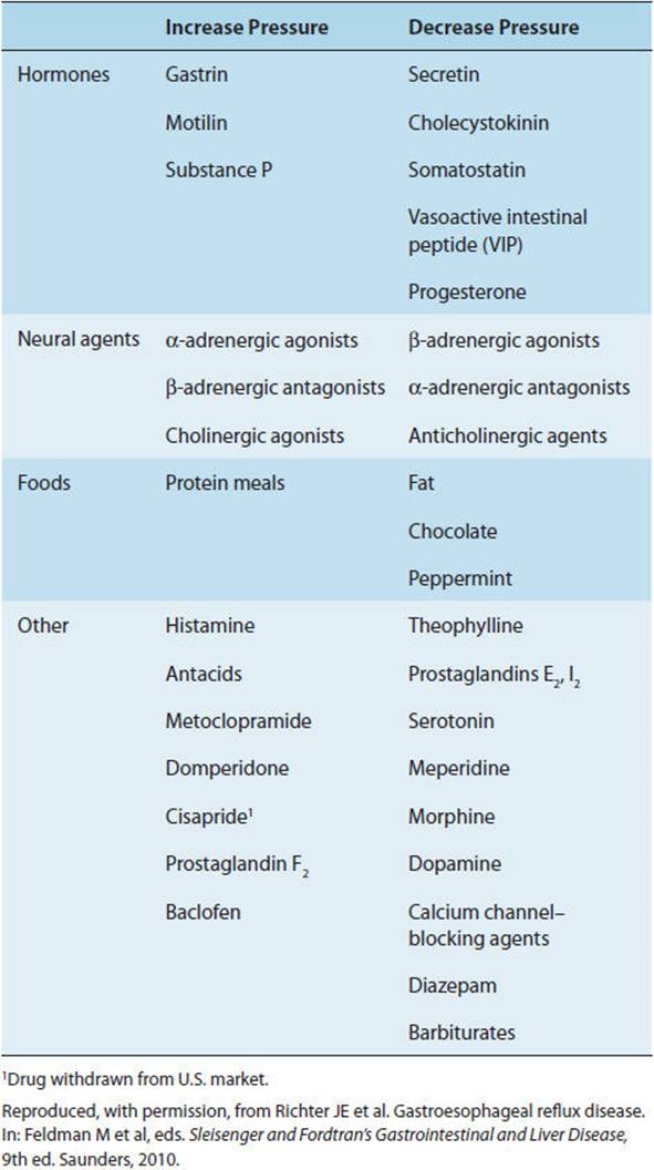
Pathology & Pathogenesis
Normally, the tonically contracted lower esophageal sphincter provides an effective barrier to reflux of acid from the stomach back into the esophagus. This is reinforced by secondary esophageal peristaltic waves in response to transient lower esophageal sphincter relaxation. Effectiveness of that barrier can be altered by loss of lower esophageal sphincter tone (ie, the opposite of achalasia), increased frequency of transient relaxations, loss of secondary peristalsis after a transient relaxation, increased stomach volume or pressure, or increased production of acid, all of which can make more likely reflux of acidic stomach contents sufficient to cause pain or erosion. Recurrent reflux can damage the mucosa, resulting in inflammation, hence the term “reflux esophagitis.” Recurrent reflux itself predisposes to further reflux because the scarring that occurs with healing of the inflamed epithelium renders the lower esophageal sphincter progressively less competent as a barrier.
Pepsin and bile can also be refluxed in addition to acid to cause esophagitis. In most cases of esophageal reflux disease, a common pathophysiologic thread can be identified (Figure 13-17). Recurrent mucosal damage results in infiltration of granulocytes and eosinophils, hyperplasia of basal cells, and eventually the development of friable, bleeding ulcers and exudates over the mucosal surface. These pathologic changes set the stage for scar formation and sphincter incompetence, predisposing to recurrent cycles of inflammation.
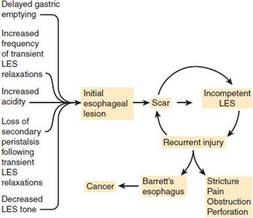
FIGURE 13-17 Pathophysiology of esophageal reflux disease. (LES, lower esophageal sphincter.)
Increased frequency of transient lower esophageal sphincter relaxations may be partly in response to increased gastric distension. Normally, transient lower esophageal sphincter relaxations are accompanied by increased esophageal peristalsis. Individuals with defects in excitatory pathways that promote peristalsis may, therefore, be at increased risk for the development of esophageal reflux. Changes in the types of prostaglandins produced by the esophagus have been noted in reflux esophagitis, perhaps contributing to impairment of healing and predisposing to recurrences. In contrast to other forms of acid-mediated injury, H pylori infection does not appear to contribute to the development of reflux or esophagitis.
Clinical Manifestations
Heartburn is the usual symptom of reflux esophagitis, typically worsening on lying prone. With recurrent reflux, a range of complications may develop. The most common complication is the development of stricture in the distal esophagus. Progressive obstruction, initially to solid food and later to liquid, presents as dysphagia. Other complications of recurrent reflux include hemorrhage or perforation; hoarseness, coughing, or wheezing; and pneumonia as a result of aspiration of gastric contents into the lungs, particularly during sleep. Chronic recurrent reflux can also result in a change in the esophageal epithelium from squamous to columnar histology (resembling that of the stomach and/or intestine). Termed Barrett esophagus, the disorder is more common in men and in smokers, and it leads to a greatly increased risk of adenocarcinoma. Adenocarcinomas in the distal esophagus and proximal (cardiac) stomach related to Barrett esophagus are among the most rapidly increasing types of cancer in young male patients in the United States.
CHECKPOINT
47. What are the roles of the lower esophageal sphincter structure in achalasia and in reflux esophagitis?
48. What are possible causes of achalasia?
49. What is the relationship of esophageal reflux to Barrett esophagus and cancer?
PATHOPHYSIOLOGY OF DISORDERS OF THE STOMACH
Common disorders involving the stomach reflect the importance of its role as a secretory organ, in particular of acid and intrinsic factor. Disorders of acid secretion result in acid-peptic disease, whereas loss of intrinsic factor secretion results in inability to absorb vitamin B12, manifesting as pernicious anemia. The major motility disorder of the stomach is gastroparesis.
ACID-PEPTIC DISEASE
Clinical Presentation
Patients with acid-peptic disease typically present with chronic, mild, gnawing or burning abdominal or chest pain resulting from superficial or deep erosion of the GI mucosa. Sudden complications include GI tract bleeding, resulting in hematemesis or melena, and perforation and infection, resulting in severe abdominal pain and signs of acute abdomen (absence of bowel sounds, guarding, rebound tenderness). The latter presentation reflects the fact that in some cases acid-peptic disease can be painless in the early stages and can be detected only when it leads to an intra-abdominal catastrophe.
Classically, duodenal ulcer presents as gnawing or burning epigastric pain occurring 1–3 hours after meals, often waking the patient at night, with antacids or food producing relief. However, many patients later documented to have duodenal ulcer do not fit this symptom profile. Elderly patients in particular often present with a complication of duodenal ulcer but no history of pain.
Etiology
Various causes of absolute or relative increased acid production (Figure 13-12) or decreased mucosal defenses (Table 13-2) predispose to acid-peptic disease. As mentioned, the bacterium H pylori is the root cause of a number of forms of acid-peptic disease, including duodenal ulcer, gastric ulcer, and gastritis (Figure 13-18).
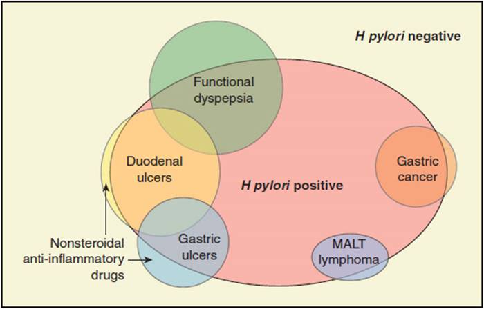
FIGURE 13-18 Relation of H pylori infection to upper GI tract conditions. The figure shows that most patients with gastroduodenal ulcers or gastric lymphoma or adenocarcinoma have also been infected with H pylori. Note, however, that the circles are not to scale, because gastric cancer occurs in less than 1% of those infected with H pylori. Note, too, that relationships among the different conditions are more complex than depicted. Despite that patients with cancer often have a prior history of ulcers, as shown in Figure 13-19, patients with a history of H pylori infection (that causes ulcers) are less likely to develop cancer. (Redrawn, with permission, from Calam J et al. Pathophysiology of duodenal and gastric ulcer and gastric cancer. BMJ. 2001;323:980.)
Pathology & Pathogenesis
Corrosive agents (acid and pepsin) secreted by the stomach play a key role in gastric ulcer, duodenal ulcer, and acute erosive gastritis. Each of these diseases has a distinctive but overlapping pathogenesis with the common themes of either excessive acid secretion or diminished mucosal defense. Exactly why one but not another form of acid-peptic disease should develop in a given individual remains unclear. H pylori infection can cause acid-peptic disease by multiple mechanisms, including direct alteration of signal transduction in mucosal and immune cells, which in turn can increase acid secretion and diminish mucosal defenses. The complex interactions of the H pylori infection and its location and virulence, along with its clinical consequences (eg, inflammation, increased or decreased acid secretion), are illustrated in Figure 13-19.
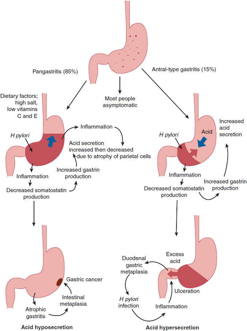
FIGURE 13-19 Patterns of chronic H pylori infection with respect to acid production and pathology. Left: Acid hyposecretion. H pylori infection of the stomach body causes suppression of parietal cells, low acid secretion, atrophic gastritis, intestinal metaplasia, and predisposition to gastric cancer. Right: Acid hypersecretion. H pylori infection primarily of the stomach antrum causes decreased somatostatin and increased gastrin secretion, increasing acid secretion and predisposition to duodenal ulceration. (Redrawn from Calam J et al. Pathophysiology of duodenal and gastric ulcer and gastric cancer. BMJ. 2001;323:980.)
H pylori is an extremely common pathogen, found in over half of the world’s population; rates of infection are even higher in the poorest countries, where sanitation facilities and standards of personal hygiene are low. The most likely route of spread from person to person is fecal-oral. As many as 90% of infected individuals show signs of inflammation (gastritis or duodenitis) on endoscopy, although many of these individuals are clinically asymptomatic. Despite this high rate of association of inflammation with H pylori infection, the important role of other factors is indicated by the fact that only about 15% of infected individuals ever develop a clinically significant ulcer. These other factors (both genetic and environmental, such as cigarette smoking) must account for the individual variations and are pathophysiologically important. Nevertheless, the role of H pylori is of particular clinical importance because, of patients who do develop acid-peptic disease, especially among those with duodenal ulcers, the vast majority have H pylori infection. Furthermore, treatment that does not eradicate H pylori is associated with rapid recurrence of acid-peptic disease in most patients. There are numerous strains of H pylori that vary in their production of toxins such as CagA and VacA that directly alter cellular signaling pathways. Variations in bacterial strains, natural variation in the balance of inflammatory mediators (eg, TH1 vs. TH2 vs. TH17 cytokines) triggered by infection, and a variety of environmental and lifestyle factors may explain why H pylori infection is asymptomatic in most patients, causes peptic ulcers in some, and increases risk for development of lymphoma and adenocarcinoma in a few.
1. Gastric Ulcer
Gastric ulcer is distinguished from erosive gastritis by the depth of the lesion, with gastric ulcers penetrating through the mucosa. The actual ulcer crater is often surrounded by an area of intact but inflamed mucosa, suggesting that gastritis is a predisposing lesion to development of gastric ulcer. Most gastric ulcers occur on the lesser curvature of the stomach. It is likely that gastric ulcer represents the outcome of a number of different abnormalities summarized next.
Some gastric ulcers are believed to be related to impaired mucosal defenses, because the acid and pepsin secretory capacity of some affected patients is normal or even below normal.
Motility defects have been proposed to contribute to development of gastric ulcer in at least three ways. First, they contribute because of a tendency of duodenal contents to reflux back through an incompetent pyloric sphincter. Bile acids in the duodenal reflux material act as an irritant and may be an important contributor to a diminished mucosal barrier against acid and pepsin. Second, they may contribute as a result of delayed emptying of gastric contents, including reflux material, into the duodenum. Third, they may contribute as a result of delayed gastric emptying and hence food retention, causing increased gastrin secretion and gastric acid production. It is not known whether these motility defects are a cause or a consequence of gastric ulcer formation.
Mucosal ischemia may play a role in the development of a gastric ulcer. Prostaglandins are known to increase mucosal blood flow as well as bicarbonate and mucus secretion and to stimulate mucosal cell repair and renewal. Thus, their deficiency, resulting from nonsteroidal anti-inflammatory drug (NSAID) ingestion or other insults, may predispose to gastritis and gastric ulcer, as might diminished bicarbonate or mucus secretion resulting from other causes. Subsets of gastric ulcer patients with each of these defects have been identified. Thus, the risk factors (NSAID ingestion, smoking, psychologic stress, H pyloriinfection) that have been associated with gastric ulcer probably act by diminishing one or more mucosal defense mechanisms.
Gastritis (inflammation of the gastric mucosa) as a result of aspirin and other NSAIDs, bile salts, alcohol, or other insults may predispose to ulcer formation by (1) attenuating the barrier created by the epithelial cells or the mucus and bicarbonate they secrete or (2) reducing the quantity of prostaglandins the epithelial cells produce that might otherwise diminish acid secretion.
2. Acute Erosive Gastritis
Acute erosive gastritis includes inflammation resulting from superficial mucosal injury, mucosal erosion, or shallow ulcers caused by a wide variety of insults, most notably alcohol, drugs, and stress. Ethanol ingestion predisposes to gastritis but not to gastric ulcer. Unlike gastric or duodenal ulcers, in erosive gastritis the submucosa and muscularis mucosa are not penetrated. Acid hypersecretion, gastric anoxia (eg, in shock), altered natural defenses (especially diminished mucus secretion), altered epithelial renewal, changes in tissue mediators (eg, prostaglandins), reduced intramucosal pH, and intramucosal energy deficits have been suggested as factors in the development of superficial gastric mucosal injury.
3. Chronic Atrophic Gastritis
Chronic atrophic gastritis is a heterogeneous group of conditions characterized by inflammatory cell infiltration with gastric mucosal atrophy that leads to death of parietal cells and ultimate dropout of gastric glands. In chronic disease, unlike acute erosive gastritis, endoscopic abnormalities may not be grossly apparent. The capacity to secrete gastric acid is progressively reduced, and the serum levels of gastrin are elevated in an attempt to restore parietal cell activity. Atrophic gastritis can be a purely autoimmune disease associated with production of autoantibodies to parietal cells, intrinsic factor, and gastrin, but it can also be the result of H pylori infection. Autoimmune gastritis can progress to pernicious anemia, whereas atrophic gastritis in the setting of H pylori infection greatly increases the risk of progression to gastric adenocarcinoma. Any condition with chronic loss of parietal cell mass or activity can lead to compensatory GI endocrine hyperplasia of reactive G cells. Progression to an autonomous gastrin-producing neuroendocrine tumor of the GI tract (gastrinoma) is a rare cause of ulcer disease. Alternatively, H pylori–mediated atrophic gastritis greatly increases the risk of progression of the inflammatory infiltrate to a lymphoma of mucosa-associated lymphoid tissue type (called a MALToma).
4. Duodenal Ulcer
Even more commonly than gastric ulcers, duodenal ulcers are sequelae of H pylori infection, caused by altered mucosal inflammatory responses and excessive acid secretion. Various other risk factors, including diet, smoking, and excessive alcohol consumption, may influence the development of duodenal ulcers, although specific associations (eg, between coffee or spicy foods and the development of ulcers) have not been demonstrated. Genetic factors also play a role; studies support the existence of a heritable component in duodenal ulcers distinct from that involved in gastric ulcer. Likewise, psychologic stress has been implicated in duodenal ulcer disease, perhaps by an autonomic-mediated influence on acid secretion (Figure 13-12). Interestingly, duodenal ulcers are associated with decreased risk for development of gastric adenocarcinoma, perhaps because chronic H pylori infection predisposes to cancer in the setting of atrophic gastritis, in which parietal cells are lost, whereas duodenal ulcers are caused by acid secretion; thus, patients with duodenal ulcers are not likely to have pronounced parietal cell atrophy (Figure 13-19).
Clinical Manifestations
Those forms of acid-peptic disease characterized by exclusively superficial mucosal lesions (eg, acute erosive gastritis) can result in either acute or chronic GI tract bleeding, accompanied by a significant drop in hematocrit and related complications (eg, precipitating angina in a patient with coronary artery disease). Patients with acute massive bleeding present with hematemesis (vomiting of blood), rectal bleeding, or melena (tarry stools from the effect of acid on blood) depending on the site of origin, the rate of transit of blood through the GI tract, and the extent of hemorrhage. Acute massive hemorrhage (>10% of blood volume over minutes to hours) is manifested by hypotension, tachycardia, and orthostatic blood pressure and heart rate changes on standing, often with dizziness.
In addition to hemorrhage, complications of duodenal ulcer and gastric ulcer include life-threatening perforation and obstruction.
CHECKPOINT
50. How does pernicious anemia result from a secretory disorder of the stomach?
51. What is the typical acid secretion status of patients with pernicious anemia?
52. In which acid-peptic disorder are diminished mucosal defenses more important than acid hypersecretion?
53. How might motility defects contribute to gastric ulcer?
54. What factors may predispose a patient to duodenal ulcer disease?
55. How do NSAIDs contribute to acid-peptic disease?
56. What evidence indicates the importance of H pylori infection in acid-peptic disease?
57. What evidence suggests that other factors besides H pylori infection contribute to acid-peptic disease?
GASTROPARESIS
Clinical Presentation
A common complication of stomach disorders is delayed gastric emptying (Table 13-6). Known as gastroparesis, it is manifested by nausea, bloating, vomiting, and either constipation or diarrhea. The condition can also occur silently, producing metabolic derangements (eg, of blood glucose in patients with diabetes mellitus) in the absence of somatic symptoms.
TABLE 13-6 Conditions producing symptomatic gastric motor dysfunction.
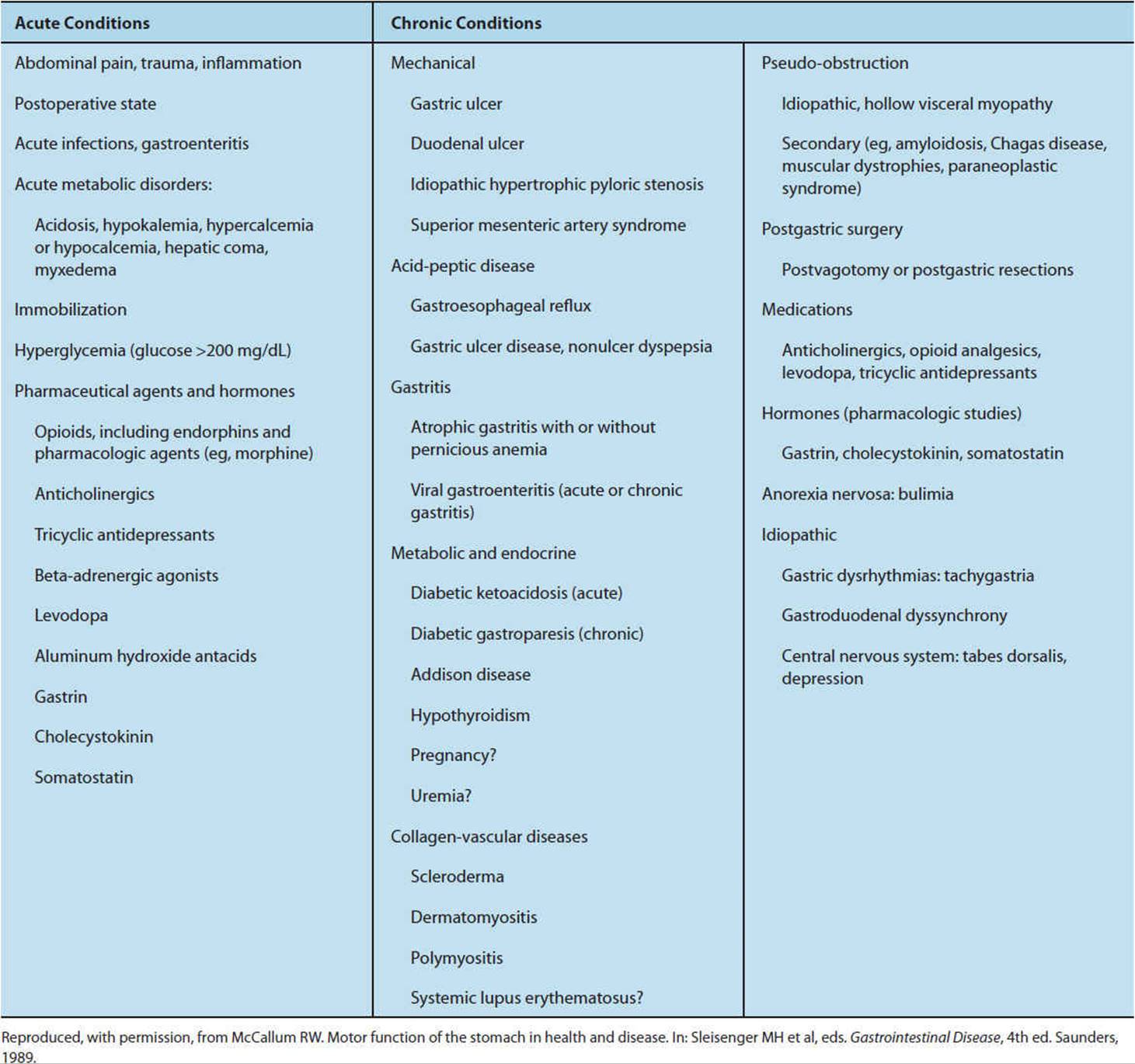
Etiology
Gastroparesis is a common complication of poorly controlled diabetes mellitus, with consequent autonomic neuropathy.
Pathology & Pathogenesis
Disorders of gastric motility result from alterations in a number of normal gastric functions. These include (1) serving as a reservoir for ingested solids and liquids (eg, alteration caused by resection of the stomach); (2) mixing and homogenizing ingested food; and (3) functioning as a barrier that allows only small spurts of well-mixed chyme beyond the pyloric sphincter. The resulting disorders span the range from partial or complete gastric outlet obstruction to excessively rapid emptying and typically result from interference with the normal mechanisms by which these functions are controlled. These include the intrinsic contractility of gastric smooth muscle, the enteric nervous system, the autonomic nervous system’s control over enteric nervous system function, and gut hormones.
Because the pyloric sphincter, like all sphincters, displays tonic contraction with intermittent transient relaxation, loss of vagal control results in excessive tonic contraction and symptoms of various degrees of gastric outlet obstruction. Disorders that affect the enteric nervous system such as the neuropathy of diabetes mellitus and surgical cutting of the stomach wall or vagal trunk typically cause delayed emptying. However, it is important to remember that, in some cases, delayed emptying can result in symptoms expected from excessively rapid emptying. For example, an excessively contracted pylorus that can open completely but does so infrequently can result in entry into the duodenum of too large a bolus of chyme from the excessively distended stomach. Such a bolus may not be efficiently handled by the small intestine, resulting in poor absorption and diarrheal symptoms characteristic of the dumping syndrome.
Hormones play an ill-defined but important role in regulation of GI motility in health and disease. For example, the antibiotic erythromycin is recognized by the receptor for the GI hormone motilin, affecting GI motility. Some patients with gastroparesis are observed to have substantial improvement with erythromycin analogs, especially when complaints related to partial gastric outlet obstruction, such as bloating, nausea, and constipation, are prominent.
Because different patients have different relative contributions of the intrinsic nervous system, enteric nervous system, autonomic nervous system, higher centers of the CNS, and hormones over control of their GI tract motility, not all treatments for gastroparesis are effective for a majority of patients even with the same initial complaints.
Clinical Manifestations
Complications of gastroparesis include the development of bezoars from retained gastric contents, bacterial overgrowth, erratic blood glucose control, and, when nausea and vomiting are profound, weight loss. Elevated blood glucose can be either a cause or a consequence of delayed gastric emptying. Bacterial overgrowth itself can result in both malabsorption and diarrhea. For unknown reasons, the symptoms of gastroparesis are variable from patient to patient as well as over time in a given patient and often correlated poorly with delayed gastric emptying. In some cases, serotonin antagonists that decrease visceral perception may be more helpful than prokinetic agents in alleviating symptoms.
CHECKPOINT
58. What are the symptoms of delayed versus rapid gastric emptying?
59. What are the complications of gastroparesis?
60. Why might erythromycin improve diabetic gastroparesis?
DISORDERS OF THE GALLBLADDER
Gallbladder disease is most commonly due to gallstones (cholelithiasis).
1. Cholelithiasis
Clinical Presentation
Gallstones are most often asymptomatic, discovered incidentally at autopsy or during surgery for an unrelated condition. Of patients who do have symptoms referable to cholelithiasis, presentations range from mild nausea or abdominal discomfort after eating fatty or fried foods to severe right upper quadrant or midepigastric abdominal pain and jaundice. A history of chronic mild symptoms with dietary association typically predates an acute episode of abdominal pain. The typical patient with gallstones is female, has a history of high dietary fat intake, has had prior pregnancies (reflecting the role of estrogens in gallstone pathogenesis), and is in her 40s (reflecting the time necessary for progression to symptomatic disease).
Etiology
Gallstones come in several varieties. Most are composed largely of cholesterol with or without calcium deposits. Occasionally, especially in patients with a chronic hemolytic disease, bilirubin stones may form. Depending on the cause and the pathophysiologic mechanism involved, patients can have one or more of the following: a few large individual stones; many smaller stones; or “sludge,” a thickened viscous gel resulting from concentration of bile that is believed to be highly prone to formation of stones.
Pathology and Pathogenesis
Cholelithiasis is of multifactorial origin. However, the formation of cholesterol gallstones usually requires the formation of bile whose cholesterol concentration is greater than its percentage solubility. The normal processes that prevent gallstone formation probably include the fact that bile does not normally stay in the gallbladder long enough to become lithogenic (prone to stone formation). Thus, loss of gallbladder muscular wall motility (resulting from either intrinsic disease of the muscle wall, altered levels of hormones such as CCK, or altered neural control) and excessive sphincteric contraction, impairing emptying, are important predisposing factors. One consequence of decreased gallbladder emptying is excessive concentration of bile, leading to heightened lithogenicity. This can occur from decreased absorption of water or altered bile composition resulting from increased cholesterol content or saturation. Other factors can cause an increased tendency to form stones at any given degree of concentration and saturation, including the presence of nucleating versus antinucleating factors in bile and the size and composition of the bile acid pool. Figure 13-20 summarizes the factors that predispose to gallstone formation, including estrogens, prostaglandins, increased mucus and glycoprotein production by the gallbladder epithelium, and chronic bacterial colonization or infection. Estrogens may play multiple roles, first affecting bile composition (increasing cholesterol and its saturation in bile) but also diminishing gallbladder motility (hence predisposing to stasis, sludge formation, and lithogenicity). Prostaglandins, which are protective in the stomach by increasing mucus production, actually may contribute to lithogenicity by the same mechanism. Thus, NSAIDs that block prostaglandin production are often beneficial for the prevention of gallstones in patients so predisposed, probably by decreasing mucus production.

FIGURE 13-20 Pathophysiology of cholelithiasis.
Clinical Manifestations
The major clinical presentation of gallstones is inflammation of the gallbladder, or cholecystitis. Cholecystitis can be either acute, chronic, or acute against a background of chronic disease. An episode of acute cholecystitis can progress to acute pancreatitis if a stone travels down the common bile duct but fails to clear the sphincter of Oddi, thereby blocking the pancreatic duct. Likewise, an inflamed gallbladder can become infected or can undergo infarction and necrosis, setting the stage for systemic sepsis if the patient does not receive systemic broad-spectrum antibiotics and undergo emergency cholecystectomy (Figure 13-21).

FIGURE 13-21 Clinical and pathologic effects of cholelithiasis. (Redrawn, with permission, from Chandrasoma P et al, eds. Concise Pathology, 3rd ed. Originally published by Appleton & Lange. Copyright © 1998 by The McGraw-Hill Companies, Inc.)
PATHOPHYSIOLOGY OF DISORDERS OF THE SMALL INTESTINE & COLON
Diseases of the small and large intestine include diarrhea, inflammatory bowel disease, and diverticular disease. Diarrhea is a symptom that has many causes and diverse pathogenetic mechanisms, including altered motility, secretion, digestion, and absorption. Although intestinal disorders are particularly prominent causes, disease of the stomach, pancreas, and biliary tract can also cause diarrhea. Inflammatory bowel diseases are poorly understood chronic autoimmune processes in the small intestine, colon, or both, with malabsorption as a prominent feature and important systemic manifestations. Diverticular disease occurs most prominently in the colon, in part as a direct or indirect consequence of altered motor function. Irritable bowel syndrome is not a disease per se but a functional disorder manifested by abdominal pain with diarrhea or constipation in the absence of organic disease or gross structural changes of the intestine.
DIARRHEA
Clinical Presentation
Symptoms of diarrhea are an increased stool frequency, increased stool volume, and a decrease in stool consistency. Any process that increases the frequency of defecation or volume of stool makes it looser, because time-dependent absorption of water is responsible for the normal soft but formed consistency of stool. Infectious diarrheas are discussed in Chapter 4. This chapter focuses on general aspects of diarrhea and diarrheas from other causes.
Patients’ subjective assessments of bowel movements are colored by their baseline bowel habits. An individual with chronic constipation, with bowel movements once every 3 days or so, may regard three soft stools in a day as diarrhea. In contrast, an individual on a high-fiber diet may normally have bowel movements twice or even three times a day.
Diarrhea can be acute (<2 weeks’ duration) or chronic (>4 weeks). Acute diarrhea is usually due to an infectious cause. The most common noninfectious causes are side effects of medications.
The simplest idea is that diarrhea is due to too much secretion or not enough absorption. Osmotic (malabsorptive) diarrhea is due to malabsorbed nutrients or poorly absorbed electrolytes that retain water in the lumen. Malabsorption occurs when the ability to digest or absorb a particular nutrient is defective and can be due to disordered mixing (altered motility), pancreatic insufficiency (altered digestion), or damage to enterocytes or their surface transporters (altered absorption). This type of diarrhea stops when the patient fasts. Secretory diarrhea results when secretagogues maintain elevated rates of fluid transport out of epithelial cells into the GI tract lumen. This type of diarrhea does not stop when the patient fasts. These physiologic distinctions are useful in both diagnosis and therapy of diarrheal disorders. In transport capacity, the small intestine far exceeds the colon (owing to the enormous surface area of the brush border). Thus, infectious, toxic, or other causes of heightened secretion in the small intestine can overwhelm absorptive mechanisms in the colon, resulting in diarrhea.
Etiology
Flow in the GI tract is a steady state involving massive fluid secretion into and absorption from the GI lumen. Each process is controlled by both extrinsic and intrinsic factors. Subtle aberrations in input or output at any of several levels can result in diarrhea with or without nutrient malabsorption. Thus, an excessive osmotic load, increased secretion, or diminished fluid resorption may result in diarrhea (Table 13-7).
TABLE 13-7 Mechanisms of diarrhea and major specific causes.
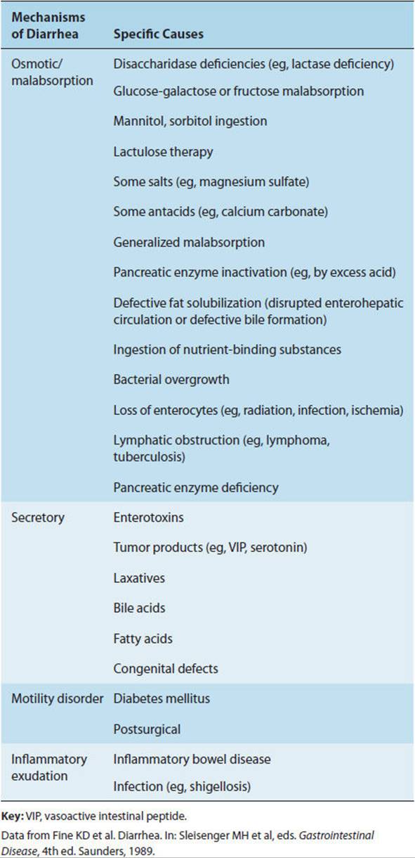
An excessive osmotic load in the GI tract may come about in three different ways: by direct oral ingestion of excessive osmoles, by ingestion of a substrate that may be converted into excessive osmoles (eg, when bacterial action on the nondigestible carbohydrate lactulose generates a diarrhea-causing osmotic load in the colon), and as a manifestation of a genetic disease such as an enzyme deficiency in the setting of a particular diet (eg, milk consumption by a lactase-deficient individual).
Secretion is increased by either blood-borne or intraluminal secretagogues. These include endogenous endocrine products (eg, overproduction of VIP by a tumor), exotoxins resulting from direct ingestion (eg, acute food poisoning) or infection (eg, cholera), or GI luminal substances (eg, bile acids) that stimulate secretion.
Absorption of fluid, electrolytes, and nutrients can be diminished by many factors, including the toxic effects of alcohol and mucosal damage from infectious agents and from cytokines and prokinetic agents. Cytokines are released by immune and other cells (eg, in response to infection). Prokinetic agents speed up GI motility, thereby diminishing the time available for absorption of any given nutrient, fluid, or electrolyte load. Finally, inflammatory and other disorders resulting in loss of mucus, blood, or protein from the GI tract may be manifested as diarrhea. Symptoms and signs suggesting specific causes of diarrhea are listed in Table 13-8.
TABLE 13-8 Clues to diagnosis of diarrhea from other symptoms and signs.
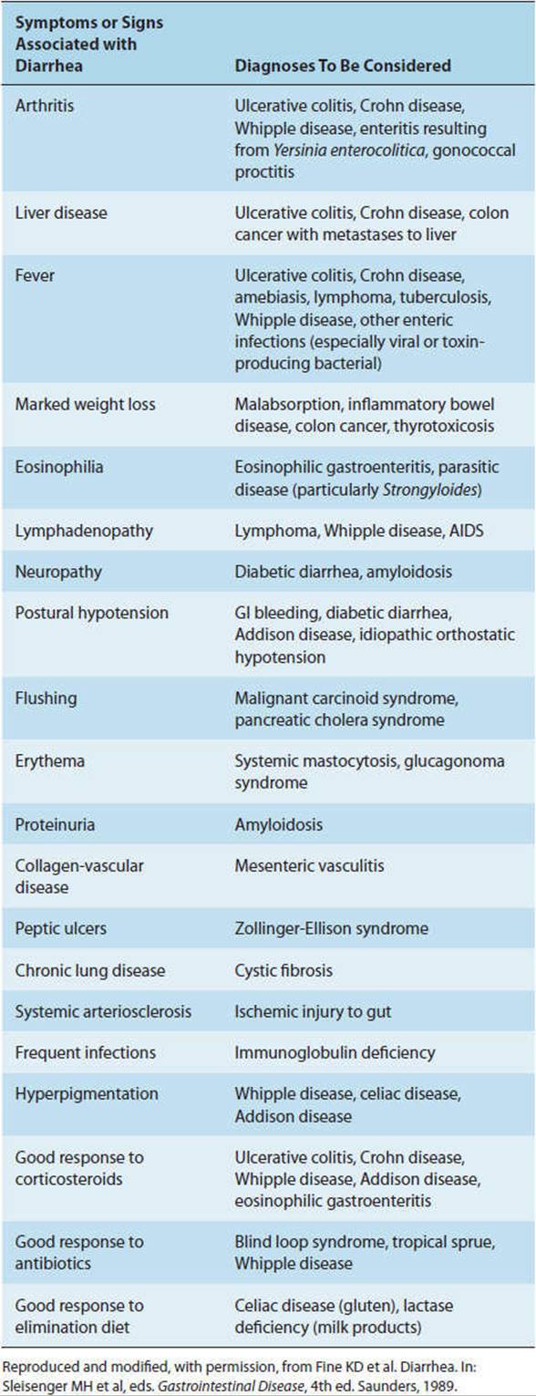
Pathology & Pathogenesis
Recognition of pathophysiologic subtypes of secretory (Tables 13–9 and 13–10) and osmotic diarrheas provides a means of approaching diagnosis and therapy of diarrheal disorders. For example, nonbloody diarrhea that continues in the absence of oral intake must be due to a secretory mechanism, whereas diarrhea that diminishes as oral intake is curtailed (eg, in a patient receiving intravenous hydration) suggests an osmotic/malabsorptive cause. Likewise, the presence of white blood cells in the stool suggests an infectious or inflammatory origin of diarrhea, although their absence does not rule out such causes.
TABLE 13-9 Clues to diagnosis of diarrhea from other symptoms and signs.

TABLE 13-10 Symptoms and signs of malabsorption and relevant pathophysiology.
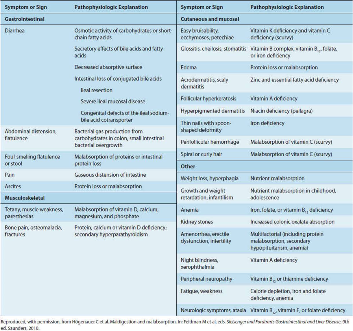
Of the many causes of diarrhea (Table 13-11), infectious agents are among the most important because they cause acute, sometimes life-threatening diseases whose pathogenesis is relatively well understood and because they are usually treatable. The symptoms of diarrhea caused by infectious agents are due to either toxins that alter small bowel secretion and absorption or direct mucosal invasion. The noninvasive toxin-producing bacteria are generally small bowel pathogens, whereas the invasive organisms are localized typically to the colon. Diarrheas caused by infectious agents are discussed in Chapter 4.
TABLE 13-11 Most likely causes of diarrhea in seven different clinical categories.
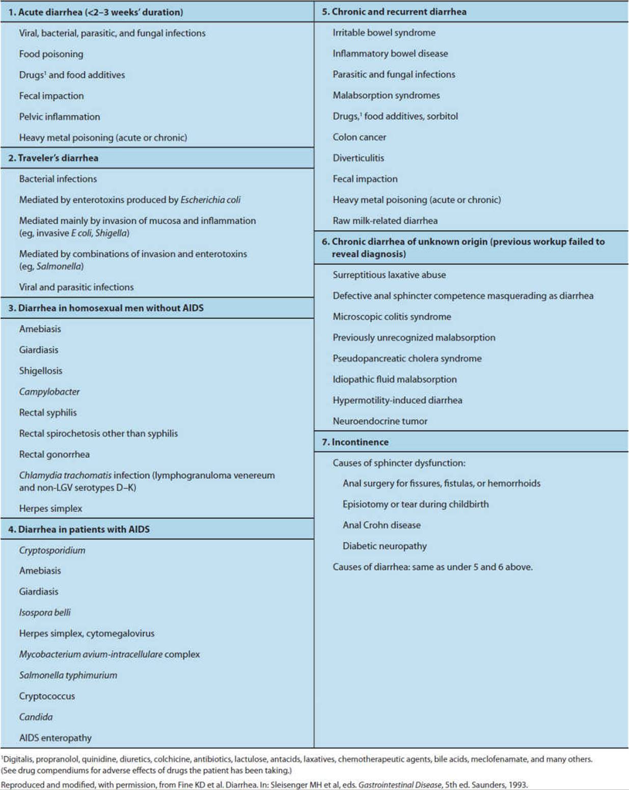
Evidence suggests that infectious causes of diarrhea can interface more intimately with normal mechanisms of secretory control than had been previously realized. Thus, in addition to its direct effect on the G protein controlling Cl−ion secretion in the crypts of the small intestinal epithelium, cholera activates the enteric nervous system to cause fluid and electrolyte secretion in the colon.
Clinical Manifestations
Dehydration, malnutrition, weight loss, and specific vitamin deficiency syndromes (eg, glossitis, cheilosis, and stomatitis) are common signs in diarrhea depending on its cause, severity, and chronicity (Tables 13–8 and 13–10). In certain circumstances (eg, in young children), viral gastroenteritis is associated with a high mortality rate from dehydration when supportive measures (ie, oral or intravenous rehydration) are not promptly provided. Some individuals with diarrhea from parasitic infections remain relatively asymptomatic, whereas others may develop more severe symptoms and complications, including intestinal perforation.
CHECKPOINT
61. By what mechanisms do infectious agents cause diarrhea?
62. Name three ways in which an excessive osmotic load can occur in the GI tract.
INFLAMMATORY BOWEL DISEASE
Clinical Presentation
Inflammatory bowel disease is distinguished from infectious entities by exclusion: recurrent episodes of mucopurulent (ie, containing mucus and white cells) bloody diarrhea characterized by lack of positive cultures for known microbial pathogens and failure to respond to antibiotics alone. Because inflammatory bowel disease is characterized by exacerbations and remissions, favorable responses to therapy are difficult to distinguish from spontaneous remissions occurring as part of the natural history of the disease.
Etiology
The trigger for inflammatory bowel disease is still unclear. There are two forms of chronic inflammatory bowel disease: Crohn disease, which is transmural and granulomatous in character, occurring anywhere along the GI tract, and ulcerative colitis, which is superficial and limited to the colonic mucosa. The causes of inflammatory bowel disease are unknown despite progress in understanding its pathogenesis.
Pathology & Pathogenesis
Genetic risk and environmental factors are recognized as two key elements in the pathogenesis of inflammatory bowel disease. An explosion of newly recognized susceptibility genes for both Crohn disease and ulcerative colitis have been discovered through genome-wide associations. These studies evaluated thousands of single nucleotide polymorphisms (SNPs) in thousands of patients with inflammatory bowel disease and compared them with thousands of people without the disease (controls). These studies found abnormalities in several categories of susceptibility genes in patients with inflammatory bowel disease. These included modulators of immune function, autophagy, and epithelial function that participate in the interaction of host and microorganism. Importantly, the relative risk of most of these susceptibility genes is low (most have a 20–30% increase in relative risk of developing disease). Therefore, most people that harbor risk alleles for inflammatory bowel disease do not develop disease.
Genetic factors are clearly not the sole contributor to inflammatory bowel disease. Many environmental factors have been found to contribute to the development of Crohn disease, including pathogenic microorganisms (bacteria and viruses), the repertoire of indigenous intestinal microbes (the microbiota), dietary factors, smoking, defective immune responses, and psychosocial factors. Moreover, recent studies suggest that patterning of the activity of certain aspects of the immune system during neonatal period strongly influences immune responses in the adult. Because the composition of the intestinal microbiota is in large part transmitted by the mother, maternal effects are thought to be a contributing factor to GI disease as well. Specifically, early exposure to intestinal microbiota may be an important component of the pathogenesis of inflammatory bowel disease.
The normal intestine is able to modulate frank inflammatory responses to its constant bombardment with dietary and microbial antigens in the lumen. This process may be defective in Crohn disease, resulting in uncontrolled inflammation. There has been considerable interest in the role of cytokines, such as interleukins and tumor necrosis factor, in Crohn disease. Cytokine profiles of TH1 and TH17 categories have been implicated in Crohn disease. Mice lacking the TH1-inhibiting cytokine interleukin-10 have a TH1 cytokine profile and develop spontaneous intestinal inflammation. Monoclonal antibodies to tumor necrosis factor (TNF) reduce inflammation in these animals and patients. Similar factors may contribute to the pathogenesis of ulcerative colitis, including infections, allergies to dietary components, immune responses to bacteria and self-antigens, and psychosocial factors. In mice, targeted disruption of the genes for the T-cell receptor and the cytokine IL-2 results in GI tract disease resembling ulcerative colitis.
The two forms of inflammatory bowel disease have characteristic differences and in many cases considerable overlap in manner of presentation (Table 13-12). The features common to all forms of inflammatory bowel disease are mucosal ulceration and inflammation of the GI tract, indistinguishable, in fact, from that which can occur acutely during invasive infectious diarrhea. Other factors besides the presence of key gene products, including infectious agents, altered host immune responses, immune-mediated intestinal damage, psychologic factors, and dietary and environmental factors, may contribute to a final common pathway of disordered immune response.
TABLE 13-12 Similarities and differences between ulcerative colitis and Crohn disease.
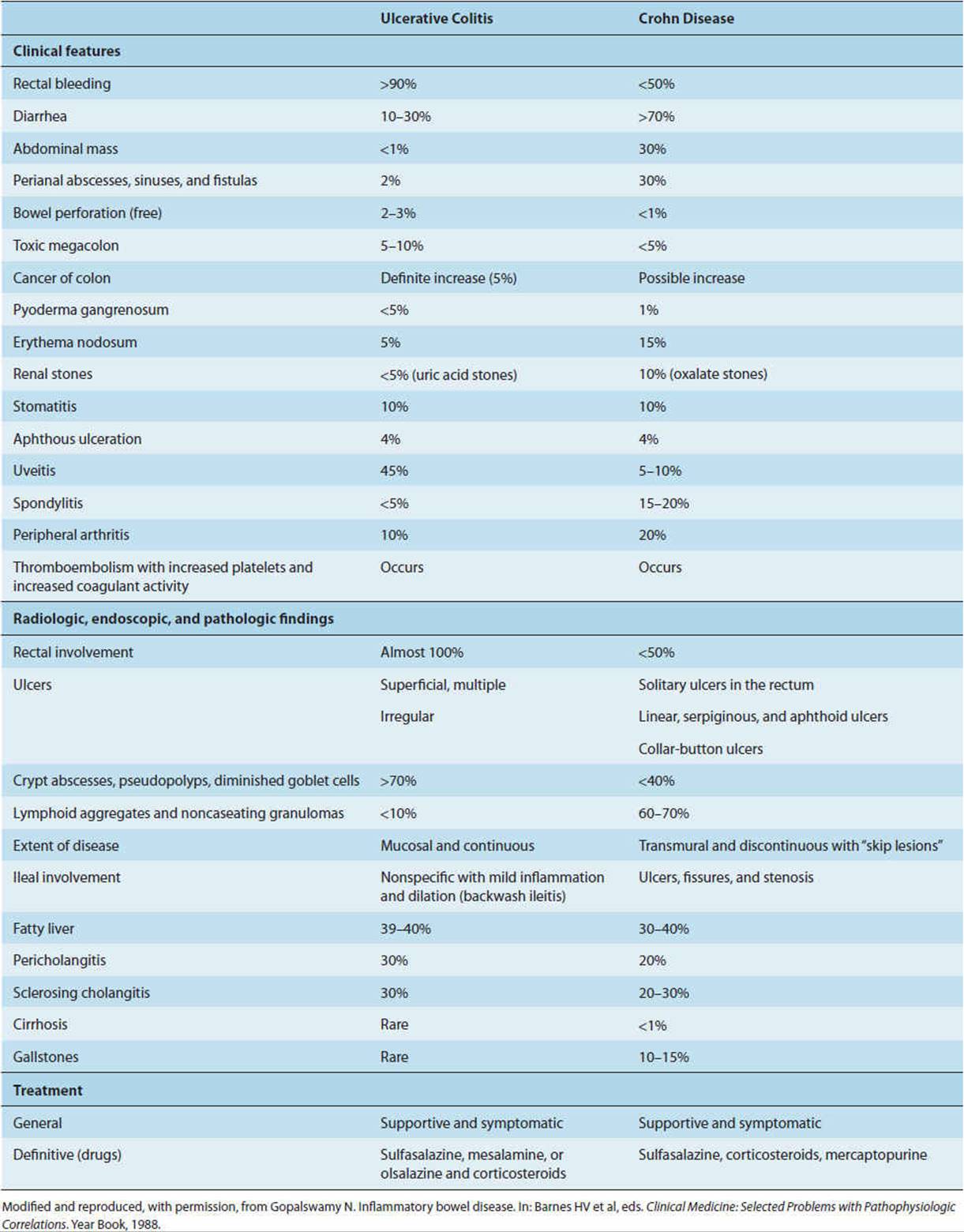
Clinical Manifestations
A. Crohn Disease
Crohn disease most typically occurs in the distal ileum. However, the distribution of the disease can also involve the colon or less commonly any other region of the GI tract (including the oral cavity, esophagus, stomach, and proximal small intestine). A characteristic feature is that areas of ulceration and inflammation occur in a discontinuous fashion and involve the entire thickness of the bowel wall. Recurrence of disease can occur in previously uninvolved regions of the intestine and can even involve adjacent mesentery and lymph nodes. The combination of deep mucosal ulceration and submucosal thickening gives the involved mucosa a characteristic “cobblestone” appearance.
Perforation, fistula formation, abscess formation, and small intestinal obstruction are frequent complications of Crohn disease, although an indolent course occurs in most patients. The full-thickness involvement of the bowel wall may predispose to these complications. Frank bleeding from the mucosal ulcerations can be either insidious or massive, as can protein-losing enteropathy. Another important complication is a possible increased incidence of intestinal cancer.
Patients with Crohn disease often manifest symptoms outside of the GI tract. Most commonly, inflammatory disorders of the joints (arthritis), skin (erythema nodosum), eye (uveitis, iritis), mucous membranes (aphthous ulcers of the buccal mucosa) bile ducts (sclerosing cholangitis), and liver (autoimmune chronic active hepatitis) are also observed in these patients. Renal disorders, especially nephrolithiasis, are observed in one-third of patients with Crohn disease, probably related to increased oxalate absorption associated with steatorrhea. Amyloidosis is a serious complication of Crohn disease, as is thromboembolic disease. Both of these complications are probably reflections of the systemic character of the inflammatory process. Patients are often malnourished and show evidence of nutrient deficiency states.
B. Ulcerative Colitis
In contrast to Crohn disease, inflammation in ulcerative colitis is restricted to the mucosa of the colon and rectum. It typically begins at the anorectal junction and extends proximally. Ulcerative colitis and Crohn disease are similar in presentation (eg, bloody diarrhea and malabsorption) and in at least some of the complications (eg, protein-losing enteropathy and malnutrition), reflecting widespread involvement of the mucosa in both entities. In both conditions, acute inflammatory cells (neutrophils) are located within the crypt epithelium (cryptitis) and crypt lumens (crypt abscesses). There is also a robust infiltration of chronic inflammatory cells in the mucosa. However, because ulcerative colitis generally is limited to the mucosa, obstruction, perforation, and fistula formation are not typical complications. Most patients have mild disease, and, as with Crohn disease, some patients will have only one or two episodes during their lifetimes. As with Crohn disease, there is an increased risk of colonic adenocarcinoma that increases with the duration of the disease. Chronic disease can also lead to damage of the muscularis propria, leading to toxic megacolon, a thin-walled dilated, poorly motile area of the colon that is susceptible to rupture. In the mucosa, chronic damage and ulceration can lead to excess granulation tissue that protrudes into the lumen of the intestine (pseudopolyps). Because of the variable response rate and the high risk of side effects, therapy with immunosuppressive agents such as mercaptopurine and azathioprine are limited to cases that have failed to respond to sulfasalazine and glucocorticoids. Both ulcerative colitis and Crohn disease can go into remission after treatment with first-line anti-inflammatory agents such as sulfasalazine and glucocorticoids. Crohn disease also responds to therapy that utilizes monoclonal antibodies against the inflammatory cytokine, TNF. These antibodies bind to and inhibit this cytokine. More recently, therapy with anti-TNF monoclonal antibodies has been used in patients with ulcerative colitis as well. Because of potential complication of serious, even life-threatening infection, these drugs are utilized only for severe cases. The natural history of both diseases is of periods of remission interrupted by active disease; medical therapy during exacerbations is directed toward supportive measures and attempts at inducing remission. Because these diseases can recur after resection of involved regions of the GI tract, operative management is generally limited to relief of life-threatening intestinal obstruction or bleeding.
CHECKPOINT
63. How is inflammatory bowel disease distinguished from infectious diarrhea?
64. What are the differences between ulcerative colitis and Crohn disease?
65. What are the complications of inflammatory bowel disease?
DIVERTICULAR DISEASE
Clinical Presentation
Nearly 80% of patients with diverticula are asymptomatic except for chronic constipation. Of those who develop other symptoms, the most common presentation is an intermittent and unpredictable griping lower abdominal pain (diverticulitis). Additional features of the presentation depend on which of the two major complications of diverticula that the patient develops.
A patient who develops diverticulitis (see later discussion) may present with fever and with symptoms and signs of peritoneal irritation (guarding, rebound tenderness, absence of bowel sounds). A patient who develops diverticular bleeding may present with either frankly bloody stools or stools that are positive for occult blood.
Etiology
Diverticulosis results from an acquired deformity of the colon in which the mucosa and submucosa herniate through the underlying muscularis (Figure 13-22). This is a disease of modern affluent life. A rarity at the turn of the century, today it afflicts 30% of adults in the U.S. population. Its incidence increases with age, starting from about 40 years. Epidemiologic studies suggest that the consumption of highly refined foods and less fiber, with resulting increased prevalence of chronic constipation, may be responsible for the increased prevalence of diverticular disease.
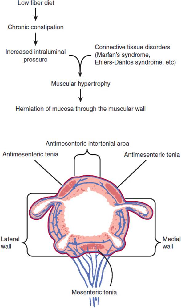
FIGURE 13-22 Pathophysiology of diverticular disease. Bottom: Cross-sectional drawing of the colon, showing principal points of diverticula formation between mesenteric and antimesenteric teniae. (Redrawn, with permission, from Goligher JC. Surgery of the Anus, Rectum and Colon, 5th ed. Baillière Tyndall, 1984.)
Pathology & Pathogenesis
A. Diverticulosis
Most acquired diverticula occur in the colon; the descending colon and sigmoid (left side) are involved in more than 90% of cases. Both structural and functional factors are believed to contribute to the development of diverticulosis. Acquired abnormalities in colonic wall connective tissue are believed to be the structural basis of diminished resistance to mucosal and submucosal herniation (Figure 13-22). The functional abnormality is believed to be related to chronic constipation and the development of a transmural pressure gradient from colonic lumen to peritoneal space as a result of vigorous muscle contraction of the colonic wall. This functional abnormality is most likely related to the change in dietary habits; decreased dietary fiber makes forward propulsion of feces at normal transmural pressures more difficult. This increased muscle contraction, which contributes to the development of diverticular disease, is also believed to cause the abdominal pain that is the cardinal symptom of uncomplicated diverticular disease. The pain may last hours to days, with sudden relief on passing flatus or feces. Constipation or diarrhea and flatulence are common findings during such episodes, leading to the suggestion that there is a relationship between irritable bowel syndrome and the development of diverticulosis. Treatment of the pain of diverticular disease with opioids is contraindicated because they directly raise intraluminal pressure and hence may increase the risk of perforation.
B. Diverticular Bleeding
Diverticula are a source of bleeding in 3–5% of patients with diverticulosis. Branches of the colonic intramural arteries (vasa recta) are closely associated with the diverticular sac, presumably leading to occasional rupture and bleeding. This is the most common cause of massive lower GI bleeding in the elderly. Diverticular bleeding is typically painless and not believed to be associated with a focus of inflammation.
The differential diagnosis of painless bleeding per rectum also includes internal hemorrhoids (dilated venous channels in the anal canal) and angiodysplasia. The latter consists of small, focal proliferations of dilated blood vessels in the mucosa, typically found in elderly patients.
C. Diverticulitis
This most common complication of diverticulosis develops when a focal area of inflammation occurs in the wall of a diverticulum in response to irritation by fecal material. The patient develops symptoms of abdominal pain and fever with a risk of progression to abscess with or without perforation. These symptoms mimic acute appendicitis. The perforations usually are self-contained, but the potential for subsequent fistula formation and intestinal obstruction is high. Approximately, 15–25% of patients who develop diverticulitis will require surgery.
Clinical Manifestations
About one-fifth of all individuals with diverticular disease develop one of the two major complications: diverticular bleeding or diverticulitis. These disorders must be distinguished from carcinoma, inflammatory bowel disease, infection, and ischemic injury. Ischemia can arise from a variety of conditions including atherosclerosis, vasculitis, hypercoagulable states, heart failure, and shock.
CHECKPOINT
66. Where in the GI tract do most diverticula occur?
67. What predisposing factors contribute to the development of diverticular disease?
68. What are the major complications of diverticular disease?
IRRITABLE BOWEL SYNDROME
Irritable bowel syndrome is the most common cause of referral to gastroenterologists. It is characterized by altered bowel habits with abdominal pain in the absence of any detectable organic pathological process or specific motility or structural abnormalities.
A change in bowel habits, commonly alternating between diarrhea and constipation, is the principal characteristic of irritable bowel syndrome. Abdominal pain, which may be caused by intestinal spasms, is also common to all patients with irritable bowel syndrome. Bloating or perceived abdominal distension is another common feature. Intraluminal gas can result from swallowing air, diminished absorption of gas, and bacterial fermentation, although the cause in irritable bowel syndrome is unknown. Stress appears to have a considerable influence on these symptoms. Symptoms of irritable bowel syndrome frequently occur during or after a stressful event, and stressful events in early life may predispose to the development of irritable bowel syndrome.
Much of our understanding of the pathophysiology of irritable bowel syndrome derives from study of motility. In normal persons, high-amplitude peristaltic contractions occur 6–8 times per day. In constipated patients with irritable bowel syndrome, the frequency of high-amplitude peristaltic contractions of the intestine is diminished compared with normal subjects, suggesting that the constipation may be due to diminished motility. Visceral hyperalgesia may also occur in patients with irritable bowel syndrome. In patients with irritable bowel syndrome, distension of the colon with a balloon, to a degree that is not painful in normal individuals, can induce pain, indicative of visceral hyperalgesia.
Irritable bowel syndrome is a complex disorder, and its cause is poorly understood. Several theories have been proposed to explain the disorder, including alterations in sensitivity of the extrinsic and intrinsic nervous systems of the intestine, which may contribute to exaggerated sensations of pain and to abnormal control of intestinal motility and secretion. An alteration in the balance of secretion and absorption is also a potential cause. Although there is no gross inflammation of the intestine, there are reports of an increased influx of inflammatory cells (lymphocytes) into the colon of affected individuals as well as destruction of enteric neurons. The intestinal microbes that normally inhabit the small intestine and colon may be altered as well, suggesting that antibiotics could have a role in treatment of this disorder. One proposed theory is that irritable bowel syndrome develops as a result of an earlier and resolved bout of interstitial inflammation. In experimental animals, induction of intestinal inflammation induces visceral hyperalgesia and altered intestinal motility and secretion that persists many months after the inflammation is resolved. A similar mechanism may occur in a subset of patients who develop irritable bowel syndrome after an infection causes intestinal inflammation.
CHECKPOINT
69. List three characteristics of irritable bowel syndrome.
70. What are possible factors in the pathogenesis of the irritable bowel syndrome?
CASE STUDIES
Yeong Kwok, MD
(See Chapter 25, p. 723 for Answers)
CASE 62
A 60-year-old man presents to the clinic with a 3-month history of gradually worsening dysphagia (difficulty swallowing). At first, he noticed the problem when eating solid food such as steak, but now it happens even with drinking water. He has a sensation that whatever he swallows becomes stuck in his chest and does not go into the stomach. He has also developed worsening heartburn, especially upon lying down, and has had to prop himself up at night to lessen the heartburn. He has lost 10 kg as a result of his swallowing difficulties. His physical examination is unremarkable. A barium swallow x-rayreveals a decrease in peristalsis of the body of the esophagus along with dilatation of the lower esophagus and tight closure of the lower esophageal sphincter. There is a beaked appearance of the distal esophagus involving the lower esophageal sphincter. There is very little passage of barium into the stomach.
Questions
A. What is the likely diagnosis in this patient, and what is the underlying pathophysiology of this condition?
B. Botulinum toxin can be used to treat this disorder. How does it help ameliorate the symptoms?
C. What are the possible complications of this disorder, and how do they arise?
CASE 63
A 32-year-old woman presents to her primary care provider complaining of a persistent burning sensation in her chest and upper abdomen. The symptoms are worse at night while she is lying down and after meals. She has tried drinking hot cocoa to help her sleep. She is a smoker and frequently relies on benzodiazepines for insomnia. She notes a sour taste in her mouth every morning. Physical examination is normal.
Questions
A. What is the pathogenetic mechanism of her GI disorder?
B. How may her lifestyle impact her symptoms?
C. What are some complications of chronic esophageal reflux disease?
CASE 64
A 74-year-old man with severe osteoarthritis presents to the emergency department reporting two episodes of melena (black stools) without hematochezia (bright red blood in the stools) or hematemesis (bloody vomitus). He takes 600 mg of ibuprofen three times a day to control his arthritis pain. He denies alcohol use. On examination his blood pressure is 150/70 Hg and his resting pulse is 96/min. His epigastrium is minimally tender to palpation. Rectal examination reveals black tarry stool in the vault, grossly positive for occult blood. Endoscopy demonstrates a 3 cm gastric ulcer. Helicobacter pylori is identified on biopsies of the ulcer site.
Questions
A. What are some of the proposed mechanisms for acid-peptic disease and specifically gastric ulcer disease?
B. How may this patient’s analgesic use predispose him to acidpeptic disease?
C. What role does H pylori infection play in the pathogenesis of ulcer disease? How should this be taken into account when treating this patient?
CASE 65
A 67-year-old man with type 2 diabetes is seen by his primary care provider for frequent nausea, bloating, and intermittent diarrhea over the preceding 2 weeks. The vomiting typically occurs approximately 1–2 hours after eating. He states that over the past year he has become increasingly depressed after the death of his wife and has been less adherent to his oral hypoglycemic regimen and evening insulin. He also reports 6 months of worsening neuropathic pain in his feet. His fasting fingerstick blood glucose level is 253 mg/dL.
Questions
A. How may diabetes contribute to the development of gastroparesis? Is his poor control a cause or consequence of gastroparesis?
B. How can delayed gastric emptying cause diarrhea?
CASE 66
A 40-year-old woman presents to the emergency department with a history of worsening right upper quadrant pain. The pain started after she had pizza for dinner 2 days ago and is described as a sharp, stabbing sensation under her right ribs. She has also felt ill, developed slight nausea, and had a low-grade fever. There has been no vomiting or diarrhea. Physical examination reveals an obese woman with a low-grade fever and tenderness to palpation of the right upper quadrant of her abdomen. An abdominal ultrasound reveals a 2 cm gallstone lodged in the cystic duct with swelling of the gallbladder and thickening of the gallbladder wall.
Questions
A. What are the mechanisms involved in gallstone formation?
B. What factors in the pathogenesis of gallstones may be responsible for the fact that it is more common in premenopausal women?
C. What local complications can ensue from gallstone disease?
CASE 67
A 45-year-old man comes to clinic with a history of excessive bloating, foul-smelling flatus, and loose stools for the past several months. He notes that about 30–60 minutes after breakfast each morning, he experiences cramping, bloating, passage of smelly flatus, and a very loose, watery bowel movement. He has not seen any blood or mucous in the stool and also denies any weight loss. This does not happen with lunch or dinner. Every day for breakfast, he eats a big bowl of cereal with milk and a yogurt smoothie. Physical exam is unremarkable with normal bowel sounds, no organomegaly, and no abdominal tenderness. He was advised to do a dietary trial of stopping dairy intake for 1 week. All his symptoms resolve, and he is diagnosed with lactose intolerance.
Questions
A. Why do people develop lactose intolerance?
B. Why does the inability to digest lactose lead to diarrhea?
CASE 68
A 42-year-old man with long-standing Crohn disease presents to the emergency department with a 1-day history of increasing abdominal distension, pain, and obstipation. He is nauseated and has vomited bilious material. He has no history of abdominal surgery and has had two exacerbations of his disease this year. He is febrile with a temperature of 38.5°C. Examination reveals multiple oral aphthous ulcers, hyperactive bowel sounds, and a grossly distended, diffusely tender abdomen without an appreciable mass. Abdominal radiographs reveal multiple air-fluid levels in the small bowel with minimal colonic gas consistent with a small bowel obstruction.
Questions
A. Describe the significance of the oral aphthous ulcers in the distribution of Crohn disease.
B. What factors are thought to be involved in the pathogenesis of Crohn disease? What is the evidence to support the role of cytokines in the pathogenesis of Crohn disease?
C. What are the GI complications of Crohn disease?
D. Describe some of the extraintestinal manifestations of Crohn disease.
CASE 69
A 76-year-old woman with chronic constipation reports a 4-day history of “achy” left lower quadrant abdominal pain, graded 7/10, accompanied by low-grade fever and nausea. A colonoscopy performed 2 years ago revealed sigmoid diverticular disease. On examination she has a temperature of 38.6°C. Her abdomen has a tender 3 × 2 cm mass in the left lower quadrant. Bowel sounds are normal. Her stool is positive for occult blood. An abdominal series shows a bowel gas pattern consistent with ileus and no evidence of free peritoneal air. A CT scan with contrast of the abdomen and pelvis shows pericolonic fat stranding with no evidence of an abscess. She is started on antibiotics and intravenous fluids with significant improvement in her symptoms.
Questions
A. Describe the pathogenesis of diverticular disease.
B. Why should opioids be avoided in the treatment of her abdominal pain?
C. What are the complications of diverticular disease?
CASE 70
A 32-year-old woman comes to the clinic complaining of a 3-month history of abdominal bloating, crampy abdominal pain, and a change in her bowel habits. Previously she had regular bowel movements, but 4 months ago, she developed gastroenteritis with nausea and vomiting after a cruise. The constant diarrhea and vomiting went away after a week, but since then she has had periods of constipation, lasting up to 3 days, alternating with periods of diarrhea. During the diarrheal episodes, she can have three to four loose bowel movements per day, though without blood or mucus in the stool. She describes diffuse abdominal cramping and bloating that are somewhat relieved by bowel movements. Her symptoms worsen during periods of stress. There has been no weight loss or fever. There is no association with particular foods (eg, wheat or dairy products). Her physical examination is unremarkable except for mild abdominal tenderness with no rebound or guarding. Serologic tests for celiac sprue are negative. Stool cultures and examinations are negative for bacterial or parasitic infections. A colonoscopy is unremarkable.
Questions
A. What is the likely diagnosis?
B. What are the theories about the pathophysiology of this condition?
REFERENCES
General
Hooper LV et al. Immune adaptations that maintain homeostasis with the intestinal microbiota. Nat Rev Immunol. 2010 Mar;10(3): 159–69. [PMID: 20182457]
Janssen P et al. Review article: the role of gastric motility in the control of food intake. Aliment Pharmacol Ther. 2011 Apr;33(8): 880–94. [PMID: 21342212]
Kozyraki R et al. Vitamin B12 absorption: mammalian physiology and acquired and inherited disorders. Biochimie. 2013 May; 95(5):1002–7. [PMID: 23178706]
Laforenza U. Water channel proteins in the gastrointestinal tract. Mol Aspects Med. 2012 Oct–Dec;33(5–6):642–50. [PMID: 22465691]
Lozupone CA et al. Diversity, stability and resilience of the human gut microbiota. Nature. 2012 Sep 13;489(7415):220–30. [PMID: 22972295]
Mills JC et al. Gastric epithelial stem cells. Gastroenterology. 2011 Feb;140(2):412–24. [PMID: 21144849]
Noah TK et al. Intestinal development and differentiation. Exp Cell Res. 2011 Nov 15;317(19):2702–10. [PMID: 21978911]
Ouellette AJ. Paneth cell alpha-defensins in enteric innate immunity. Cell Mol Life Sci. 2011 Jul;68(13):2215–29. [PMID: 21560070]
Saqui-Salces M et al. A high-fat diet regulates gastrin and acid secretion through primary cilia. FASEB J. 2012 Aug;26(8):3127–39. [PMID: 22516298]
van der Flier LG et al. Stem cells, self-renewal, and differentiation in the intestinal epithelium. Annu Rev Physiol. 2009;71:241–60. [PMID: 18808327]
Zhao CM et al. The ECL cell: relay station for gastric integrity. Curr Med Chem. 2012;19(1):98–108. [PMID: 22300082]
GI Disease
Bornschein J et al. Gastric cancer: clinical aspects, epidemiology and molecular background. Helicobacter. 2011 Sep;16(Suppl 1):45–52. [PMID: 21896085]
Cho I et al. The human microbiome: at the interface of health and disease. Nat Rev Genet. 2012 Mar 13;13(4):260–70. [PMID: 22411464]
Goldenring JR et al. Spasmolytic polypeptide-expressing metaplasia and intestinal metaplasia: time for reevaluation of metaplasias and the origins of gastric cancer. Gastroenterology. 2010 Jun; 138(7):2207–10. [PMID: 20450866]
Kandulski A et al. Helicobacter pylori infection: a clinical overview. Dig Liver Dis. 2008 Aug;40(8):619–26. [PMID: 18396114]
Rugge M et al. Autoimmune gastritis: histology phenotype and OLGA staging. Aliment Pharmacol Ther. 2012 Jun;35(12):1460–6. [PMID: 22519568]
Schulzke JD et al. Disorders of intestinal secretion and absorption. Best Pract Res Clin Gastroenterol. 2009;23(3):395–406. [PMID: 19505667]
Taylor ND et al. Infantile hypertrophic pyloric stenosis: has anything changed? J Paediatr Child Health. 2013 Jan;49(1):33–7. [PMID: 23198903]
Achalasia & Esophageal Reflux
Burke ZD et al. Barrett’s metaplasia as a paradigm for understanding the development of cancer. Curr Opin Genet Dev. 2012 Oct;22(5):494–9. [PMID: 22981230]
Chuah SK et al. 2011 update on esophageal achalasia. World J Gastroenterol. 2012 Apr 14;18(14):1573–8. [PMID: 22529685]
Ghoshal UC et al. Pathogenesis of achalasia cardia. World J Gastroenterol. 2012 Jun 28;18(24):3050–7. [PMID: 22791940]
Lenglinger J et al. Review on the annual cancer risk of Barrett’s esophagus in persons with symptoms of gastroesophageal reflux disease. Anticancer Res. 2012 Dec;32(12):5465–73. [PMID: 23225453]
Phillips WA et al. Barrett’s esophagus. J Gastroenterol Hepatol. 2011 Apr;26(4):639–48. [PMID: 21166712]
Souza RF. The role of acid and bile reflux in oesophagitis and Barrett’s metaplasia. Biochem Soc Trans. 2010 Apr;38(2):348–52. [PMID: 20298181]
Acid-Peptic Disease
de Vries AC et al. Helicobacter pylori infection and nonmalignant diseases. Helicobacter. 2010 Sep;15(Suppl 1):29–33. [PMID: 21054650]
Ghoshal UC et al. Gastroesophageal reflux disease and Helicobacter pylori: what may be the relationship? J Neurogastroenterol Motil. 2010 Jul;16(3):243–50. [PMID: 20680162]
Hunt RH et al. Acid-NSAID/aspirin interaction in peptic ulcer disease. Dig Dis. 2011;29(5):465–8. [PMID: 22095011]
Ng SC et al. NSAID-induced gastrointestinal and cardiovascular injury. Curr Opin Gastroenterol. 2010 Nov;26(6):611–7. [PMID: 20948372]
Sung JJ. Marshall and Warren Lecture 2009: peptic ulcer bleeding: an expedition of 20 years from 1989–2009. J Gastroenterol Hepatol. 2010 Feb;25(2):229–33. [PMID: 20136987]
Gastroparesis & Ileus
Hasler WL. Gastroparesis: pathogenesis, diagnosis and management. Nat Rev Gastroenterol Hepatol. 2011 Jul 19;8(8):438–53. [PMID: 21769117]
Knowles CH et al. New perspectives in the diagnosis and management of enteric neuropathies. Nat Rev Gastroenterol Hepatol. 2013 Apr;10(4):206–18. [PMID: 23399525]
Mostafa RM et al. Interstitial cells of Cajal, the Maestro in health and disease. World J Gastroenterol. 2010 Jul 14;16(26):3239–48. [PMID: 20614479]
Oh JH et al. Recent advances in the pathophysiology and treatment of gastroparesis. J Neurogastroenterol Motil. 2013 Jan;19(1):18–24. [PMID: 23350043]
Quigley EM. What we have learned about colonic motility: normal and disturbed. Curr Opin Gastroenterol. 2010 Jan;26(1):53–60. [PMID: 19786868]
van Bree SH et al. New therapeutic strategies for postoperative ileus. Nat Rev Gastroenterol Hepatol. 2012 Nov;9(11):675–83. [PMID: 22801725]
Gallstone Disease
Reshetnyak VI. Concept of the pathogenesis and treatment of cholelithiasis. World J Hepatol. 2012 Feb 27;4(2):18–34. [PMID: 22400083]
Van Erpecum KJ. Pathogenesis of cholesterol and pigment gallstones: an update. Clin Res Hepatol Gastroenterol. 2011 Apr;35(4):281–7. [PMID: 21353662]
Diarrhea
Ivanov AI et al. Cytoskeletal regulation of epithelial barrier function during inflammation. Am J Pathol. 2010 Aug;177(2):512–24. [PMID: 20581053]
Marchiando AM. Epithelial barriers in homeostasis and disease. Annu Rev Pathol. 2010;5:119–44. [PMID: 20078218]
Tack J. Functional diarrhea. Gastroenterol Clin North Am. 2012 Sep;41(3):629–37. [PMID: 22917168]
Inflammatory Bowel Disease
Abraham C et al. Inflammatory bowel disease. N Engl J Med. 2009 Nov 19;361(21):2066–78. [PMID: 19923578]
Ciorba MA et al. Probiotic therapy in radiation-induced intestinal injury and repair. Ann N Y Acad Sci. 2009 May;1165:190–4. [PMID: 19538306]
Khor B et al. Genetics and pathogenesis of inflammatory bowel disease. Nature. 2011 Jun 15;474(7351):307–17. [PMID: 21677747]
Packey CD et al. Commensal bacteria, traditional and opportunistic pathogens, dysbiosis and bacterial killing in inflammatory bowel diseases. Curr Opin Infect Dis. 2009 Jun;22(3):292–301. [PMID: 19352175]
Rubino SJ et al. Nod-like receptors in the control of intestinal inflammation. Curr Opin Immunol. 2012 Aug;24(4):398–404. [PMID: 22677577]
Sun L et al. Host genetic susceptibility, dysbiosis, and viral triggers in inflammatory bowel disease. Curr Opin Gastroenterol. 2011 Jul;27(4):321–7. [PMID: 21483258]
Ziegler SF et al. Sensing the outside world: TSLP regulates barrier immunity. Nat Immunol. 2010 Apr;11(4):289–93. [PMID: 20300138]
Diverticular Disease
Strate LL et al. Diverticular disease as a chronic illness: evolving epidemiologic and clinical insights. Am J Gastroenterol. 2012 Oct;107(10):1486–93. [PMID: 22777341]
Touzios JG et al. Diverticulosis and acute diverticulitis. Gastroenterol Clin North Am. 2009 Sep;38(3):513–25. [PMID: 19699411]
Irritable Bowel Syndrome
Almansa C et al. Intestinal microbiota, pathophysiology and translation to probiotic use in patients with irritable bowel syndrome. Expert Rev Gastroenterol Hepatol. 2012 Jun;6(3):383–98. [PMID: 22646259]
Camilleri M. Peripheral mechanisms in irritable bowel syndrome. N Engl J Med. 2012 Oct 25;367(17):1626–35. [PMID: 23094724]