
J. Ben Davoren, MD, PhD, & Sunny Wang, MD
NORMAL STRUCTURE & FUNCTION
Blood is an extremely complex fluid, composed of both formed elements (red cells, white cells, platelets) and plasma. Red blood cells (erythrocytes) are the most common formed elements, carrying oxygen to the cells of the body via their main component, hemoglobin. White blood cells are generally present at about 1/700th the number of erythrocytes and function as mediators of immune responses to infection or other stimuli of inflammation. Platelets are the formed elements that participate in coagulation. Plasma is largely water, electrolytes, and plasma proteins. The plasma proteins most important in blood clotting are the coagulation factors. Because blood circulates throughout the body, alterations in normal blood physiology—either formed elements or plasma proteins—may have widespread adverse consequences.
FORMED ELEMENTS OF BLOOD
Anatomy
A. Bone Marrow and Hematopoiesis
Although the mature formed elements of blood are quite different from each other in both structure and function, all of these cells develop from a common hematopoietic stem cell population, which resides in the bone marrow. The developmental process is called hematopoiesis and represents an enormous metabolic task for the body. More than 100 billion cells are produced every day. This makes the bone marrow one of the most active organs in the body. In adults, most of the active marrow resides in the vertebrae, sternum, and ribs. In children, the marrow is more active in the long bones.
The process of differentiation from stem cell to mature erythrocyte, granulocyte, lymphocyte, monocyte, or platelet is shown in Figure 6-1. It is not clear exactly what early events lead dividing stem cells down a particular path of development, but many different peptides, called cytokines, are clearly involved (Table 6-1); see also Chapter 3. Perhaps because mature white blood cells have a much shorter half-life in the circulation, white blood cell precursors usually outnumber red blood cell precursors by a ratio of 3:1 in the bone marrow.

FIGURE 6-1 Hematopoiesis: development of the formed elements of blood from bone marrow stem cells. Cells below the horizontal line are found in normal peripheral blood. The principal cytokines that stimulate each cell lineage to differentiate are shown. (EPO, erythropoietin; TPO, thrombopoietin; CSF, colony-stimulating factor; G, granulocyte; M, macrophage; IL, interleukin; SCF, stem cell factor.) See Table 6-1 for details. (Redrawn, with permission, from Ganong WF. Review of Medical Physiology, 22nd ed. McGraw-Hill, 2005.)
TABLE 6-1 Cytokines that regulate hematopoiesis.

The major hormone that stimulates the production of erythrocytes (erythropoiesis) is erythropoietin. This peptide is produced by the kidneys and regulates red blood cell production by a feedback system: When blood hemoglobin levels fall (anemia), oxygen delivery to the kidneys falls, and they produce more erythropoietin, causing the marrow to produce more red cells. When hemoglobin levels rise, the kidney produces less erythropoietin and the marrow fewer red cells.
For white blood cells, the situation is more complex. The most common cells are the granulocytes, so named because their cytoplasms are filled with granules. Of these, the neutrophils are the most prevalent and the most important cells in producing inflammation. Granulocyte production (myelopoiesis) can be affected by many cytokines at different stages of development. Figure 6-1 shows that interleukin-3 (IL-3), granulocyte colony-stimulating factor (G-CSF), and granulocyte-macrophage colony-stimulating factor (GM-CSF) are the most important. All three proteins have been purified, sequenced, and cloned. The latter two proteins are used therapeutically. Unlike G-CSF, GM-CSF also stimulates the maturation of a different white blood cell line, the monocyte-macrophage line. These cells are part of the immune system as well (eg, ingesting foreign bacteria) and can reside in skin and other tissues, not just blood. Their function, along with that of the B- and T-lymphocyte populations, is discussed more fully in Chapter 3.
Platelets are not cells but fragments of larger multinucleated cells in the marrow called megakaryocytes. Platelets are crucial to normal blood clotting. Platelet production is also stimulated by multiple cytokines but is dependent mainly on the action of IL-3, IL-6, and IL-11 and thrombopoietin (TPO). This peptide is produced by the liver, kidney, skeletal muscle, and marrow stroma. One model of thrombopoiesis proposes that the production of TPO occurs at a constant rate. However, the amount of this hormone free to interact with platelet precursors rises and falls, probably as a result of uptake by TPO receptors (c-Mpl) on existing platelets in the blood. Therefore, a low platelet count (with a lower mass of c-Mpl) stimulates thrombopoiesis as a result of increased circulating levels of TPO. A second model proposes that low platelet levels can induce increased production of TPO in marrow stromal cells, via various cytokines including platelet-derived growth factor (PDGF) and fibroblast growth factor (FGF). These two models are not necessarily mutually exclusive. Inflammation can also lead to thrombocytosis via IL-6–mediated increases in TPO production by the liver.
For all its complexity and metabolic activity, there is tremendous regulation of the marrow through the interaction of various cytokines. Normally, only the most mature elements in each cell lineage are released into the general circulation, demonstrating this exquisite control over development. Complex negative-feedback mechanisms must be at work to maintain circulating quantities of each formed element at the consistent levels at which they are found.
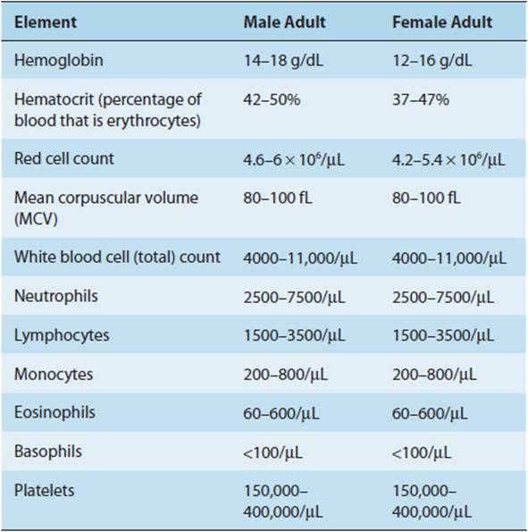
Examination of the appropriateness of blood cell development is best undertaken with the microscope, using the thin blood smear (Figure 6-2). Modern technical equipment, which can optically sort cells by size and various optical reflective parameters, gives important information, especially about whether cell numbers are out of the normal ranges (Table 6-2). However, microscopic examination of the blood smear, usually using Wright stain, gives additional information once an abnormality is detected and should always be done when a blood disorder is suspected on clinical grounds.
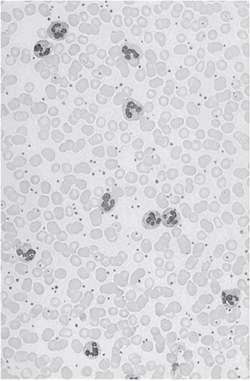
FIGURE 6-2 Normal thin blood smear, seen at low power (40×) with Wright stain. Erythrocytes predominate and can be seen to be thin disks with central pallor (see text). Platelets are the numerous small, dark bodies. Larger cells with lobulated nuclei are mature neutrophils. Lymphocytes and monocytes are not present on this smear.
TABLE 6-2 Normal values obtained on automated blood count—formed elements of blood.
Physiology
A. Erythrocytes
Mature red blood cells are biconcave disk-shaped cells filled with hemoglobin, which function as the oxygen-carrying component of the blood. In contrast to most other cells, they do not have nuclei at maturity; their nuclei are extruded during the final phase of erythrocyte development. The presence of erythrocytes with nuclei in the peripheral blood smear suggests an underlying disease state. Normal red cells are about 8 μm in diameter, a size that is larger than the smallest capillaries. However, their biconcave shape gives them enough flexibility to slip through small capillaries and deliver oxygen to the tissues. Once extruded from the bone marrow, individual erythrocytes function for about 120 days before they are removed from the circulation by the spleen.
In a typical blood smear (stained with Wright stain), erythrocytes dominate the microscopic field, and their biconcave disk shape resembles that of a doughnut. There is a thicker outer rim that appears red owing to the hemoglobin present and an area of central pallor where the disk is thinnest. Young erythrocytes (reticulocytes) appear bluer (basophilic) because they still contain some ribosomes and mitochondria for a few days after the nuclei are extruded.
Hemoglobin is the most important substance in the erythrocyte. This protein is actually a tetramer, made of two β-protein subunits and two α-protein subunits (in normal adult hemoglobin, called hemoglobin A). Each β- or α-subunit contains the actual oxygen-binding portion of the complex, heme. Heme is a compound whose centrally important atom is iron; it is this atom that actually binds oxygen in the lungs and subsequently releases it in the tissues of the body. A low level of hemoglobin in the blood, from a variety of causes (see later discussion), is anemia, the most common general blood disorder.
B. Granulocytes: Neutrophils, Eosinophils, and Basophils
The granulocytes are the most common white blood cells; of these, neutrophils are most abundant, followed by eosinophils and basophils (Table 6-2). Developmentally, all three types are similar: As they mature, their nuclei become more convoluted and multilobed, and each develops a cytoplasm filled with granules. These granules contain a variety of enzymes, prostaglandins, and mediators of inflammation, with specific factors dependent on the cell type. Early progenitor cells for each type of granulocyte (“blasts”) are indistinguishable on microscopic examination of the bone marrow, but under the influence of different cytokines, they become morphologically distinct cell types.
Basophils contain very dark blue or purple granules when stained with either Giemsa or Wright stain. Basophil granules are large and usually obscure the nucleus because of their density. Normally, basophils function in hypersensitivity reactions (as described in Chapter 3). However, their numbers can be increased in diseases not associated with hypersensitivity, such as chronic myelogenous leukemia.
Eosinophils contain large, strikingly “eosinophilic” granules (staining red with Wright or Giemsa stain). Eosinophil nuclei are usually bilobed. Normally, eosinophils function as part of the inflammatory response to parasites too large to be engulfed by individual immune cells. They are also involved in some allergic reactions.
Neutrophils contain granules that are “neutrophilic” (ie, neither eosinophilic nor basophilic). Although they predominate in the blood, their major function is actually in the tissues; they must leave the blood by inserting themselves between the endothelial cells of the vasculature to reach sites of injury or infection. Their granules contain highly active enzymes such as myeloperoxidase, which, along with the free radical oxygen ions produced by membrane enzymes such as nicotinamide adenine dinucleotide phosphate (NADPH) oxidase, kill bacteria that neutrophils ingest via endocytosis or phagocytosis. They are the “first line of defense” against bacterial pathogens, and low numbers of them (leukopenia) lead directly to a high incidence of significant bacterial infections (see later discussion). Of all the cells produced by the bone marrow, the neutrophils comprise the greatest fraction. Their life span in blood, only 8 hours, is much shorter than that of any other cell type. Evidence of their importance and their short survival is commonly manifested, because examination of the blood smear under the microscope in a patient with an active infection may show not only increased numbers of mature, multilobed neutrophils (neutrophilia) but also increased numbers of less mature cells. These less mature cells, released from a large storage pool in the bone marrow, are called bands and have a characteristic horseshoe-shaped nucleus that is not yet fully lobulated. The phenomenon of finding these cells in the peripheral blood is called a left shift of the granulocyte lineage.
C. Other White Blood Cells: Monocytes and Lymphocytes
Both monocytes and lymphocytes arise from the common stem cell. It is the widespread pluripotential ability of stem cells to differentiate into these cells in addition to the granulocytes, erythrocytes, and platelets that makes bone marrow transplantation a therapeutic option for immune system disorders and malignancies. Monocytes have a very long life span, probably several months, but spend only about 3 days in the circulation. They mostly reside in tissues and act there as immune cells that engulf (phagocytose) bacteria and subsequently can “present” components of these bacteria to lymphocytes in a way that further amplifies and refines the immune response (Chapter 3). On blood smear evaluation, monocytes are the largest cells seen, with irregular but not multilobed nuclei and pale blue cytoplasm, often with prominent vacuoles.
Lymphocyte precursors leave the marrow early and require extramedullary (outside of the marrow) maturation to become normally functioning immune cells in either the blood or the lymphatic system (Figure 6-3). Their crucial roles in recognizing “self” versus “nonself” and in modulating virtually all aspects of the immune response are described in Chapter 3. On microscopic examination of the blood smear, lymphocytes are small cells, slightly larger than an erythrocyte, with dark nuclei essentially filling the entire cell; only a thin rim of light blue cytoplasm is normally seen. Granules are sparse or absent.
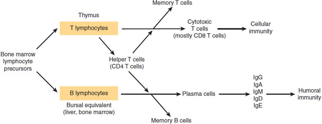
FIGURE 6-3 Development of the immune system from the common bone marrow stem cell. (Redrawn, with permission, from Ganong WF. Review of Medical Physiology, 22nd ed. McGraw-Hill, 2005.)
D. Platelets
Platelets are the smallest formed elements in the blood. They are fragments of larger, multinucleated cells, which are the largest discrete constituents of the bone marrow (megakaryocytes), but platelets have no nuclei of their own. Most platelets remain in the circulation, but a substantial minority is trapped in the spleen; this phenomenon becomes important in a variety of immune-mediated decreases in platelet count (thrombocytopenia; see later discussion). In the setting of a normal platelet count, they have a circulatory half-life of about 10 days. In cases of thrombocytopenia, their half-life decreases, as they are consumed in the routine maintenance of vascular integrity.
Platelets are integral components of the coagulation system. Their membranes provide an important source of phospholipids (PLs), which are required for the function of the coagulation system proteins (Figure 6-4), and contain important receptors that allow attachment to endothelial cells (platelet adhesion) so that a platelet plug can be formed in response to blood vessel injury. This prevents further blood loss after trauma and limits the coagulation response to the site of injury rather than letting coagulation proceed inappropriately.
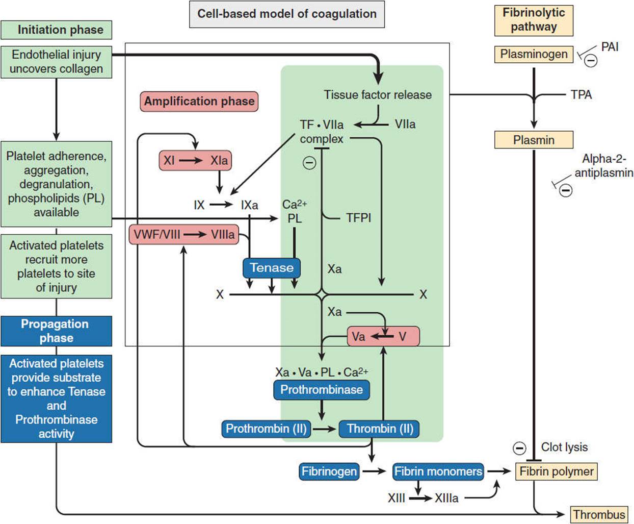
FIGURE 6-4 Coagulation and thrombolytic systems, showing balanced activity between them.
The cytoplasm is also important for platelet function, particularly the intracellular dense granules and alpha granules. The phenomenon of platelet activation is also called “degranulation” and can be initiated by exposure of platelets to the activated blood coagulation factor thrombin, adenosine 5′-diphosphate (ADP), or collagen. This last reaction is probably the most important, occurring when collagen, normally in the basement membrane below the endothelial cells, is exposed to the blood after injury. Platelet activation can also be induced by exposure to platelet-activating factor (PAF) (a neutrophil-derived phospholipid cytokine), thromboxane A2, serotonin, and epinephrine.
During platelet activation, the dense and alpha granules release further activators of platelet activity, such as ADP, and platelet factor 4, which can also bind to endothelial cells. It is important because it binds to the most commonly used therapeutic anticoagulant, heparin (see later discussion). After activation, platelets change shape from discoid to spherical with filopodial extensions and finally to a flat shape that allows for adequate coverage of the site of vessel injury. The last step in platelet activity is platelet aggregation, where platelets stick to each other, firming up the platelet plug. On examination of the blood smear, platelets are small, irregularly shaped blue or purple granular bodies. In conditions in which platelet numbers are rising as a result of increased marrow activity, more immature platelets can be identified by their larger size.
CHECKPOINT
1. What is the ratio of red blood cells to white blood cells in the bloodstream?
2. What is the number of cells produced daily by the bone marrow?
3. What are the different formed elements of blood and how can they and their subtypes be distinguished?
COAGULATION FACTORS & THE COAGULATION SYSTEM
Anatomy
The coagulation system, diagrammed in Figure 6-4, is a highly complex, regulated interaction of cells and plasma proteins. The coagulation system provides for immediate activation when control of bleeding (hemostasis) is required and confines its activity to the site of blood loss. Otherwise, coagulation might occur throughout the entire circulatory system, which would be incompatible with life.
The major components of hemostasis are platelets (discussed previously), endothelial cells (lining the blood vessels), other tissue factor (TF)–bearing cells and the coagulation factors, which are plasma proteins. The end result of the activated coagulation system is the formation of a complex of cross-linked fibrin molecules and platelets that terminate hemorrhage after injury. To maintain a well-regulated balance between prothrombotic and antithrombotic factors, the sophisticated coagulation system provides several points of control (Figure 6-4).
The coagulation factors do not generally circulate in active forms. Most of them are enzymes (serine proteases) and remain dormant until they are needed. This is accomplished by having other enzymes (the other proteases in the coagulation system) available that can cleave the inactive factors into active ones. All of the factors have roman numerals, and the inactive forms are written without annotation (eg, factor II, also known as prothrombin). The activated forms of the factors are signified by the letter “a” (eg, factor IIa, also known as thrombin).
Most of the coagulation factors are made by the liver, but factor XIII derives from platelets and factor VIII is made by endothelial cells. Factors II, VII, IX, and X are particularly important factors (Table 6-3) because they are all dependent on the liver enzyme γ-carboxylase. Gammacarboxylase is dependent on vitamin K, and the oral anticoagulant warfarin acts by interfering with vitamin K activity. Two of the anticoagulant proteins, protein S and protein C (see later discussion), are also vitamin K dependent.
TABLE 6-3 Coagulation factors of plasma.

Physiology
Hemostasis is divided into three major processes: primary hemostasis, secondary hemostasis, and fibrinolysis.
Primary hemostasis involves vasoconstriction and platelet adhesion and activation at sites of endothelial injury. Collagen and thrombin activate platelets, leading to an increase in intracellular calcium, secretion of platelet granules, and activation of various signaling pathways.
Secondary hemostasis is the process whereby fibrin is formed. The classical coagulation cascade, involving the intrinsic, extrinsic, and common pathways, better describes coagulation in vitro, as tested by the coagulation assays: activated partial thromboplastin time (aPTT) and prothrombin time (PT). The cell-based model of coagulation has replaced the coagulation cascade as a more accurate depiction of the in vivo coagulation process (Figure 6-4). Secondary hemostasis is further divided into three overlapping phases: initiation, amplification, and propagation.
Initiation occurs at the surface of injured cells. It starts with the release of TF by the injured cells. TF, also called thromboplastin, is a lipid-rich protein material that is exposed to plasma upon injury to the vascular wall. It directly activates factor VII, forming the complex TF-VIIa, which activates both factor IX and factor X. Together factor Xa (an enzyme) and Va (a cofactor, activated from factor V by Factor Xa) on the surface of the injured cell catalyze the conversion of prothrombin (II) to thrombin (IIa). Thrombin, a serine protease, cleaves the ubiquitous plasma protein fibrinogen into fibrin monomers, small insoluble proteins that can polymerize with each other to form the complex fibrin; however, the amount of thrombin formed at the site of the injured cell is insufficient by itself to produce enough fibrin to stabilize the platelet plug.
Amplification, unlike the initiation phase, occurs at the surface of platelets. During this phase, thrombin produced in the initiation phase activates platelets and coagulation factors V, VIII, and XI found on the platelet surface. Factor VIII is normally complexed to von Willebrand factor (vWF), the protein that allows platelets to adhere to endothelial cells. Thrombin activates Factor VIII by releasing it from vWF. It also activates both V and XI, which allows them to bind to the platelet surface. Factor XIa then catalyzes the activation of IX to IXa, providing supplemental factor IXa at the platelet surface.
Propagation involves activated platelets recruiting other circulating platelets to the site of vessel injury and the formation of two major complexes: tenase and prothrombinase, which are crucial to fibrin production. Factors VIIIa and IXa form the tenase complex on the surface of platelets in the presence of PLs and calcium (VIIIa-IXa-Ca2+-PL). Together, they activate factor X on the platelet surface. Factor Xa then forms the prothrombinase complex with factor Va on the platelet surface, again in the presence of PLs and calcium (Xa-Va-Ca2+-PL). This complex catalyzes the cleavage of prothrombin (II) to thrombin (IIa) and can convert multiple molecules per complex. As the activated platelets recruit more circulating platelets to the site of injury, a critical mass of platelets leads to a surge of thrombin generation. This, in turn, leads to enough fibrin formation to stabilize the platelet plug. This fibrin polymer is further solidified by chemical cross-links catalyzed by factor XIIIa, which itself is activated by thrombin. Factor XIIIa also incorporates β2-antiplasmin into the clot to protect it from fibrinolytic proteases.
Fibrinolysis involves the process of breaking down fibrin into its degradation products. Plasmin is the main catalytic enzyme in this process. It is a serum protease that cleaves fibrin, resulting in breakup of the clot and creating fibrin degradation products that inhibit thrombin. Thrombin, working in a negative feedback fashion, actually helps catalyze the formation of plasmin from the inactive precursor protein, plasminogen. Plasminogen can also be cleaved by tissue plasminogen activator (t-PA) to form plasmin; t-PA and related proteins are used clinically to break up clots that form in coronary arteries in patients with a new myocardial infarction, as well as in cerebral arteries in patients with a new stroke. Inhibitors of fibrinolysis include plasminogen activator inhibitor and β2-antiplasmin.
In addition to the fibrinolytic pathway, checks on the coagulation system (ie, the anticoagulant system) also involve various feedback loops and inhibitors. Factor Xa binds to another plasma (and lipid-bound) protein called tissue factor pathway inhibitor (TFPI). TFPI not only inhibits further activity of factor Xa itself but also prevents Xa from binding to the platelet surface, and the combination of factor Xa and TFPI greatly inhibits TF-VIIa complex. Furthermore, downstream prothrombinase activity can be sustained only if the initial injury continues to generate enough factor IXa and VIIIa (in the form of the tenase complex) to activate more factor X on platelet surfaces.
Other anticoagulants include a group of inhibitors of the coagulation factors. They are composed of antithrombin (AT), protein S, and protein C (see later discussion). AT is a protease inhibitor and physically blocks the action of the serine proteases in the coagulation system. Its activity is enhanced up to 2000-fold by heparin. Protein C, activated by thrombin, cleaves factor Va into an inactive form so that the prothrombinase complex cannot cleave prothrombin (II) into thrombin. Protein C requires protein S as a cofactor. This complex also inactivates factor VIIIa.
LABORATORY TESTING OF THE COAGULATION PROCESS
Assays are available for determining both the absolute level and the activity of each of the coagulation factors, but in practice there are two common in vitro tests of coagulation function, both reported as “seconds required to form a clot”: the PT and the aPTT. The tests are designed in such a way that the results will be prolonged out of the normal range in different pathologic states, but significant alterations in the coagulation pathway inevitably lead to changes in both tests because of the multiple interactions of the involved factors.
PT assesses the “extrinsic” TF-dependent and common pathways of the classical coagulation cascade and is used clinically to monitor the effects of warfarin. Because all vitamin K–dependent factor levels are lowered by warfarin, eventually the aPTT will also become abnormal with high enough doses; but factor VII has the shortest half-life of those factors, so its levels fall first. Because of its critical role in clotting, thrombin is the principal factor whose activity must be reduced to achieve and maintain therapeutic anticoagulation.
The aPTT assesses the “intrinsic” non–TF-dependent and common pathways and is prolonged most easily when there are reduced levels of factor VIII or factor IX activity, regardless of whether these factors are present at low concentrations or are present at normal concentrations but are being actively inhibited by other molecules. The aPTT is also very sensitive to the presence of heparin bound to AT and is used to monitor the anticoagulant effects of unfractionated heparin. Low-molecular-weight heparins (a specific purified subset of unfractionated heparin) in combination with AT preferentially inhibit factor Xa. In the doses of low-molecular-weight heparins usually given for prevention or treatment of thrombosis, the aPTT will not be prolonged (at least not into the usual “therapeutic range” for unfractionated heparin) despite good evidence of anticoagulation efficacy if factor Xa activity is measured directly.
CHECKPOINT
4. Name the vitamin K–dependent clotting factors and the organ in which they are synthesized.
5. What are the two major complexes found on the surface of activated platelets that are important to the surge in thrombin production? Describe the coagulation factors that form these complexes and what the complexes specifically activate.
6. What is the main catalytic enzyme in fibrinolysis? Name two inhibitors of fibrinolysis.
OVERVIEW OF BLOOD DISORDERS
FORMED ELEMENT DISORDERS
Disorders of red cells, white cells, and platelets are separated for discussion because one or the other is found to be the most abnormal during laboratory testing. However, because of the clonal nature of hematopoiesis, many disorders affect all the formed elements of the blood. This is perhaps best demonstrated in the “blast crisis” phase of chronic myelogenous leukemia, in which the majority of both myeloid and lymphoid cells in the blood may be shown to express an identical gene rearrangement, called bcr-abl or Philadelphia chromosome, that has arisen in a single abnormal progenitor cell.
1. Red Cell Disorders
There are many red cell abnormalities, but the principal ones are a variety of anemias. Anemia is defined as an abnormally low hemoglobin concentration in the blood. There are several methods of classification, but the prevailing systems are based on red cell size and shape.
In normal persons, erythrocytes are of uniform size and shape, and the automated blood count shows a mean corpuscular volume (MCV) near 90 fL, which is the estimated volume of a single cell. Automated systems usually report abnormalities of red cells as changes in hemoglobin concentration, red cell number, and MCV. Small cells (with low MCVs) are termed microcytic, and cells larger than normal are termed macrocytic. The relative nonuniformity of cell shapes (poikilocytosis) or sizes (anisocytosis) can further aid in subclassifying erythrocyte disorders.
The morphologic classification of anemias is set forth in Table 6-4 and Figure 6-5. In general, the microcytic anemias are due to abnormalities in hemoglobin production, either in number of hemoglobin molecules per cell or in type of hemoglobin molecules (hemoglobinopathies). Iron deficiency anemia resulting from chronic blood loss and the thalassemias are examples of microcytic anemia.
TABLE 6-4 Morphologic classification and common causes of anemia.
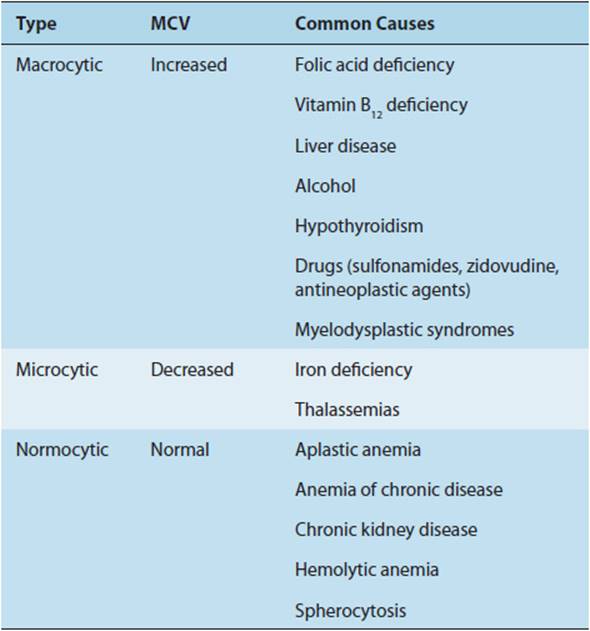
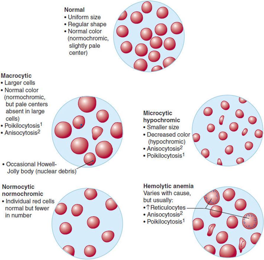
FIGURE 6-5 Thin blood smear appearance of erythrocytes in the different morphologic types of anemias. (1, Poikilocytosis [variation in shape]; 2, anisocytosis [variation in size].) (Redrawn, with permission, from Chandrasoma P et al. Concise Pathology, 3rd ed. Originally published by Appleton & Lange. Copyright © 1998 by The McGraw-Hill Companies, Inc.)
The macrocytic anemias reflect either abnormal nuclear maturation or a higher fraction of young, large red cells (reticulocytes). When the nuclei of maturing red cells appear too young and large for the amount of hemoglobin in the cytoplasm, the macrocytic anemia is termed megaloblastic. These anemias are most often due either to vitamin deficiencies (vitamin B12 or folic acid) or drugs that interfere with DNA synthesis. Abnormal nuclear maturation can also be due to clonal proliferation in the bone marrow, producing preleukemic states termed the myelodysplastic syndromes.
The normocytic anemias can be due to multiple causes: decreased numbers of red cell precursors in the marrow (primary failure called aplastic anemia, replacement of marrow elements with cancer, certain viral infections, or autoimmune inhibition called pure red cell aplasia), low levels of erythropoietin (resulting from chronic kidney disease), or chronic inflammatory diseases that affect the availability of iron in the marrow. Other normocytic anemias can be secondary to decreased life span of the cells that are produced. Examples of this phenomenon are acute blood loss; autoimmune hemolytic anemias, in which antibodies or complement bind to red cells and cause their destruction; sickle cell anemia, in which the abnormal hemoglobin polymerizes and obliterates the usual resilience of the red cell; and hereditary spherocytosis or hereditary elliptocytosis, in which defects in the erythrocyte membrane affect their ability to squeeze through the capillary microcirculation.
Anemias are very common. In contrast, an elevated hemoglobin concentration, termed erythrocytosis, is uncommon. Elevations in hemoglobin concentration can occur as a secondary phenomenon because of increased erythropoietin levels, such as that found in smokers or people who live at high altitudes (whose low blood oxygen levels stimulate erythropoietin production). Some tumors, especially renal tumors, can also make erythropoietin. Primary polycythemia is an abnormality of the bone marrow itself. This myeloproliferative syndrome leads to an increased red cell mass and consequent low erythropoietin levels by the negative-feedback mechanism discussed previously.
2. White Blood Cell Disorders
Abnormalities in white cell numbers occur commonly (Table 6-5), whereas abnormalities of function are rare. Neoplastic transformation in the form of leukemia (granulocytes and monocytes) or lymphoma (lymphocytes) is fairly common. The leukemias are discussed in Chapter 5.
TABLE 6-5 Causes of abnormal neutrophil counts.

Changes in neutrophil count are the most common white cell abnormality detected on the automated blood count. Increased numbers of neutrophils (leukocytosis) suggest acute or chronic infection or inflammation but can be a sign of many conditions. These include stress, because adrenal corticosteroids cause demargination of neutrophils from blood vessel walls.
Decreased numbers of neutrophils (neutropenia) can be seen in overwhelming infection and benign diseases such as cyclic neutropenia (see later discussion) but can also be seen when the bone marrow is infiltrated with tumor or involved by the myelodysplastic syndromes. Many drugs can also directly suppress marrow production, and because neutrophils have the shortest half-life in the blood of any cell produced by the marrow, their numbers may fall quickly.
Lymphocyte numbers can vary substantially (Table 6-6). Lymphocyte counts are classically elevated in viral infections, such as infectious mononucleosis. However, persistent elevations suggest malignancies, particularly chronic lymphocytic leukemia, which may not cause any symptoms and be incidentally discovered on a routine blood count.
TABLE 6-6 Causes of abnormal lymphocyte counts.
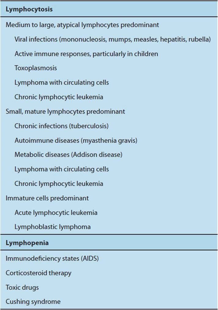
Decreased lymphocyte counts (lymphopenia) are a common complication of corticosteroid therapy but are most worrisome for immunodeficiency states; HIV directly infects lymphocytes, and the likelihood of opportunistic infections increases as lymphocyte counts fall, resulting in AIDS.
3. Platelet Disorders
Abnormalities in platelet number are fairly common, particularly low counts (thrombocytopenia). Causes are listed in Table 6-7. Decreased production of platelets occurs when the marrow is affected by a variety of diseases or when TPO production by the liver is impaired, as in cirrhosis. Increased destruction of platelets is much more prevalent. There are three general mechanisms. Because a significant number of platelets normally reside in the spleen, any increase in spleen size or activity (hypersplenism) leads to lower platelet counts. Platelet consumption that is due to ongoing clotting will also lower counts. Most commonly, however, there is immune-mediated consumption caused by either drugs or autoantibodies. The latter are usually directed against the platelet membrane antigen gpIIb/IIIa.
TABLE 6-7 Causes of platelet abnormalities.
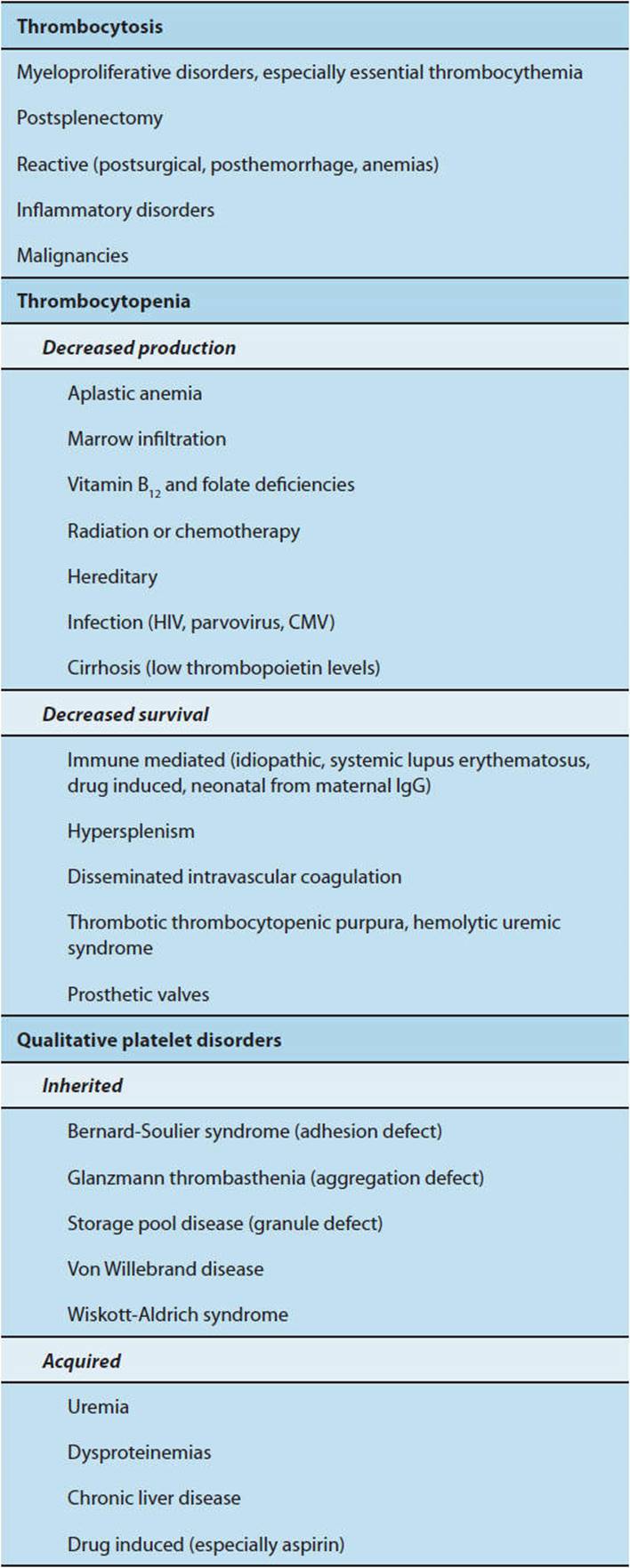
Functional platelet disorders are common, especially the acquired disorders resulting from uremia (renal failure) or aspirin, which inhibits the platelet enzyme cyclooxygenase and decreases platelet aggregability. Inherited abnormalities are unusual with the exception of von Willebrand disease, which results from either quantitative or qualitative defect of vWF, the carrier protein for factor VIII. vWF also acts as a bridge between platelets and the endothelium and thus is crucial for formation of the platelet plug in the coagulation cascade.
Elevations in the platelet count above normal (thrombocytosis) are relatively common and are especially apt to occur in recovery from iron deficiency anemia upon iron repletion. In the myeloproliferative disorders, such as polycythemia, platelet counts are often high. In essential thrombocythemia, platelet counts may be higher than 1,000,000/μL.
COAGULATION FACTOR DISORDERS
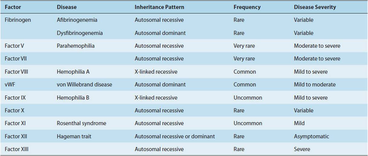
The most important coagulation factor disorders are quantitative rather than qualitative and usually hereditary rather than acquired (Table 6-8). Exceptions to this rule are acquired factor inhibitors, which are antibodies that bind to one of the coagulation factors, most often factor VIII. These may or may not cause clinical bleeding problems, but they can be extremely difficult to treat. The quantitative disorders that most commonly cause bleeding are hemophilia A (deficiency of factor VIII) and hemophilia B (deficiency of factor IX). Both are X chromosome–linked recessive traits, and affected males have very low levels of factor VIII or IX. It is not clear why all affected males do not have complete absence of factor VIII or IX activity. Hemophilia A is more common, with a prevalence of 1:10,000 males worldwide. Both disorders lead to spontaneous and excessive post-traumatic bleeding, particularly into joints and muscles. Females with the trait have 50% of the normal amount of either factor and tend not to have any bleeding problems; in general, one needs only half of the normal quantities of most coagulation factors to clot normally. The aPTT test is usually designed to become abnormal when factor VIII or IX activities fall below 50% of normal.
TABLE 6-8 Coagulation factor deficiencies.
Vitamin K deficiency also leads to quantitative declines in the levels of factors II, VII, IX, and X and proteins C and S; prolongation of the prothrombin time may result.
Quantitative inherited abnormalities of the anticoagulation systems also occur. Protein S deficiency, protein C deficiency, and AT deficiency all occur and lead to abnormal clotting problems, as discussed in the next section.
Finally, the condition of consumptive coagulopathy or disseminated intravascular coagulation (DIC) needs to be included. This condition is generally due to overwhelming infection, specific leukemias or lymphomas, or massive hemorrhage. In DIC, the coagulation factors become depleted. Often there is simultaneous activation of the fibrinolytic system as well, and uncontrolled bleeding may occur throughout the entire circulatory system. PT and aPTT are usually both abnormal.
CHECKPOINT
7. Define anemia and suggest three causes each for macrocytic and microcytic anemia.
8. What are some categories of explanations for a white blood cell count that is substantially increased or decreased compared with the normal range?
9. What are the three general mechanisms of thrombocytopenia?
10. What is the nature of the defects in hemophilia A and B?
PATHOPHYSIOLOGY OF SELECTED BLOOD DISORDERS
RED CELL DISORDERS
1. Iron Deficiency Anemia
Etiology
Iron deficiency anemia is the most common form of anemia. Although in many developing countries dietary deficiency of iron can occur, in developed nations the main cause is loss of iron, almost always through blood loss from the GI or genitourinary tracts.
Because of recurrent menstrual blood loss, premenopausal women represent the population with the highest incidence of iron deficiency. The incidence in this group is even higher because of iron losses during pregnancy, because the developing fetus efficiently extracts maternal iron for use in its own hematopoiesis. In men or in postmenopausal women with iron deficiency, GI bleeding is usually the cause. Blood loss in this case may be due to relatively benign disorders, such as peptic ulcer, arteriovenous malformations, or angiodysplasia (small vascular abnormalities along the intestinal walls). More serious causes are inflammatory bowel disease or malignancy. Endoscopic investigation to exclude malignancy is mandatory in patients without a known cause of iron deficiency.
There are other less common causes of iron deficiency, but most are related to blood loss: Bleeding disorders and hemoptysis are the chief possibilities. When no source of bleeding is uncovered, GI malabsorption should be considered as a possible cause of iron deficiency anemia. Such malabsorption occurs in patients with celiac disease, Helicobacter pylori infection, partial gastrectomy, or gastric bypass surgery. Other mechanisms of iron deficiency anemia include intravascular hemolysis (paroxysmal nocturnal hemoglobinuria or cardiac valvular disease) and response to erythropoietin treatment.
Pathogenesis
Body iron stores are generally sufficient to last several years, but there is a constant loss of iron in completely healthy persons, such that iron balance depends on adequate intake and absorption. Dietary iron is primarily absorbed in the duodenum. Absorption is increased in the setting of anemia, hypoxia, and systemic iron deficiency. Iron is also recycled from senescent erythrocytes via macrophage phagocytosis and lysis. The export of iron to plasma from these cellular sites is regulated by hepcidin, a 25-amino acid peptide produced by the liver. Hepcidin binds to ferroportin, a transmembrane protein, inducing its internalization and lysosomal degradation. When iron stores are low, hepcidin production is reduced and ferroportin molecules are expressed on the basolateral membrane of enterocytes, where they transfer iron from the cytoplasm of enterocytes to plasma transferrin. Conversely, when iron stores are adequate or elevated, hepcidin production is increased, resulting in the internalization of ferroportin and reduced export of iron into plasma. In inflammatory states, hepcidin production is increased, leading to the internalization of ferroportin on macrophages and the trapping of recycled iron within macrophage stores.
Iron is stored in most body cells as ferritin, a combination of iron and the protein apoferritin. It is also stored as hemosiderin, which is ferritin partly stripped of the apoferritin protein shell. Iron is transported in blood bound to its carrier protein transferrin. Because of the complex interactions between these molecules, a simple measurement of serum iron rarely reflects body iron stores (see later discussion).
Iron is found predominantly in hemoglobin and is present also in myoglobin, the oxygen-storing protein of skeletal muscle. The main role for iron is as the ion in the center of the body’s oxygen-carrying molecule, heme. Held stably in the ferrous form by the other atoms in heme, iron reversibly binds oxygen. Each protein subunit of hemoglobin contains one heme molecule; because hemoglobin exists as a tetramer, four iron molecules are needed in each hemoglobin unit. When there is iron deficiency, the final step in heme synthesis is interrupted (Figure 6-6). In this step, ferrous iron is inserted into protoporphyrin IX by the enzyme ferrochelatase; when heme synthesis is interrupted, there is inadequate heme production. Globin biosynthesis is inhibited by heme deficiency through a heme-regulated translational inhibitor (HRI). Elevated HRI activity (a result of heme deficiency) inhibits a key transcription initiation factor for heme synthesis, eIF2. Thus, less heme and fewer globin chains are available in each red cell precursor. This directly causes anemia, a decrease in the hemoglobin concentration of the blood.

FIGURE 6-6 Heme synthesis, emphasizing the role of iron and the insertion of heme into individual globin chains to make hemoglobin, and the role of the heme-regulated translational inhibitor (HRI) of globin synthesis. Normal concentrations of heme keep the activity of HRI low, preserving normal globin synthesis.
As noted, heme is also the oxygen acceptor in myoglobin; therefore, iron deficiency will also lead to decreased myoglobin production. Other proteins also are dependent on iron; most of these are enzymes. Many use iron in the heme molecule, but some use elemental iron. Although the exact implications of iron deficiency on their activity is not known, these enzymes are crucial to metabolism, energy production, DNA synthesis, and even brain function.
Pathology
As iron stores are depleted, the peripheral blood smear pattern evolves. In early iron deficiency, the hemoglobin level of the blood falls but individual erythrocytes appear normal. In response to a falling oxygen level, erythropoietin levels rise and stimulate the marrow, but the hemoglobin level cannot rise in response because of the iron deficiency. Other hormones are presumably also stimulated, however, and the resulting “revved-up” marrow usually causes an elevated blood platelet count. An elevated white cell count is less common. Reticulocytes are notably absent.
Eventually, the hemoglobin concentration of individual cells falls, leading to the classic picture of microcytic, hypochromic erythrocytes (Figure 6-5). This is most commonly found as an abnormally low MCV of red cells on the automated hemogram. There is also substantial anisocytosis and poikilocytosis, seen on the peripheral smear, and target cells may be seen. The target shape occurs because there is a relative excess of red cell membrane compared with the amount of hemoglobin within the cell, so that the membrane bunches up in the center.
Laboratory results are often confusing. A low serum ferritin level is diagnostic of iron deficiency, but even in obvious cases, levels can be normal; ferritin levels rise in acute or chronic inflammation or significant illnesses, which can themselves be the cause of iron (blood) loss. Serum iron levels fall in many illnesses, and levels of its serum carrier, transferrin, fluctuate as well, so neither of them is a consistent indicator of iron deficiency, nor is their ratio, the transferrin saturation. If ferritin levels are not diagnostic, measuring serum soluble transferrin receptor (sTfR) can help. TfRs are membrane glycoproteins that facilitate iron transport from plasma transferrin into body cells. Erythroid precursors increase their expression of membrane TfR in the setting of iron deficiency but not anemia of chronic disease. Some membrane TfR is released into the serum as sTfR. The amount of sTfR in the serum reflects the amount of membrane TfR. A high ratio of sTfR to ferritin predicts iron deficiency when ferritin is not diagnostically low. Though helpful, this test has had limited adoption in clinical practice.
Other than observing a hematologic response to empiric iron supplementation, bone marrow biopsy can confirm a diagnosis of iron deficiency. Iron is normally found in the macrophages of the marrow, where it supplies erythrocyte precursors; intracellular hemosiderin is easily visualized with Prussian blue stain. These macrophages do not stain at all if there is iron deficiency.
Clinical Manifestations
All anemias lead to classic symptoms of decreased oxygen-carrying capacity (ie, fatigue, weakness, and shortness of breath, particularly dyspnea on exertion), and iron deficiency is no exception. Decreased oxygen-carrying capacity leads to decreased oxygen delivery to metabolically active tissues, which nonetheless must have oxygen; this leads directly to fatigue. The compensatory mechanisms of the body lead to additional symptoms and signs of anemia. Some patients appear pale not only because there is less hemoglobin per unit of blood (oxygenated hemoglobin is red and gives color to the skin) but also because superficial skin blood vessels constrict, diverting blood to more vital structures. Patients may also respond to the anemia with tachycardia. This increased cardiac output is appropriate because one way to increase oxygen delivery to the tissues is to increase the number of times each hemoglobin molecule is oxygenated in the lungs every hour. This tachycardia may cause benign cardiac murmurs due to the increased blood flow.
Abnormalities of the GI tract occur because iron is also needed for proliferating cells. Glossitis, where the normal tongue papillae are absent, can occur, as can gastric atrophy with achlorhydria (absence of stomach acid). The achlorhydria may compound the iron deficiency because iron is best absorbed in an acidic environment, but this complication is quite unusual.
In children, there may be significant developmental problems, both physical and mental. Iron-deficient children, mostly in developing regions, perform poorly on tests of cognition compared with iron-replete children. Iron therapy can reverse these findings if started early enough in childhood. The exact mechanism of cognitive loss in iron deficiency is not known. Another unexplained but often observed phenomenon in severe iron deficiency is pica, a craving for nonnutritive substances such as clay or dirt.
Many patients have no specific symptoms or findings at all, and their iron deficiency is discovered because of anemia noted on a blood count obtained for another purpose. It is of interest that mild anemias (hemoglobins of 11–12 g/dL) may be tolerated very well because they develop slowly. In addition to the physiologic compensatory mechanisms discussed previously (increased cardiac output, diversion of blood flow from less metabolically active areas), there is a biochemical adaptation as well. The ability to transfer oxygen from hemoglobin to cells is partly dependent on a small molecule in erythrocytes called 2,3-biphosphoglycerate (2,3-BPG). In high concentrations, the ability to unload oxygen in the tissues is increased. Chronic anemia leads to elevated 2,3-BPG concentrations in erythrocytes.
Other patients who do not present with symptoms directly related to the anemia present instead with symptoms or signs related directly to blood loss. Because the most common site of unexpected (nonmenstrual) blood loss is the GI tract, patients often have visible changes in the stool. There may be gross blood (hematochezia), which is more common with bleeding sites near the rectum, or black, tarry, metabolized blood (melena) from more proximal sites. Significant blood loss from the urinary tract is very uncommon.
CHECKPOINT
11. What is the most common form of anemia and its most likely cause in a premenopausal woman? In a man?
12. Why is the serum ferritin level often not a good indicator of whether anemia is due to iron deficiency?
13. What are some disorders associated with iron deficiency anemia?
14. What are the physiologic adaptations to slowly developing iron deficiency anemia?
2. Pernicious Anemia
Etiology
Pernicious anemia is a megaloblastic anemia in which there is abnormal erythrocyte nuclear maturation. Unlike in many other types of anemia such as that resulting from iron deficiency, hemoglobin synthesis is normal. Pernicious anemia is the end result of a cascade of events that are autoimmune in origin. The ultimate effect is a loss of adequate stores of vitamin B12 (cobalamin), which is a cofactor involved in DNA synthesis. Rapidly proliferating cells are those most often affected, predominantly bone marrow cells and those of the GI epithelium. The nervous system is also affected, demonstrating that this is a systemic disease. Anemia is merely the most common manifestation.
Besides pernicious anemia, cobalamin deficiency can also be due to bacterial overgrowth in the intestine (because bacteria compete with the host for cobalamin), intestinal malabsorption of vitamin B12involving the terminal ileum (such as in Crohn disease), surgical removal of the antrum of the stomach (gastrectomy), and, rarely, dietary deficiency, which occurs only in strict vegetarians. In the diet, cobalamin is found mostly in animal products.
Pernicious anemia is most common in older patients of Scandinavian descent and is more commonly found in those of European and African than Asian descent. In the United States, black females are one of the most common groups. Pernicious anemia accounts for only a small percentage of patients with anemia, however.
Pathogenesis
The initial events in the pathogenetic cascade begin in the stomach (Figure 6-7). The gastric parietal cells are initially affected by an autoimmune phenomenon that leads to two discrete effects: loss of gastric acid (achlorhydria) and loss of intrinsic factor. Pernicious anemia interferes with both the initial availability and the absorption of vitamin B12: Stomach acid is required for the release of cobalamin from foodstuffs, and intrinsic factor is a glycoprotein that binds cobalamin and is required for the effective absorption of cobalamin in the terminal ileum. Both stomach acid and intrinsic factor are made exclusively by parietal cells.
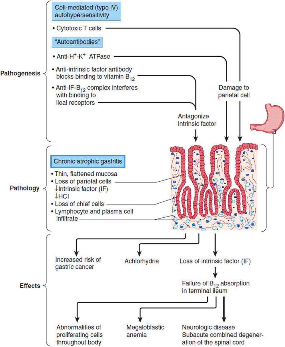
FIGURE 6-7 Pathogenesis and effects of pernicious anemia (autoimmune atrophic gastritis). (Redrawn, with permission, from Chandrasoma P et al. Concise Pathology, 3rd ed. Originally published by Appleton & Lange. Copyright © 1998 by The McGraw-Hill Companies, Inc.)
Evidence for the autoimmune destruction of parietal cells is strong: Patients with pernicious anemia have atrophy of the gastric mucosa, and pathologic specimens show infiltrating lymphocytes, which are predominantly antibody-producing B cells. In addition, 90% or more of patients have antibodies in their serum directed against parietal cell membrane proteins. The major protein antigen appears to be H+-K+ATPase, the proton pump, which is responsible for the production of stomach acid. Cytotoxic T cells whose receptors recognize H+-K+ ATPase may also contribute to the gastric atrophy. More than half of patients also have antibodies to intrinsic factor itself or the intrinsic factor-cobalamin complex. Furthermore, patients with pernicious anemia have a higher incidence of other autoimmune diseases, such as Graves disease. Lastly, corticosteroid therapy, used as first-line therapy for many autoimmune disorders, may reverse the pathologic findings in pernicious anemia. Despite this evidence, the exact mechanism of the inciting event remains unknown.
Complete vitamin B12 deficiency develops slowly, even after total achlorhydria and loss of intrinsic factor occur. Liver stores of vitamin B12 are adequate for several years. However, the lack of this vitamin eventually leads to alterations in DNA synthesis and in the nervous system, altered myelin synthesis.
In DNA synthesis, cobalamin, along with folic acid, is crucial as a cofactor in the synthesis of deoxythymidine from deoxyuridine (Figure 6-8). Cobalamin accepts a methyl group from methyltetrahydrofolate, which leads to the formation of two important intracellular compounds. The first is methylcobalamin, which is required for the production of the amino acid methionine from homocysteine. The second is reduced tetrahydrofolate, which is required as the single-carbon donor in purine synthesis. Thus, cobalamin deficiency depletes stores of reduced tetrahydrofolate and impairs DNA synthesis because of lowered purine production. In cobalamin deficiency, other reduced folates may substitute for tetrahydrofolate (and may explain why pharmacologic doses of folic acid can partially reverse the megaloblastic blood cell changes, but not the neurologic changes, seen in pernicious anemia). However, methyltetrahydrofolate, normally the methyl donor to cobalamin, accumulates. This folate cannot be retained intracellularly because it cannot be polyglutamated; the addition of multiple glutamate residues leads to a charged compound that does not freely diffuse out of the cell. Therefore, there is relative folate deficiency in pernicious anemia as well. In addition, methionine may serve as a principal donor of methyl groups to these other “substituting” reduced folates; because methionine cannot be produced in cobalamin deficiency, this compounds the problems in purine synthesis.
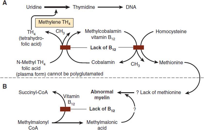
FIGURE 6-8 Role of cobalamin (vitamin B12) and folic acid in nucleic acid and myelin metabolism. Lack of either cobalamin or folic acid retards DNA synthesis (A) and lack of cobalamin leads to loss of folic acid, which cannot be held intracellularly unless polyglutamated. Lack of cobalamin also leads to abnormal myelin synthesis, probably via a deficiency in methionine production (B). (Redrawn, with permission, from Chandrasoma P et al. Concise Pathology, 3rd ed. Originally published by Appleton & Lange. Copyright © 1998 by The McGraw-Hill Companies, Inc.)
The exact mechanism of the neurologic consequences of pernicious anemia, with demyelination (loss of the myelin sheaths around nerves), is not known. Defects in the methionine synthase pathway have been suggested but not proven experimentally. Instead, observations in cobalamin-deficient gastrectomized rats implicate an imbalance of cytokines and growth factors as a potential mediator of nerve damage. The synthesis of the cytokine tumor necrosis factor (TNF) is regulated by S-adenosyl-methione, a product of methionine. Deficiency of methionine may indirectly lead to neuropathy via unregulated production of TNF, a myelinolytic cytokine, among other mechanisms.
The production of succinyl-coenzyme A (CoA) is also dependent on the presence of cobalamin. It is not clear whether a decrease in the production of succinyl-CoA, which may affect fatty acid synthesis, is also involved in the demyelinating disease.
Pathology
The gastric disorders associated with pernicious anemia are dominated by the picture of chronic atrophic gastritis (Figure 6-7). The normally tall columnar epithelium is replaced by a very thin mucosa, and there is obvious infiltration of plasma cells and lymphocytes. Pernicious anemia also increases the risk for gastric adenocarcinoma. Thus, pathologic examination may also reveal cancer.
The peripheral blood smear picture (Figure 6-5) varies, depending on the length of time the patient has been cobalamin deficient. In early stages, the patient may have mild macrocytic anemia, and large ovoid erythrocytes (macro-ovalocytes) are commonly seen. In full-blown megaloblastic anemia, however, there are abnormalities in all cell lines. The classic picture reveals significant anisocytosis and poikilocytosis of the red cell line, and there are hypersegmented neutrophils, revealing the nuclear dysgenesis from abnormal DNA synthesis (Figure 6-9). In severe cases of pernicious anemia, the red and white cell series are easily mistaken for acute leukemia because the cells look so atypical.

FIGURE 6-9 Megaloblastic hematopoiesis: morphologic changes visible with microscopic examination of bone marrow or peripheral blood. (Redrawn, with permission, from Chandrasoma P et al. Concise Pathology, 3rd ed. Originally published by Appleton & Lange. Copyright © 1998 by The McGraw-Hill Companies, Inc.)
Bone marrow aspiration and biopsy are not necessary in the diagnosis and may be misleading, because the marrow pathology can be confused with acute leukemia, with hypercellularity, increased erythroblasts, and even cytogenetic changes. Typical findings in B12 deficiency include megaloblastic changes—nuclei that are too large and immature in cells with mature, hemoglobin-filled cytoplasm—that are seen at each stage of erythrocyte development. These cells are not seen in the peripheral blood because the abnormal erythrocytes generally are destroyed in the marrow (intramedullary hemolysis) by unexplained processes. This compounds the anemia. Megaloblastic changes can be seen in the marrow even in the absence of obvious changes on the peripheral blood smear.
Spinal cord abnormalities consist of demyelination of the posterolateral spinal columns, called subacute combined degeneration. Peripheral nerves may also show demyelination. Demyelination eventually results in neuronal cell death, which is also obvious on pathologic examination. Because neurons do not divide, new neurons cannot replace the dead ones.
Laboratory findings include elevated lactate dehydrogenase (LDH) and, sometimes, indirect bilirubin consistent with the hemolysis occurring in the bone marrow. LDH is directly released from lysed red cells, and free hemoglobin is metabolized to bilirubin. Serum vitamin B12 levels are usually low, revealing the deficient state. Yet there remain high rates of both false positive and false negative test results because only 20% of total measured serum B12 is bound to the cellular delivery protein, transcobalamin; the rest is bound to haptocorrin, which is not available for cells to utilize. Antibodies to intrinsic factor are usually detectable. Serum elevations of methylmalonic acid (MMA) and/or homocysteine (see Figure 6-8) are highly predictive of B12 deficiency. The Schilling test, which assesses the oral absorption of vitamin B12 with and without added intrinsic factor, is no longer used, because of lack of availability of radioactively labeled vitamin B12. Typically, the approach is to first measure serum B12 and, if equivocal, to obtain serum levels of MMA and/or homocysteine.
Clinical Manifestations
The clinical presentation consists of one or more symptoms related to the underlying deficiency. Anemia is the most commonly encountered abnormality and is often very severe; hemoglobin levels of 4 g/dL (less than a third of normal) can be seen. This degree of anemia is rare with other causes, such as iron deficiency. Typical symptoms are fatigue, dyspnea, or dizziness, because a decreased red cell mass equals decreased oxygen-carrying capacity of the blood. High-output heart failure is relatively common, with tachycardia and signs of left ventricular failure (Chapter 10). Because oxygen demands are constant (or rise with exercise) and oxygen-carrying capacity is falling, the only way to maintain tissue oxygenation in anemia is to increase cardiac output (ie, the number of times per minute each red cell is fully oxygenated by the lungs). Eventually, however, the left ventricle fails.
However, symptoms may be mild because the anemia develops slowly as a result of the extensive liver storage of vitamin B12. Patients with anemia usually adapt over time to slow changes in oxygen-carrying capacity. The same changes in 2,3-BPG that encourage oxygen delivery to the tissues from the hemoglobin in red cells in other anemias occur in vitamin B12 deficiency.
GI symptoms are less prevalent and include malabsorption, muscle wasting (unusual), diarrhea (more common), and glossitis (most common). In glossitis, the normal tongue papillae are absent regardless of whether the tongue is painful, red, and “beefy” or pale and smooth.
Neurologic symptoms are least likely to improve with cobalamin replacement therapy. As with other neuropathies involving loss of myelin from large peripheral sensory nerves, numbness and tingling (paresthesias) occur frequently and are the most common symptoms. Demyelination and neuronal cell death in the posterolateral “long tracts” of the spinal cord interfere with delivery of positional information to the brainstem, cerebellum, and sensory cortex. Patients, therefore, complain of loss of balance and coordination. Examination reveals impaired proprioception (position sense) and vibration sense. True dementia may also occur when demyelination involves the brain. Importantly but somewhat unexpectedly, neurologic symptoms may occur in the absence of any changes in the peripheral blood smear suggestive of pernicious anemia.
Less commonly, vitamin B12 deficiency can manifest with thrombosis and possibly at unusual sites such as cerebral venous sinuses. The prothrombotic state may be secondary to hyperhomocysteinemia seen in severe vitamin B12deficiency.
CHECKPOINT
15. Name two crucial cofactors in DNA synthesis whose deficiency results in pernicious anemia. In what specific biochemical pathways do they participate?
16. What neurologic defects are observed in prolonged pernicious anemia?
17. What symptoms of pernicious anemia are usually relatively mild?
18. Are changes in the peripheral blood smear necessary for neurologic effects of vitamin B12 deficiency?
WHITE CELL DISORDERS
1. Malignant Disorders
The most important leukocyte abnormalities are the malignant disorders leukemia and lymphoma. They are discussed in Chapter 5.
2. Cyclic Neutropenia
Absolute neutropenia, characterized by neutrophil counts less than 1500–2000/μL (>2 SD below the mean in normals), is a commonly encountered problem in medicine and can be due to a large number of disease entities (Table 6-5). Cyclic neutropenia, however, is rare. It is of interest because it provides insight into normal neutrophil production and function. It is characterized by a lifetime history of neutrophil counts that decrease to zero or near zero for 3–5 days at a time every 3 weeks and then rebound. Interestingly, the peripheral blood neutrophil counts and monocyte counts oscillate in opposite phases on this 3-week cycle.
Etiology
Classic, childhood-onset cyclic neutropenia results from heterozygous germline mutations in the gene ELANE (ELAstase, neutrophil expressed), formerly known as ELA2, which encodes for a single enzyme, neutrophil elastase (NE). NE is found in the primary azurophilic granules of neutrophils and monocytes. There are approximately 100 cases in the literature, most of which are consistent with an autosomal dominant inheritance. However, sporadic adult cases also occur, and these are associated with neutrophil elastase mutations. There does not seem to be a racial predilection or gender bias in incidence.
Pathogenesis
The neutrophil count in blood is stable in normal individuals, reflecting the fact that there is a large storage pool of granulocytes in the marrow. The marrow reserve exceeds the circulating pool of neutrophils by 5- to 10-fold. This large pool is necessary because it takes nearly 2 weeks for the full development of a neutrophil from an early stem cell within the bone marrow, yet the average life span of a mature neutrophil in blood is less than 12 hours.
In cyclic neutropenia, the storage pool is not adequate. Daily measurements of neutrophil counts in the blood reveal striking variations in their number. Studies of neutrophil kinetics in affected patients reveal that the defect is in abnormal production, rather than abnormal disposition of neutrophils. Neutrophil production occurs in discrete waves even in normal individuals. As neutrophils differentiate from an early progenitor cell, they produce neutrophil elastase, which is thought to inhibit the differentiation of myeloblasts in a negative feedback loop. This results in an oscillatory wave with peaks and troughs of neutrophil production. As neutrophil numbers increase in the marrow, a peak is obtained where enough neutrophil elastase causes a drop in neutrophil differentiation. Then, as the number of neutrophils drops again to a nadir, the production of neutrophil elastase also declines, allowing the number of neutrophils to climb once again. In cyclic neutropenia, it is hypothesized that the mutant neutrophil elastase may have an excessive inhibitory effect, causing prolonged trough periods and inadequate storage pools to maintain a normal peripheral neutrophil count. However, once they are extruded from the marrow, the neutrophils appear to have a normal life span (Figure 6-10).

FIGURE 6-10 Feedback loop hypothesis to explain hematopoietic cycling. Neutrophil elastase (NE) is postulated to inhibit further differentiation by a myeloblast. Gray sinewave denotes neutrophil count oscillations. In this model, NE is produced by the terminally differentiating cohort of neutrophils and ultimately feeds back to inhibit further production of neutrophils, which results in loss of the inhibitory cycle—at least for a while, until production of the neutrophils resumes, followed again by the inhibitory action of NE in a cyclic manner. (Redrawn from Horwitz MS et al. Neutrophil elastase in cyclic and severe congenital neutropenia. Blood. 2007 Mar 1;109(5):1817–24. Copyright American Society of Hematology.)
The myeloid progenitor for neutrophil can also produce monocytes. Therefore, during neutrophil nadirs, the myeloid progenitor cell can preferentially differentiate to the monocyte lineage, giving the opposing oscillatory waves of neutrophils and monocytes seen in these patients (see Figure 6-11).

FIGURE 6-11 Regular cyclic variation of monocytes, reticulocytes, and neutrophils in a patient with cyclic neutropenia. Note that monocytes and reticulocytes tend to rise when the neutrophils fall. (Redrawn, with permission, from Dale D et al. Cyclic neutropenia: a clinical review. Blood Rev. 1988;2:178.)
The waves are remarkably constant in their periodicity. Almost every patient has a cycle between 19 and 22 days, and each patient’s cycle length is constant during his or her lifetime. Neutrophils and monocytes are not the only marrow elements that cycle. Platelet and reticulocyte counts also cycle with the same cycle length, but in contrast to the blood neutrophil count, clinically significant decreases are not observed. This is presumably because the blood life spans of these elements are so much longer than the life span of neutrophils. Because multiple cell lines are seen to cycle, it is believed that neutrophil elastase mutations accelerate the process of apoptosis (programmed cell death) in early progenitor cells, as well, unless they are “rescued” by G-CSF.
Clinically, administration of pharmacologic doses of G-CSF (filgrastim) to affected individuals has three interesting effects that partially overcome the condition. First, although cycling continues, mean neutrophil counts increase at each point in the cycle, such that patients are rarely neutropenic. Second, cycling periodicity decreases immediately from 21 days to 14 days. Third, other cell line fluctuations change in parallel; their cycle periodicity also decreases to 14 days, suggesting that an early progenitor cell is indeed at the center of this illness. However, the fact that cycling does not disappear demonstrates that there are other abnormalities yet to be discovered. It also suggests that there may be an inherent cycling of all stem cells in normal individuals that is modulated by multiple cytokines in the marrow.
Pathology
The pathologic features of cyclic neutropenia are seen mostly in the laboratory. The peripheral blood smear appears normal except for the paucity of neutrophils—mature or immature—during the nadirs of each cycle. Individual neutrophils appear normal. The bone marrow, however, shows striking differences depending on the day of the cycle on which it is examined. During the nadir of each cycle, there are increased numbers of early myeloid precursors such as promyelocytes and myelocytes, and mature neutrophils are rare. This picture is similar to that seen in acute leukemia, but 10 days later, as circulating neutrophil counts are rising, an entirely normal-appearing marrow is typical.
Clinical Manifestations
In general, neutropenia from any cause places patients at risk for severe bacterial infections, generally from enteric organisms, because of the alteration in host defenses in the gastrointestinal tract. This is especially true when the neutropenia is due to administration of chemotherapeutic agents, because chemotherapy also affects the lining of the GI tract. Neutrophils, with their ability to engulf bacteria and deliver toxic enzymes and oxidizing free radicals to sites of infection, normally serve as the first line of host defenses against the bacteria that inhabit the gut. Such patients are also at risk for fungal infections if the neutropenia lasts more than several days; this is because it takes longer for fungi to reproduce and invade the bloodstream. Untreated infections of either type can be rapidly fatal, particularly if the neutrophil count is less than about 250/μL.
In cyclic neutropenia, then, recurrent infections are to be expected, and deaths from infections with intestinal organisms have been reported. Each cycle is characterized by malaise and fever coincident with the time neutrophil counts are falling. Cervical lymphadenopathy is almost always present as are oral ulcers. These symptoms usually last for about 5 days and then subside until the next cycle.
When infections occur, the site is usually predictable. Skin infections, specifically small superficial pyogenic abscesses (furunculosis) or bacterial invasion of the dermis or epidermis (cellulitis), are the most common and respond to antibiotic therapy with few sequelae. The next most common infection site is usually the gums, and chronic gingivitis is evident in about half of patients. It is also the most noticeably improved problem when patients receive therapy with filgrastim. Other infections are unusual, but any neutropenic patient is at risk for infection from organisms that reside in the GI system. In the few patients who have required abdominal surgery during their neutropenia, ulcers similar to those seen in the mouth have been noted; this destruction of the normal mucosal barrier presumably eases entry of intestinal bacteria into the bloodstream. Because the period of greatest susceptibility to infection is only a few days in each cycle, most patients grow and develop normally.
CHECKPOINT
19. How long does it take for a neutrophil to develop from a stem cell in the bone marrow? Once fully mature, what is its life span?
20. At what level of neutropenia does the incidence of infection dramatically increase?
21. What are the most common sites and types of infections observed in neutropenic patients?
22. What is the probable underlying abnormality in cyclic neutropenia?
PLATELET DISORDERS
1. Drug-Associated Immune Thrombocytopenia
Etiology
Thrombocytopenia, defined as the occurrence of platelet levels below the normal laboratory range, is a commonly encountered abnormality. Although there are many causes (Table 6-7), the possibility of a drug-induced immune thrombocytopenia should always be considered.
Many drugs have been associated with this phenomenon, and the most common ones are listed in Table 6-9. In practice, the association between a given drug and thrombocytopenia is usually made clinically rather than with specific tests. Thrombocytopenia usually occurs at least 5–7 days after exposure to the drug, if given for the first time. The suspect drug is stopped and platelet counts rebound within a few days. Rechallenge with the drug, which is rarely done, almost always reproduces the thrombocytopenia.
TABLE 6-9 Common drugs that may cause thrombocytopenia.

Heparin is the most important cause of thrombocytopenia because of its frequent use in hospitalized patients; its use also carries the potential to cause a life-threatening thrombotic syndrome. The pathophysiology of the thrombocytopenia caused by heparin is also the most completely described.
Pathogenesis
Although the phenomenon of drug-induced thrombocytopenia has been known for decades to be immune in nature, the specific mechanisms have long been controversial. The association of antibodies with platelets leads to their destruction via the spleen. The spleen acts as the major “blood filter” and recognizes platelets bound to antibodies as abnormal and thus removes them. Spleen removal also occurs in autoimmune (idiopathic) thrombocytopenia, which is relatively common and difficult to distinguish clinically from drug-induced thrombocytopenia.
There are various mechanisms underlying drug-induced immune thrombocytopenia. Quinine- or NSAID-induced thrombocytopenia involves the tight binding of antibody to normal platelets only in the presence of the sensitizing drug. The antibody usually targets epitopes on the glycoprotein IIb/IIIa or Ib/IX complexes, the major platelet receptors for fibrinogen and vWF, respectively. Penicillin and cephalosporin antibiotics are believed to lead to platelet destruction via hapten-dependent antibodies. The drug acts as a hapten, a small molecule that only elicits an immunologic response when it is bound to a large carrier molecule or protein. Some drugs (gold salts, procainamide, and possibly sulfonamides) can induce autoantibodies that are capable of binding to and destroying platelets even in the absence of the sensitizing drug. Finally, antithrombotic agents that block the binding of fibrinogen to gpIIb/IIIa receptors (abciximab, tirofiban, or eptifibatide) can cause an acute immune-mediated thrombocytopenia, where patients develop severe thrombocytopenia within hours of the exposure. The mechanism involves either naturally occurring antibodies that recognize the murine component of abciximab or structural changes to the gpIIb/IIIa receptor caused by the binding of tirofiban or eptifibatide.
For heparin, there is clear evidence of binding to a platelet protein, platelet factor 4 (PF4). PF4 resides in the alpha granules of platelets and is released when they are activated. It binds back onto the platelet surface through a specific PF4 receptor molecule, further increasing platelet activation. It also binds with high affinity to heparin and to heparin-like glycosaminoglycan molecules present on the vascular endothelium. This non–immune-based adhesion to PF4 can lead to mild thrombocytopenia via promotion of platelet binding to fibrinogen and subsequent aggregation, known as heparin-induced thrombocytopenia (HIT) type I. This can happen in 30% of patients exposed to heparins without clinical sequelae. However, the combination of heparin with PF4 can also act as an antigenic stimulus that provokes the production of immunoglobulin G (IgG) directed against the combination. This immunologic response is known as heparin-induced thrombocytopenia (HIT) type II. About 10–20% of these patients with heparin-PF4 antibodies will develop a serious clinical syndrome, HIT(T) (heparin-induced thrombocytopenia [and thrombosis]), which paradoxically involves both thrombocytopenia 5–10 days after drug exposure and a prothrombotic state via increased platelet activation. There is a 10-fold increased risk for HIT in patients receiving unfractionated heparin (UFH) compared with those receiving low-molecular-weight heparins. Cardiac or orthopedic surgery patients have a higher risk for clinical HIT (1–5%) than medical or obstetric patients (0.1–1%) when receiving UFH. Women have twice the risk for HIT as men.
Thrombocytopenia occurs in HIT type II after a series of steps. First, PF4 is released from platelets either by heparin itself or by other stimuli. Heparin then binds to PF4, forming an antigenic complex that results in the production of IgG antibodies that can bind directly to this compound. The new complex of IgG-heparin-PF4 binds to platelets through the platelet Fc receptor, via its IgG end. Platelets bound with this antibody complex are then destroyed by the spleen.
Despite the resulting thrombocytopenia, HIT type II leads to a prothrombotic state via the additional binding of the heparin-PF4 portion to the PF4 receptor on platelets, promoting platelet cross-linking, activation, and aggregation (Figure 6-12).
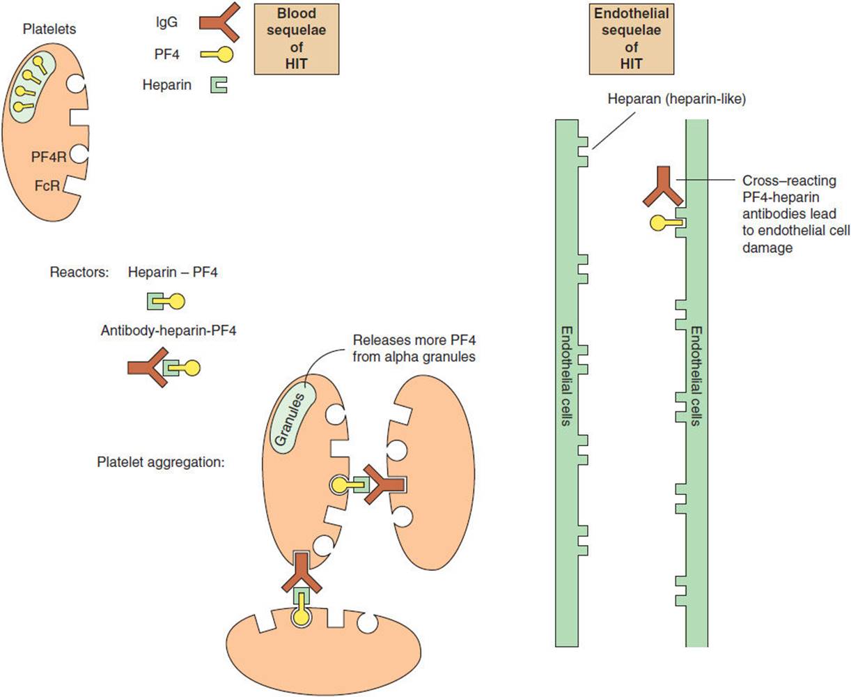
FIGURE 6-12 Pathogenesis of heparin-induced thrombocytopenia (HIT). IgG is the autoantibody against the heparin-PF4 complex. Platelets can bind to each other and become activated via either the IgG-Fc receptor interaction or the PF4-PF4 receptor interaction or both. Aggregation and thrombus formation may thus occur. Furthermore, IgG may bind to endothelial cell bound heparan-PF4 construct and cause vascular damage, which may also provoke thrombus formation.
Because each end of this IgG-heparin-PF4 molecule can bind to a platelet, it is possible that platelets can become cross-linked by a single molecule. Many platelets could actually interact in this fashion, leading to further platelet aggregation and activation. Clinically, this decreases the numbers of circulating platelets, but it may also lead to creation of a thrombus at the site of activation. Thus, despite the fact that heparin is the most commonly used anticoagulant, in this case it may actually provoke coagulation. Furthermore, the activation of platelets via this mechanism leads to increased amounts of circulating PF4, which can bind to more heparin and continue the cycle. The excess PF4 can also bind to the endothelial surface via the heparin-like glycosaminoglycans described earlier. It is thus possible that the antibodies to the heparin-PF4 construct could bind to the endothelial cells as well, which may lead to endothelial cell injury, further increasing the risk of local thrombosis by release of TF and ultimately thrombin. Lastly, there is some evidence that macrophages may release TF in response to these antibodies, further stimulating coagulation.
Pathology
The peripheral blood smear is not strikingly abnormal unless platelet counts are less than about 75,000/μL, and then it is usually abnormal only because relatively few platelets are seen. Platelet morphology, however, is usually normal, although large platelets can be seen. These large platelets are less mature and are a bone marrow compensation for a low peripheral platelet count, with platelet production from megakaryocytes being increased. Although drugs—heparin in particular—may cause platelet aggregation in vivo and in vitro, this is usually not apparent on review of the blood smear.
The bone marrow usually appears normal, although the megakaryocyte number may be relatively increased, presumably reflecting an attempt to increase the number of platelets (megakaryocyte fragments) in the circulation. In a few cases of immune-mediated thrombocytopenia, however, there may be decreased numbers of megakaryocytes. There are many hypotheses as to why this may occur, but it most likely means that the antigenic combination of drug-platelet protein is also occurring on megakaryocytes, so that they as well as the platelets in the peripheral circulation are being immunologically destroyed. This destruction would not involve the spleen, of course, but would require antibody-dependent cell killing.
In patients who develop heparin-induced thrombocytopenia and thrombosis, thrombi are seen that are relatively rich in platelets when compared with “typical” thrombi seen in other situations. They are described as “white clots.” The thrombi may be either arterial or venous.
Clinical Manifestations
Despite the fact that the platelet count in immune-mediated thrombocytopenia can be extremely low (<10,000/μL, compared with a normal value of over 150,000/μL), severe bleeding is unusual. More often there is easy bruising with minimal trauma. With platelet counts of less than about 5000/μL, pinpoint hemorrhages (petechiae) may spontaneously occur in the skin or mucous membranes. These are self-limited because the plasma coagulation factors are still intact, and only a small number of aggregated platelets are needed to provide adequate PL for clotting.
The relationship between the likelihood of bleeding and the platelet count is not linear. The bleeding time, a test used clinically to evaluate platelet function, does not even begin to be abnormally prolonged until the platelet count is less than 90,000/μL. Spontaneous bleeding is unlikely until platelet counts are less than 20,000/μL but is still uncommon until counts are less than about 5000/μL, assuming that patients do not have other abnormalities of hemostasis. For example, aspirin inhibits platelet aggregation and increases the likelihood of bleeding. When bleeding from thrombocytopenia does occur, it is most often mucosal or superficial in the skin. This is most commonly seen as a nosebleed (epistaxis), but bleeding of the gums, GI tract, or bladder mucosa may be seen.
As mentioned, however, when immune thrombocytopenia occurs as a result of heparin, paradoxical clotting may occur instead of bleeding. This may cause a very confusing picture, because the heparin may have been given therapeutically for another thrombosis; it may be difficult to determine whether the new thrombosis is an extension of the initial clot or a new one due to the heparin exposure. However, the occurrence of the simultaneous thrombocytopenia provides a clue.
When heparin-induced thrombocytopenia and thrombosis do occur, the clinical manifestation of the new thrombosis will depend on the site of the thrombus. Most studies of this disorder suggest that when thrombosis occurs, it is at the site of previous vascular injury or abnormality. Thus, in patients with atherosclerotic vascular disease, arterial thromboses are much more common than venous clots. Patients have the rapid onset of severe pain, usually in an extremity, with a cool, pale limb. Pulses are absent. This can be life-threatening (5–10% mortality rate) or at least extremity threatening because oxygen flow to the affected area is cut off, and emergency clot removal or vascular bypass surgery may be necessary. Venous clots also occur in a manner similar to typical venous clots (see later discussion). In addition to stopping heparin, patients with type II HIT need anticoagulation to prevent and treat thrombosis formation. Direct thrombin inhibitors, (argatroban, lepirudin, or bivalirudin) provide a direct means of blocking the effects of thrombin, a primary mediator of the coagulation system.
CHECKPOINT
23. What is the most common category of cause of thrombocytopenia?
24. Name the antibodies to which platelet protein are implicated in the pathogenesis of heparin-induced thrombocytopenia?
25. By what mechanism can heparin-induced thrombocytopenia actually increase clot formation?
26. Why is major bleeding unusual in drug-induced thrombocytopenia?
COAGULATION DISORDERS
1. Inherited Hypercoagulable States
Etiology
The formation of blood clots in otherwise normal vessels is distinctly abnormal because the coagulation system in mammalian species is both positively and negatively balanced by so many factors. Nonetheless, there are a number of diseases that result in abnormal clotting (thrombosis). Abnormal clotting states may be either primary, in that the abnormalities are due to genetic predispositions involving the coagulation factors themselves, or secondary (ie, acquired) because of changes in coagulation factors, blood vessels, or blood flow.
As first noted by the pathologist Virchow more than 150 years ago, there are three possible contributors to formation of an abnormal clot (thrombus): decreased blood flow, vessel injury or inflammation, and changes in the intrinsic properties of the blood. Persistent physiologic changes in any of these three factors (the Virchow triad) are referred to as the “hypercoagulable states.”
The primary, or inherited, hypercoagulable states are all autosomal dominant genetic defects. This means that carriers (heterozygotes) are affected. Except for hyperprothrombinemia, all lead to only moderate (50%) decreases in the levels of the relevant factors. Despite the relatively modest fall, affected individuals are predisposed to abnormal thrombosis. These disorders are relatively rare in the general population, but they do account for a significant percentage of young patients who come to medical attention with thromboses. The specific states to be discussed are activated protein C (APC) resistance (the most commonly encountered abnormality), protein C deficiency, protein S deficiency, AT deficiency, and the prothrombin 20210 AG abnormality. Hyperhomocystinemia, an inborn error of metabolism, is also an inherited hypercoagulable state, but because it does not involve the coagulation cascade, it is not further discussed here.
Pathogenesis
In the coagulation cascade, activated factor V (Va) plays a pivotal role (Figure 6-13). It is required for the formation of the prothrombinase complex with factor Xa, which leads to the thrombin burst and fibrin generation during hemostasis. Factor Va thus makes an excellent negative control point, so that once clot formation has begun, it does not go on unchecked.

FIGURE 6-13 Central role of factor V in the control of the coagulation cascade. The action of each of the negative control factors—protein S, protein C, and antithrombin—is shown in color.
Protein C is the major inhibitor of factor Va. Although it is an anticoagulation factor, its production is contingent on vitamin K–dependent γ-carboxylation, just like the coagulation factors II, VII, IX, and X. Protein C, when activated by the presence of clotting that generates thrombin, cleaves factor Va into an inactive form, and activation of factor X is thus slowed. By itself, however, protein C only weakly influences factor Va; its negative effect on factor Va is enhanced by a protein cofactor, protein S.
Factor V does not provide the only negative control point, however. Protein C also inhibits activated factor VIIIa, which is critical to forming the tenase-factor IXa complex, which is needed to activate factor X on activated platelets leading to prothrombinase generation. Factors II, IX, X, and XI (the serine proteases) are inhibited by a different molecule, AT. The action of AT itself is also regulated and is highly dependent on the binding of an accelerator, heparin, or similar molecules that are present in abundance along the endothelial cells that line the vasculature. Evidence suggests that AT may also inhibit the TF-VIIa complex.
The fact that deficiencies of protein S, protein C, and AT activity cause clinically significant thrombosis demonstrates an important concept: It is the lack of adequate anticoagulant activity rather than the overproduction of procoagulant activity that characterizes most of the hypercoagulable states.
A. Activated Protein C Resistance—APC resistance is the most common inherited hypercoagulable state, with as many as 3–7% of the general population heterozygous for the abnormality. Up to 25% of patients who have venous thrombosis without an inciting event are found to have APC resistance in a large patient series. Most of the cases are due to a single DNA base pair mutation in the factor V gene, where guanine (G) is replaced by adenine (A). This single base change leads to substitution of the amino acid glutamine for arginine at position 506, and the altered factor V is referred to as “factor V Leiden,” named for the town in the Netherlands where it was discovered. This amino acid change alters the three-dimensional conformation of the cleavage site within factor Va, where APC normally binds to inactivate it. Thus, factor Va molecules can continue to enhance factor Xa’s conversion of prothrombin to thrombin (factor IIa), via the prothrombinase complex, and coagulation is not inhibited. This mutation also leads to loss of a cleavage product that is normally formed when factor V is inactivated by APC, a cofactor that is important in APC’s inactivation of factor VIIIa. Therefore, loss of this cofactor leads to decreased anticoagulant activity and contributes to the hypercoagulable state.
B. Protein C Deficiency—Protein C deficiency is common; up to 1 of every 200 individuals in the population is a heterozygote. Yet, thrombosis is uncommon among these individuals. The families that are thrombosis prone are thought to carry additional genetic factors, in addition to protein C deficiency, that increase their risk for thrombosis.
As noted earlier, protein C inactivates factors Va and VIIIa but requires protein S for its own action. Protein C is also dependent on the presence of platelet PL and calcium. In protein C deficiency, there is less inhibition of the prothrombinase complex, leading to relatively unrestricted clot formation. Normally, some of the thrombin generated binds to an endothelial cell protein, thrombomodulin, and this complex activates protein C in the first place. This “negative feedback loop” is thus lost in protein C deficiency.
Protein C deficiency is not all one disease, however, unlike the factor V Leiden abnormality discussed previously. Type I deficiency refers to individuals with decreased levels of protein C. Type II deficiency denotes cases with normal protein C levels but low protein C activity.
C. Protein S Deficiency—Protein S deficiency is also an uncommon heterogeneous disorder. Type I protein S deficiency refers to cases with low free and total protein S levels. Type II deficiency, which is the least encountered, refers to an abnormal functioning protein S. Type III deficiency refers to only low levels of free protein S. In the coagulation cascade, when factors Va and Xa are complexed together, the inactivation site on factor Va is “hidden” from protein C. Protein S, not a protease itself, exposes this site so that protein C can cleave Va. Because protein S is so crucial, deficiency of protein S also leads to the unregulated procoagulant action of factor Xa.
D. Antithrombin Deficiency—AT deficiency is less common than any of the previously discussed disorders, with approximately 1 in 2000 cases in the general population. AT binds to and inhibits not just thrombin (whence its name) but also the activated forms of factors IX, X, XI, and XII and perhaps the factor VII–TF complex as well. Unlike protein C’s proteolytic cleavage of factor Va, AT binds to each factor, directly blocking their activity; it is not an enzyme. This action is accelerated—up to 2000 times—in a reversible manner by the anticoagulant molecule heparin, which binds to AT via its pentasaccharide sequence. The anticoagulant fondaparinux is a synthetic version of this five-saccharide sequence, and thus, it can also bind to AT. In AT deficiency, then, multiple coagulation steps are unbalanced, and the coagulation cascade may proceed unrestrained. More than 100 different AT mutations have been reported. Type I molecular defects involve a parallel decrease in antigen and activity, while Type II defects involve a dysfunctional molecule that has decreased activity, but normal or near-normal antigen levels.
E. Hyperprothrombinemia—A mutation in the untranslated region of the prothrombin gene (a single base pair mutation, called 20210 AG) is associated with elevated plasma prothrombin (II) levels and an increased risk of thrombosis. Presumably, this leads to excess thrombin generation when the prothrombinase complex is activated. This is probably the second most common hereditary hypercoagulable state after factor V Leiden. It is the first hereditary thrombophilia associated with overproduction of procoagulant factors.
Pathology
The pathologic features of thrombi in hypercoagulable states are indistinguishable from those of genetically normal individuals on a gross anatomic or microscopic basis, except that there is a greater likelihood in hypercoagulable states of having a clot in unusual sites. (See Clinical Manifestations section.)
Most of the pathologic features of the hereditary hypercoagulable states consist of laboratory abnormalities. In the evaluation of patients suspected of having a hereditary hypercoagulable state, there are two basic types of laboratory abnormalities. The first type is quantitative: Specific immunologic assays can define the relative amount of protein C, protein S, AT, or fibrinogen present in a given patient’s serum, but they do not evaluate the function of any of these molecules. The second type is qualitative: The assays for protein C or protein S activity (rather than amount) measure the ability (or inability) of the patient’s protein C or S to prolong a clotting time in vitro. APC resistance can be evaluated with a different clotting assay, but generally the presence of the specific mutation in factor V Leiden is assessed by the polymerase chain reaction, because the full sequence of the molecule is known. The polymerase chain reaction is also used for detecting the 20210 AG prothrombin abnormality. Prothrombin levels can also be measured and are consistently in the highest quartile of prothrombin levels found.
Clinical Manifestations
Most thromboembolic events encountered in clinical practice are secondary, not primary. Patients have blood clots usually in the deep veins of the legs for two reasons: (1) because of sluggish blood flow (in high-capacity, low-flow veins) compared with other sites, particularly when inactive (bedridden after surgery or as a result of illness); and (2) because the extremities are more likely to sustain injury than the trunk. Trauma causes blood vessel compression or injury; thus, two elements of the Virchow triad are more readily observed in the legs than elsewhere.
These venous clots in the legs (commonly referred to as deep venous thromboses [DVTs]) usually present with pain, swelling, and redness below the level of the thrombus, with normal arterial pulses and distal extremity perfusion. Because blood return to the central circulation is blocked in these high-capacity vessels, superficial collateral veins just under the skin may be prominent and engorged. The swelling is mechanical, because normal arterial blood flow continues to the extremity while venous return is compromised, leading to engorgement. Pain occurs primarily as a result of the swelling alone but can also occur from lactic acid buildup in the muscles of the legs. This happens when the pressure in the legs increases to the point that it compromises arterial blood flow and adequate oxygen delivery to those muscles.
Pulmonary emboli, the major source of morbidity and mortality after DVT of the lower extremity, typically present with acute-onset shortness of breath, hypoxemia, and a history suggesting an initial DVT that has now broken off and migrated through the right side of the heart to the pulmonary arterial system. The presence of the clot blocks blood flow from the heart to a portion of lung, leading to hypoxemia, which can be exacerbated by any underlying lung disease.
The clinical presentations of all of the hypercoagulable states are similar, but there are some interesting differences. DVTs tend to occur (whether there is a hypercoagulable state or not) in patients with a history of trauma, pregnancy, oral contraceptive use, or immobility but rarely in adolescents or young adults. The inherited hypercoagulable states are suspected in patients who present with a thromboembolic event, usually because they are young or have recurrent clots. Events that occur without any specific risks are particularly suspect. Because of the dominant pattern of inheritance, suspicion is aroused when other family members have had clotting problems, underscoring the importance of taking a family history.
Despite the distinct coagulation abnormalities, most thromboses still occur in usual sites (ie, the deep veins of the legs with or without pulmonary embolism). Other unusual sites (the sagittal sinus of the skull or the mesenteric veins in the abdomen) are more likely to be found in patients with underlying coagulation disorders than in those without. Arterial thromboses, however, are extremely rare.
Interestingly, only a minority of patients with an inherited hypercoagulable state develop symptomatic thromboses; this is particularly true for heterozygotes. Each disorder is slightly different, presumably because of the redundancy of the factors in the coagulation cascade, and the penetrance of each state varies in individual patients because of factors we do not yet understand. Heterozygotes who develop thrombosis usually present in the setting of a “typical” risk factor: sustaining an injury, having an extremity immobilized, having surgery, or being pregnant.
Homozygous protein C or protein S deficiencies have the highest likelihood of causing illness. Both conditions usually result in thrombosis, which is fatal in early life (neonatal purpura fulminans), although some patients may not present until their teens. Heterozygotes for protein C deficiency are unlikely to develop a thrombosis over their lifetimes, although they are about 4–6 times more likely to do so than members of the general population. Heterozygotes for protein S deficiency have a 1- to 10-fold increased relative risk of thrombosis.
AT deficiency is another significant defect in terms of the likelihood of developing thrombosis. These patients have a lifetime 5- to 10-fold increased relative risk for thrombosis.
The situation is complex in the case of APC resistance. Heterozygotes for APC resistance probably represent more than one third of all patients with familial thromboses. Although there is a 3- to 5-fold increased relative risk of thrombosis for heterozygotes of this mutation, heterozygosity rarely leads to thrombosis unless there is an additional risk factor for hypercoagulability. In heterozygotes, proteins C and S can still cleave factor VIIIa and the factor V abnormality is a relative rather than an absolute insensitivity to APC. There is still negative control of the clotting cascade at the factor X step by TFPI as well.
Even homozygous factor V Leiden does not inevitably cause thrombosis. Families in which homozygous females have had repeated pregnancies without difficulty have been carefully described. This is somewhat surprising because pregnancy, a hypercoagulable state itself, leads to decreases in protein S concentration, which would be expected to amplify the resistance to protein C. Nevertheless, there is at least a 20- to 50-fold increased risk of thrombosis versus the general population for homozygotes for factor V Leiden.
Persons with the prothrombin 20210 AG mutation are nearly all heterozygotes, with about a 2- to 3-fold higher risk of thrombosis than the general population.
CHECKPOINT
27. What constitutes the Virchow triad of factors predisposing to formation of intravascular clots?
28. Deficiencies in what proteins can result in clinically significant thromboses?
29. What is the basis for activated protein C resistance?
CASE STUDIES
Yeong Kwok, MD
(See Chapter 25, p. 705 for Answers)
CASE 23
A 65-year-old previously well man presents to the clinic with complaints of fatigue of 3-months’ duration. Questioning reveals diffuse weakness and feeling winded when walking uphill or climbing more than one flight of stairs. All of the symptoms have slowly worsened over time. There are no other complaints, and the review of systems is otherwise negative. The patient has no significant medical history, social history, or family history. On physical examination, he appears somewhat pale, with normal vital signs. The physical examination is unremarkable except for his rectal examination, which reveals brown, guaiac-positive stool (suggests the presence of blood in the stool). A blood test reveals anemia.
Questions
A. What is the most likely form of anemia in this man? What is the probable underlying cause?
B. What is the mechanism by which this disorder results in anemia?
C. What might one expect to see in the peripheral blood smear?
D. What other tests might be ordered to confirm the diagnosis?
E. What is the pathophysiologic mechanism of this patient’s fatigue, weakness, and shortness of breath? Why is he pale?
CASE 24
A 58-year-old black woman presents to the emergency department with complaints of progressive fatigue and weakness for the past 6 months. She is short of breath after walking several blocks. On review of systems, she mentions mild diarrhea. She has noted intermittent numbness and tingling of her lower extremities and a loss of balance while walking. She denies other neurologic or cardiac symptoms and has no history of black or bloody stools or other blood loss. On physical examination she is tachycardic to 110 bpm; other vital signs are within normal limits. Head and neck examination is notable for pale conjunctivas and a beefy red tongue with loss of papillae. Cardiac examination shows a rapid regular rhythm with a grade 2/6 systolic murmur at the left sternal border. Lung, abdominal, and rectal examination findings are normal. Neurologic examination reveals decreased sensation to light touch and vibration in the lower extremities. The hematology consultant on call is asked to see this patient because of a low hematocrit level.
Questions
A. What vitamin deficiency is the probable cause of this woman’s anemia? How does this result in anemia?
B. What might one expect the peripheral blood smear to look like? What other blood tests may be ordered, and what results are anticipated? What test might differentiate the various causes of this vitamin deficiency?
C. Workup reveals pernicious anemia. What is the pathogenesis of this disease? What is the evidence to support an autoimmune origin?
D. What is the pathophysiologic mechanism of this woman’s symptoms of tachycardia, paresthesias, and impaired proprioception?
CASE 25
A 6-year-old boy presents to the pediatric emergency department. His mother states that he has had 3 days of general malaise and fevers to 38.5°C. He has no other localizing symptoms. Medical history is remarkable for multiple febrile illnesses. His mother says, “It seems like he gets sick every month.” Physical examination is notable for cervical lymphadenopathy and oral ulcers. Blood tests reveal a neutrophil count of 200/μL. The patient is admitted to the hospital. Blood, urine, and cerebrospinal fluid cultures are negative, and over 48 hours, his neutrophil counts return to normal. He is then discharged.
Questions
A. What is the likely pathogenesis of cyclic neutropenia? What evidence supports this theory?
B. What aspects of this case presentation support the diagnosis of cyclic neutropenia? What is the expected clinical course?
C. Assuming that the diagnosis of cyclic neutropenia is correct, what would one expect the peripheral blood smear to look like? What would the bone marrow examination results be at this second admission? What would they be in 2 weeks?
CASE 26
A 36-year-old man was admitted to hospital after sustaining multiple fractures to the lower extremities by jumping from a three-story building in a suicide attempt. His fractures required surgical repair. He has no significant medical history. Current medications include morphine for pain and subcutaneous heparin for prophylaxis against deep venous thrombosis. Consultation with a hematologist is requested because of a dropping platelet count. On physical examination, the patient has multiple bruises, and his lower extremities are casted bilaterally. Examination is otherwise normal. Laboratory tests from the last several days reveal a platelet count that has dropped from 170,000/μL on admission to 30,000/μL 5 days later.
Questions
A. What is the most likely cause of this man’s thrombocytopenia?
B. By what mechanisms does heparin sometimes cause thrombocytopenia?
C. What are the possible clinical consequences of this patient’s thrombocytopenia?
CASE 27
A 23-year-old woman presents to the emergency department with a chief complaint of acute onset of shortness of breath. It is associated with right-sided chest pain, which increases with inspiration. She denies fever, chills, cough, or other respiratory symptoms. She has had no lower extremity swelling. She has not been ill, bedridden, or immobile for prolonged periods. Her medical history is notable for an episode about 2 years ago of deep venous thrombosis in the right lower extremity while taking oral contraceptives. She has been otherwise healthy and is currently taking no medications. The family history is notable for a father who died of a pulmonary embolism. On physical examination she appears anxious and in mild respiratory distress. She is tachycardic to 110 bpm, with a respiratory rate of 20/min. She has no fever, and blood pressure is stable. The remainder of the physical examination is normal. Chest x-ray film is normal. Ventilation-perfusion scan reveals a high probability of pulmonary embolus. Given her history of deep vein thrombosis, a hypercoagulable state is suspected.
Questions
A. What constitutes the Virchow triad of predisposing factors for venous thrombosis? Which components of the triad may be present in this patient?
B. What are some causes of inherited hypercoagulable states specifically associated with the coagulation cascade? How do they result in hypercoagulability?
C. How might this woman be evaluated for the presence of an inherited hypercoagulable state?
REFERENCES
General Hematology
Baker DC et al. Review of continuing education course on hemostasis. Toxicol Pathol. 2011 Jan;39(1):281–8. [PMID: 21131603]
Beutler E et al. Williams Hematology, 8th ed. McGraw-Hill, 2010.
Colman RW et al. Hemostasis and Thrombosis: Basic Principles and Clinical Practice, 5th ed. Lippincott Williams & Wilkins, 2007.
Gale AJ. Continuing education course #2: current understanding of hemostasis. Toxicol Pathol. 2011 Jan;39(1):273–80. [PMID: 21119054]
Geddis A. Megakaryopoiesis. Semin Hematol. 2010 Jul;47(3):212–9. [PMID: 20620431]
Kaushansky K. Historical review of megakaryopoiesis and thrombopoiesis. Blood. 2008 Feb 1;111(3):981–6. [PMID: 18223171]
Ott I. Inhibitors of the initiation of coagulation. Br J Clin Pharmacol. 2011 Oct;72(4):547–52. [PMID: 21392058]
Romney G et al. An updated concept of coagulation with clinical implications. J Am Dent Assoc. 2009 May;140(5):567–74. [PMID: 19411526]
Iron-Deficiency Anemia
Goodnough LT. Iron deficiency syndromes and iron-restricted erythropoiesis. Transfusion. 2012 Jul;52(7):1584–92. [PMID: 22211566]
Koulaouzidis A et al. Soluble transferrin receptors and iron deficiency, a step beyond ferritin. A systematic review. J Gastrointestin Liver Dis. 2009 Sep;18(3):345–52. [PMID: 19795030]
Liu K et al. Iron deficiency anaemia: a review of diagnosis, investigation and management. Eur J Gastroenterol Hepatol. 2012 Feb;24(2):109–16. [PMID: 22157204]
Muñoz M et al. Disorders of iron metabolism. Part II: iron deficiency and iron overload. J Clin Pathol. 2011 Apr;64(4):287–96. [PMID: 21177268]
Pernicious Anemia
Dali-Youcef N et al. An update on cobalamin deficiency in adults. QJM. 2009 Jan;102(1):17–28. [PMID: 18990719]
Froese DS et al. Genetic disorders of vitamin B12 metabolism: eight complementation groups—eight genes. Expert Rev Mol Med. 2010 Nov 29;12:e37. [PMID: 21114891]
Stabler SP. Clinical Practice. Vitamin B12 deficiency. N Engl J Med. 2013 Jan 10;368(2):149–60. [PMID: 23301732]
Cyclic Neutropenia
Dale DC et al. Cyclic and chronic neutropenia. Cancer Treat Res. 2011;157:97–108. [PMID: 21052952]
Dingli D et al. Progenitor cell self-renewal and cyclic neutropenia. Cell Prolif. 2009 June;42(3):330–8. [PMID: 19397594]
Newburger PE et al. Cyclic neutropenia and severe congenital neutropenia in patients with a shared ELANE mutation and paternal haplotype: evidence for phenotype determination by modifying genes. Pediatr Blood Cancer. 2010 Aug;55(2):314–7. [PMID: 20582973]
Salipante SJ et al. Contributions to neutropenia from PFAAP5 (N4BP2L2), a novel protein mediating transcriptional repressor cooperation between Gfi1 and neutrophil elastase. Mol Cell Biol. 2009 Aug;29(16):4394–405. [PMID: 19506020]
Drug-Induced Thrombocytopenia
Aster RH. Drug-induced immune thrombocytopenia: pathogenesis, diagnosis, and management. J Thromb Haemost. 2009 Jun;7(6):911–8. [PMID: 19344362]
George JN et al. Drug-induced thrombocytopenia: pathogenesis, evaluation, and management. Hematology Am Soc Hematol Educ Program. 2009:153–8. [PMID: 20008194]
Linkins LA et al. Treatment and prevention of heparin-induced thrombocytopenia: Antithrombotic Therapy and Prevention of Thrombosis, 9th ed: American College of Chest Physicians Evidence-Based Clinical Practice Guidelines. Chest. 2012 Feb;141(2 Suppl):e495S–530S. [PMID: 22315270]
McFarland J et al. Improving the specificity of the PF4 ELISA in diagnosing heparin-induced thrombocytopenia. Am J Hematol. 2012 Aug;87(8):776–81. [PMID: 22641378]
Sakr Y. Heparin-induced thrombocytopenia in the ICU: an overview. Crit Care. 2011;15(2):211. [PMID: 21457505]
Warkentin TE. How I diagnose and manage HIT. Hematology Am Soc Hematol Educ Program. 2011;2011:143–9. [PMID: 22160026]
Warkentin TE et al; Scientific and Standardization Committee of the International Society on Thrombosis and Haemostasis. Laboratory testing for heparin-induced thrombocytopenia: a conceptual framework and implications for diagnosis. J Thromb Haemost. 2011 Dec;9(12):2498–500. [PMID: 22947414]
Hypercoagulable States
Baglin T. Unraveling the thrombophilia paradox: from hypercoagulability to the prothrombotic state. J Thromb Haemost. 2010 Feb;8(2):228–33. [PMID: 19943876]
Middeldorp S. Is thrombophilia testing useful? Hematology Am Soc Hematol Educ Program. 2011;2011:150–5. [PMID: 22160027]
Reitsma PH et al. Mechanistic view of risk factors for venous thromboembolism. Arterioscler Thromb Vasc Biol. 2012 Mar;32(3):563–8. [PMID: 22345594]
Shaheen K et al. Factor V Leiden: how great is the risk of venous thromboembolism? Cleve Clin J Med. 2012 Apr;79(4):265–72. [PMID: 22473726]
Smalberg JH. Hypercoagulability and hypofibrinolysis and risk of deep vein thrombosis and splanchnic vein thrombosis: similarities and differences. Arterioscler Thromb Vasc Biol. 2011 Mar;31(3):485–93. [PMID: 21325670]