
Catherine Lomen-Hoerth, MD, PhD
The major functions of the nervous system are to detect, analyze, and transmit information. Information is gathered by sensory systems, integrated by the brain, and used to generate signals to motor and autonomic pathways for control of movement and of visceral and endocrine functions. These actions are controlled by neurons, which are interconnected to form signaling networks that comprise motor and sensory systems. In addition to neurons, the nervous system contains neuroglial cells that serve a variety of immunologic and support functions and modulate the activity of neurons. Understanding the pathophysiology of nervous system disease requires knowledge of neural and glial cell biology and the anatomy of neural networks. The first part of this chapter reviews several basic aspects of histology, cellular physiology, and anatomy of the nervous system.
Understanding the causes of neurologic diseases requires knowledge of molecular and biochemical mechanisms. Discoveries in the fields of molecular biology and genetics have made available important information about the mechanisms of several disease states. Several neurologic disorders in which some of the molecular mechanisms of pathogenesis are known are discussed later in this chapter including motor neuron disease, Parkinson disease, myasthenia gravis, epilepsy, Alzheimer disease, and stroke. Exciting advances in our understanding and overlap of these diseases are leading to new therapeutic targets and the hope of better treating these devastating diseases.
NORMAL STRUCTURE & FUNCTION OF THE NERVOUS SYSTEM
HISTOLOGY & CELL BIOLOGY
Neurons
The major function of neurons is to receive, integrate, and transmit information to other cells. Neurons consist of three parts: dendrites, which are elongated processes that receive information from the environment or from other neurons; the cell body, which contains the nucleus; and the axon, which may be up to 1 m long and conducts impulses to muscles, glands, or other neurons (Figure 7-1). Most neurons are multipolar, containing one axon and several dendrites. Bipolar neurons have one dendrite and one axon and are found in the cochlear and vestibular ganglia, retina, and olfactory mucosa. Spinal sensory ganglia contain pseudounipolar neurons that have a single process that emanates from the cell body and divides into two branches, one extending to the spinal cord and the other extending to the periphery. Axons and dendrites usually branch extensively at their ends. Dendritic branching can be very complex, with the result that a single neuron may receive thousands of inputs. Axon branching allows several target cells to simultaneously receive a message from one neuron. Each branch of the axon terminates on the next cell at a synapse, which is a structure specialized for information transfer from the axon to muscle, to glands, or to another neuron. Synapses between neurons most often occur between axons and dendrites but may occur between an axon and a cell body, between two axons, or between two dendrites.
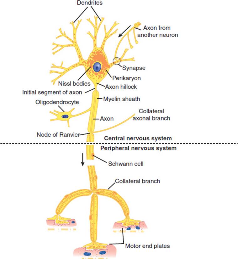
FIGURE 7-1 Schematic drawing of a Nissl-stained motor neuron. The myelin sheath is produced by oligodendrocytes in the central nervous system and by Schwann cells in the peripheral nervous system. Note the three motor end plates, which transmit the nerve impulse to striated skeletal muscle fibers. (Redrawn, with permission, from Mescher AL. Junqueira’s Basic Histology, 12th ed. McGraw-Hill, 2009.)
Signals are propagated electrically along axons. Like other cells, neurons maintain cell size and osmolarity primarily through the action of Na+-K+ ATPase, which actively pumps Na+ out of cells in exchange for K+. This results in the formation of concentration gradients for Na+ and K+ across the cell membrane. The membrane is practically impermeable to Na+, but the presence of K+ leak channels permits the flow of K+ out of cells. This produces a difference in electrical charge across the membrane that counters transport of K+ from the cell. The flow of ions continues until the opposing electrical force reaches a value that balances the diffusional force and the membrane reaches the equilibrium potential for K+ (EK). EK is calculated by the Nernst equation:

where
R = gas constant (2 kcal mol−1°−1)
T = absolute temperature (°K)
F = Faraday constant (2.3 × 10⁴ kcal V−1 mol−1)
[K+]o = concentration of K+ outside the cell
[K+]i = concentration of K+ inside the cell
In most neurons, the resting membrane potential (Em) is 50−100 mV and lies close to EK since the leak of K+ is the major determinant of the charge difference across the membrane.
The membrane potential may be altered by increasing the permeability of the membrane to another ion, which drives the resting membrane potential toward the equilibrium potential for that ion. Neurons are highly specialized to use rapid changes in membrane potential to generate electrical signals. This is accomplished by ligand-gated and voltage-gated ion channels that allow the passage of Na+, K+, Ca2+, or Cl− ions in response to electrical or chemical stimuli. These channels are composed of protein complexes embedded in the lipid membrane to form aqueous pores to the inside of the cell. In general, channels are selective for a particular species of ion. An array of charged amino acids within voltage-dependent channels detects changes in voltage and induces a conformational change in the channel to alter ion permeability. Binding sites for neurotransmitters such as glutamate, γ-aminobutyric acid (GABA), glycine, and acetylcholine exist on ligand-gated channels and, when occupied, induce a conformational change to open the channel.
Electrical signals are propagated in neurons because a voltage change across the membrane in one part of a neuron is propagated to other parts. Passive spread of a voltage disturbance weakens with increasing distance from the source unless energy-dependent processes amplify the signal. Passive spread of electrical signals works well over short distances and is a major mechanism of signal propagation in dendrites. However, long-distance communication down axons to nerve terminals requires amplification. This is accomplished through the generation of self-propagating waves of excitation known as action potentials.
An action potential arises primarily from voltage-dependent changes in membrane permeability to Na+ and K+ (Figure 7-2). If a depolarizing stimulus raises the membrane potential to about −45 mV, voltage-gated Na+ channels open, allowing influx of Na+ and further depolarization toward ENa (±50 mV). Nearby areas of membrane are depolarized to the threshold for Na+ channel activation, propagating a wave of depolarization from the initial site. The resting potential is restored quickly by a combination of events. First, Na+ channels close rapidly and remain in an inactive state until the membrane potential returns to negative levels for several milliseconds. Voltage-dependent K+channels open as the membrane potential peaks, speeding the efflux of K+ from cells and driving the membrane potential back to EK. K+ channels are also inactivated, but more slowly than Na+ channels, and this may transiently hyperpolarize cells. Plasma membrane ion exchangers and ion pumps then counteract the ion fluxes and eventually restore the resting state.

FIGURE 7-2 Conduction of the nerve impulse through an unmyelinated nerve fiber. In the resting axon, there is a difference of 70 mV between the interior of the axon and the outer surface of its membrane (resting potential). During the impulse passage, more Na+ (thick arrow) passes into the axon interior than the amount of K+ (thin arrow) that migrates in the opposite direction. In consequence, the membrane polarity changes (the membrane becomes relatively positive on its inner surface), and the resting potential is replaced by an action potential (+35 mV here). (Redrawn, with permission, from Junqueira LC et al, eds. Basic Histology, 10th ed. McGraw-Hill, 2003.)
Neurons transmit signals chemically to other cells at synapses (Figure 7-3). Presynaptic and postsynaptic cells are electrically isolated from each other and separated by a narrow synaptic cleft. Signaling across the cleft occurs through the release of neurotransmitters from the terminal of the presynaptic neuron. Most neurotransmitters are stored in membrane-bound synaptic vesicles and are released into the synaptic cleft by Ca2+-dependent exocytosis. Depolarization of the nerve terminal opens voltage-gated Ca2+ channels, stimulating Ca2+ influx and neurotransmitter release. Neurotransmitters diffuse across the cleft and bind to receptors on ligand-gated ion channels concentrated at the postsynaptic membrane. This produces local permeability changes, altering the membrane potential of the postsynaptic cell. If the response is depolarizing, an action potential may be generated if there are enough voltage-gated Na+ channels nearby and the membrane potential has been raised to the threshold for their activation. Receptor-gated ion channels are highly selective for a particular neurotransmitter and for the type of ions they pass, which determines whether they generate excitatory or inhibitory responses. In general, excitatory neurotransmitters, such as glutamate, open cation channels that allow influx of Na+ or Ca2+ and generate a depolarizing excitatory postsynaptic potential. Inhibitory neurotransmitters such as GABA and glycine open Cl− channels and generate an inhibitory postsynaptic potential, keeping the postsynaptic membrane near ECl (= −70 mV). Termination of the signal is achieved by removal of the neurotransmitter from the synaptic cleft. Acetylcholine is hydrolyzed by acetylcholinesterase at the postsynaptic membrane. Other neurotransmitters such as glutamate are removed by specific membrane transporters on nerve terminals or glial cells.

FIGURE 7-3 Schematic drawing of a synaptic terminal. Vesicles pass through the presynaptic membrane and release a transmitter substance into the synaptic cleft. (Redrawn, with permission, from Waxman SG. Clinical Neuroanatomy, 25th ed. McGraw-Hill, 2003.)
Not all neurotransmitter receptors are ion channels. Many receptors are coupled to cellular enzymes that regulate levels of intracellular second messengers to modulate the function of ion channels and many other cell proteins. A major mechanism by which messengers regulate ion channels is by promoting phosphorylation of channel subunits. For example, binding of the neurotransmitter norepinephrine to β-adrenergic receptors activates the enzyme adenylyl cyclase and stimulates the production of cyclic adenosine monophosphate (cAMP). The cAMP, in turn, activates a cAMP-dependent protein kinase that can phosphorylate voltage-gated calcium channels. In many cases, this increases the duration of time the channel remains open once it is activated, resulting in increased Ca2+ influx through the channel. Other neurotransmitter receptors, such as α1-adrenergic, muscarinic cholinergic, or metabotropic glutamate receptors, are coupled to the enzyme phospholipase C, which catalyzes the hydrolysis of the membrane lipid phosphatidylinositol-4,5-bisphosphate. Binding of neurotransmitter to the receptor activates phospholipase C to produce two second messengers: 1,2-diacylglycerol and inositol-1,4,5-trisphosphate. Diacylglycerol activates several enzymes of the protein kinase C family, some of which phosphorylate ion channels and either enhance or suppress their function. Inositol-1,4,5-trisphosphate binds an intracellular receptor that is itself a calcium ionophore, allowing release of calcium from intracellular stores into the cytosol. This calcium signal activates several calcium-dependent enzymes, including phosphatases and kinases that can alter the phosphorylation state and function of several ion channels and other cell proteins.
Astrocytes
Astrocytes serve a variety of metabolic, immunologic, structural, and nutritional support functions required for normal function of neurons. They possess numerous processes that radiate from the cell body, surrounding blood vessels and covering the surfaces of the brain and spinal cord (Figure 7-4). Astrocytes express voltage- and ligand-gated ion channels and regulate K+ and Ca2+ concentrations within the interstitial space. Many synapses are invested with astrocytic processes, and this allows astrocytes to modulate neurotransmission by regulating extracellular concentrations of these cations. Astrocytes provide structural and trophic support for neurons through the production of extracellular matrix molecules such as laminin and through release of growth factors such as nerve growth factor, fibroblast growth factors, and brain-derived neurotrophic factor. End-feet of astrocytic processes at blood vessels provide sites for release of cytokines and chemoattractants during central nervous system (CNS) injury. Astrocytes respond to brain injury by increasing in size—and in some cases in number—through a process called reactive astrocytosis. This phenotypic change is characterized by an increase in cells expressing glial-fibrillary acidic protein and by synthesis and release of cytokines that regulate inflammatory responses and entry of hematogenous cells into the CNS. Astrocytes play an important role also in terminating neuronal responses to glutamate, the most abundant excitatory neurotransmitter in the brain. In cell cultures, neurons die in the presence of high levels of glutamate unless astrocytes are present. Glutamate transporters present on astrocyte cell membranes remove glutamate from the synapse. Astrocytes also contain glutamine synthase, which converts glutamate to glutamine, detoxifying the CNS of both glutamate and ammonia.

FIGURE 7-4 Drawings of neuroglial cells as seen in slides stained by metallic impregnation. Observe that only astrocytes exhibit vascular end-feet, which cover the walls of blood capillaries. (Redrawn, with permission, from Mescher AL. Junqueira’s Basic Histology, 12th ed. McGraw-Hill, 2009.)
Oligodendrocytes & Schwann Cells
Plasma membranes of oligodendrocytes in the CNS and Schwann cells in the peripheral nervous system envelop axons. For many axons, the membranes of these glial cells are wrapped layer on layer around the axon, forming a myelin sheath (Figure 7-5). Gaps form between myelin sheaths from neighboring glia and produce nodes of Ranvier where a small portion of the axon is exposed to the interstitial space and where voltage-dependent Na+ channels are clustered in the axonal membrane. Between the nodes, myelin insulates the axon from the extracellular space, allowing efficient spread of depolarization from one node to another. This allows action potentials to propagate rapidly by jumping from node to node in a process called saltatory conduction.

FIGURE 7-5 Myelination of axons. Top left: Unmyelinated axon. Top right: Myelinated axon. Note that the cell membrane of the Schwann cell has wrapped itself around the axon. Bottom: Myelination of several axons in the CNS by an oligodendrogliocyte. (Redrawn, with permission, from Ganong WF. Review of Medical Physiology, 22nd ed. McGraw-Hill, 2005.)
Microglia
Although peripheral blood lymphocytes and monocytes enter from the circulation and patrol the CNS, microglia, which reside in the CNS, function as the main immune effector cells. They appear to be derived from bone marrow precursors of macrophage-monocyte lineage and invade the CNS during the perinatal period. Microglia cells are activated by brain injury, infection, or neuronal degeneration. Activation is characterized by proliferation, migration into damaged tissue, increased or de novo expression of surface receptors, including CD45 (leukocyte common antigen), MHC class I and class II and immunoglobulin Fc receptors, and secretion of several cytokines, reactive oxygen intermediates, and proteinases. This response functions to remove dead tissue and destroy invading organisms but may contribute to CNS damage, particularly in certain CNS inflammatory and degenerative diseases.
CHECKPOINT
1. What are the primary functions of neurons, astrocytes, and microglia?
2. What role does myelin play in axonal conduction?
3. What is responsible for the resting membrane potential and for the generation of action potentials?
4. What are some of the major neurotransmitters in the nervous system, and what effects do they produce when they bind to their receptors?
FUNCTIONAL NEUROANATOMY
To understand neuroanatomy, it is useful to study structures as parts of functional systems.
MOTOR SYSTEM
Large alpha motor neurons of the spinal cord ventral horns and brainstem motor nuclei (facial nucleus, trigeminal motor nucleus, nucleus ambiguus, hypoglossal nucleus) extend axons into spinal and cranial nerves to innervate skeletal muscles. Damage to these lower motor neurons results in loss of all voluntary and reflex movement because they comprise the output of the motor system. Neurons in the precentral gyrus and neighboring cortical regions (upper motor neurons) send axons to synapse with lower motor neurons. Axons from these upper motor neurons comprise the corticospinal and corticobulbar tracts. The motor cortex and spinal cord are connected with other deep cerebral and brainstem motor nuclei, including the caudate nucleus, putamen, globus pallidus, red nuclei, subthalamic nuclei, substantia nigra, reticular nuclei, and neurons of the cerebellum. Neurons in these structures are distinct from cortical motor (pyramidal) neurons and are referred to as extrapyramidal neurons. Many parts of the cerebral cortex are connected by fiber tracts to the primary motor cortex. These connections are important for complex patterns of movement and for coordinating motor responses to sensory stimuli.
1. Lower Motor Neurons & Skeletal Muscles
Anatomy
Each alpha motor neuron axon contacts up to about 200 muscle fibers, and together they constitute the motor unit (Figure 7-6). Axons of the motor neurons intermingle to form spinal ventral roots, plexuses, and peripheral nerves. Muscles are innervated from specific segments of the spinal cord, and each muscle is supplied by at least two roots. Motor fibers are rearranged in the plexuses so that most muscles are supplied by one peripheral nerve. Thus, the distribution of muscle weakness differs in spinal root and peripheral nerve lesions.

FIGURE 7-6 Anatomic components of the motor unit. (Redrawn, with permission, from Greenberg DA et al, eds. Clinical Neurology, 8th ed. McGraw-Hill, 2012.)
Physiology
The lower motor neurons are the final common pathway for all voluntary movement. Therefore, damage to lower motor neurons or their axons causes flaccid weakness of innervated muscles. In addition, muscle tone or resistance to passive movement is reduced, and deep tendon reflexes are impaired or lost. Tendon reflexes and muscle tone depend on the activity of alpha motor neurons (Figure 7-7), specialized sensory receptors known as muscle spindles, and smaller gamma motor neurons whose axons innervate the spindles. Some gamma motor neurons are active at rest, making the spindle fibers taut and sensitive to stretch. Tapping on the tendon stretches the spindles, which causes them to send impulses that activate alpha motor neurons. These in turn fire, producing the brief muscle contraction observed during the myotactic stretch reflex. Alpha motor neurons of antagonist muscles are simultaneously inhibited. Both alpha and gamma motor neurons are influenced by descending fiber systems, and their state of activity determines the level of tone and activity of the stretch reflex.

FIGURE 7-7 Diagram illustrating the pathways responsible for the stretch reflex and the inverse stretch reflex. Stretch stimulates the muscle spindle, and impulses pass up the Ia fiber to excite the motor neuron. It also stimulates the Golgi tendon organ, and impulses passing up the Ib fiber activate the interneuron to release the inhibitory mediator glycine. With strong stretch, the resulting hyperpolarization of the motor neuron is so great that it stops discharging. (Redrawn, with permission, from Barnett KE et al, eds. Ganong’s Review of Medical Physiology, 24th ed. McGraw-Hill, 2012.)
Each point of contact between nerve terminal and skeletal muscle forms a specialized synapse known as a neuromuscular junction composed of the presynaptic motor nerve terminal and a postsynaptic motor end plate (Figure 7-8). Presynaptic terminals store synaptic vesicles that contain the neurotransmitter acetylcholine. The amount of neurotransmitter within a vesicle constitutes a quantum of neurotransmitter. Action potentials depolarize the motor nerve terminal, opening voltage-gated calcium channels and stimulating calcium-dependent release of neurotransmitter from the terminal. Released acetylcholine traverses the synaptic cleft to the postsynaptic (end plate) membrane, where it binds to nicotinic cholinergic receptors. These receptors are ligand-gated cation channels, and, on binding to acetylcholine, they allow entry of extracellular sodium into the motor end plate. This depolarizes the motor end plate, which in turn depolarizes the muscle fiber. After activation, cholinergic receptors are rapidly inactivated, reducing sodium entry. They remain inactive until acetylcholine dissociates from the receptor. This is facilitated by the enzyme acetylcholinesterase, which hydrolyzes acetylcholine and is present in the postsynaptic zone.
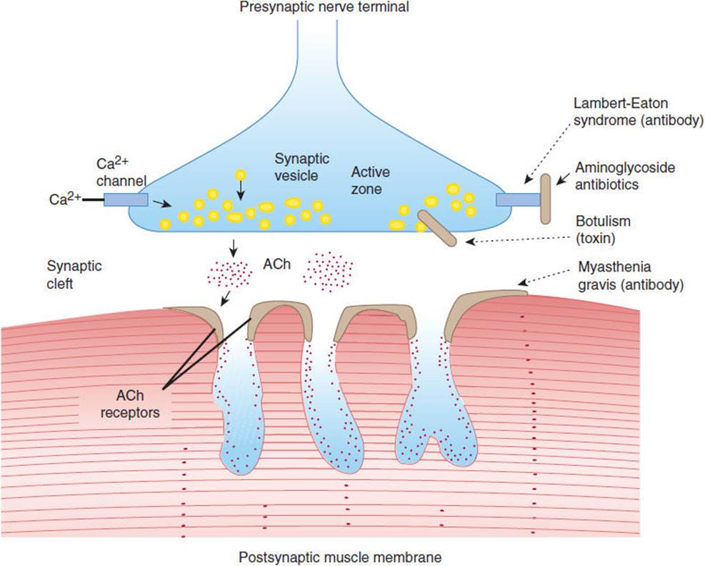
FIGURE 7-8 Sites of involvement in disorders of neuromuscular transmission. Left: Normal transmission involves depolarization-induced influx of calcium (Ca2+) through voltage-gated channels. This stimulates release of acetylcholine (ACh) from synaptic vesicles at the active zone and into the synaptic cleft. ACh binds to ACh receptors and depolarizes the postsynaptic muscle membrane. Right:Disorders of neuromuscular transmission result from blockage of Ca2+ channels (Lambert-Eaton syndrome or aminoglycoside antibiotics), impairment of Ca2+-mediated ACh release (botulinum toxin), or antibody-induced internalization and degradation of ACh receptors (myasthenia gravis). (Redrawn, with permission, from Greenberg DA et al, eds. Clinical Neurology, 8th ed. McGraw-Hill, 2012.)
Neuromuscular transmission may be disturbed in several ways (Figure 7-8). In the Lambert-Eaton myasthenic syndrome, antibodies to calcium channels inhibit calcium entry into the nerve terminal and reduce neurotransmitter release. In these cases, repetitive nerve stimulation facilitates accumulation of calcium in the nerve terminal and increases acetylcholine release. Clinically, limb muscles are weak, but if contraction is maintained, power increases. Electrophysiologically, there is an increase in the amplitude of the muscle response to repetitive nerve stimulation. Aminoglycoside antibiotics also impair calcium channel function and cause a similar syndrome. Proteolytic toxins produced by Clostridium botulinum cleave specific presynaptic proteins, preventing neurotransmitter release at both neuromuscular and parasympathetic cholinergic synapses. As a result, patients with botulism develop weakness, blurred vision, diplopia, ptosis, and large unreactive pupils. In myasthenia gravis, autoantibodies to the nicotinic acetylcholine receptor (AChR) block neurotransmission by inhibiting receptor function and activating complement-mediated lysis of the postsynaptic membrane. Myasthenia gravis is discussed in greater detail later in this chapter.
Motor nerves exert trophic influences on the muscles they innervate. Denervated muscles undergo marked atrophy, losing more than half of their original bulk in 2–3 months. Nerve fibers are also required for organization of the muscle end plate and for the clustering of cholinergic receptors to that region. Receptors in denervated fibers fail to cluster and become spread across the muscle membrane. Muscle fibers within a denervated motor unit may then discharge spontaneously, giving rise to a visible twitch (fasciculation) within a portion of a muscle. Individual fibers may also contract spontaneously, giving rise to fibrillations, which are not visible to the examiner but can be detected by electromyography. Fibrillations usually appear 7–21 days after damage to lower motor neurons or their axons.
CHECKPOINT
5. From where do lower motor neurons emanate, and to where do they send axons?
6. Describe four mechanisms that can disturb the function of the neuromuscular junction.
2. Upper Motor Neurons
Anatomy
The motor cortex is the region from which movements can be elicited by electrical stimuli (Figure 7-9). This includes the primary motor area (Brodmann area 4), premotor cortex (area 6), supplementary motor cortex (medial portions of 6), and primary sensory cortex (areas 3, 1, and 2). In the motor cortex, groups of neurons are organized in vertical columns, and discrete groups control contraction of individual muscles. Planned movements and those guided by sensory, visual, or auditory stimuli are preceded by discharges from prefrontal, somatosensory, visual, or auditory cortices, which are then followed by motor cortex pyramidal cell discharges that occur several milliseconds before the onset of movement.

FIGURE 7-9 Lateral aspect of the cerebrum. The cortical areas are shown according to Brodmann, with functional localizations. (Redrawn, with permission, from Waxman SG. Clinical Neuroanatomy, 26th ed. McGraw-Hill, 2010.)
Cortical motor neurons contribute axons that converge in the corona radiata and descend in the posterior limb of the internal capsule, cerebral peduncles, ventral pons, and medulla. These fibers constitute the corticospinal and corticobulbar tracts and together are known as upper motor neuron fibers (Figure 7-10). As they descend through the diencephalon and brainstem, fibers separate to innervate extrapyramidal and cranial nerve motor nuclei. The lower brainstem motor neurons receive input from crossed and uncrossed corticobulbar fibers, although neurons that innervate lower facial muscles receive primarily crossed fibers.

FIGURE 7-10 Schematic illustration of upper motor neuron pathways. (Redrawn, with permission, from Ropper AH et al, eds. Adams and Victor’s Principles of Neurology, 9th ed. McGraw-Hill, 2009.)
In the ventral medulla, the remaining corticospinal fibers course in a tract that is pyramidal in shape in cross section—thus, the name pyramidal tract. At the lower end of the medulla, most fibers decussate, although the proportion of crossed and uncrossed fibers varies somewhat between individuals. The bulk of these fibers descend as the lateral corticospinal tract of the spinal cord.
Different groups of neurons in the cortex control muscle groups of the contralateral face, arm, and leg. Neurons near the ventral end of the central sulcus control muscles of the face, whereas neurons on the medial surface of the hemisphere control leg muscles (Figure 7-10). Because the movements of the face, tongue, and hand are complex in humans, a large share of the motor cortex is devoted to their control. A somatotopic organization is also apparent in the lateral corticospinal tract of the cervical cord, where fibers to motor neurons that control leg muscles lie laterally and fibers to cervical motor neurons lie medially.
Physiology
Upper motor neurons are the final common pathway between cortical and subcortical structures, such as the basal ganglia, in the planning, initiation, sequencing, and modulation of all voluntary movement. Much has been learned about the normal function of upper motor neurons through the study of animals and humans with focal brain lesions. Upper motor neuron pathways can be interrupted in the cortex, subcortical white matter, internal capsule, brainstem, or spinal cord. Unilateral upper motor neuron lesions spare muscles innervated by lower motor neurons that receive bilateral cortical input, such as muscles of the eyes, jaw, upper face, pharynx, larynx, neck, thorax, and abdomen. Unlike paralysis resulting from lower motor neuron lesions, paralysis from upper motor neuron lesions is rarely complete for a prolonged period of time. Acute lesions, particularly of the spinal cord, often cause flaccid paralysis and absence of spinal reflexes at all segments below the lesion. With spinal cord lesions, this state is known as spinal shock. After a few days to weeks, a state known as spasticity appears, which is characterized by increased tone and hyperactive stretch reflexes. A similar but less striking sequence of events can occur with acute cerebral lesions.
Upper motor neuron lesions cause a characteristic pattern of limb weakness and change in tone. Antigravity muscles of the limbs become more active relative to other muscles. The arms tend to assume a flexed, pronated posture, and the legs become extended. In contrast, muscles that move the limbs out of this posture (extensors of the arms and flexors of the legs) are preferentially weakened. Tone is increased in antigravity muscles (flexors of the arms and extensors of the legs), and if these muscles are stretched rapidly, they respond with an abrupt catch, followed by a rapid increase and then a decline in resistance as passive movement continues. This sequence constitutes the “clasp knife” phenomenon. Clonus—a series of involuntary muscle contractions in response to passive stretch—may be present, especially with spinal cord lesions.
Pure pyramidal tract lesions in animals cause temporary weakness without spasticity. In humans, lesions of the cerebral peduncles also cause mild paralysis without spasticity. It appears that control of tone is mediated by other tracts, particularly corticorubrospinal and corticoreticulospinal pathways. This may explain why the degrees of weakness and spasticity often do not correspond in patients with upper motor neuron lesions.
The distribution of paralysis resulting from upper motor neuron lesions varies with the location of the lesion. Lesions above the pons impair movements of the contralateral lower face, arm, and leg. Lesions below the pons spare the face. Lesions of the internal capsule often impair movements of the contralateral face, arm, and leg equally, because motor fibers are packed closely together in this region. In contrast, lesions of the cortex or subcortical white matter tend to differentially affect the limbs and face because the motor fibers are spread over a larger area of brain. Bilateral cerebral lesions cause weakness and spasticity of cranial, trunk, and limb muscles, which leads to dysarthria, dysphonia, dysphagia, bifacial paresis, and sometimes reflexive crying and laughing (pseudobulbar palsy).
CHECKPOINT
7. Define the motor cortex and describe its organization.
8. Fibers from which nuclei and in which tracts constitute upper motor neurons? What is their path?
9. Describe the somatotopic organization of motor neurons in the cortex.
10. What are the characteristics of weakness and tone in upper motor neuron lesions?
11. How is the distribution of paralysis and spasticity affected by the location of an upper motor neuron lesion?
3. Cerebellum
Anatomy
The cerebellar cortex can be divided into three anatomic regions (Figure 7-11B). The flocculonodular lobe, composed of the flocculus and the nodulus of the vermis, has connections to vestibular nuclei and is important for the control of posture and eye movement. The anterior lobe (Figure 7-11A) lies rostral to the primary fissure and includes the remainder of the vermis. It receives proprioceptive input from muscles and tendons via the dorsal and ventral spinocerebellar tracts and influences posture, muscle tone, and gait. The posterior lobe, which comprises the remainder of the cerebellar hemispheres, receives major input from the cerebral cortex via the pontine nuclei and middle cerebellar peduncles and is important for the coordination and planning of voluntary skilled movements initiated from the cerebral cortex.

FIGURE 7-11 Anatomic divisions of the cerebellum in midsagittal view: (A) unfolded (arrows) and (B) viewed from behind. (Redrawn, with permission, from Greenberg DA et al, eds. Clinical Neurology, 5th ed. McGraw-Hill, 2002.)
Efferent fibers from these lobes project to deep cerebellar nuclei, which in turn project to the cerebrum and brainstem through two main pathways (Figure 7-12). The fastigial nucleus receives input from the vermis and sends fibers to bilateral vestibular nuclei and reticular nuclei of the pons and medulla via the inferior cerebellar peduncles. Other regions of the cerebellar cortex send fibers to the dentate, emboliform, and globose nuclei, whose efferents form the superior cerebellar peduncles, enter the upper pons, decussate completely in the lower midbrain, and travel to the contralateral red nucleus. At the red nucleus, some fibers terminate, whereas others ascend to the ventrolateral nucleus of the thalamus, whence thalamic neurons send ascending efferent fibers to the motor cortex of the same side. A smaller group of fibers descend after decussation in the midbrain and terminate in reticular nuclei of the lower brainstem. Thus, the cerebellum controls movement through connections with cerebral motor cortex and brainstem nuclei.
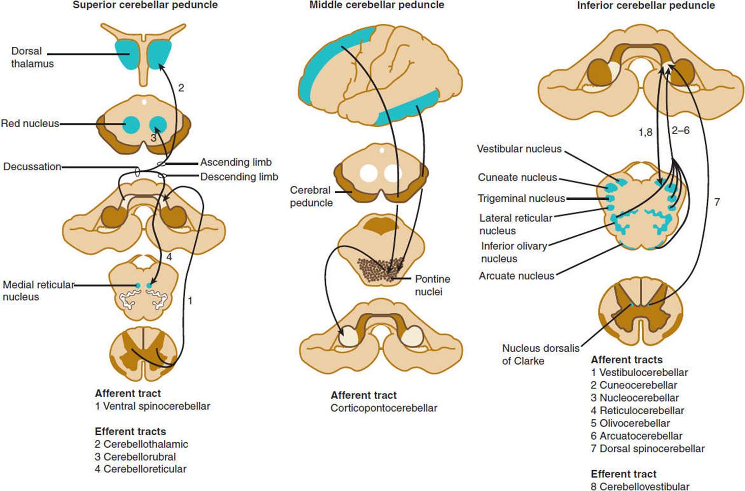
FIGURE 7-12 Cerebellar connections in the superior, middle, and inferior cerebellar peduncles. The peduncles are indicated by gray shading and the areas to and from which they project by blue shading. (Redrawn, with permission, from Greenberg DA et al, eds. Clinical Neurology, 8th ed. McGraw-Hill, 2012.)
Physiology
The cerebellum is responsible for the coordination of muscle groups, control of stance and gait, and regulation of muscle tone. Rather than causing paralysis, damage to the cerebellum interferes with the performance of motor tasks. The major manifestation of cerebellar disease is ataxia, in which simple movements are delayed in onset and their rates of acceleration and deceleration are decreased, resulting in intention tremor and dysmetria (“overshooting”). Lesions of the cerebellar hemispheres affect the limbs, producing limb ataxia, whereas midline lesions affect axial muscles, causing truncal and gait ataxia and disorders of eye movement. Cerebellar lesions are often associated with hypotonia as a result of depression of activity of alpha and gamma motor neurons. If a lesion of the cerebellum or cerebellar peduncles is unilateral, the signs of limb ataxia appear on the same side as the lesion. However, if the lesion lies beyond the decussation of efferent cerebellar fibers in the midbrain, the clinical signs are on the side opposite the lesion.
CHECKPOINT
12. What is the overall role of the cerebellum?
13. What are the anatomic regions of the cerebellum, what do they control, and through which other regions of the brain do they make connections?
14. What are the consequences of damage to the cerebellum, and what symptoms and signs are seen in patients with cerebellar lesions?
15. Below what point do unilateral cerebellar lesions manifest on the opposite side?
4. Basal Ganglia
Anatomy
Several subcortical, thalamic, and brainstem nuclei are critical for regulating voluntary movement and maintaining posture. These include the basal ganglia (ie, the caudate nucleus and putamen [corpus striatum]), globus pallidus, substantia nigra, and subthalamic nuclei. They also include the red nuclei and the mesencephalic reticular nuclei. The major pathways that involve the basal ganglia form three neuronal circuits (Figure 7-13). The first is the cortical-basal ganglionic-thalamic-cortical loop. Inputs mainly from premotor, primary motor, and primary sensory cortices (areas 1, 2, 3, 4, and 6) project to the corpus striatum, which sends fibers to the medial and lateral portions of the globus pallidus. Fibers from the globus pallidus form the ansa and fasciculus lenticularis, which sweep through the internal capsule and project onto ventral and intralaminar thalamic nuclei. Axons from these nuclei project to the premotor and primary motor cortices (areas 4 and 6), completing the loop. In the second loop, the substantia nigra sends dopaminergic fibers to the corpus striatum, which has reciprocal connections with the substantia nigra. The substantia nigra also projects to the ventromedial thalamus. The third loop is composed of reciprocal connections between the globus pallidus and the subthalamic nucleus. The subthalamic nucleus also sends efferents to the substantia nigra and corpus striatum.

FIGURE 7-13 Diagrammatic representation of the principal connections of the basal ganglia. Solid lines indicate excitatory pathways; dashed lines indicate inhibitory pathways. The transmitters are indicated in the pathways, where they are known. Glu, glutamate; DA, dopamine. Acetylcholine is the transmitter produced by interneurons in the striatum (ie, the putamen and the caudate nucleus, which have similar connections). SNPR, substantia nigra, pars reticulata; SNPC, substantia nigra, pars compacta; ES, external segment; IS, internal segment. The subthalamic nucleus also projects to the pars compacta of the substantia nigra; this pathway has been omitted for clarity. (Redrawn, with permission, from Barnett KE et al, eds. Ganong’s Review of Medical Physiology, 24th ed. McGraw-Hill, 2012.)
Physiology
Basal ganglia circuits regulate the initiation, amplitude, and speed of movements. Diseases of the basal ganglia cause abnormalities of movement and are collectively known as movement disorders. They are characterized by motor deficits (bradykinesia, akinesia, loss of postural reflexes) or abnormal activation of the motor system, resulting in rigidity, tremor, and involuntary movements (chorea, athetosis, ballismus, and dystonia).
Several neurotransmitters are found within the basal ganglia, but their role in disease states is only partly understood. Acetylcholine is present in high concentrations within the corpus striatum, where it is synthesized and released by large Golgi type 2 neurons (Figure 7-14). Acetylcholine acts as an excitatory transmitter at medium-sized spiny striatal neurons that synthesize and release the inhibitory neurotransmitter GABA and project to the globus pallidus. Dopamine is synthesized by neurons of the substantia nigra, whose axons form the nigrostriatal pathway that terminates in the corpus striatum. Dopamine released by these fibers inhibits striatal GABAergic neurons. In Parkinson disease, degeneration of nigral neurons leads to loss of dopaminergic inhibition and a relative excess of cholinergic activity. This increases GABAergic output from the striatum and contributes to the paucity of movement that is a cardinal manifestation of the disease. Anticholinergics and dopamine agonists tend to restore the normal balance of striatal cholinergic and dopaminergic inputs and are effective in treatment. The pathogenesis of Parkinson disease is discussed later in this chapter.

FIGURE 7-14 Simplified neurochemical anatomy of the basal ganglia. Dopamine (DA) neurons exert a net inhibitory effect and acetylcholine (ACh) neurons a net excitatory effect on the GABAergic output from the striatum. In Parkinson disease, DA neurons degenerate. The net effect is to increase GABAergic output from the striatum. (Redrawn, with permission, from Greenberg DA et al, eds. Clinical Neurology, 5th ed. McGraw-Hill, 2002.)
Huntington disease is inherited as an autosomal dominant disorder. When disease onset occurs later in life, patients develop involuntary, rapid, jerky movements (chorea) and slow writhing movements of the proximal limbs and trunk (athetosis). When disease onset occurs earlier in life, patients develop signs of parkinsonism with tremor (cogwheeling) and stiffness. The spiny GABAergic neurons of the striatum preferentially degenerate, resulting in a net decrease in GABAergic output from the striatum. This contributes to the development of chorea and athetosis. Dopamine antagonists, which block inhibition of remaining striatal neurons by dopaminergic striatal fibers, reduce the involuntary movements. Neurons in deep layers of the cerebral cortex also degenerate early in the disease, and later this extends to other brain regions, including the hippocampus and hypothalamus. Thus, the disease is characterized by cognitive defects and psychiatric disturbances in addition to the movement disorder.
The gene for the disease is located on chromosome 4p and encodes for a 3144-amino acid protein, huntingtin, which is widely expressed and interacts with several proteins involved in intracellular trafficking and endocytosis, gene transcription, and intracellular signaling. The protein contains a trinucleotide (CAG) repeat of 11–34 copies that encodes a polyglutamine domain and is expanded in patients with the disease. Deletion of the gene in mice causes embryonic death, whereas heterozygous animals are healthy. Transgenic mice with an expanded repeat develop a neurodegenerative disorder, suggesting that the disease results from the toxic effect of a gain of function mutation.
The mechanisms by which mutant huntingtin causes disease are not certain. The mutant protein is degraded, and the resulting fragments that contain the glutamine repeats form aggregates, which are deposited in nuclear and cytoplasmic inclusions. These fragments may bind abnormally to other proteins and interfere with normal protein processing or disrupt mitochondrial function. Nuclear fragments may interfere with nuclear functions such as gene expression. For example, in the cerebral cortex, mutant huntingtin reduces the production of brain-derived neurotrophic factor by suppressing its transcription. In addition, normal huntingtin is protective for cortical and striatal neurons and blocks the processing of procaspase 9, thereby reducing apoptosis (programmed cell death). Therefore, both loss of neurotrophic support and enhanced caspase activity could promote striatal cell loss in Huntington disease.
CHECKPOINT
16. Which are the component nuclei of the basal ganglia, and what is their functional role?
17. What are the clinical consequences of lesions in the basal ganglia?
18. What are some of the neurotransmitters within the basal ganglia, and what is their role in disorders of basal ganglia function?
SOMATOSENSORY SYSTEM
Somatosensory pathways confer information about touch, pressure, temperature, pain, vibration, and the position and movement of body parts. This information is relayed to thalamic nuclei and integrated in the sensory cortex of the parietal lobes to provide conscious awareness of sensation. Information is also relayed to cortical motor neurons to adjust fine movements and maintain posture. Some ascending sensory fibers, particularly pain fibers, enter the midbrain and project to the amygdala and limbic cortex, where they contribute to emotional responses to pain. In the spinal cord, painful stimuli activate local pathways that induce the firing of lower motor neurons and cause a reflex withdrawal. Thus, somatosensory pathways provide tactile information, guide movement, and serve protective functions.
Anatomy
A variety of specialized end organs and free nerve endings transduce sensory stimuli into neural signals and initiate the firing of sensory nerve fibers. Fibers that mediate cutaneous sensation from the trunk and limbs travel in sensory or mixed sensorimotor nerves to the spinal cord (Figure 7-15). Cutaneous sensory nerves contain small myelinated Aδ fibers that transmit information about pain and temperature, larger myelinated fibers that mediate touch and pressure sensation, and more numerous unmyelinated pain and autonomic C fibers. Myelinated proprioceptive fibers and afferent and efferent muscle spindle fibers are carried in the larger sensorimotor nerves. The cell bodies of the sensory neurons are in the dorsal root ganglia, and their central projections enter the spinal cord via the dorsal spinal roots. Innervation of the skin, muscles, and surrounding connective tissue is segmental, and each root innervates a region of skin known as a dermatome (Figure 7-16). Cell bodies of the sensory neurons that innervate the face reside in the trigeminal ganglion and send their central projections in the trigeminal nerve to the brainstem. The trigeminal innervation of the face is subdivided into three regions, each innervated by one of the three divisions of the trigeminal nerve.

FIGURE 7-15 Schematic illustration of a spinal cord segment with its dorsal root, ganglion cells, and sensory organs. Sensory organs shown (from top to bottom) are the pacinian corpuscle, muscle spindle, tendon organ, encapsulated ending, and free nerve endings. The somatotopic arrangement of fibers in the dorsal columns, spinothalamic tract, and corticospinal tract is also shown. (Redrawn, with permission, from Waxman SG. Clinical Neuroanatomy, 26th ed. McGraw-Hill, 2010.)

FIGURE 7-16 Segmental distribution of the body viewed in the approximate quadruped position, including sensory distribution of the trigeminal (V) cranial nerve. (Redrawn, with permission, from Waxman SG. Clinical Neuroanatomy, 26th ed. McGraw-Hill, 2010.)
The dorsal roots enter the dorsal horn of the spinal cord (Figure 7-15). Large myelinated fibers divide into ascending and descending branches and either synapse with dorsal gray neurons within a few cord segments or travel in the dorsal columns, terminating in the gracile or cuneate nuclei of the lower medulla on the same side. Secondary neurons of the dorsal horn also send axons up the dorsal columns. Fibers in the dorsal columns are displaced medially as new fibers are added, so that in the cervical cord, leg fibers are located medially and arm fibers laterally (Figure 7-15). The gracile and cuneate nuclei send fibers that cross the midline in the medulla and ascend to the thalamus as the medial lemniscus (Figure 7-17). The dorsal column-lemniscal system carries information about pressure, limb position, vibration, direction of movement, recognition of texture and shape, and two-point discrimination.

FIGURE 7-17 Sensory pathways conveying touch, pressure, vibration, joint position, pain, and temperature sensation. (Redrawn, with permission, from Greenberg DA et al, eds. Clinical Neurology, 5th ed. McGraw-Hill, 2002.)
Thinly myelinated and unmyelinated fibers enter the lateral portion of the dorsal horn and synapse with dorsal spinal neurons within one or two segments. The majority of secondary fibers from these cells cross in the anterior spinal commissure and ascend in the anterolateral spinal cord as the lateral spinothalamic tracts. Crossing fibers are added to the inner side of the tract, so that in the cervical cord the leg fibers are located superficially and arm fibers are deeper. These fibers carry information about pain, temperature, and touch sensation.
Sensation from the face is carried by trigeminal sensory fibers that enter the pons and descend to the medulla and upper cervical cord (Figure 7-18). Fibers carrying information about pain and temperature sensation terminate in the nucleus of the spinal tract of cranial nerve V, which is continuous with the dorsal horn of the cervical cord. Touch, pressure, and postural information is conveyed by fibers that terminate in the main sensory and mesencephalic nuclei of the trigeminal nerve. Axons arising from trigeminal nuclei cross the midline and ascend as the trigeminal lemniscus just medial to the spinothalamic tract. Fibers from the spinothalamic tract, medial lemniscus, and trigeminal lemniscus merge in the midbrain and terminate along with sensory fibers ascending from the spinal cord in the posterior thalamic nuclei, mainly in the nucleus ventralis posterolateralis. These thalamic nuclei project to the primary somatosensory cortex (Brodmann areas 3, 1, and 2) and to a second somatosensory area on the upper bank of the sylvian fissure (lateral cerebral sulcus). The primary somatosensory region is organized somatotopically like the primary motor cortex.

FIGURE 7-18 Schematic drawing of the trigeminal system. (Redrawn, with permission, from Waxman SG. Clinical Neuroanatomy, 26th ed. McGraw-Hill, 2010.)
Physiology
A. Pain
Free nerve endings of unmyelinated C fibers and small-diameter myelinated Aδ fibers in the skin convey sensory information in response to chemical, thermal, and mechanical stimuli. Intense stimulation of these nerve endings evokes the sensation of pain. In contrast to skin, most deep tissues are relatively insensitive to chemical or noxious stimuli. However, inflammatory conditions can sensitize sensory afferents from deep tissues to evoke pain on mechanical stimulation. This sensitization appears to be mediated by bradykinin, prostaglandins, and leukotrienes released during the inflammatory response. Information from primary afferent fibers is relayed via sensory ganglia to the dorsal horn of the spinal cord and then to the contralateral spinothalamic tract, which connects to thalamic neurons that project to the somatosensory cortex.
Damage to these pathways produces a deficit in pain and temperature discrimination and may also produce abnormal painful sensations (dysesthesias) usually in the area of sensory loss. Such pain is termed neuropathic pain and often has a strange burning, tingling, or electric shocklike quality. It may arise from several mechanisms. Damaged peripheral nerve fibers become highly mechanosensitive and may fire spontaneously without known stimulation. They also develop sensitivity to norepinephrine released from sympathetic postganglionic neurons. Electrical impulses may spread abnormally from one fiber to another (ephaptic conduction), enhancing the spontaneous firing of multiple fibers. Neuropeptides released by injured nerves may recruit an inflammatory reaction that stimulates pain. In the dorsal horn, denervated spinal neurons may become spontaneously active. In the brain and spinal cord, synaptic reorganization occurs in response to injury and may lower the threshold for pain. In addition, inhibition of pathways that modulate transmission of sensory information in the spinal cord and brainstem may promote neuropathic pain.
Pain-modulating circuits exert a major influence on the perceived intensity of pain. One such pathway (Figure 7-19) is composed of cells in the periaqueductal gray matter of the midbrain that receive afferents from frontal cortex and hypothalamus and project to rostroventral medullary neurons. These in turn project in the dorsolateral white matter of the spinal cord and terminate on dorsal horn neurons. Additional descending pathways arise from other brainstem nuclei (locus ceruleus, dorsal raphe nucleus, and nucleus reticularis gigantocellularis). Major neurotransmitters utilized by these systems include endorphins, serotonin, and norepinephrine, providing the rationale for the use of opioids, serotonin agonists, and serotonin and norepinephrine reuptake inhibitors in the treatment of pain.
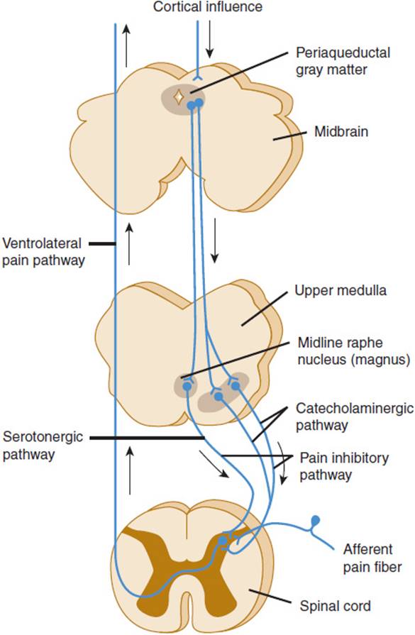
FIGURE 7-19 Schematic illustration of the pathways involved in pain control. (Courtesy A Basbaum.)
B. Proprioception and Vibratory Sense
Receptors in the muscles, tendons, and joints provide information about deep pressure and the position and movement of body parts. This allows one to determine an object’s size, weight, shape, and texture. Information is relayed to the spinal cord via large Aα and Aβ myelinated fibers and to the thalamus by the dorsal column-lemniscal system. Detecting vibration requires sensing touch and rapid changes in deep pressure. This depends on multiple cutaneous and deep sensory fibers and is impaired by lesions of multiple peripheral nerves, the dorsal columns, medial lemniscus, or thalamus but rarely by lesions of single nerves. Vibratory sense is often impaired together with proprioception.
C. Discriminative Sensation
Primary sensory cortex provides awareness of somatosensory information and the ability to make sensory discriminations. Touch, pain, temperature, and vibration sense are considered the primary modalities of sensation and are relatively preserved in patients with damage to sensory cortex or its projections from the thalamus. In contrast, complex tasks that require integration of multiple somatosensory stimuli and of somatosensory stimuli with auditory or visual information are impaired. These include the ability to distinguish two points from one when touched on the skin (two-point discrimination), localize tactile stimuli, perceive the position of body parts in space, recognize letters or numbers drawn on the skin (graphesthesia), and identify objects by their shape, size, and texture (stereognosis).
D. Anatomy of Sensory Loss
The patterns of sensory loss often indicate the level of nervous system involvement. Symmetric distal sensory loss in the limbs, affecting the legs more than the arms, usually signifies a generalized disorder of multiple peripheral nerves (polyneuropathy). Sensory symptoms and deficits may be restricted to the distribution of a single peripheral nerve (mononeuropathy) or two or more peripheral nerves (mononeuropathy multiplex). Symptoms limited to a dermatome indicate a spinal root lesion (radiculopathy).
In the spinal cord, segregation of fiber tracts and the somatotopic arrangement of fibers give rise to distinct patterns of sensory loss. Loss of pain and temperature sensation on one side of the body and of proprioception on the opposite side occurs with lesions that involve one half of the cord on the side of the proprioceptive deficit (Brown-Séquard syndrome; Figure 7-20). Compression of the upper spinal cord causes loss of pain, temperature, and touch sensation first in the legs, because the leg spinothalamic fibers are most superficial. More severe cord compression compromises fibers from the trunk. In patients with spinal cord compression, the lesion is often above the highest dermatome involved in the deficit. Thus, radiographic studies should be tailored to visualize the cord at and above the level of the sensory deficit detected on examination. Intrinsic cord lesions that involve the central portions of the cord often impair pain and temperature sensation at the level of the lesion because the fibers crossing the anterior commissure and entering the spinothalamic tracts are most centrally situated. Thus, enlargement of the central cervical canal in syringomyelia typically causes loss of pain and temperature sensation across the shoulders and upper arms (Figure 7-21).
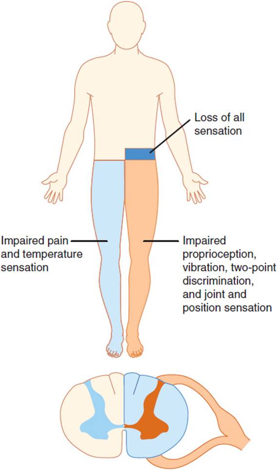
FIGURE 7-20 Brown-Séquard syndrome with lesion at left tenth thoracic level (motor deficits not shown). (Redrawn, with permission, from Waxman SG. Neuroanatomy with Clinical Correlations, 25th ed. McGraw-Hill, 2003.)
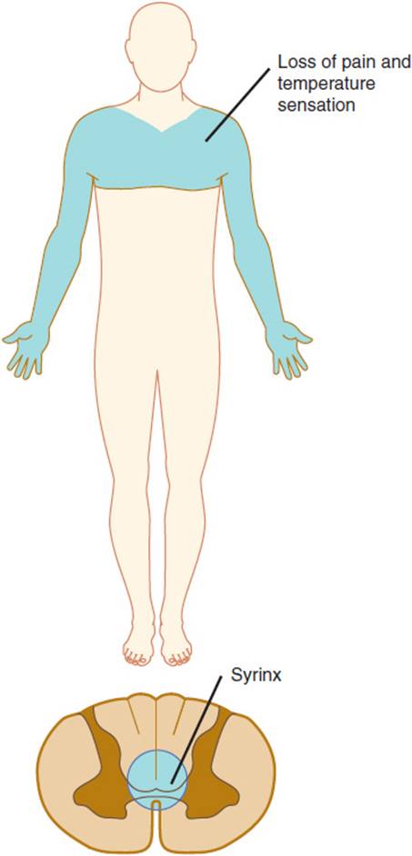
FIGURE 7-21 Syringomyelia (the presence of a cavity in the spinal cord resulting from breakdown of gliomatous new formations, presenting clinically with pain and paresthesias followed by muscular atrophy of the hands) involving the cervicothoracic portion of the cord. (Redrawn, with permission, from Waxman SG. Clinical Neuroanatomy, 26th ed. McGraw-Hill, 2010.)
Brainstem lesions involving the spinothalamic tract cause loss of pain and temperature sensation on the opposite side of the body. In the medulla, such lesions can involve the neighboring spinal trigeminal nucleus, resulting in a “crossed” sensory deficit involving the ipsilateral face and contralateral limbs. Above the medulla, the spinothalamic and trigeminothalamic tracts lie close together, and lesions there cause contralateral sensory loss of the face and limbs. In the midbrain and thalamus, medial lemniscal fibers run together with pain and temperature fibers, and lesions are more likely to impair all primary sensation contralateral to the lesion. Because sensory fibers converge at the thalamus, lesions there tend to cause fairly equal loss of pain, temperature, and proprioceptive sensation on the contralateral half of the face and body. Lesions of the sensory cortex in the parietal lobe impair discriminative sensation on the opposite side of the body, whereas detection of the primary modalities of sensation may remain relatively intact.
CHECKPOINT
19. What fibers carry pain, and how are they segregated from fibers that carry proprioception information in the spinal cord?
20. What are the differences in characteristics of sensory loss at different levels of the nervous system?
21. What is the function of the sensory cortex in the parietal lobe, and what are the clinical features of damage to this region?
VISION & CONTROL OF EYE MOVEMENTS
The visual system provides our most important source of sensory information about the environment. The visual system and pathways for the control of eye movements are among the best characterized pathways in the nervous system. Familiarity with these neuroanatomic features is often extremely valuable in localization of neurologic disease.
Anatomy
The cornea and lens of the eye refract and focus images on the photosensitive posterior portion of the retina. The posterior retina contains two classes of specialized photoreceptor cells, rods and cones, which transduce photons into electrical signals. At the retina, the image is reversed in the horizontal and vertical planes so that the inferior visual field falls on the superior portions of the retina and the lateral field is detected by the nasal half of the retina.
Fibers from the nasal half of the retina traverse the medial portion of the optic nerve and cross to the other side at the optic chiasm (Figure 7-22). Each optic tract contains fibers from the same half of the visual field of both eyes. The optic tracts terminate in the lateral geniculate nuclei of the thalamus. Lateral geniculate neurons send fibers to the primary visual cortex in the occipital lobe (area 17, calcarine cortex; see Figure 7-9). These fibers form the optic radiations, which extend through the white matter of the temporal lobes and the inferior portion of the parietal lobes.

FIGURE 7-22 Common visual field defects and their anatomic bases. (1) Central scotoma caused by inflammation of the left optic disk (optic neuritis) or optic nerve (retrobulbar neuritis). (2) Total blindness of the right eye from a complete lesion of the right optic nerve. (3) Bitemporal hemianopia caused by pressure exerted on the optic chiasm by a pituitary tumor. (4) Right nasal hemianopia caused by a perichiasmal lesion (eg, calcified internal carotid artery). (5) Right homonymous hemianopia from a lesion of the left optic tract. (6) Right homonymous superior quadrantanopia caused by partial involvement of the optic radiation by a lesion in the left temporal lobe (Meyer loop). (7) Right homonymous inferior quadrantanopia caused by partial involvement of the optic radiation by a lesion in the left parietal lobe. (8) Right homonymous hemianopia from a complete lesion of the left optic radiation. A similar defect may also result from lesion. (9) Right homonymous hemianopia (with macular sparing) resulting from posterior cerebral artery occlusion. (Redrawn, with permission, from Greenberg DA et al, eds. Clinical Neurology, 8th ed. McGraw-Hill, 2012.)
Eye movements are produced by the extraocular muscles, which function in pairs to move the eyes along three axes (Figure 7-23). These muscles are innervated by the oculomotor (III), trochlear (IV), and abducens (VI) nerves. The oculomotor nerve innervates the ipsilateral medial, superior, and inferior rectus muscles and the inferior oblique muscles. It also supplies the ipsilateral levator palpebrae, which elevates the eyelid. The oculomotor nerve also carries parasympathetic fibers that mediate pupillary constriction (see later discussion). Trochlear nerve fibers decussate before leaving the brainstem, and each trochlear nerve supplies the contralateral superior oblique muscle. The abducens nerve innervates the lateral rectus muscle of the same side.
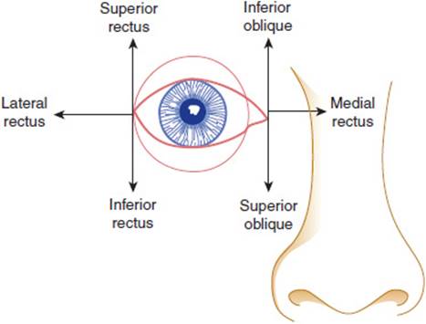
FIGURE 7-23 Extraocular muscles subserving the six cardinal positions of gaze. The eye is adducted by the medial rectus and abducted by the lateral rectus. The adducted eye is elevated by the inferior oblique and depressed by the superior oblique; the abducted eye is elevated by the superior rectus and depressed by the inferior rectus. (Redrawn, with permission, from Greenberg DA et al, eds. Clinical Neurology, 8th ed. McGraw-Hill, 2012.)
Cortical and brainstem gaze centers innervate the extraocular motor nuclei and provide for supranuclear control of gaze. A vertical gaze center is located in the midbrain tegmentum, and lateral gaze centersare present in the pontine paramedian reticular formation. Each lateral gaze center sends fibers to the neighboring ipsilateral abducens nucleus and, via the medial longitudinal fasciculus, to the contralateral oculomotor nucleus. Therefore, activation of the right lateral gaze center stimulates conjugate deviation of the eyes to the right. Rapid saccadic eye movements are initiated by the frontal eye fields in the premotor cortex that stimulate conjugate movement of the eyes to the opposite side. Slower eye movements involved in pursuit of moving objects are controlled by parieto-occipital gaze centers, which stimulate conjugate gaze to the side of the gaze center. These cortical areas control eye movements through their connections with the brainstem gaze centers.
The size of the pupils is determined by the balance between parasympathetic and sympathetic discharge to the pupillary muscles. The parasympathetic oculomotor nuclei of Edinger-Westphal send fibers in the oculomotor nerves that synapse in the ciliary ganglia within the orbits and innervate the pupillary constrictor muscles.
The motor portion of pupillary dilation is controlled by a three-neuron system (Figure 7-24). It is composed of axons from neurons in the posterolateral hypothalamus that descend through the lateral brainstem tegmentum and the intermediolateral column of the cervical spinal cord to the level of T1. There they terminate on preganglionic sympathetic neurons within the lateral gray matter of the thoracic cord. These neurons send axons that synapse with postganglionic neurons in the superior cervical ganglion. Postganglionic neurons send fibers that travel with the internal carotid artery and the first division of the trigeminal nerve to innervate the iris. The fibers also innervate the tarsal muscles of the eyelids. Damage to these pathways causes Horner syndrome, which consists of miosis, ptosis, and sometimes impaired sweating ipsilateral to the lesion.
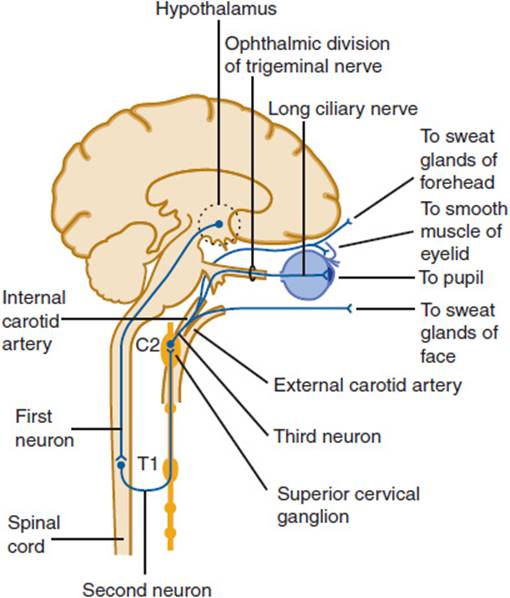
FIGURE 7-24 Oculosympathetic pathways. This three-neuron pathway projects from the hypothalamus to the intermediolateral column of the spinal cord, then to the superior cervical (sympathetic) ganglion, and finally to the pupil, the smooth muscle of the eyelids, and the sweat glands of the forehead and face. Interruption of these pathways results in Horner syndrome. (Redrawn, with permission, from Greenberg DA et al, eds. Clinical Neurology, 5th ed. McGraw-Hill, 2002.)
Physiology
A. Vision
The rods are sensitive to low levels of light and are most numerous in the peripheral regions of the retina. In retinitis pigmentosa, there is degeneration of the retina that begins in the periphery. Poor twilight vision is thus an early symptom of this disorder. Cones are responsible for perception of stimuli in bright light and for discrimination of color. They are concentrated in the macular region, which is crucial for visual acuity. In disorders of the retina or optic nerve that impair acuity, diminished color discrimination is often an early sign.
Visual processing begins in the retina, where information gathered from rods and cones is modified by interactions among bipolar, amacrine, and horizontal cells. Amacrine and bipolar cells send their output to ganglion cells, whose axons comprise the optic nerve. Photoreceptors convey information about the absolute level of illumination. Retinal processing renders ganglion cells sensitive to simultaneous differences in contrast for detection of edges of objects.
Ganglion cell axons terminate in a highly ordered fashion in well-defined layers of the lateral geniculate nuclei. Because of the separation of fibers in the optic chiasm, the receptive fields of cells in the lateral geniculate lie in the contralateral visual field. Geniculate neurons are arranged in six layers, and ganglion cell axons from each eye terminate in separate layers. Cells in different layers are in register, so that the receptive fields of cells in the same part of each layer are in corresponding regions of the two retinas. A greater proportion of cells are devoted to the macular region of both retinas. This reflects use of the central retina for high acuity and color vision. Some visual processing occurs in the geniculate, particularly for contrast and edge perception and detection of movement.
In the primary visual cortex, visual fields from the eyes are also represented in a topographic projection. Cortical neurons are functionally organized in columns perpendicular to the cortical surface. Geniculate fibers terminate within layer IV of the visual cortex, and cells within a column above and below layer IV show the same eye preference and similar receptive fields. Narrow alternating columns of cells supplied by one eye or the other lie next to each other (ocular dominance columns). A tremendous amount of visual processing occurs in primary visual cortex, including the synthesis of complex receptive fields and determination of axis orientation, position, and color. The retina is not simply represented as a map on the cortex; rather, each area of the retina is represented in multiple columns and analyzed with respect to position, color, and orientation of objects. As in the geniculate, a major portion of the primary visual cortex is devoted to analysis of information derived from the macular regions of both retinas. Cortical areas 18 and 19 (and many other areas) provide higher levels of visual processing.
The anatomic organization of the visual system is useful for localizing neurologic disease (Figure 7-22). Lesions of the retina or optic nerves (prechiasmal lesions) impair vision from the ipsilateral eye. Lesions that compress the central portion of the chiasm, such as pituitary tumors, disrupt crossing fibers from the nasal halves of both retinas, causing bitemporal hemianopia. Lesions involving structures behind the chiasm (retrochiasmal lesions) cause visual loss in the contralateral field of both eyes. Lesions that completely destroy the optic tract, lateral geniculate nucleus, or optic radiations on one side produce a contralateral homonymous hemianopia. Selective destruction of temporal lobe optic radiations causes superior quadrantanopia, and lesions of the parietal optic radiations cause inferior quadrantanopia. The posterior portions of the optic radiations and the calcarine cortex are supplied mainly by the posterior cerebral artery, although the macular region of the visual cortex receives some collateral supply from the middle cerebral artery. Therefore, a lesion of primary visual cortex generally causes contralateral homonymous hemianopia, but if it is due to posterior cerebral artery occlusion it may spare macular vision.
B. Eye Movements
Conjugate eye movements are regulated by proprioceptive information from neck structures and information about head movement and position from the vestibular system. This information is used to maintain fixation on a stationary point when moving the head. In a comatose patient, the integrity of these oculovestibular and oculocephalic pathways can be assessed by the “doll’s eye” maneuver. This is elicited by briskly turning the head, which normally results in conjugate movement of the eyes in the opposite direction in a comatose patient. Irrigation of the ear with 10–20 mL of cold water reduces the activity of the labyrinth on that side and elicits jerk nystagmus, with the fast component away from the irrigated ear in a conscious individual. In coma, the fast saccadic component is lost, and the vestibular influence on eye movements dominates. Cold-water irrigation then results in deviation of the eyes toward the irrigated ear. These caloric responses are lost with midbrain or pontine lesions, with damage to the labyrinths, or with drugs that inhibit vestibular function.
C. Pupillary Function
The size of the pupils is controlled by the amount of ambient light sensed by the retina (Figure 7-25). Fibers from each retina terminate within midbrain pretectal nuclei that send fibers to both Edinger-Westphal nuclei. The fibers mediate pupillary constriction in bright light. In dim light, this reflex is inhibited and the influence of sympathetic fibers predominates, causing pupillary dilatation. The pupillary constrictor fibers release acetylcholine, which activates muscarinic AChRs and thus stimulates contraction of the pupillary sphincter muscle of the iris. Sympathetic pupillary fibers release norepinephrine, which activates α1-adrenergic receptors, causing contraction of the radial muscle of the iris. Drugs that inhibit muscarinic receptors, such as atropine, or that stimulate α1-adrenergic receptors, such as epinephrine, dilate the pupils, whereas drugs that stimulate muscarinic receptors or block α1-adrenergic receptors cause pupillary constriction.

FIGURE 7-25 Anatomic basis of the pupillary light reflex. The afferent visual pathways from the retina to the pretectal nuclei of the midbrain are represented by dashed lines, the efferent pupilloconstrictor pathways from the midbrain to the retinas by solid lines. Note that illumination of one eye results in bilateral pupillary constriction. (Redrawn, with permission, from Greenberg DA et al, eds. Clinical Neurology, 8th ed. McGraw-Hill, 2012.)
CHECKPOINT
22. What is the pathway of fibers from the retina to the visual cortex?
23. What is the innervation of the extraocular muscles?
24. Describe how lesions in various parts of the visual pathways produce characteristic visual field defects.
HEARING & BALANCE
Anatomy
Structures of the middle ear serve to amplify and transmit sounds to the cochlea, where specialized sensory cells (hair cells) are organized to detect ranges in amplitude and frequency of sound. The semicircular canals contain specialized hair cells that detect movement of endolymphatic fluid contained within the canals. Similar hair cells in the saccule and utricle detect movement of the otolithic membrane, which is composed of calcium carbonate crystals embedded in a matrix. The semicircular canal hair cells detect angular acceleration, whereas the hair cells of the utricle and saccule detect linear acceleration. Axons from auditory and vestibular neurons comprise the eighth cranial nerve, which traverses the petrous bone, is joined by the facial nerve, and enters the posterior fossa through the auditory canal. Auditory fibers terminate in the cochlear nuclei of the pons, and vestibular fibers terminate in the vestibular nuclear complex.
Cochlear neurons send fibers bilaterally to a network of auditory nuclei in the midbrain, and impulses are finally relayed through the medial geniculate thalamic nuclei to the auditory cortex in the superior temporal gyri. Vestibular nuclei have connections with the cerebellum, red nuclei, brainstem gaze centers, and brainstem reticular formation. The vestibular nuclei exert considerable control over posture through descending vestibulospinal, rubrospinal, and reticulospinal pathways.
Physiology
A. Hearing
There are three types of hearing loss: (1) conductive deafness, which is due to diseases of the external or middle ear that impair conduction and amplification of sound from the air to the cochlea; (2) sensorineural deafness, resulting from diseases of the cochlea or eighth cranial nerve; and (3) central deafness, resulting from diseases affecting the cochlear nuclei or auditory pathways in the CNS. Because of the redundancy of central pathways, almost all cases of hearing loss are due to conductive or sensorineural deafness. Besides hearing loss, auditory diseases may cause tinnitus, the subjective sensation of noise in the ear. Tinnitus resulting from disorders of the cochlea or eighth cranial nerve sounds like a constant nonmusical tone and may be described as ringing, whistling, hissing, humming, or roaring. Transient episodes of tinnitus occur in most individuals and are not associated with disease. When persistent, tinnitus is often associated with hearing loss.
Conductive and sensorineural deafness may be distinguished by examining hearing with a vibrating 512-Hz tuning fork. In the Rinne test, the tuning fork is held on the mastoid process behind the ear and then is placed at the auditory meatus. If the sound is louder at the meatus, the test is positive. Normally the test is positive because sound transmitted through air is amplified by middle-ear structures. In sensorineural deafness, although sound perception is reduced, the Rinne test is still positive because middle-ear structures are intact. In conductive deafness, sounds are heard less well through air and the test is negative. In the Weber test, the tuning fork is applied to the forehead at the midline. In conductive deafness, the sound is heard best in the abnormal ear, whereas with sensorineural deafness the sound is heard best in the normal ear. Audiometry can distinguish types of hearing loss. In general, sensorineural deafness causes greater loss of high-pitched sounds, whereas conductive deafness causes more loss of low-pitched sounds.
B. Vestibular Function
In contrast to hearing, vestibular function is commonly disturbed by small brainstem lesions. The vestibular nuclei occupy a large portion of the lateral brainstem, extending from medulla to midbrain. Although there are extensive bilateral connections between vestibular nuclei and other motor pathways, these connections are not redundant but are highly lateralized and act in concert to control posture, balance, and conjugate eye movement.
Patients with diseases of the vestibular system complain of disequilibrium and dizziness. Cerebellar disease also causes disequilibrium, but this is often described as a problem with coordination rather than a feeling of dizziness in the head. Interpretation of the complaint of dizziness can often be difficult. Many patients use the term loosely to describe sensations of light-headedness, weakness, or malaise. Directed questioning is often required to establish whether there is truly an abnormal sensation of movement (vertigo).
Vertigo may be due to disease of the labyrinth or vestibular nerve (peripheral vertigo) or to dysfunction of brainstem and CNS pathways (central vertigo). In general, peripheral vertigo is more severe and associated with nausea and vomiting, especially if the onset is acute. Diseases of the semicircular canal neurons or their fibers frequently cause rotational vertigo, whereas diseases involving the utricle or saccule cause sensations of tilting or listing, as on a boat. Traumatic and ischemic lesions may cause associated hearing loss. Dysfunction of one labyrinth often causes horizontal and rotatory jerk nystagmus. The slow phase of the nystagmus is caused by the unopposed action of the normal labyrinth, which drives the eyes to the side of the lesion. The fast-jerk phase is due to a rapid saccade, which maintains fixation.
Vertigo resulting from lesions of the CNS is usually less severe than peripheral vertigo and is often associated with other findings of brainstem dysfunction. In addition, nystagmus associated with central lesions may be present in vertical or multiple directions of gaze. Common causes of central vertigo include brainstem ischemia, brainstem tumors, and multiple sclerosis.
CONSCIOUSNESS, AROUSAL, & COGNITION
Anatomy
Consciousness is awareness of self and the environment. It has two aspects: arousal, which is the state of wakefulness, and cognition, which is the sum of mental activities. This distinction is useful because neurologic disorders can affect arousal and cognition differently. Arousal is generated by activity of the ascending reticular activating system (Figure 7-26), which is composed of neurons within the central mesencephalic brainstem, the lateral hypothalamus, and the medial, intralaminar, and reticular nuclei of the thalamus. Widespread projections from these nuclei synapse on distal dendritic fields of large pyramidal neurons in the cerebral cortex and generate an arousal response. Cognition is the chief function of the cerebral cortex, particularly of prefrontal cortex and cortical association areas of the occipital, temporal, and parietal lobes. Some specialized mental functions are localized to specific cortical regions. Several subcortical nuclei in the basal ganglia and thalamus are intimately linked with cortical association areas, and damage to these nuclei or their interconnections with cortex may give rise to cognitive deficits similar to those observed with cortical lesions.

FIGURE 7-26 Brainstem reticular activating system and its ascending projections to the thalamus and cerebral hemispheres. (Redrawn, with permission, from Greenberg DA et al, eds. Clinical Neurology, 8th ed. McGraw-Hill, 2012.)
Physiology
A. Arousal
The reticular activating system is excited by a wide variety of stimuli, especially somatosensory stimuli. It is most compact in the midbrain and can be damaged by central midbrain lesions, resulting in failure of arousal, or coma. Higher nuclei and projections are less localized, and lesions rostrad to the midbrain, therefore, must be bilateral to cause coma.
Less severe dysfunction causes confusional states in which consciousness is clouded and the patient is sleepy, inattentive, and disoriented. Alertness is reduced, and the patient appears drowsy or falls asleep easily without frequent stimulation. More awake patients perceive stimuli slowly but are distractible, assigning important and irrelevant stimuli equal value. Perceptions may be distorted, leading to hallucinations, and the patient may be unable to organize and interpret a complex set of stimuli. The inability to perceive properly interferes with learning and memory and with problem solving. Thoughts become disorganized and tangential, and the confused patient may maintain false beliefs even in the face of evidence of their falsity (delusions). In some cases, the confusional state presents as delirium, which is characterized by heightened alertness, disordered perception, agitation, delusions, hallucinations, convulsions, and autonomic hyperactivity (sweating, tachycardia, hypertension).
Coma may result from structural or metabolic causes. Some structural lesions of the cerebral hemispheres, such as hemorrhages, large areas of ischemic infarction, abscesses, or tumors can expand over minutes or a few hours and cause brain tissue to herniate into the posterior fossa (Figure 7-27). If lateral within the temporal lobe, the expanding mass may drive the uncus of the temporal lobe into the ambient cistern surrounding the midbrain, compressing the ipsilateral third cranial nerve (uncal herniation). This causes pupillary dilation and impaired function of eye muscles innervated by that nerve. Continued pressure distorts the midbrain, and the patient lapses into coma with posturing of the limbs. With continued herniation, pontine function is impaired, causing loss of oculovestibular responses. Eventually, medullary function is lost and breathing ceases. Hemispheric lesions closer to the midline compress the thalamic reticular formation structures and can cause coma before eye findings develop (central herniation). With continued pressure, midbrain function is affected, causing the pupils to dilate and the limbs to posture. With progressive herniation, pontine vestibular and then medullary respiratory functions are lost.
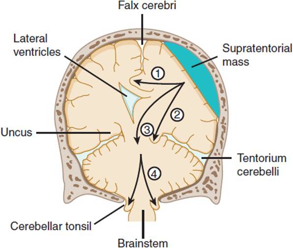
FIGURE 7-27 Anatomic basis of herniation syndromes. An expanding supratentorial mass lesion may cause brain tissue to be displaced into an adjacent intracranial compartment, resulting in (1) cingulate herniation under the falx cerebri, (2) downward transtentorial (central) herniation, (3) uncal herniation over the edge of the tentorium, or (4) cerebellar tonsillar herniation into the foramen magnum. Coma and ultimately death result when (2), (3), or (4) produces brainstem compression. (Redrawn, with permission, from Greenberg DA et al, eds. Clinical Neurology, 5th ed. McGraw-Hill, 2002.)
Several nonstructural disorders that diffusely disturb brain function can produce a confusional state or, if severe, coma (Table 7-1). Most of these disorders are acute, and many, particularly those caused by drugs and metabolic toxins, are reversible. Clues to the cause of these “metabolic” encephalopathies are provided by general physical examination, drug screens, and certain blood studies. When these disorders cause coma, pupillary light responses are usually preserved despite impaired oculovestibular or respiratory function. This finding is of great help in distinguishing metabolic from structural causes of coma.
TABLE 7-1 Nonstructural causes of confusional states and coma.

Neurons in the dorsal midbrain and especially nuclei within the pontine reticular formation are important for sleep. Thus, lesions involving the pons may preserve consciousness but disturb sleep. In contrast, diffuse lesions of the neocortex, such as those resulting from global cerebral ischemia, may preserve the reticular activating system and brainstem sleep centers, resulting in a patient with preserved sleep-wake cycles who cannot interact in any meaningful way with the environment (coma vigil or apallic state).
B. Cognition
Several disorders disturb cognition rather than the level of consciousness. Specific cortical regions generally mediate different cognitive functions, although there is considerable overlap and interconnection between cortical and subcortical structures in all mental tasks. When several of these abilities are impaired, the patient is said to suffer from dementia. Dementia is discussed in more detail later in this chapter.
The prefrontal cortex (Figure 7-9) generally refers to areas 9, 10, 11, 12, 45, 46, and 47 of Brodmann on the superior and lateral surfaces of the frontal lobes and the anterior cingulate, parolfactory, and orbitofrontal cortex inferiorly and mesially. These regions are essential for orderly planning and sequencing of complex behaviors, attending to several stimuli or ideas simultaneously, concentrating and flexibly altering the focus of concentration, grasping the context and meaning of information, and controlling impulses, emotions, and thought sequences. Damage to the frontal lobes or connections to the caudate and dorsal medial nuclei of the thalamus causes the frontal lobe syndrome. Patients may suffer dramatic alterations in personality and behavior, whereas most sensorimotor functions remain intact. Some patients become vulgar in speech, slovenly, grandiose, and irascible, whereas others lose interest, spontaneity, curiosity, and initiative. The affect may become apathetic and blunted (abulia). Some patients lose the capacity for creativity and abstract reasoning and the ability to solve problems while becoming excessively concrete in their thinking. Often they are distractible and unable to focus attention when presented with multiple stimuli. The most dramatic manifestations are seen after bilateral frontal lobe damage; unilateral damage can lead to subtle alterations in behavior that may be difficult to detect. Involvement of premotor areas may lead to incontinence, inability to perform learned motor tasks (apraxia), variable increases in muscle tone (paratonia), and appearance of primitive grasp and oral reflexes (sucking, snouting, and rooting).
In about 90% of people, language is a function of the left hemisphere. Whereas 99% of right-handed people are left hemisphere dominant, about 40% of left-handed people are right hemisphere dominant for language. In most left-handed people, hemispheric dominance for language is incomplete, and damage to the dominant hemisphere tends to disturb language less severely than in right-handed individuals. The cortical regions most critical for language include Broca area (area 44), Wernicke area (area 22), the primary auditory cortex (areas 41 and 42), and neighboring frontal and temporoparietal association areas (Figure 7-9). Injury to these areas or their connections to other cortical regions result in aphasia. Lesions in the frontal speech areas cause nonfluent, dysarthric, halting speech, whereas lesions of the temporal speech area cause fluent speech that contains many errors or may be totally devoid of understandable words. Patients with damage to temporal speech areas also lack comprehension of spoken words. Isolation of the temporal speech area from the occipital lobes causes an inability to read (alexia). Portions of the parietal lobe adjacent to the temporal lobe are important for retrieval of previously learned words, and damage here may result in anomia. The inferior parietal region is important for the translation of linguistic messages generated in the temporal language areas into visual symbols. Damage to this region may result in an inability to write (agraphia).
Memory requires that information be registered by the primary somatosensory, auditory, or visual cortex. Posterior cortical areas involved in comprehension of language are needed for immediate processing of spoken or written events and recalling them immediately. The hippocampi and their connections to the dorsal medial nuclei of the thalamus and the mammillary nuclei of the hypothalamus constitute a limbic system network crucial for learning and processing of events for long-term storage. When these areas are damaged, the patient is unable to learn new material or retrieve memories from the recent past. The most severe symptoms occur with bilateral lesions; unilateral disease causes more subtle learning deficits. Memories that remain with a person for years are considered remote memories and are stored in corresponding association cortex areas (eg, visual cortex for scenes). Remote memories remain intact in patients with damage to limbic structures required for learning. However, they may be lost by damage to cortical association areas. Understanding the mechanisms by which recent memories are transferred from the limbic memory network to association cortex for long-term storage is a major goal of current research.
The parietal association cortex is the region principally involved in visuomotor integration of constructional tasks. The visual cortex is required for observation, whereas the auditory cortex and the temporal language cortex are necessary for drawing objects on command. The inferior parietal cortex (areas 39 and 40) integrates visual and auditory information, and the output from this region is translated into motor patterns by motor cortex. Thus, lesions to the parietal lobes commonly cause constructional impairment. Damage to either hemisphere may result in constructional errors. Drawings may show rotation of objects, disorientation of objects on the background, fragmentation of design, inability to draw angles properly, or omission of parts of a figure presented for copying. It is often difficult to determine which side is damaged, although if language is preserved, a nondominant parietal deficit is more likely.
Calculation ability, abstract reasoning, problem solving, and several other aspects of intelligence are difficult to localize because they require integration of several cortical regions. They are frequently disturbed by diseases that cause widespread cortical dysfunction, such as those that cause dementia.
CHECKPOINT
25. What is the network of neurons that maintain normal arousal and consciousness?
26. What are the symptoms and signs of cerebral herniation caused by focal brain lesions?
27. Which cognitive functions are controlled by the frontal lobes and by the parietal association cortex?
28. What regions of the cortex are important for language and memory?
PATHOPHYSIOLOGY OF SELECTED NEUROLOGIC DISORDERS
Nervous system disease may be caused by a wide variety of degenerative, metabolic, structural, neoplastic, or inflammatory conditions that affect neurons, glia, or both. The resultant dysfunction is expressed by either neuronal hyperactivity, as seen during seizures, or decreased activity of neurons, as observed after a stroke. The specific functional abnormalities observed depend on the network of neurons affected. For example, because amyotrophic lateral sclerosis is a disorder of upper and lower motor neurons, neurologic deficits are limited to the motor system. In Parkinson disease, dopaminergic neurons of the substantia nigra degenerate, causing symptoms of extrapyramidal motor system dysfunction. In patients with ischemic stroke, the particular constellation of deficits is determined by the vascular territory affected. Therefore, an understanding of the pathophysiology of neurologic disease requires an analysis of events occurring at both the cellular level and the level of neural networks.
MOTOR NEURON DISEASE
Clinical Presentation
Motor neuron diseases predominantly affect the anterior horn cells of the spinal cord and are characterized by wasting and weakness of skeletal muscles. Spontaneous discharges of degenerating motor nerve fibers occur, giving rise to muscle twitches known as fasciculations (see prior discussion). Electromyography characteristically shows features of denervation, including increased numbers of spontaneous discharges (fibrillations) in resting muscle and a reduction in the number of motor units detected during voluntary contraction. Sprouting of remaining healthy motor fibers may occur, leading to the appearance of large, polyphasic motor unit potentials (reinnervation).
The spinal muscular atrophies (SMAs) are a heterogeneous group of genetic diseases characterized by selective degeneration of lower motor neurons. The most common form is autosomal recessive with childhood onset and has a frequency of between 1:6,000 and 1:10,000. Childhood SMA has been divided into three types depending on age of onset and clinical progression. SMA I is infantile spinal muscular atrophy (Werdnig-Hoffman disease), a disorder that manifests usually within the first 3 months of life. Infants with this condition have difficulty sucking, swallowing, and breathing. Atrophy and fasciculations are found in the tongue and limb muscles. SMA I is rapidly progressive, leading to death from respiratory complications usually by age 3. SMA II begins in the latter half of the first year of life. It progresses more slowly than the infantile form, and patients may survive into adulthood. SMA III (Kugelberg-Welander disease) is a juvenile form that develops after age 2. Patients develop weakness of proximal limb muscles with relative sparing of bulbar muscles. The pattern of weakness can falsely suggest a myopathy such as limb-girdle dystrophy rather than a motor neuron disease. The course is gradually progressive, leading to disability in adulthood. All three forms of SMA are due to deletions or mutations in the survival motor neuron 1 (SMN1) gene on chromosome 5q13. The SMN gene product is expressed in all tissues and appears to be involved in RNA metabolism. Loss of SMN function promotes apoptosis of lower motor neurons. It is not yet known why motor neurons are selectively affected. Recent clinical trials were aimed at adjusting the levels of the SMN protein to try to modulate disease progression using drugs such as hydroxyurea and valproic acid, but unfortunately these studies failed to show any improvement in the disease. Recent focus has turned to antisense oligonucleotides and stem cell therapies to attempt to slow disease progression.
In adults, motor neuron disease usually begins between the ages of 20 and 80 years, with an average age at onset of 56 years. It is commonly sporadic but is familial in up to 10% of cases. Several varieties have been described, depending on relative involvement of upper or lower motor neurons and bulbar or spinal anterior horn cells. For example, X-linked spinobulbar atrophy is an X-linked recessive disorder that typically manifests clinically in the fourth or fifth decade and is associated with an expanded CAG repeat in the androgen receptor gene. As with other genetic disorders associated with triplet repeat expansions, the neurodegeneration is associated with neuronal inclusions. Testosterone promotes the development of inclusions, and women homozygous for the mutation develop only mild symptoms. Moreover, female mice carrying the mutation show motor impairment after testosterone administration, whereas castration reduces impairment in male mice. These findings led to testing of gonadotropin-releasing hormone antagonists, which reduce testosterone release from the testes, as treatments for the disease. Unfortunately, the treatments did not improve function and resulted in significantly reduced quality of life secondary to the low testosterone. Current work is focusing on RNAi targeting of the polyQ-AR transcript to reduce the expression and toxicity of the expanded repeat.
The most common form of motor neuron disease in adults is amyotrophic lateral sclerosis (ALS), in which mixed upper and lower motor neuron deficits are found in limb and bulbar muscles. In 80% of patients, the initial symptoms are due to weakness of limb muscles. Complaints are often bilateral but asymmetric. Involvement of bulbar muscles causes difficulty with swallowing, chewing, speaking, breathing, and coughing. Neurologic examination reveals a mixture of upper and lower motor neuron signs. There is usually no involvement of extraocular muscles or sphincters. The disease is progressive and generally fatal within 3–5 years, with death usually resulting from pulmonary infection and respiratory failure.
Pathology & Pathogenesis
In ALS, there is selective degeneration of motor neurons in the primary motor cortex and the anterolateral horns of the spinal cord. Many affected neurons show cytoskeletal disease with accumulations of intermediate filaments in the cell body and in axons. There is only a subtle glial cell response and little evidence of inflammation. The cause is unknown, but biochemical and genetic studies have provided several clues.
A. Glutamate Signaling and RNA Processing
Glutamate (Figure 7-28) is the most abundant excitatory neurotransmitter in the CNS. Glutamate activates a large family of receptors that either open cation channels (ionotropic receptors) or activate phospholipase C (metabotropic receptors), which catalyzes the formation of the second messenger, inositol-1,4,5-trisphosphate (IP3). Influx of Na+ and Ca2+ through glutamate-gated cation channels depolarizes cells, whereas IP3 stimulates release of Ca2+ from intracellular storage sites. The net effect of these events is to generate an excitatory postsynaptic potential and raise the concentration of free intracellular Ca2+ in the cytosol of the postsynaptic neuron. This Ca2+ signal activates calcium-sensitive enzymes and is quickly terminated by removal of glutamate from the synapse and by mechanisms for calcium sequestration and extrusion in the postsynaptic cell. Breakdown of normal mechanisms for terminating the excitatory signal leads to sustained elevations of intracellular Ca2+ that cause cell death.

FIGURE 7-28 Glutamatergic neurotransmission. Depolarization stimulates release of glutamate from presynaptic terminals into the synaptic cleft, where it binds to ionotropic or metabotropic glutamate receptors, stimulating Ca2+influx and activation of phospholipase C (PLC). PLC catalyzes hydrolysis of phosphatidylinositol-4,5-bisphosphate (PIP2) to produce inositol-1,4,5-trisphosphate (IP3), which causes release of Ca2+ from storage sites in smooth endoplasmic reticulum (SER). Synaptic actions of glutamate are terminated mainly by uptake through Na+-dependent glutamate transporters (GT) on glia. In astrocytes, glutamate is converted into glutamine by glutamine synthetase.
Glutamate is removed from synapses by transport proteins on surrounding astrocytes and nerve terminals. In astrocytes, it is metabolized to glutamine and can be shuttled back to neurons for reconversion into glutamate. In 60% of patients with sporadic ALS, there is a large decrease in glutamate transport activity in the motor cortex and spinal cord but not in other regions of the CNS. This has been associated with a loss of the astrocytic glutamate transporter protein excitatory amino acid transporter 2 (EAAT2), perhaps resulting from a defect in splicing of its messenger RNA. In cultured spinal cord slices, pharmacologic inhibition of glutamate transport induces motor neuron degeneration. Thus, selective loss of a glutamate transporter may cause excitotoxicity in ALS by increasing extracellular levels of glutamate.
A second alteration in glutamate signaling has been found recently in spinal motor neurons from five patients with ALS. RNA editing is a process whereby gene-specified codons are altered by RNA-dependent deaminases. In GluR2 receptor subunits, this process is virtually 100% efficient, resulting in conversion of a glutamine to arginine in the second transmembrane domain of this subunit, which markedly reduces the calcium permeability of a major subclass of glutamate receptors. Editing efficiency was reduced in more than 50% of neurons from the patients with ALS. Because transgenic mice that express GluR2 made artificially more permeable to calcium develop a motor neuron disease late in life, it is possible that defective editing of GluR2 contributes to ALS pathogenesis. These findings suggest that sporadic ALS may be caused by a defect in RNA metabolism.
B. Free Radicals
About 10% of ALS cases are familial and 20% of these familial cases are due to missense mutations in the cytosolic copper-zinc superoxide dismutase (SOD1) gene on the long arm of chromosome 21. SOD1 catalyzes the formation of hydrogen peroxide from superoxide anion. Hydrogen peroxide is then detoxified by catalase or glutathione peroxidase to form water. Not all mutations reduce SOD1 activity, and the disorder is typically inherited as an autosomal dominant trait, suggesting that familial ALS results from a gain rather than a loss of function. This is supported by the finding that transgenic mice expressing mutant SOD1 develop motor neuron disease analogous to human familial ALS, whereas mice lacking SOD1 do not develop motor neuron disease. One hypothesis suggests that the mutant enzyme has an altered substrate specificity catalyzing the reduction of hydrogen peroxide to yield hydroxyl radicals and utilizing peroxynitrite to produce nitration of tyrosine residues in proteins. This is consistent with elevated levels of carbonyl proteins in the brain and elevated levels of free nitrotyrosine in the spinal cord of ALS patients. EAAT2 may also be inactivated by mutant SOD1, thereby promoting excitotoxicity. Some mutations also promote the formation of SOD aggregates, which may be neurotoxic.
C. Cytoskeletal Proteins
Motor neurons tend to be very large, with extremely long axons, and cytoskeletal proteins that maintain axonal structure may be critical targets for motor neuron injury. A role for neurofilament dysfunction in ALS is supported by the finding that neurofilamentous inclusions in cell bodies and proximal axons are an early feature of ALS pathology. In addition, mutations in the heavy chain neurofilament subunit (NF-H) have been detected in some patients with sporadic ALS, suggesting that NF-H variants may be a risk factor for ALS. Peripherin is another intermediate filament protein found with neurofilaments in neuronal inclusions in ALS and in mice with SOD1 mutations. Peripherin expression is increased in response to cell injury, and overexpression of peripherin causes a late-onset motor neuron disease in mice. Inclusions containing peripherin and neurofilaments may interfere with axonal transport, resulting in failure to maintain axonal structure and transport of macromolecules such as neurotrophic factors required for motor neuron survival.
D. TDP-43
An exciting discovery of the protein transactive response DNA-binding protein 43 (TDP-43) may offer new clues to the etiology of this disorder. This newly discovered protein is the major component of the ubiquitinated, tau-negative, inclusions that are the pathological hallmark of sporadic and familial ALS and frontotemporal dementia (FTD). It is also found in some cases of Alzheimer disease and Parkinson disease. Mutations in this gene, which is located on chromosome 1, co-segregate with disease in familial forms of ALS and FTD and are not found in SOD1 familial ALS. FTD and ALS overlap in approximately 15–25% of cases and these disorders are starting to be referred to as “TDP-43 proteinopathies.” Several other genes and gene regions have been identified to cause both FTD and ALS such as TARDBP on chromosome 1p36.2, MAPT on chromosome 7q21, and DCTN1 on chromosome 2p13.
E. C9ORF72
The major genetic cause of ALS and/or FTD was recently discovered. Two independent groups identified hexanucleotide repeats in an intron of C9ORF72 on chromosome 9 in 34% of familial ALS cases, 6% of sporadic ALS cases, 26% of familial FTD cases, and 5% of sporadic FTD cases. The protein is of unknown function. These mutations likely induce a gain-of-function mutation similar to other noncoding repeat expansion disorders. This discovery of another disorder caused by nucleotide repeats may provide additional rationale for a new drug development paradigm focused on decreasing expression of these toxic repeats.
CHECKPOINT
29. What are the clinical features of motor neuron disease?
30. What gene is responsible for some cases of familial ALS, and what is a postulated molecular mechanism by which the mutation causes disease?
31. What two other mechanisms may play a role in motor neuron degeneration?
PARKINSON DISEASE
Clinical Presentation
Parkinsonism is a clinical syndrome of rigidity, bradykinesia, tremor, and postural instability. Most cases are due to Parkinson disease, an idiopathic disorder with a prevalence of about 1–2 per 1000. In the first half of the last century, parkinsonism was a common sequela of von Economo encephalitis. Parkinsonism can also result from exposure to certain toxins such as manganese, carbon disulfide, 1-methyl-4-phenyl-1,2,3,6-tetrahydropyridine (MPTP), and carbon monoxide. Several drugs, particularly butyrophenones, phenothiazines, metoclopramide, reserpine, and tetrabenazine, can cause reversible parkinsonism. Parkinsonism may also result from repeated head trauma or may be a feature of several basal ganglia diseases, including Wilson disease, some cases of early onset Huntington disease, Shy-Drager syndrome, striatonigral degeneration, and progressive supranuclear palsy. In these disorders, other symptoms and signs are present along with parkinsonism.
Pathology & Pathogenesis
In Parkinson disease, there is selective degeneration of monoamine-containing cell populations in the brainstem and basal ganglia, particularly of pigmented dopaminergic neurons of the substantia nigra. In addition, scattered neurons in basal ganglia, brainstem, spinal cord, and sympathetic ganglia contain eosinophilic, cytoplasmic inclusion bodies (Lewy bodies). These contain filamentous aggregates of α-synuclein, along with parkin, synphilin, neurofilaments, and synaptic vesicle proteins.
Important clues about the pathogenesis of Parkinson disease have been discovered through study of the potent neurotoxin MPTP. MPTP is a by-product of synthesis of a synthetic opioid derivative of meperidine. Illicit use of opioid preparations heavily contaminated with MPTP led to several cases of parkinsonism in the early 1980s. MPTP selectively injures dopaminergic neurons in the brain and produces a clinical syndrome very similar to Parkinson disease.
MPTP enters the brain (Figure 7-29) and is converted by monoamine oxidase B present in glia and serotonergic nerve terminals to N-methyl-4-phenyldihydropyridine (MPDP+), which diffuses across glial membranes and then undergoes nonenzymatic oxidation and reduction to the active metabolite N-methyl-4-phenylpyridinium (MPP+). Plasma membrane transporters that normally act to terminate the action of monoamines by removing them from synapses take up MPP+. Internalized MPP+ inhibits oxidative phosphorylation by interacting with complex I of the mitochondrial electron transport chain. This inhibits ATP production and reduces metabolism of molecular oxygen, allowing for increased formation of peroxide, hydroxyl radicals, and superoxide radicals that react with lipids, proteins, and nucleic acids that cause cell injury. In support of a role for mitochondrial dysfunction and oxidative damage in the pathogenesis of Parkinson disease is evidence that the insecticide rotenone, which inhibits mitochondrial complex I, produces parkinsonism in animals with degeneration of nigrostriatal dopaminergic neurons and cytoplasmic inclusions that resemble Lewy bodies. Exposure to paraquat, a common herbicide that is structurally similar to MPP+ and also inhibits complex I, can lead to selective degeneration of dopaminergic neurons and aggregation of α-synuclein. Furthermore, impaired complex I activity has been observed in cell lines derived from Parkinson disease patients, and one genetic variant of NADH dehydrogenase 3 in complex I is associated with a reduced risk of the disease among Caucasians. Thus, alterations in mitochondrial complex I activity appear to play an important role in the pathogenesis of Parkinson disease.
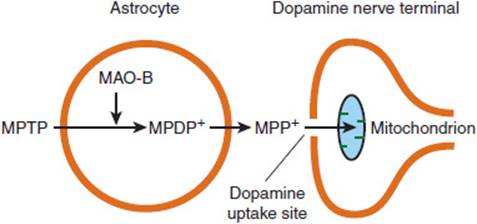
FIGURE 7-29 Proposed mechanism of MPTP-induced parkinsonism. MPTP enters brain astrocytes and is converted to MPDP+ through the action of monoamine oxidase type B (MAO-B). MPDP+ is then metabolized extracellularly to MPP+, which is taken up through dopamine uptake sites on dopamine nerve terminals and concentrated in mitochondria. The resulting disturbance of mitochondrial function can lead to neuronal death. (Redrawn, with permission, from Greenberg DA et al, eds. Clinical Neurology, 5th ed. McGraw-Hill, 2002.)
The reasons why dopaminergic neurons appear selectively vulnerable to complex I inhibition are not clear. Although controversial, some evidence suggests that dopamine can promote neurotoxicity. Addition of exogenous dopamine is toxic to neurons in culture. Dopamine undergoes autooxidation to generate superoxide radicals or is metabolized by monoamine oxidase to generate hydrogen peroxide. Superoxide dismutase catalyzes the conversion of superoxide to H2O2, which is converted by glutathione peroxidase and catalase to water. However, H2O2 can also react with ferrous iron to form highly reactive hydroxyl radicals. Thus, dopamine within dopaminergic neurons may provide a source of reactive oxygen species, which, when coupled with reduced complex I function, may promote cell death.
Approximately 5% of Parkinson disease cases are familial. Genetic studies have identified causative mutations in five genes that provide important information about molecular pathways involved in the disease. These genes include the genes for α-synuclein (PARK1), parkin (PARK2), DJ-1 (PARK7), ubiquitin-C-hydrolase-L1 (PARK5), PTEN (phosphatase and tensin homolog deleted on chromosome 10)-induced kinase 1 (PINK1), and leucine-rich repeat kinase 2 (LRRK2).
Mutations in the gene for α-synuclein on chromosome 4q21-23 cause autosomal dominant Parkinson disease. Even in sporadic disease, α-synuclein is the largest single genetic risk factor. Alpha-synuclein is found in nerve terminals in close proximity to synaptic vesicles. Its normal function is not known. Overexpression of nonmutant human α-synuclein in transgenic mice results in formation of Lewy bodies, reduced dopaminergic terminals in the striatum, and impaired motor performance due to formation of abnormal complexes at the synapse with SNARE proteins. Genomic triplication of α-synuclein leading to overexpression has been documented in a human family with autosomal dominant Parkinson disease. This suggests that it is the production of neuronal inclusions containing α-synuclein rather than a change in α-synuclein function that contributes to degeneration of dopaminergic neurons. Interestingly, mice lacking α-synuclein are resistant to the toxic effects of the complex I inhibitor MPTP, suggesting that mitochondrial dysfunction generates an environment that favors α-synuclein aggregation and neurodegeneration.
Misfolded, damaged, or unassembled proteins are generally degraded by a process involving covalent attachment of ubiquitin. Ubiquitin is a 76-residue protein that marks proteins for processing by a proteolytic complex (proteasome). A missense mutation in one component of the ubiquitin-proteasome system, ubiquitin carboxyl terminal hydrolase L1, has been found in one family with autosomal dominant Parkinson disease. Mutations in parkin on chromosome 6q25 have been identified in cases of autosomal recessive juvenile parkinsonism. Parkin is a ubiquitin E3 ligase that catalyzes the addition of ubiquitin to specific proteins to target them for degradation. Known mutations cause loss of function, which presumably leads to a disturbance in protein degradation. However, most patients with parkinmutations lack Lewy bodies, suggesting that other mechanisms, such as increased oxidative stress, cause neurodegeneration in these patients. In support of this mechanism is the finding that Drosophilamutants that lack parkin show mitochondrial pathology.
The most common known genetic form of Parkinson disease was recently discovered. Mutations in the glucocerebrosidase (GCase) enzyme account for 3% of sporadic Parkinson disease cases and 25% of juvenile-onset Parkinson disease cases. This enzyme is involved in lysosomal processing. The enzyme activity is reduced by 58% in the substantia nigra of heterozygous patients and 33% lower in patients with sporadic Parkinson disease. Inhibiting this enzyme leads to accumulation of α-synuclein, which leads to further inhibition of this enzyme.
CHECKPOINT
32. What are the clinical features of parkinsonism?
33. What are some of the causes of this syndrome?
34. What are two major mechanisms proposed to explain the pathophysiology of Parkinson disease?
MYASTHENIA GRAVIS
Clinical Presentation
Myasthenia gravis is an autoimmune disorder of neuromuscular transmission. The major clinical features are fluctuating fatigue and weakness that improve after a period of rest and after administration of acetylcholinesterase inhibitors. Muscles with small motor units, such as ocular muscles, are most often affected. Oropharyngeal muscles, flexors and extensors of the neck, proximal limb muscles, and the erector spinae muscles are involved less often. In severe cases, all muscles are weak, including the diaphragm and intercostal muscles, and death may result from respiratory failure.
About 5% of patients have coexistent hyperthyroidism. Rheumatoid arthritis, systemic lupus erythematosus, and polymyositis are also more common in patients with myasthenia gravis than in the general population, and up to 30% of patients have a maternal relative with an autoimmune disorder. These associations suggest that patients with myasthenia gravis share a genetic predisposition to autoimmune disease.
Pathology & Pathogenesis
The major structural abnormality in myasthenia gravis is a simplification of the postsynaptic region of the neuromuscular synapse. The muscle end plate shows sparse, shallow, and abnormally wide or absent synaptic clefts. In contrast, the number and size of the presynaptic vesicles are normal. Scattered collections of lymphocytes, some within the vicinity of motor end plates, may be present. IgG and the C3 component of complement are present at the postsynaptic membrane.
Electrophysiologic studies indicate that the postsynaptic membrane has a decreased response to applied acetylcholine. Studies with iodine-125–labeled α-bungarotoxin, which binds with high affinity to muscle nicotinic AChRs, show a 70–90% decrease in the number of receptors per end plate in affected muscles. Circulating antibodies to the receptor are present in 90% of patients, and the disorder may be passively transferred to animals by administration of IgG from affected patients. Moreover, immunization with AChR protein from muscle can produce myasthenia in experimental animals. The antibodies block acetylcholine binding and receptor activation (Figure 7-30). In addition, the antibodies cross-link receptor molecules, increasing receptor internalization and degradation. Bound antibody also activates complement-mediated destruction of the postsynaptic region, resulting in simplification of the end plate. Many patients who lack antibodies to the AChR have autoantibodies instead against the muscle-specific receptor tyrosine kinase (MuSK), which is an important mediator of AChR clustering at the end plate. These antibodies inhibit clustering of receptors in muscle cell culture.
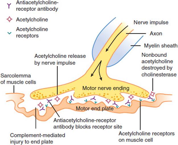
FIGURE 7-30 Pathogenesis of myasthenia gravis. Acetylcholine released at the nerve ending by the nerve impulse normally binds with acetylcholine receptors. This evokes the action potential in the muscle. In myasthenia gravis, antiacetylcholine receptor antibody binds to the acetylcholine receptor and inhibits the action of acetylcholine. Bound antibody evokes immune-mediated destruction of the end plate. (Redrawn, with permission, from Chandrasoma P et al, eds. Concise Pathology, 3rd ed. Originally published by Appleton & Lange. Copyright © 1998 by The McGraw-Hill Companies, Inc.)
During repetitive stimulation of a motor nerve, the number of quanta released from the nerve terminal declines with successive stimuli. Normally, this causes no clinical impairment because a sufficient number of AChR channels are opened by the reduced level of neurotransmitter. However, in myasthenia gravis, where there is a deficiency in the number of functional receptors, neuromuscular transmission fails at lower levels of quantal release. Electrophysiologically, this is measured as a decremental decline in the compound muscle action potential during repetitive stimulation of a motor nerve. Clinically, this is manifested by muscle fatigue with sustained or repeated activity.
Treatment has reduced the mortality rate from approximately 30% to 5% in generalized myasthenia gravis. The two basic strategies for treatment that stem from knowledge of the pathogenesis are to increase the amount of acetylcholine at the neuromuscular junction and to inhibit immune-mediated destruction of AChRs.
By preventing metabolism of acetylcholine, cholinesterase inhibitors can compensate for the normal decline in released neurotransmitter during repeated stimulation. Therapy with cholinesterase inhibitors can also cause a paradoxical increase in weakness known as a cholinergic crisis. This is due to an excess of acetylcholine. At the molecular level, binding of acetylcholine first opens nicotinic cation channels, but with continued exposure to the agonist the channels desensitize and shut down again. The desensitized channels recover their sensitivity to acetylcholine only after the neurotransmitter is removed. Removal of acetylcholine is impaired when cholinesterase activity is inhibited. This can result in depolarization block of neurotransmission similar to the effect of the depolarizing paralytic agent succinylcholine or organophosphate insecticides and nerve gases that markedly inhibit acetylcholinesterase. Therefore, the dose of cholinesterase inhibitors must be carefully regulated to reduce myasthenia but avoid a cholinergic crisis.
Plasmapheresis, corticosteroids, and immunosuppressant drugs are effective in reducing levels of autoantibody to AChRs and suppressing disease. The thymus is thought to play an important role in the pathogenesis of the disease by supplying helper T cells sensitized against thymic proteins that cross-react with AChRs. In most patients with myasthenia gravis, the thymus is hyperplastic, and 10–15% have thymomas. Thymectomy is indicated if a thymoma is suspected. In patients with generalized myasthenia without thymoma, thymectomy induces remission in 35% and improves symptoms in another 45% of patients.
For patients with AChR antibody–negative myasthenia gravis who test positive for the MuSK antibody, the clinical features and treatment are different. Patients tend to be younger women with bulbar weakness, and muscle atrophy is often seen, particularly in the tongue, making it difficult to distinguish from motor neuron disease. Results of repetitive stimulation studies and single-fiber EMG studies in the limbs are often normal, necessitating facial studies to make a diagnosis. Cholinesterase inhibitors often make these patients worse, but plasma exchange is very effective, as is less conventional immunosuppressive therapy. Thymectomy is not clearly beneficial in this population.
Lastly, there are myasthenia gravis patients with no antibodies for either AChR antibodies or MuSK, referred to as double sero-negative patients. Recently, a new antibody has been found in 50% of these patients. Antibodies to lipoprotein-related protein 4 (LRP4), which is the agrin-binding receptor of the MuSK complex, disrupt agrin-induced AChR clustering, causing the disease symptoms. The clinical presentation of these patients is similar to that of those with AChR-myasthenia gravis without thymoma.
CHECKPOINT
35. What is the clinical presentation of myasthenia gravis?
36. What causes this disorder?
37. What is the pathophysiology of symptoms in myasthenia gravis?
EPILEPSY
Clinical Presentation
Seizures are paroxysmal disturbances in cerebral function caused by an abnormal synchronous discharge of cortical neurons. The epilepsies are a group of disorders characterized by recurrent seizures. Approximately 0.6% of people in the United States suffer from recurrent seizures, and idiopathic epilepsy accounts for more than 75% of all seizure disorders. In some forms of idiopathic epilepsy, a genetic basis is apparent. Other forms are secondary to brain injury from stroke, trauma, a mass lesion, or infection. About two thirds of new cases arise in children, and most of these cases are idiopathic or caused by trauma. In contrast, seizures or epilepsy with onset in adult life is more often due to underlying brain lesions or metabolic causes.
Seizures are classified by behavioral and electrophysiologic data (Table 7-2). Generalized tonic-clonic seizures are attacks characterized by sudden loss of consciousness followed rapidly by tonic contraction of muscles, causing limb extension and arching of the back. The tonic phase lasts 10–30 seconds and is followed by a clonic phase of limb jerking. The jerking builds in frequency to a peak after 15–30 seconds and then slows gradually over another 15–30 seconds. Thereafter, the patient remains unconscious for several minutes. As consciousness is regained, there is a period of postictal confusion lasting several minutes. In patients with recurrent seizures or an underlying structural or metabolic abnormality, confusion may persist for a few hours. Focal abnormalities may be present on neurologic examination during the postictal period. Such findings suggest a focal brain lesion requiring further laboratory and radiologic study.
TABLE 7-2 Simplified classification of seizures.

Typical absence seizures begin in childhood and usually remit by adulthood. Seizures are characterized by brief lapses in consciousness lasting several seconds without loss of posture. These spells may be associated with eyelid blinking, slight head movement, or brief jerks of limb muscles. Immediately after the seizure, the patient is fully alert. The spells may occur several times through the day and impair school performance. The electroencephalogram (EEG) shows characteristic runs of spikes and waves at a rate of three per second, particularly after hyperventilation (Figure 7-31). The disorder is transmitted as an autosomal dominant trait with incomplete penetrance.

FIGURE 7-31 EEG of a patient with typical absence (petit mal) seizures, showing a burst of generalized 3-Hz spike-and-wave activity (center of record) that is bilaterally symmetric and bisynchronous. Odd-numbered leads indicate electrode placements over the left side of the head; even numbers, those over the right side. (Redrawn, with permission, from Greenberg DA et al, eds. Clinical Neurology, 8th ed. McGraw-Hill, 2012.)
Some forms of epilepsy cause seizures with only a tonic or clonic phase. In others, the seizure is manifested by sudden loss of muscle tone (atonic seizures). In myoclonic epilepsy, sudden, brief contractions of muscles occur. Myoclonic seizures are found in certain neurodegenerative diseases or after diffuse brain injury, as occurs during global cerebral ischemia.
Focal seizures are caused by focal brain disease. Therefore, in general, patients with simple or focal dyscognitive seizures should be investigated for underlying brain lesions. Simple focal seizures begin with motor, sensory, visual, psychic, or autonomic phenomena depending on the location of the seizure focus. Consciousness is preserved unless the seizure discharge spreads to other areas, producing a tonic-clonic seizure (secondary generalization). Focal dyscognitive seizures are characterized by the sudden onset of impaired consciousness with stereotyped, coordinated, involuntary movements (automatisms). Immediately before impairment of consciousness, there may be an aura consisting of unusual abdominal sensations, olfactory or sensory hallucinations, unexplained fear, or illusions of familiarity (déjà vu). Seizures usually last for 2–5 minutes and are followed by postictal confusion. Secondary generalization may occur. The seizure focus usually lies in the temporal or frontal lobe.
Pathogenesis
Normal neuronal activity occurs in a nonsynchronized manner, with groups of neurons inhibited and excited sequentially during the transfer of information between different brain areas. Seizures occur when neurons are activated synchronously. The kind of seizure depends on the location of the abnormal activity and the pattern of spread to different parts of the brain.
Interictal spike discharges are often observed on EEG recordings from epileptic patients. These are due to synchronous depolarization of a group of neurons in an abnormally excitable area of brain. Experimentally, this is known as the paroxysmal depolarizing shift and is followed by a hyperpolarizing afterpotential that is the cellular correlate of the slow wave that follows spike discharges on the EEG. The shift is produced by depolarizing currents generated at excitatory synapses and by subsequent influx of sodium or calcium through voltage-gated channels.
Normally, discharging excitatory neurons activate nearby inhibitory interneurons that suppress the activity of the discharging cell and its neighbors. Most inhibitory synapses utilize the neurotransmitter GABA. Voltage-gated and calcium-dependent potassium currents are also activated in the discharging neuron to suppress excitability. In addition, adenosine generated from adenosine triphosphate (ATP) released during excitation further suppresses neuronal excitation by binding to adenosine receptors present on nearby neurons. Disruption of these inhibitory mechanisms by alterations in ion channels, or by injury to inhibitory neurons and synapses, may allow for the development of a seizure focus. In addition, groups of neurons may become synchronized if local excitatory circuits are enhanced by reorganization of neural networks after brain injury.
Spread of a local discharge occurs by a combination of mechanisms. During the paroxysmal depolarizing shift, extracellular potassium accumulates, depolarizing nearby neurons. Increased frequency of discharges enhances calcium influx into nerve terminals, increasing neurotransmitter release at excitatory synapses by a process known as posttetanic potentiation. This involves increased calcium influx through voltage-gated channels and through the N-methyl-d-aspartate (NMDA) subtype of glutamate receptor-gated ion channels. NMDA receptor-gated channels preferentially pass calcium ions but are relatively quiescent during normal synaptic transmission because they are blocked by magnesium ions. Magnesium block is relieved by depolarization. In contrast, the effect of inhibitory synaptic neurotransmission appears to decrease with high-frequency stimulation. This may be partly due to rapid desensitization of GABA receptors at high concentrations of released GABA. The net effect of these changes is to recruit neighboring neurons into a synchronous discharge and cause a seizure.
In secondary epilepsy, loss of inhibitory circuits and sprouting of fibers from excitatory neurons appear to be important for the generation of a seizure focus. In several of the idiopathic epilepsies, genetic studies have identified mutations in ion channels. For example, benign familial neonatal convulsions have been linked to mutations in two homologous voltage-gated K+ channels: KCNQ2 encoded by a gene on chromosome 20q13.3 and KCNQ3 encoded by a gene on chromosome 8q24. Two forms of generalized epilepsy associated with febrile seizures have been linked to mutations in voltage-gated Na+ channel subunits. Another rare condition, autosomal dominant nocturnal frontal lobe epilepsy, is associated with mutations on chromosome 20q13.2 in the gene for the α4 subunit of neuronal nicotinic cholinergic receptors. Lastly, a genome-wide association study in idiopathic generalized epilepsy revealed the first common genetic risk variants. These variants were found in genes with largely unknown pathways: CHRM3, VRK2, ZEB2, PNPO, and SCN1A.
Animal models have provided clues to the pathogenesis of absence seizures. Absence seizures arise from synchronous thalamic discharges that are mediated by activation of low-threshold calcium currents (T or “transient” currents) in thalamic neurons. The anticonvulsant ethosuximide blocks T channels and suppresses absence seizures in humans. T channels are more likely to be activated after hyperpolarization of the cell membrane. Activation of GABABreceptors hyperpolarizes thalamic neurons and facilitates T-channel activation. Lethargic (lh/lh) mice demonstrate frequent absence spells accompanied by 5- to 6-Hz spike-wave discharges on the EEG and respond to drugs used in human absence epilepsy. A single mutation in a gene on chromosome 2 results in this autosomal recessive disorder. There is an increase in the number of GABAB receptors in the cerebral cortex in these mice, and the GABABagonist baclofen worsens the seizures, whereas antagonists alleviate them. This suggests that abnormal regulation of GABAB receptor function or expression may be important in the pathogenesis of absence seizures. This is supported by the finding that γ-hydroxybutyrate, which causes behavioral and electroencephalographic alterations similar to those seen during absence attacks, activates GABAB receptors and that GABAB agonists increase and GABAB antagonists reduce spike-wave discharges in rats genetically susceptible to absence seizures (GAERS rats).
The main targets for currently available anticonvulsants are (1) voltage-gated ion channels that are involved in the generation of action potentials and in neurotransmitter release and (2) ligand-gated channels that modulate synaptic excitation and inhibition. Many agents act by more than one mechanism. Several anticonvulsants and some of their presumed mechanisms of action are listed in Table 7-3.
TABLE 7-3 Known mechanisms of action of some anticonvulsant drugs.

CHECKPOINT
38. What is the clinical presentation of the major types of seizures?
39. What are some disorders that lead to secondary epilepsy and what changes in brain structure lead to secondary epilepsy?
40. What kinds of mutations have been associated with idiopathic epilepsies?
DEMENTIA & ALZHEIMER DISEASE
1. Clinical Features of Dementia
Dementia is an acquired decline in intellectual function resulting in loss of social independence. There is impairment of memory and at least one other area of cortical function, such as language, calculation, spatial orientation, decision making, judgment, and abstract reasoning. In contrast to patients with confusional states, symptoms progress over months to years, and alertness is preserved until the very late stages of disease. Dementia affects 5–20% of persons over age 65, and, although not part of normal aging, its incidence increases with age. The most common causes, which are listed in Table 7-4, account for almost 90% of cases. Treatable causes are important to recognize and include hypothyroidism, vitamin B12 deficiency, neurosyphilis, brain tumor, normal pressure (communicating) hydrocephalus, and chronic subdural hematoma. In addition, although not curable, dementia associated with HIV infection may be slowed by antiretroviral treatment. About 10–15% of patients referred for evaluation of dementia suffer from depression (“pseudodementia”), which may also respond to treatment.
TABLE 7-4 Major causes of dementia.

Cerebrovascular disease is the second most common cause of dementia (after Alzheimer disease). Dementia results from either multiple infarctions in the territory of major cerebral vessels (multi-infarct dementia) or from subcortical infarctions in the distributions of deep penetrating arterioles (lacunar state, Binswanger disease, subcortical arteriosclerotic encephalopathy). There is usually a history of stepwise progression of neurologic deficits, focal signs on neurologic examination, and multiple infarctions on brain imaging studies. Patients generally have a history of hypertension or other risk factors for atherosclerosis.
Chronic drug intoxication is often listed as a cause of dementia but actually produces a confusional state. The existence of alcohol-induced dementia is controversial. Although animal and cell culture studies provide evidence for a direct neurotoxic effect of alcohol, dementia in alcoholic patients also results from associated nutritional deficiency, from recurrent head trauma, and (rarely) from acquired hepatocerebral degeneration, a complication of chronic hepatic insufficiency caused by alcoholic cirrhosis.
2. Alzheimer Disease
Clinical Features
Alzheimer disease is the most common cause of dementia and accounts for more than 50% of cases. It is a slowly progressive disorder that runs a course of 5–10 years and typically begins with impairment of learning and recent memory. Anomia, aphasia, and acalculia eventually develop, causing loss of employment and inability to manage finances. Spatial disorientation causes patients to become lost easily, and apraxias lead to difficulty with cooking, cleaning, and self-care. A frontal lobe gait disorder may appear, with short, shuffling steps, flexed posture, difficulty turning, and a tendency to fall backward (retropulsion) similar to that seen in Parkinson disease. In later stages, social graces are lost, and psychiatric symptoms such as paranoia, hallucinations, and delusions may appear. Cholinesterase treatments such as donepezil, rivastigmine, and galantamine may help for a couple of years to improve memory, but eventually the neuronal degeneration progresses and these medications are no longer effective. Terminally ill patients are bedridden, mute, and incontinent.
Pathology
The pathology of Alzheimer disease is characterized by extracellular neuritic plaques in the cerebral cortex and in walls of meningeal and cerebral blood vessels (Figure 7-32). These plaques contain a dense core of amyloid material surrounded by dystrophic neurites (axons, dendrites), reactive astrocytes, and microglia. Other structural changes include the formation of intraneuronal neurofibrillary tangles, neuronal and synaptic loss, reactive astrocytosis, and microglial proliferation. Controversy exists as to which features are most related to the pathogenesis of the disease. Formation of neuritic plaques is particularly characteristic for Alzheimer disease, but there is little evidence that the course or onset of disease correlates with plaque number. Neurofibrillary tangles are paired helical filaments composed of a hyperphosphorylated form of the microtubule protein tau. They are not specific for Alzheimer disease and occur in several other neurodegenerative disorders. In general, all pathologic changes are most prominent in the hippocampus, entorhinal cortex, association cortex, and basal forebrain. This accounts for the early symptoms of memory loss and disturbance of higher cortical functions, with preservation of primary sensory and motor function until later in the course.

FIGURE 7-32 Amyloid plaques in cerebral cortex in Alzheimer disease.
Pathophysiology
A. Amyloid β-Peptide—The major protein in neuritic plaques is amyloid β-peptide (Aβ), which is proteolytically derived from a membrane protein, the β-amyloid precursor protein (APP) encoded by a gene on chromosome 21q21.3-22.05. APP interacts with extracellular matrix and supports the growth of neurites in neuronal cultures. Genetic evidence implicates Aβ in the pathogenesis of Alzheimer disease. Almost all patients with trisomy 21 (Down syndrome) develop pathologic changes indistinguishable from those seen in Alzheimer disease, suggesting that having an increased copy of the APP gene increases the metabolism of APP to Aβ. About 10% of cases of Alzheimer disease are familial, with early onset (before age 65 years) and autosomal dominant inheritance. In approximately 5% of these families, Alzheimer disease is strongly linked to missense mutations immediately flanking the Aβ sequence in the APP gene. Transgenic mice expressing human APP with these mutations show elevated levels of Aβ, behavioral abnormalities, and neuritic plaques. The APP mutations result in either increased production of all forms of Aβ or mainly in the long 42-amino-acid form, Aβ42, which self-aggregates and promotes plaque formation. Aβ is toxic to cultured neurons and stimulates production of cytokines from microglial cells. Aβ also triggers the release of glutamate from glial cells and may injure neurons through excitotoxicity. This evidence links increased production of Aβ, particularly Aβ42, to Alzheimer disease and suggests that Aβ causes the neurodegeneration. Transgenic mice that express mutant forms of familial human APP develop synaptic dysfunction before plaque deposition, indicating that diffusible forms of Aβ are neurotoxic. This may explain why plaque number and disease severity correlate poorly.
B. Presenilins—The enzymatic pathways that regulate Aβ formation are critical areas of current research that may lead to new treatments. Some clues have come from analysis of additional families with Alzheimer disease. APP is cleaved at the amino terminal of the Aβ sequence by the membrane-anchored protease BACE, or beta-amyloid precursor protein cleaving enzyme, which is also known as beta-secretase. This cleavage generates a 99-amino-acid carboxyl terminal fragment. A second enzymatic activity termed γ-secretase cleaves this fragment to yield Aβ. Almost 70% of familial cases of Alzheimer disease have been linked to missense mutations in the gene PS-1/S182, which encodes a seven-trans-membrane protein (presenilin 1) on chromosome 14q24.3. Another 20% of cases have been linked to mutations in another gene, STM2 (presenilin 2), on chromosome 1q31-42. The proteins encoded by these genes are 67% identical in amino acid sequence and presumably have similar functions. Current evidence indicates that the presenilins are subunits of γ-secretase, because mutant mice lacking either presenilin show reduced γ-secretase function, and mutations designed to inhibit the predicted aspartyl protease function of presenilins eliminate γ-secretase activity. Mutant variants of presenilins associated with familial Alzheimer disease increase the production of Aβ42. This suggests that these mutations produce Alzheimer disease by selectively altering γ-secretase activity to favor production of the longer, amyloid-producing form of A. In addition, γ-secretase is important for processing Notch proteins and other substrates critical for neuronal function, and mice deficient in presenilins show deficiencies in spatial memory and synaptic plasticity. Thus, γ-secretase deficiency may contribute to neurodegeneration in patients with presenilin mutations.
C. Apolipoprotein E—The majority of patients with Alzheimer disease are older than 60 years, and in about 50% of these patients the e4 isoform of apolipoprotein E (apoE4) has been identified as a risk factor. ApoE is a 34-kDa protein that mediates the binding of lipoproteins to the low-density lipoprotein (LDL) receptor and the LDL receptor-related protein (LRP). It is synthesized and secreted by astrocytes and macrophages and is thought to be important for mobilizing lipids during normal development of the nervous system and during regeneration of peripheral nerves after injury. There are three major isoforms (apoE2, apoE3, and apoE4), which arise from different alleles (e2, e3, and e4) of a single gene on chromosome 19q13.2. The e3 allele is the most common, accounting for about 75% of all alleles, whereas e2 and e4 account for roughly 10% and 15%, respectively. The e4 allele is associated with increased risk and earlier onset of both familial and sporadic late-onset Alzheimer disease. In contrast, inheritance of e2 is associated with decreased risk and later onset. It is important to note that Alzheimer disease develops in the absence of e4 and also that many persons with e4 escape disease. Therefore, genotyping is not currently recommended as a useful genetic test.
The mechanism by which apoE alleles alter disease risk is not certain. In cultured neurons, apoE3 increases neurite outgrowth in the presence of very low-density lipoproteins, whereas apoE4 inhibits outgrowth. Alzheimer patients who are homozygous for the e4 allele have larger and denser senile plaques than patients homozygous for the e3 allele. ApoE is found in neuritic plaques, and apoE4 binds Aβ more readily than does apoE3. Therefore, apoE4 may facilitate plaque formation or reduce the clearance of Aβ from brain tissue. In addition, apoE enters neurons and binds the microtubule-associated protein tau, which is the major constituent of neurofibrillary tangles. ApoE3 binds tau much more avidly than apoE4. Binding of apoE3 to tau may prevent the formation of neurofibrillary tangles and support normal microtubule assembly required for neurite outgrowth.
CHECKPOINT
41. What are the treatable causes of dementia?
42. What are the clinical features of Alzheimer disease?
43. In which proteins are there mutations associated with familial forms of Alzheimer disease?
44. What is the association between apolipoprotein E and Alzheimer disease?
STROKE
Clinical Presentation
Stroke is a clinical syndrome characterized by the sudden onset of a focal neurologic deficit that persists for at least 24 hours and is due to an abnormality of the cerebral circulation. It is the third leading cause of death in the United States. The incidence of stroke increases with age and is higher in men than in women. Significant risk factors include hypertension, hypercholesterolemia, diabetes, smoking, heavy alcohol consumption, and oral contraceptive use. Advances in neuroimaging have had a great impact on treatment and outcomes.
Pathophysiology
A. Vascular Supply
The focal symptoms and signs that result from stroke correlate with the area of brain supplied by the affected blood vessel. Strokes may be classified into two major categories based on pathogenesis: ischemic stroke and hemorrhage (Table 7-5). In ischemic stroke, vascular occlusion interrupts blood flow to a specific brain region, producing a fairly characteristic pattern of neurologic deficits resulting from loss of functions controlled by that region. The pattern of deficits resulting from hemorrhage is less predictable because it depends on the location of the bleed and also on factors that affect the function of brain regions distant from the hemorrhage (eg, increased intracranial pressure, brain edema, compression of neighboring brain tissue, and rupture of blood into ventricles or subarachnoid space).
TABLE 7-5 Classification of stroke.
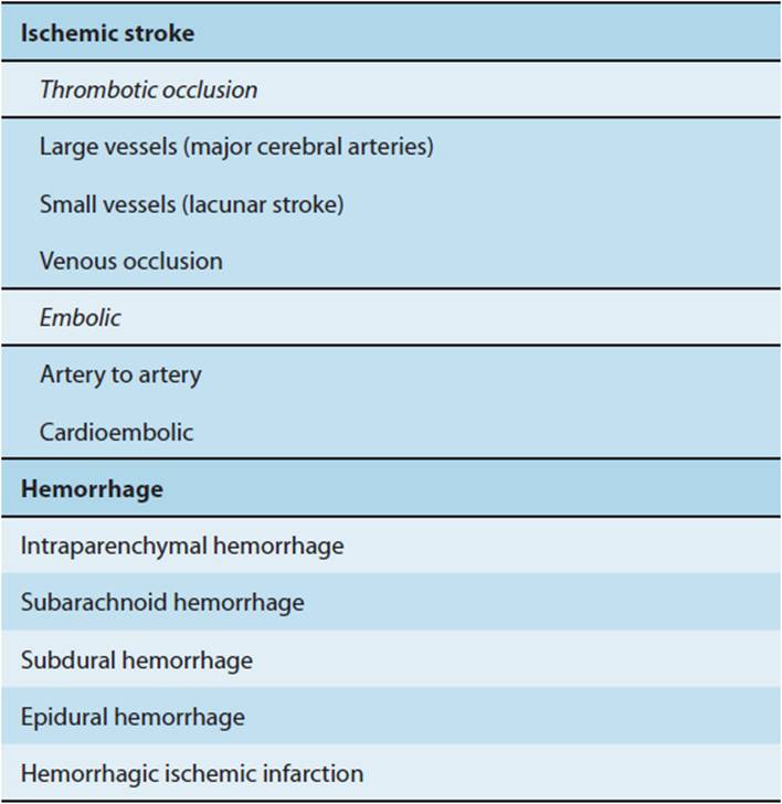
B. Ischemic Stroke
Ischemic strokes result from thrombotic or embolic occlusion of cerebral vessels. Neurologic deficits caused by occlusion of large arteries (Figure 7-33) result from focal ischemia to the area of brain supplied by the affected vessel (Figure 7–34) and produce recognizable clinical syndromes (Table 7-6). Not all signs are present in every patient, because the extent of the deficit depends on the presence of collateral blood flow, individual variations in vascular anatomy, blood pressure, and exact location of the occlusion. Thrombosis usually involves the internal carotid, middle cerebral, or basilar arteries. Symptoms typically evolve over several minutes and may be preceded by brief episodes of reversible focal deficits known as transient ischemic attacks. Emboli from the heart, aortic arch, or carotid arteries usually occlude the middle cerebral artery, because it carries more than 80% of blood flow to the cerebral hemisphere. Emboli that travel in the vertebral and basilar arteries commonly lodge at the apex of the basilar artery or in one or both posterior cerebral arteries.

FIGURE 7-33 Major cerebral arteries. A: Anterior view. B: Inferior view showing the circle of Willis and principal arteries of the brainstem.(Redrawn, with permission, from Waxman SG. Clinical Neuroanatomy, 26th ed. McGraw-Hill, 2010.)
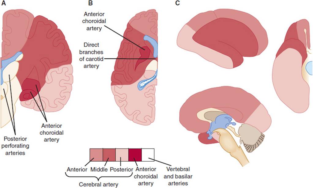
FIGURE 7-34 Vascular territories of the major cerebral arteries. A: Coronal section through the cerebrum. B: Horizontal section through the cerebrum. C: Vascular supply to the cerebral cortex. (Redrawn, with permission, from Chusid JG. Correlative Neuroanatomy and Functional Neurology, 19th ed. Originally published by Appleton & Lange. Copyright © 1985 by The McGraw-Hill Companies, Inc.)
TABLE 7-6 Vascular territories and clinical features in ischemic stroke.

Ischemic strokes involving occlusion of small arteries occur at select locations, where perfusion depends on small vessels that are end arteries. Most result from a degenerative change in the vessel, described pathologically as lipohyalinosis, that is caused by chronic hypertension and predisposes to occlusion. The most common vessels involved are the lenticulostriate arteries, which arise from the proximal middle cerebral artery and perfuse the basal ganglia and internal capsule. Also commonly affected are small branches of the basilar and posterior cerebral arteries that penetrate the brainstem and thalamus. Occlusion of these vessels causes small areas of tissue damage known as lacunar infarctions. These typically occur in the putamen, caudate, thalamus, pons, and internal capsule and less commonly in subcortical white matter and cerebellum. Lacunar infarctions produce several fairly stereotyped clinical syndromes. The two most common are pure motor stroke and pure sensory stroke. In pure motor stroke, the infarction is usually within the internal capsule or pons contralateral to the weak side. In pure sensory stroke, the infarction is usually in the contralateral thalamus.
Several vascular, cardiac, and hematologic disorders can cause focal cerebral ischemia (Table 7-7). The most common is atherosclerosis of the large arteries of the neck and base of the brain (Figure 7-35). Atherosclerosis is thought to arise from injury to vascular endothelial cells by mechanical, biochemical, or inflammatory insults (see Chapter 11). Endothelial injury stimulates attachment of circulating monocytes and lymphocytes that migrate into the vessel wall and stimulate proliferation of smooth muscle cells and fibroblasts. This leads to the formation of a fibrous plaque. Damaged endothelial cells also provide a nidus for aggregation and activation of platelets. Activated platelets secrete growth factors that encourage further proliferation of smooth muscle and fibroblasts. The plaque may eventually enlarge to occlude the vessel or may rupture, releasing emboli.
TABLE 7-7 Conditions associated with focal cerebral ischemia.


FIGURE 7-35 Sites of predilection (dark red areas) for atherosclerosis in the intracranial arterial circulation. (Redrawn, with permission, from Greenberg DA et al, eds. Clinical Neurology, 8th ed. McGraw-Hill, 2012.)
C. Hemorrhage
Epidural and subdural hematomas typically occur as sequelae of head injury. Epidural hematomas arise from damage to an artery, typically the middle meningeal artery, which can be ruptured by a blow to the temporal bone. Blood dissects the dura from the skull and compresses the hemisphere lying below. Initial loss of consciousness from the injury is due to concussion and may be transient. Neurologic symptoms then return a few hours later as the hematoma exerts a mass effect that may be severe enough to cause brain herniation (Figure 7-27). Subdural hematomas usually arise from venous blood that leaks from torn cortical veins bridging the subdural space. These may be ruptured by relatively minor trauma, particularly in the elderly. The blood is under low pressure, and symptoms resulting from mass effect may not appear for several days.
Subarachnoid hemorrhage may occur from head trauma, extension of blood from another compartment into the subarachnoid space, or rupture of an arterial aneurysm. Cerebral dysfunction occurs because of increased intracranial pressure and from poorly understood toxic effects of subarachnoid blood on brain tissue and cerebral vessels. The most common cause of spontaneous (nontraumatic) subarachnoid hemorrhage is rupture of a berry aneurysm, which is thought to arise from a congenital weakness in the walls of large vessels at the base of the brain. The aneurysms become symptomatic in adulthood, usually after the third decade. Rupture suddenly elevates intracranial pressure, which can interrupt cerebral blood flow and cause a generalized concussive injury. This results in loss of consciousness in about half of patients. With very large hemorrhages, global cerebral ischemia can cause severe brain damage and prolonged coma. Focal ischemia may later result from vasospasm of arteries at or near the site of rupture. Recurrence of hemorrhage within the first few days is a common and often fatal complication.
Intraparenchymal hemorrhage may result from acute elevations in blood pressure or from a variety of disorders that weaken vessels. The resultant hematoma causes a focal neurologic deficit by compressing adjacent structures. In addition, metabolic effects of extravasated blood disturb the function of surrounding brain tissue, and nearby vessels are compressed, causing local ischemia. Chronic hypertension is the most common predisposing factor. In hypertensive patients, small Charcot-Bouchard aneurysms appear in the walls of small penetrating arteries and are thought to be the major sites of rupture. Most vulnerable are the small vessels that are also involved in lacunar infarction. Hypertensive hemorrhages occur mainly in the basal ganglia, thalamus (Figure 7-36), pons, and cerebellum and less commonly in subcortical white matter. Other causes of intraparenchymal hemorrhage include vascular malformations, which contain abnormally fragile vessels susceptible to rupture at normal arterial pressures, and certain brain tumors, such as glioblastoma multiforme, which induce proliferation of fragile vessels within the tumor. Certain platelet and coagulation disorders may predispose to intracerebral hemorrhage by inhibiting coagulation. Cocaine and amphetamines cause rapid elevation of blood pressure and are common causes of intraparenchymal hemorrhage in young adults. Hemorrhage may be related to spontaneous bleeding from the acute elevation in blood pressure, rupture of an occult vascular abnormality, or drug-induced vasculitis. Cerebral amyloid angiopathy is a disorder that occurs mainly in the elderly and may be associated with Alzheimer disease. Deposition of amyloid weakens the walls of small cortical vessels and causes lobar hemorrhage, often at several sites.

FIGURE 7-36 CT scan in hypertensive intracerebral hemorrhage. Blood is seen as a high-density signal at the site of hemorrhage in the thalamus (left arrow) and its extension into the third ventricle (top arrow) and the occipital horns of the ipsilateral (bottom arrow) and contralateral (right arrow) lateral ventricles. (Reproduced, with permission, from Greenberg DA et al, eds. Clinical Neurology, 8th ed. McGraw-Hill, 2012.)
D. Excitotoxicity
Most efforts to intervene in stroke have focused on the vasculature. In ischemic stroke, these efforts include restoring circulation through surgical endarterectomy and reducing thrombosis with anticoagulant, antiplatelet, and thrombolytic drugs. A complementary approach is to attempt to reduce the vulnerability of brain tissue to ischemic damage. This is based on observations that CNS glutamate homeostasis is markedly altered during ischemia, leading to increased and toxic levels of extra-cellular glutamate.
Neurons deep within an ischemic focus die from energy deprivation. However, at the edge of the ischemic region, neurons appear to die because of excessive stimulation of glutamate receptors (Figure 7-37). As noted, glutamate is released at excitatory synapses, and glutamate levels in the extracellular space are normally tightly regulated by sodium-dependent reuptake systems in neurons and glia. In glia, glutamate is further detoxified by conversion to glutamine via the ATP-dependent enzyme glutamine synthetase. Glutamine is then released by glia and taken up by neurons, where it is repackaged into synaptic vesicles for subsequent release. Ischemia deprives the brain of oxygen and glucose, and the resultant disruption in cellular metabolism depletes neurons and glia of energy reserves required to maintain normal transmembrane ion gradients. This leads to accumulation of intracellular Na+ and collapse of the transmembrane Na+ gradient, which in turn inhibits glutamate uptake. Declining energy reserves also reduce conversion of glutamate to glutamine in glia. Both events promote accumulation of extracellular glutamate, which stimulates glutamate receptors on surrounding neurons, causing entry of Ca2+ and Na+. The influx of cations depolarizes these neurons, stimulating additional Ca2+ influx through voltage-gated channels.

FIGURE 7-37 Excitotoxicity in neuronal ischemia. Depletion of energy supplies inhibits Na + -K + ATPase, leading to accumulation of extracellular K + and a decline in extracellular Na +. The rise in extracellular K + depolarizes nerve terminals, causing release of glutamate. The reduction in extracellular Na + reduces Na + -dependent glutamate uptake, potentiating synaptic eff ects of released glutamate. This generates a sustained increase in intracellular Ca 2+ in the postsynaptic cell, leading to cell death. Bold “X” denotes inhibition of Na + -K + ATPase (left ), glutamate transporters (right ), and glutamine synthetase (bottom ). Other abbreviations are defined in the legend to Figure 7-28.
Ischemia also disrupts K+ homeostasis, leading to an increase in the concentration of extracellular K+ ([K+]o). Neuronal activity can rapidly increase [K+]o, and one major function of glial cells is to keep [K+]oat about 3 mmol/L to help neurons maintain their resting membrane potential. Two energy-dependent transporters are particularly important for removal of extracellular K+ by glia: a Na+-K+ ATPase and an anion transporter that cotransports K+ and Na+with Cl-. In ischemia, these energy-dependent mechanisms fail, and K+ released into the extracellular space can no longer be taken up by glia. This depolarizes neurons because the gradient of K+ across neuronal membranes determines the level of the resting membrane potential. Depolarization activates release of neurotransmitters, increasing accumulation of glutamate at excitatory synapses and in the extracellular space.
The net effect of these events is a tremendous influx of Na+ and Ca2+ into neurons through glutamate- and voltage-gated ion channels. The resultant overload in intracellular Ca2+ appears to be especially toxic and may exceed the ability of the neuron to extrude or sequester the cation. This results in sustained activation of a variety of calcium-sensitive enzymes, including proteases, phospholipases, and endonucleases, leading to cell death. In support of an excitotoxic mechanism of cell death in stroke are animal studies that demonstrate a reduction in the size of ischemic lesions after treatment with glutamate receptor antagonists.
CHECKPOINT
45. What are the differences between the clinical presentation of stroke resulting from ischemia and stroke caused by spontaneous hemorrhage?
46. What are the most common causes of stroke?
47. What role does glutamate play in neuronal injury during ischemia?
CASE STUDIES
Yeong Kwok, MD
(See Chapter 25, p. 708 for Answers)
CASE 28
A 43-year-old right-handed man presents to the clinic with gradual onset of right hand and arm weakness. He had been in good health and an avid golfer until a few weeks ago when he noted that he was having trouble keeping his club steady during his swing. His driving distance has markedly decreased, and he would drop things that he would be holding with his right hand. There is no numbness or other sensory symptoms. On physical examination, he appears well and has normal vital signs. He has mild wasting and fasciculations along his right brachioradialis muscle. His grip strength is 4 out of 5 on the right and 5 out of 5 on the left. He has absent reflexes in his right arm and 1+ reflexes on the left. An electromyelogram shows features of denervation, including increased numbers of spontaneous discharges in resting muscle, and a reduction in the number of motor units detected during voluntary contraction. A diagnosis of amyotrophic lateral sclerosis (ALS) is entertained.
Questions
A. What are the presenting clinical symptoms and progression of the clinical course in ALS?
B. Which cells are affected in ALS?
C. What are some possible molecular mechanisms responsible for the pathologic changes?
CASE 29
A 63-year-old man comes to the clinic with a several-month history of difficulty with his gait and coordination. He finds walking difficult and has almost fallen on a number of occasions, especially when trying to change directions. He has also found that using his hands is difficult, and other people have noticed that his hands shake. Physical examination is notable for a resting tremor in the hands that disappears with intentional movement. He has a shuffling gait with difficulty turning. There is so-called cogwheeling rigidity in his arms, a jerky sensation with passive flexion and extension of the arms.
Questions
A. What is the likely diagnosis? What clinical factors make this diagnosis likely?
B. What are the underlying pathologic changes responsible for the clinical presentation?
C. What are some possible molecular mechanisms responsible for the pathologic changes?
CASE 30
A 35-year-old woman presents to the clinic with a chief complaint of double vision. She reports intermittent and progressively worsening double vision for approximately 2 months, rarely at first but now every day. She works as a computer programmer, and the symptoms increase the longer she stares at the computer screen. She has also noted a drooping of her eyelids, which seems to worsen with prolonged working at the screen. Both symptoms subside with rest. She is generally fatigued but has noted no other weakness or neurologic symptoms. Her medical history is unremarkable. Physical examination is notable only for the neurologic findings. Cranial nerve examination discloses impaired lateral movement of the right eye and bilateral ptosis, which worsen with repetitive eye movements. Motor, sensory, and reflex examinations are otherwise unremarkable.
Questions
A. What is the likely diagnosis? What is the pathogenesis of this disease?
B. What other neurologic manifestations might one expect to see?
C. What is the mechanism by which this patient’s ocular muscle weakness increases with prolonged activity?
D. What associated conditions should be investigated in this patient?
E. What treatments should be considered?
CASE 31
A 73-year-old man is brought in by his wife with concerns about his worsening memory. He is a retired engineer who has recently been getting lost in the neighborhood where he has lived for 30 years. He has been found wandering and has often been brought home by neighbors. When asked about this, he becomes upset and defensive and states that he was just trying to get some exercise. He has also had trouble dressing himself and balancing his checkbook. A physical examination is unremarkable, except that he scores 12 points out of 30 on the Mini-Mental Status Examination, a test of cognitive function. A metabolic workup is normal. A computed tomography scan of the head shows generalized brain atrophy, though perhaps only what would be expected for his age. He is diagnosed with dementia, likely from Alzheimer disease.
Questions
A. If a brain biopsy is done, what is likely to be found?
B. Where in the brain are the changes most prominent, and how does that explain the progression of symptoms?
C. What is the role of the amyloid peptide in Alzheimer disease?
D. Is there a role for genetic testing to determine risk for development of Alzheimer disease at this time?
CASE 32
A middle-aged man is transported to the emergency department unconscious and accompanied by a nurse from the medical floor. The nurse states that the patient was in line in front of her in the hospital cafeteria when he suddenly fell to the floor. He then had a “generalized tonic-clonic seizure.” She called for assistance and accompanied him to the emergency department. No other historical information is available. On physical examination, the patient is confused and unresponsive to commands. He is breathing adequately and has oxygen in place via nasal prongs. His vital signs are as follows: temperature, 38°C; blood pressure, 170/90 mm Hg; heart rate, 105 bpm; respiratory rate, 18/min. Oxygen saturation is 99% on 2 L of oxygen. Neurologic examination is notable for reactive pupils of 3 mm, intact gag reflex, decreased movement of the left side of the body, and Babinski reflexes bilaterally. Examination is otherwise unremarkable.
Questions
A. Describe what is meant by a generalized tonic-clonic seizure.
B. What are some of the underlying causes of seizure disorders? Which cause might you be most concerned about in this patient?
C. What is the likely pathophysiology of seizures in this patient?
CASE 33
A 72-year-old man presents to the emergency department with acute onset of right-sided weakness. The patient was eating breakfast when he suddenly lost strength in the right side of his body such that he was unable to move his right arm or leg. He also noted a loss of sensation in the right arm and leg and difficulty speaking. His wife called 911, and he was brought to the emergency department. His medical history is remarkable for long-standing hypertension, hypercholesterolemia, and recently diagnosed coronary artery disease. On physical examination, his blood pressure is 190/100 mm Hg. Neurologic examination is notable for right facial droop and a dense right hemiparesis. The Babinski reflex is present on the right. CT scan of the brain shows no evidence of hemorrhage. He is admitted to the neurologic ICU.
Questions
A. What is the diagnosis? Which artery or vascular territory is apt to be involved?
B. What are some risk factors for this condition?
C. What are the possible mechanisms by which this man developed these focal neurologic deficits? Which are most likely in this patient? Why?
D. What underlying disorder may be responsible? How does it result in stroke?
REFERENCES
General
Hille B. Ion Channels of Excitable Membranes, 3rd ed. Sinauer, 2001.
Kandel ER et al, eds. Principles of Neural Science, 4th ed. McGraw-Hill, 2000.
Ropper AH et al, eds. Adams and Victor’s Principles of Neurology, 9th ed. McGraw-Hill, 2009.
Rosenberg RN et al, eds. The Molecular and Genetic Basis of Neurologic and Psychiatric Disease, 4th ed. Lippincott Williams & Wilkins, 2007.
Functional Neuroanatomy
Greenberg DA et al, eds. Clinical Neurology, 8th ed. McGraw-Hill, 2012.
Haerer A. De Jong’s The Neurologic Exam, 5th ed. Lippincott Williams & Wilkins, 1992.
Parent A. Carpenter’s Human Neuroanatomy, 9th ed. Williams & Wilkins, 1996.
Patten J. Neurological Differential Diagnosis, 2nd ed. Springer, 1998.
Motor Neuron Disease
Turner MR et al. Controversies and priorities in amyotrophic lateral sclerosis. Lancet Neurol. 2013 Mar;12(3):310–22. [PMID: 23415570]
van Blitterswijk M et al. How do C9ORF72 repeat expansions cause amyotrophic lateral sclerosis and frontotemporal dementia: can we learn from other noncoding repeat expansion disorders? Curr Opin Neurol. 2012 Dec;25(6):689–700. [PMID: 23160421]
Van Damme P et al. Recent advances in motor neuron disease. Curr Opin Neurol. 2009 Oct;22(5):486–92. [PMID: 19593125]
Parkinson Disease
Berardelli A et al. EFNS/MDS-ES recommendations for the diagnosis of Parkinson’s disease. Eur J Neurol. 2013 Jan;20(1):16–34. [PMID: 23279440]
Foltynie T et al. Parkinson’s disease: an update on pathogenesis and treatment. J Neurol. 2013 May;260(5):1433–40. [PMID: 23589196]
Goedert M et al. 100 years of Lewy pathology. Nat Rev Neurol. 2013 Jan;9(1):13–24. [PMID: 23183883]
Myasthenia Gravis
Cavalcante P et al. Autoimmune mechanisms in myasthenia gravis. Curr Opin Neurol. 2012 Oct;25(5):621–9. [PMID: 22941261]
Niks EH et al. Pre- and postsynaptic neuromuscular junction abnormalities in MuSK myasthenia. Muscle Nerve. 2010 Aug;42(2):283–8. [PMID: 20544919]
Silvestri NJ et al. Myasthenia gravis. Semin Neurol. 2012 Jul;32(3):215–26. [PMID: 23117946]
Epilepsy
Helbig I et al. Genetics of the epilepsies: where are we and where are we going? Curr Opin Neurol. 2013 Apr;26(2):179–85. [PMID: 23429546]
Korff CM et al. Epilepsy classification: a cycle of evolution and revolution. Curr Opin Neurol. 2013 Apr;26(2):163–7. [PMID: 23406910]
Pitkänen A et al. Molecular and cellular basis of epileptogenesis in symptomatic epilepsy. Epilepsy Behav. 2009 Jan;14(Suppl 1):16–25. [PMID: 18835369]
Dementia and Alzheimer Disease
Bettens K et al. Genetic insights in Alzheimer’s disease. Lancet Neurol. 2013 Jan;12(1):92–104. [PMID: 23237904]
Hunter S et al. The senescence hypothesis of disease progression in Alzheimer disease: an integrated matrix of disease pathways for FAD and SAD. Mol Neurobiol. 2013 Apr 3. [Epub ahead of print] [PMID: 23546742]
Raina P et al. Effectiveness of cholinesterase inhibitors and memantine for treating dementia: evidence review for a clinical practice guideline. Ann Intern Med. 2008 Mar 4;148(5):379–97. [PMID: 18316756]
Stroke
Caplan LR. Basic Pathology, anatomy, and pathophysiology of stroke. In: Caplan’s Stroke: A Clinical Approach, 4th ed. Saunders, 2009.
Deb P et al. Pathophysiologic mechanisms of acute ischemic stroke: an overview with emphasis on therapeutic significance beyond thrombolysis. Pathophysiology. 2010 Jun;17(3):197–218. [PMID: 20074922]
Sanelli PC et al. Imaging and treatment of patients with acute stroke: an evidence-based review. AJNR Am J Neuroradiol. 2013 Apr 18. [Epub ahead of print] [PMID: 23598836]