
... For it is the heart by whose virtue and pulse the blood is moved, perfected, made apt to nourish and is preserved from corruption and coagulation. ... It is indeed the fountain of life, the source of all action.
William Harvey (1578-1657)
General Considerations
The heart does not rest for more than a fraction of a second at a time. During a lifetime, it contracts more than 4 billion times. To support this active state, the coronary arteries supply more than 10 million liters of blood to the myocardium and more than 200 million liters to the systemic circulation. Cardiac output can vary under physiologic conditions from 3 to 30 L/minute, and regional blood flow can vary by 200%. This wide range occurs without any loss of efficiency in the normal state.
Diseases of the heart are common. The major disease categories are coronary heart disease (CHD), hypertension, rheumatic heart disease, bacterial endocarditis, and congenital heart disease. The clinical consequences of these conditions are usually serious.
Nearly 65 million Americans have one or more forms of cardiovascular disease (CVD), and nearly 1 million die from such conditions each year. Although CVD death rates in the United States are declining, the illness is still the leading cause of death, by far, in the nation. It accounts for approximately 38.5% of all deaths, or 1 of every 2.6 deaths. CVD kills more Americans than the next seven causes combined, including cancer. CHD, stroke, high blood pressure, and congestive heart failure have been the leading causes of death in the United States every year since 1900, with the exception of 1918, when there was a worldwide flu pandemic. The National Center for Health Statistics reported that in 2004, there were 79,400,000 Americans with one or more forms of CVD. There are 72 million Americans with high blood pressure, 15.8 million with CHD, and 5.7 million who had suffered a stroke. There are 15.8 million people alive today who have a history of heart attack, angina pectoris, or both: about 8.5 million male patients and 7.3 million female patients. The 2004 death rates from CVD were 335.7 white male patients, 448.9 African-American male patients, 239.3 white female patients, and 331.6 African-American female patients.*
*Death rates are per 100,000 population. For these rates, the year 2000 standard U.S. population is used as the base for age adjustment.
CHD is the leading cause of death in the United States. In 2004, there were 871,500 deaths from CVD (36.3% of all deaths), including 452,300 deaths related to ischemic heart disease, equivalent to 1 death every 33 seconds. By the age of 60 years, nearly one per five American men has symptomatic CHD caused by coronary atherosclerosis. The 2004 death rates from CHD were 194.4 white male patients, 222.2 black African-American male patients, 115.4 white female patients, and 148.6 African-American female patients.* Autopsy studies during the Korean War showed that 40% of all American soldiers who were killed in their early 20s had atheromatous involvement of one or more of their coronary arteries.
*Death rates are per 100,000 population. For these rates, the year 2000 standard U.S. population is used as the base for age adjustment.
CHD is also the leading cause of mortality in women in the United States and is responsible for the deaths of nearly 250,000 women yearly. It claims more lives than the next 14 causes of death combined. In women, CHD is highly age dependent; one per eight or nine women from the ages of 45 to 64 years has clinical evidence of CHD, whereas one per three women older than 65 years is affected. Among women from the ages of 55 to 64 years with clinically significant CHD, 36% are disabled by its symptoms; this disability increases to 55% for women older than 75 years. Numerous studies have shown that the mortality rate from a myocardial infarction is worse for women than for men, although more men die suddenly from myocardial infarction. The lifetime risk of death from CHD in women is 31%; the lifetime risk of death from breast cancer is 2.8%.
In 2004 in the United States, 15.8 million people had suffered a myocardial infarction, angina pectoris, or both. About 325,000 people per year die of a myocardial infarction in the emergency department or without ever reaching a medical facility. In 2007, an estimated 1.2 million Americans had a new or recurrent myocardial infarction. Unlike other forms of cardiac disease, CHD may be severe and life-threatening despite normal results on physical examination, electrocardiography, and chest radiography.
The good news, however, is that from 1994 to 2004, the death rate from CHD declined 33%, perhaps because of better control of hypertension, better cholesterol management, and cigarette smoking cessation.
Systemic arterial hypertension affects approximately 20% of the American population. It is a major risk factor for coronary artery disease, as well as a prime cause of congestive heart failure and strokes. It has been well established that among patients with higher systolic or diastolic pressures, there is a greater incidence of morbidity and mortality.
Since the implementation of antibiotic therapy, the incidence of rheumatic heart disease has been decreasing in the more affluent countries. In 1998, there were 4792 deaths related to rheumatic fever and rheumatic heart disease. In areas of overcrowding and in less affluent areas, rheumatic fever and the valvular heart disease that results from it are still a major cause of cardiac morbidity and mortality.
Bacterial endocarditis remains a significant medical problem despite the wide use of antibiotics. The increasing number of cases is related to intravenous use of street drugs. The existence of endocarditis is often not suspected in a patient until serious sequelae develop. In addition to causing valvular damage, the persistent bacteremia can spread to the brain, myocardium, spleen, kidneys, and other sites in the body.
The incidence of congenital heart disease averages 5 per 1000 live births. If other commonly found congenital cardiovascular conditions, such as bicuspid aortic valve and mitral valve prolapse, are included, the incidence approaches 1 per 100 live births.
It is clear that the magnitude of cardiac disease is enormous, and the cost of the morbidity and mortality is directly proportional. In 2005, the cost of heart disease and stroke in the United States exceeded $394 billion: $242 billion for health-care expenditures and $152 billion for lost productivity resulting from death and disability. In 2005, $60 billion in health-care spending was attributed to high blood pressure alone.
Structure and Physiology
The principal function of the cardiovascular system is to deliver nutrients to and remove metabolites from every cell in the body. This metabolic exchange system is produced by a high-pressure delivery system, an area of exchange, and a low-pressure return system. The high-pressure delivery system is the left side of the heart and arteries, and the low-pressure return system includes the veins and the right side of the heart. The circulation of blood through the heart is illustrated in Figure 14-1.
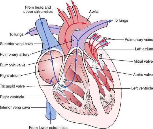
Figure 14-1 Circulation of blood through the heart.
The heart is enveloped by a thin pericardial sac. The bottom of this sac is adherent to the diaphragm, and the top is loosely attached to the upper portion of the sternum. The visceral pericardium is the epicardial, or outermost, layer of cells of the heart. The parietal pericardium is the outer sac. Between these two surfaces, a small amount of pericardial fluid in the pericardial sac provides a lubricating interface for the constantly moving heart. The parietal pericardium is innervated by the phrenic nerve, which contains pain fibers. The visceral pericardium is insensitive to pain.
The synchronous contraction of the heart results from the conduction of impulses generated by the sinoatrial (SA) node and propagated through the conduction system. The SA node is located at the juncture of the superior vena cava and the right atrium. The SA impulse spreads from its point of origin concentrically. When the impulse reaches the atrioventricular (AV) node, in the interatrial septum near the entrance of the coronary sinus, the impulse is slowed. It is then transmitted to the specialized conducting tissue known as the right and left bundle branches, which conduct the impulse to the specialized conducting pathways in the ventricles, Purkinje's fibers. The impulse spreads from the endocardial to the epicardial surface of the heart. These conducting pathways are illustrated in Figure 14-2.
The heart is innervated extensively by branches of the autonomic nervous system. Both sympathetic and parasympathetic fibers are present in the SA and atrioventricular nodes. The atrial muscle is also innervated by both types of fibers. The ventricular musculature is innervated predominantly by the sympathetic nervous system.
The parasympathetic fibers travel along the vagus, or 10th cranial, nerve. The sympathetic fibers descend in the spinal cord to the level of T1 to T5, where they emerge through the ventral roots to form a synapse in the thoracic and cervical sympathetic ganglia. The postganglionic fibers travel through the cervical cardiac nerves to join the parasympathetic fibers in forming the cardiac plexus, which is located near the aortic arch and the tracheal bifurcation. These neural pathways are illustrated in Figure 14-3.
Sympathetic stimulation by norepinephrine produces marked increases in heart rate and contractility. Parasympathetic stimulation mediated by acetylcholine slows the heart rate and decreases contractility.
In addition, several receptor sites provide circulatory information to the medullary cardiovascular center in the brain. This center has cardioexcitatory and cardioinhibitory areas that regulate the neural output to the sympathetic and parasympathetic fibers. Stretch receptors in the aortic arch and in the carotid sinus monitor blood pressure. These baroreceptors respond to a decrease in blood pressure by decreasing their impulses to the medullary center. The center senses this decreased activity and increases its sympathetic efferent activity and decreases its parasympathetic efferent activity. The net result is to increase the heart rate and contractility. An increase in blood pressure causes an increase in afferent activity to the center, and the opposite changes occur.
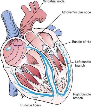
Figure 14-2 Conducting pathways of the heart.
To describe physical signs, the examiner must be able to identify the important surface topographic landmarks. Chapter 13, The Chest, describes the major areas. These areas should be reviewed at this time.
The surface projection of the heart and great vessels is illustrated in Figure 14-4. Most of the anterior cardiac surface is the right ventricle. The right atrium forms a narrow border from the third to the fifth ribs to the right of the sternum. The left ventricle lies to the left and behind the right ventricle. The left ventricular apex is normally in the fifth intercostal space at the midclavicular line. This location is commonly written as 5ICS-MCL. The apical impulse is called the point of maximum impulse (PMI). The other chambers and vessels of the heart are usually not identifiable on examination.
The four classic auscultatory areas correspond to points over the precordium, at which events originating at each valve are best heard. The areas are not necessarily related to the anatomic position of the valve, nor are all sounds heard in the area directly produced by the valve for which the area is named. The normal areas are as follows:
Aortic: Second intercostal space, right sternal border (2ICS-RSB)
Pulmonic: Second intercostal space, left sternal border (2ICS-LSB)
Tricuspid: Left lower sternal border (LLSB)
Mitral: Cardiac apex (5ICS-MCL)
In addition to these four areas, the third left intercostal space, known as Erb's point, is frequently the area at which pulmonic or aortic sounds are best heard. The five areas are illustrated in Figure 14-5. The second intercostal space to the right and left of the sternum is called the base.
Remember that the left atrium is the most posterior portion of the heart. When the left atrium enlarges, it extends posteriorly and to the right.
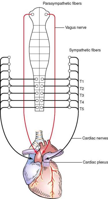
Figure 14-3 Autonomic neural pathways of the heart.
The Cardiac Cycle
To understand the cardiac cycle, the motion of the valves and the pressures within the chambers should be reviewed. The interrelationships of valve motion are critically important and must be understood. Only with the knowledge of these cycles can the clinician fully comprehend the cardiac physical examination and heart sounds. The pressure tracings and valve motions are shown in Figure 14-6.
Normally, only the closing of the heart valves can be heard. The closure of the atrioventricular valves, the tricuspid and the mitral, produces the first heart sound (Si). The closure of the semilunar valves, the aortic and the pulmonic, produces the second heart sound (S2).
The opening of the valves can be heard only if they are damaged. When an atrioventricular valve is narrowed, or stenotic, the opening of the valve may be heard and is termed an opening snap. If a semilunar valve is stenotic, the opening may be heard and is termed an ejection click. It should be noted that in Figure 14-6, the term opening snap refers to the opening of a pathologically damaged atrioventricular valve that occurs during diastole, and the term ejection click refers to the opening of a damaged semilunar valve that occurs during systole.
The sequence of the opening and closing of the four valves is as follows:
MVcTVcPVoAVoAVcPVcTVoMVo,
in which MV = mitral valve, TV = tricuspid valve, PV = pulmonic valve, AV = aortic valve, c = closing, and o = opening.
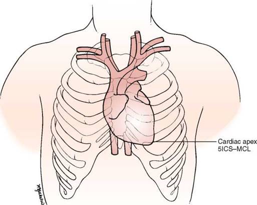
Figure 14-4 Surface topography of the heart. 5ICS-MCL, fifth intercostal space at the midclavicular line.
The mitral component of S1 occurs as a result of the closure of the mitral valve when the left ventricular pressure rises above the left atrial pressure; it is written M1. The tricuspid component of S1 occurs as a result of closure of the tricuspid valve when right ventricular pressure rises above right atrial pressure; it is written T1.
The time between the closure of the atrioventricular valves and the opening of the semilunar valves is the period of isovolumetric contraction. When the pressure in the right ventricle exceeds the diastolic pressure in the pulmonary artery, the pulmonic valve opens. A pulmonic
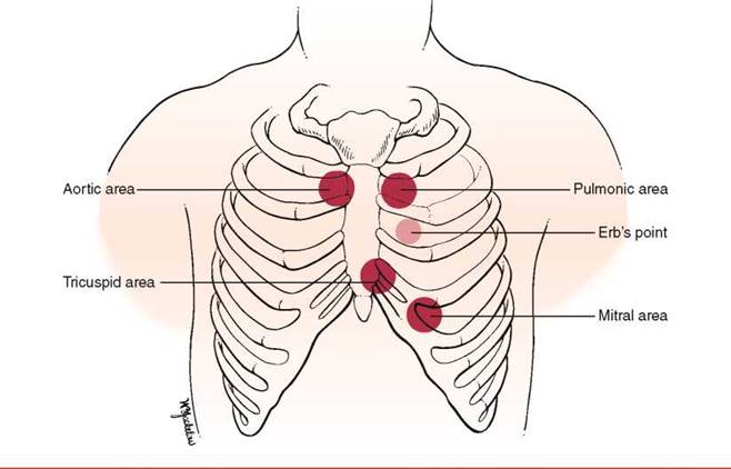
Figure 14-5 Auscultatory areas.

Figure 14-6 The cardiac cycle. TVc, tricuspid valve closing; PVo, pulmonic valve opening; MVc, mitral valve closing; AVo, aortic valve opening; EC, ejection click; OS, opening snap; S1 to S4, first to fourth heart sounds.
ejection click is heard at this time if the pulmonic valve is stenotic. When the pressure in the left ventricle exceeds the diastolic pressure in the aorta, the aortic valve opens. An aortic ejection click is heard at this time if the aortic valve is stenotic.
The time between the opening and the closing of the semilunar valves is the systolic period of ejection. The point at which ejection is completed and the aortic and left ventricular curves separate is called the incisura, or dicrotic notch, and is simultaneous with the aortic component of S2, or closure of the aortic valve; this is written A2. The pulmonic valve closes at the point
when the right ventricular pressure falls below the pulmonary diastolic pressure. This is the pulmonic component of S2 and is commonly written P2.
The time between the closure of the semilunar valves and the opening of the atrioventricular valves is called isovolumetric relaxation. The tricuspid valve opens when the pressure in the right atrium exceeds right ventricular pressure. A tricuspid opening snap may be heard if the tricuspid valve is stenotic. The mitral valve opens when the pressure in the left atrium exceeds left ventricular pressure. A mitral opening snap may occur at this time if the mitral valve is stenotic.
With the opening of the atrioventricular valves, the period of rapid filling of the ventricles occurs. Approximately 80% of ventricular filling occurs at this point. At the end of the rapid filling period, a third heart sound (S3) maybe heard. An S3 occurs 120 to 170 msec after S2. This period is approximately the same time as it takes to say ''me too.'' The ''me'' is the S2, and the ''too'' is the S3. An S3 is normal in children and young adults. When present in individuals older than 30 years, it signifies a volume overload to the ventricle. Regurgitant valvular lesions and congestive heart failure may be responsible.
At the end of diastole, atrial contraction and the additional 20% of ventricular filling occur. A fourth heart sound (S4) may be heard. The interval from the S4 to the S1 is approximately the time it takes to say ''middle.'' The ''mid-'' is the S4, and the ''-dle'' is the S1. Note that the ''mid'' is much softer than the ''dle,'' which is quite similar to the S4-S1 cadence. An S4 is normal in children and young adults. When present in individuals older than 30 years, it is indicative of a noncompliant, or ''stiff,'' ventricle. Pressure overload on a ventricle causes concentric hypertrophy, which produces a noncompliant ventricle. In addition, CHD is a major cause of a stiff ventricle.
Two useful mnemonics for remembering the cadence and pathophysiology of the third and fourth heart sounds are as follows:

The presence of an S3 or an S4 creates a cadence resembling the gallop of a horse. These sounds are therefore called gallop sounds or rhythms.
The first heart sound is loudest at the cardiac apex. Splitting of the first heart sound may be heard in the tricuspid area. The second heart sound is loudest at the base.
The terms A2 and P2 indicate the aortic component and the pulmonic component of S2, respectively. A2 normally precedes P2, meaning that the aortic valve closes before the pulmonic valve. With inspiration, the intrathoracic pressure lowers. This causes more blood to be drawn from the superior and inferior venae cavae into the right chambers of the heart. The right ventricle enlarges, and it takes longer for all the blood to be ejected into the pulmonary artery; thus the pulmonic valve stays open longer. P2 occurs later in inspiration, and the split between A2 and P2 is widened during inspiration in comparison with expiration. This is the cause of physiologic splitting of S2, which is diagrammed in Figure 14-7.
The blood in the right ventricle is then pumped into the large-capacitance bed of the lungs. Therefore, the return of blood from the lungs to the left side of the heart is decreased, and the left atrium and left ventricle become smaller. Atrial receptors trigger a reflex tachycardia that compensates for the decreased left ventricular volume. This increase in heart rate with inspiration is termed sinus arrhythmia. It is a misnomer because it is not really an arrhythmia but a normal physiologic response to a decreased left ventricular volume during inspiration.
The Arterial Pulse
The arterial pulse is produced by the ejection of blood into the aorta. The normal configuration of the pulse consists of a smooth and rapid upstroke that begins about 80 msec after the first component of S1. There is sometimes a slight notch in the arterial pulsation toward the end of the rapid ejection period. This is called the anacrotic notch. The peak of the pulse is smooth and dome-shaped and occurs about 100 msec after the onset of the pulse. The descent from the peak is less steep. There is a gradual descent to the dicrotic notch, which represents the closure of the aortic valve. The contour and volume of the arterial pulse are determined by several factors, including the left ventricular stroke volume, the ejection velocity, the relative compliance and capacity of the arteries, and the pressure waves that result from the antegrade flow of blood. Figure 14-8 illustrates a characteristic arterial pulse.
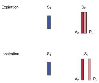
Figure 14-7 Physiologic splitting of the second heart sound.

Figure 14-8 The arterial pulse.
As the arterial pulse travels to the periphery, there are several changes. The initial upstroke becomes steeper, the systolic peak is higher, and the anacrotic notch becomes less evident. In addition, the dicrotic notch occurs later in the peripheral pulse, approximately 300 msec after the onset of the pulse. The positive wave that follows the dicrotic notch is called the dicrotic wave.
Commonly, two waves may be present in the arterial pulse, which precedes the dicrotic notch. The percussion wave is the earlier wave and is associated with the rate of flow in the artery. The percussion wave occurs during peak velocity of flow. The tidal wave is the second wave, is related to pressure in the vessel, and occurs during peak systolic pressure. The tidal wave is usually smaller than the percussion wave, but it may be increased in hypertensive or elderly patients.
Blood Pressure
Arterial blood pressure is the lateral pressure exerted by a column of blood against the arterial wall. It is the result of cardiac output and peripheral vascular resistance. Blood pressure depends on the volume of blood ejected, its velocity, the distensibility of the arterial wall, the viscosity of the blood, and the pressure within the vessel after the last ejection.
Systolic blood pressure is the peak pressure in the arteries. It is regulated by the stroke volume and the compliance of the blood vessels. Diastolic blood pressure is the lowest pressure in the arteries and depends on peripheral resistance. The difference in the systolic and diastolic pressures is the pulse pressure. Systolic blood pressure in the legs is 15 to 20 mm Hg greater than in the arms, even while the individual is lying flat. This is in part related to Poiseuille's law, according to which the total resistance of vessels connected in parallel is greater than the resistance of a single large vessel. The blood pressure in the aorta is less than the blood pressure in the branched arteries of the lower extremities.
Blood pressure varies greatly, according to the patient's degree of excitement, degree of activity, smoking habits, pain, bladder distention, and dietary pattern. There is normally an inspiratory decline of up to 10 mm Hg in systolic blood pressure during quiet respiration.
Jugular Venous Pulse
The jugular venous pulse provides direct information about the pressures in the right side of the heart because the jugular system is in direct continuity with the right atrium. During diastole, when the tricuspid valve is open, the jugular veins are continuous with the right ventricle as well. If there is no stenotic lesion at the pulmonic or mitral valves, the right ventricle indirectly monitors the pressures in the left atrium and left ventricle. The most common cause of right-sided heart failure is left-sided heart failure. Examination of the neck veins also provides information about the cardiac rhythm.
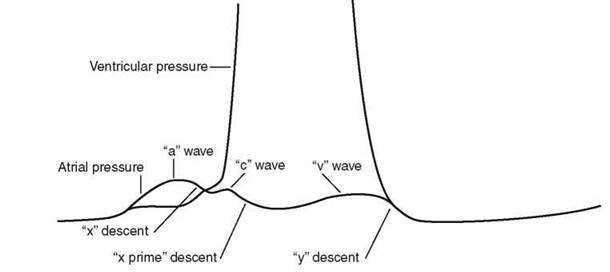
Figure 14-9 The atrial and ventricular pressure curves enlarged.
The understanding of the normal physiology is important in the consideration of the jugular venous pulsation. Figure 14-9 is an enlargement of the atrial and ventricular pressure curves in Figure 14-6.
The “a” wave of the jugular venous pulse is produced by right atrial contraction. When the ''a'' wave is timed with the electrocardiogram, it is found to occur about 90 msec after the onset of the P wave. This time delay is related to the time from electrical stimulation of the atria to atrial contraction and to the resultant wave propagated in the neck. The “x" descent is caused by atrial relaxation, which occurs just before ventricular contraction. This drop in right atrial pressure is terminated by the “c" wave. The resulting increase in right atrial pressure is caused by tricuspid valve closure secondary to right ventricular contraction. The descent of the atrioventricular valve rings, also known as the descent of the base of the heart, produces the next change in right atrial pressure, called the ''x prime'' descent. As the free wall of the right ventricle approaches the septum during contraction, the atrioventricular valve rings descend toward the apex as contraction progresses. This increases the size of the atrium, causing a fall in its pressure (hence the ''x prime'' descent). During ventricular systole, the right atrium begins to fill with blood returning through the venae cavae. This increase in right atrial pressure as a result of its filling produces the ascending limb of the v'' wave. At the end of ventricular systole, right ventricular pressure falls rapidly. When it falls below the right atrial pressure, the tricuspid valve opens. This drop in right atrial pressure produces the ‘‘y'' descent.
Normally, only the ''a'' and ''v'' waves are visible on examination. Because the ''c'' wave is frequently not observed, the ''x'' and ''x prime'' descents are summated into a single ''x'' descent. On occasion, the later portion of the ''c'' wave may be enlarged by a carotid artery pulsation artifact.
Evaluation of the jugular venous pulse provides information about the level of venous pressure and the type of venous wave pattern. These are described later in the section The Jugular Venous Pulse.
Review of Specific Symptoms
The important symptoms of cardiac disease are the following:
• Chest pain
• Palpitations
• Dyspnea
• Syncope
• Fatigue
• Dependent edema
• Hemoptysis
• Cyanosis
Chest Pain
Chest pain is probably the most important symptom of cardiac disease. It is not, however, pathognomonic for heart disease. It is well known that chest pain may result from pulmonary, intestinal, gallbladder, and musculoskeletal disorders. Ask the following questions of any patients complaining of chest pain:
Where is the pain?''
''How long have you had the pain?''
Do you have recurrent episodes of pain?''
''What is the duration of the pain?''
''How often do you get the pain?''
''What do you do to make it better?''
''What makes the pain worse? breathing? lying flat? moving your arms or neck?''
''How would you describe the pain:* burning? pressing? crushing? dull? aching? throbbing? knifelike? sharp? constricting? sticking?''
''Does the pain occur at rest? with exertion? after eating? when moving your arms? with emotional strain? while sleeping? during sexual intercourse?''
''Is the pain associated with shortness of breath? palpitations? nausea or vomiting? coughing? fever? coughing up blood? leg pain?''
Angina pectoris is the true symptom of CHD. Angina is commonly the consequence of hypoxia of the myocardium resulting from an imbalance of coronary supply and myocardial demand. Table 14-1 lists the characteristics that differentiate angina pectoris from the other types of chest pain.
Commonly, a patient may describe the angina by clenching the fist and placing it over the sternum. This is a pathognomonic sign of angina commonly referred to as Levine's sign. Figure 14-10 demonstrates this body language.
When chest pain is related to a cardiac cause, coronary atherosclerosis and aortic valvular disease are the most common ones. Table 14-2 lists some common causes of chest pain.
Palpitations
Palpitations are the uncomfortable sensations in the chest associated with a range of arrhythmias. Patients may describe palpitations as ''fluttering, skipped beats, pounding,'' ''jumping, stopping,'' or ''irregularity.''
Table 14-1 Characteristics of Chest Pain*
|
Feature |
Angina |
Not Angina |
|
Location |
Retrosternal, diffuse |
Left inframammary, localized |
|
Radiation |
Left arm, jaw, back |
Right arm |
|
Description |
''Aching, dull/’‘'pressing, squeezing, viselike'' |
''Sharp, shooting, cutting'' |
|
Intensity |
Mild to severe |
Excruciating |
|
Duration |
Minutes |
Seconds, hours, days |
|
Precipitated by |
Effort, emotion, eating, cold |
Respiration, posture, motion |
|
Relieved by |
Rest, nitroglycerin |
Nonspecific |
*Angina and other chest pain may manifest in a variety of ways. The characteristics listed here are the common manifestations. This list, however, is not exhaustive and should be used only as a guide.
*In general, it is best to allow the patient to describe the character of the pain. These descriptions are provided for the interviewer to use only when the patient is unable to characterize the pain.

Figure 14-10 Levine's sign.
Table 14-2 Common Causes of Chest Pain
|
Organ System |
Cause |
|
Cardiac |
Coronary artery disease Aortic valvular disease Pulmonary hypertension Mitral valve prolapse Pericarditis Idiopathic hypertrophic subaortic stenosis |
|
Vascular |
Dissection of the aorta |
|
Pulmonary |
Pulmonary embolism Pneumonia Pleuritis Pneumothorax |
|
Musculoskeletal |
Costochondritis* Arthritis Muscular spasm Bone tumor |
|
Neural |
Herpes zoster* |
|
Gastrointestinal |
Ulcer disease Bowel disease Hiatal hernia Pancreatitis Cholecystitis |
|
Emotional |
Anxiety Depression |
|
*Tietze's syndrome, which is an inflammation of the costal cartilages. *Shingles, which is a viral invasion of the peripheral nerves in a dermatomal distribution. |
|
Determine whether the patient has had similar episodes and what was done to extinguish them. Palpitations are common and do not necessarily indicate serious heart disease. Any condition in which there is an increased stroke volume, as in aortic regurgitation, may be associated with a sensation of ''forceful contraction.'' When a patient complains of palpitations, ask the following questions:
''How long have you had palpitations?''
''Do you have recurrent attacks?'' If so, ''How frequently do they occur?''
''When did the current attack begin?''
''How long did it last?''
‘‘What did it feel like?''
''Did any maneuvers or positions stop it?''
''Did it stop abruptly?''
''Could you count your pulse during the attack?''
''Can you tap out on the table what the rhythm was like?''
‘‘Have you noticed palpitations after strenuous exercise? on exertion? while lying on your left side? after a meal? when tired?''
''During the palpitations, have you ever fainted? had chest pain?''
''Was there an associated flush, headache, or sweating associated with the palpitations?''*
Have you noticed an intolerance to heat? cold?''
What kind of medications are you taking?''
Do you take any medications for your lungs?''
''Are you taking any thyroid medications?''
Have you ever been told that you had a problem with your thyroid?''
How much tea, coffee, chocolate, or cola sodas do you consume a day?''
''Do you smoke?'' If yes, ‘‘What do you smoke?''
Do you drink alcoholic beverages?''
''Did you notice that after the palpitations you had to urinate?''*
In addition to primary cardiovascular causes, thyrotoxicosis, hypoglycemia, fever, anemia, pheochromocytoma, and anxiety states are commonly associated with palpitations. Hyperthyroidism is an important cause of rhythm disturbances that originate outside the cardiovascular system. Caffeine, tobacco, and drugs are also important factors in arrhyth- mogenicity. Sympathomimetic amines used in the treatment of bronchoconstriction are potent stimuli for arrhythmia as well. In patients with panic disorders and other anxiety states, the sensation of palpitations may occur during periods of normal rate and rhythm.
Patients who have had previous attacks of palpitations should be asked the following:
How was your previous attack terminated?''
How often do you get the attacks?''
Are you able to terminate them?'' If so, How?''
''Have you ever been told that you have Wolff-Parkinson-White syndrome?''*
*Symptoms associated with a pheochromocytoma.
*After an attack of paroxysmal atrial tachycardia, patients often have an urge to urinate. The pathophysiologic process is not well understood, but the association is present.
*The use of this technical term is appropriate because a patient having this form of preexcitation may have been told of this condition and may recognize the name.
Table 14-3 Common Causes of Palpitations
|
Extrasystoles |
|
Atrial premature beats* |
|
Nodal premature beats |
|
Ventricular premature beats* |
|
Tachyarrhythmias |
|
Paroxysmal supraventricular tachycardia |
|
Atrial flutter |
|
Atrial fibrillation |
|
Multifocal atrial tachycardia |
|
Ventricular tachycardia |
|
Bradyarrhythmias |
|
Heart block |
|
Sinus arrest |
|
Drugs |
|
Bronchodilators |
|
Digitalis |
|
Antidepressants |
|
Smoking |
|
Caffeine |
|
Thyrotoxicosis |
|
*Also known as atrial premature contractions or premature atrial contractions. *Also known as ventricular premature contractions or premature ventricular contractions. |
Table 14-3 outlines the common causes of palpitations.
Dyspnea
The complaint of dyspnea is important. Patients report that they have ''shortness of breath'' or that they ''can't get enough air.'' Dyspnea is commonly related to cardiac or pulmonary conditions. The questions relating to dyspnea are discussed in Chapter 13, The Chest. This section further delineates dyspnea as a cardiac symptom.
Paroxysmal nocturnal dyspnea (PND) occurs at night or when the patient is supine. This position increases the intrathoracic blood volume, and a weakened heart may be unable to handle this increased load; congestive heart failure may result. The patient is awakened about 2 hours after having fallen asleep, is markedly dyspneic, is often coughing, and may seek relief by running to a window to ''get more air.'' Episodes of PND are relatively specific for congestive heart failure.
The symptom of PND is often associated with the symptom of orthopnea, the need for using more pillows on which to sleep. Inquire of all patients, ''How many pillows do you need in order to sleep?'' To help quantify the orthopnea, you can state, for example, ''3-pillow orthopnea for the past 4 months.''
Dyspnea on exertion (DOE) is usually caused by chronic congestive heart failure or severe pulmonary disease. Quantify the severity of the dyspnea by asking, ''How many level blocks can you walk now?'' and ''How many level blocks could you walk 6 months ago?'' You can now attempt to quantify the dyspnea: for example, ''The patient has had 1-block DOE for the past 6 months. Before 6 months ago, the patient was able to walk 4 blocks without becoming short of breath. In addition, during the last 3 months, the patient has noted 4-pillow orthopnea.''
Trepopnea is a rare form of positional dyspnea in which the dyspneic patient has less dyspnea while lying on the left or right side. The pathophysiologic process of trepopnea is not well understood.
Table 14-4 Common Causes of Dyspnea
|
Organ System or Condition |
Cause |
|
Cardiac |
Left ventricular failure Mitral stenosis |
|
Pulmonary |
Obstructive lung disease Asthma Restrictive lung disease Pulmonary embolism Pulmonary hypertension |
|
Emotional |
Anxiety |
|
High-altitude exposure |
Decreased oxygen pressure |
|
Anemia |
Decreased oxygen-carrying capacity |
Table 14-4 lists the common causes of dyspnea.
Syncope
Fainting, or syncope, is the transient loss of consciousness that results from inadequate cerebral perfusion. Ask patients what they mean by ''fainting'' or ''dizziness.'' Syncope may have cardiac or noncardiac causes. When a patient describes fainting, ask the following questions:
‘‘What were you doing just before you fainted?''
Have you had recurrent fainting spells?'' If so, How often do you have these attacks?''
Was the fainting sudden?''
Did you lose consciousness?''
''In what position were you when you fainted?''
‘‘Was the fainting preceded by any other symptom? nausea? chest pain? palpitations? confusion? numbness? hunger?''
Did you have any warning that you were going to faint?''
Did you have any black, tarry bowel movements after the faint?''
The activity that preceded the syncope is important because some cardiac causes are associated with syncope during exercise (e.g., valvular aortic stenosis, idiopathic hypertrophic subaortic stenosis, and primary pulmonary hypertension). If a patient describes palpitations before the syncope, an arrhythmogenic cause may be present. Cardiac output may be reduced by arrhythmias or obstructive lesions.
The position of the patient just before fainting is important because this information may help determine the cause of the syncope. For example, if a patient fainted after rising suddenly from bed in the middle of the night (e.g., to run to answer the telephone), orthostatic hypotension may be the cause. Orthostatic hypotension is a common form of postural syncope and is the result of a peripheral autonomic limitation. There is a sudden fall in systemic blood pressure, resulting from a failure of adaptive reflexes to compensate for an erect posture. Symptoms of orthostatic hypotension include dizziness, blurring of vision, profound weakness, and syncope. Many drugs can cause orthostatic hypotension by leading to changes in intravascular volume or tone. Older patients are most prone to orthostatic hypotension. Micturition syncope usually occurs in men during straining with nocturnal urination. It may occur after considerable alcohol consumption.
Vasovagal syncope is the most common type of fainting and is one of the most difficult to manage. It has been estimated that 40% of all syncopal events are vasovagal in nature. Vasovagal syncope occurs during periods of sudden, stressful, or painful experiences, such as receiving bad news, surgical manipulation, trauma, the loss of blood, or even the sight of blood. It is often preceded by pallor, nausea, weakness, blurred vision, lightheadedness, perspiring, yawning, diaphoresis, hyperventilation, epigastric discomfort, or a ''sinking feeling.''
Table 14-5 Common Causes of Syncope
|
Organic System or Condition |
Cause |
|
Cardiac |
Decreased cerebral perfusion secondary to cardiac rhythm disturbance Left ventricular output obstruction |
|
Metabolic |
Hypoglycemia Hyperventilation Hypoxia |
|
Psychiatric |
Hysteria |
|
Neurologic |
Epilepsy Cerebrovascular disease |
|
Orthostatic hypotension |
Volume depletion Antidepressant medications Antihypertensive medications |
|
Vasovagal |
Vasodepression |
|
Micturition |
Visceral reflex (vasodepressor) |
|
Cough |
Chronic lung disease |
|
Carotid sinus |
Vasodepressor response to carotid sinus sensitivity |
There is a sudden fall in systemic vascular resistance without a compensatory increase in cardiac output as a result of an increased vagotonia. If the patient sits or lies down promptly, frank syncope can be aborted.
Carotid sinus syncope is associated with a hypersensitive carotid sinus and is most common in the elderly population. Whenever a patient with carotid sinus syncope wears a tight shirt collar or turns the neck in a certain way, there is an increased stimulation of the carotid sinus. This causes a sudden fall in systemic pressure, and syncope results. Two types of carotid sinus hypersensitivity exist: a cardioinhibitory (bradycardia) type and a vasodepressor (hypotension without bradycardia) type. Post-tussive syncope usually occurs in patients with chronic obstructive lung disease. Several mechanisms have been postulated to explain its occurrence. It is generally accepted that coughing produces an increase in intrathoracic pressure, which decreases both venous return and cardiac output. There may also be a rise in cerebrospinal fluid pressure, producing a decreased perfusion to the brain.
There are other suggested questions to ask a patient with syncope that direct attention to a neurologic cause. These are summarized in Chapter 21, The Nervous System. Table 14-5 lists the common causes of syncope.
Fatigue
Fatigue is a common symptom of decreased cardiac output. Patients with congestive heart failure and mitral valvular disease frequently complain of fatigue. Fatigue, however, is not specific for cardiac problems. The most common causes of fatigue are anxiety and depression. Other conditions associated with fatigue include anemia and chronic diseases. You must attempt to differentiate organic from psychogenic fatigue. Ask the following questions:
‘‘How long have you been tired?''
Did the fatigue start suddenly?''
Do you feel tired all day? in the morning? in the evening?''
When do you feel least tired?''
''Do you feel more tired at home than at work?''
Is the fatigue relieved by rest?''
Patients with psychogenic fatigue are tired ''all the time.'' They are often more tired at home than at work but occasionally describe being more tired in the morning. They may feel their best at the end of the day, which is when most patients with organic causes feel the worst.
Dependent Edema
Swelling of the legs, a form of dependent edema, is a frequent complaint of patients. Ask the following questions:
''When was the swelling first noted?''
''Are both legs swollen equally?''
''Did the swelling appear suddenly?''
‘‘Is the swelling worse at any time of the day?''
‘‘Does it disappear after a night's sleep?''
''Does elevation of your feet reduce the swelling?''
''What kind of medications are you taking?''
''Is there a history of kidney, heart, or liver disease?''
''Do you have shortness of breath?'' If so, ''Which came first, the edema or the shortness of breath?''
‘‘Do you have pain in the legs?''
‘‘Do you have any ulcers on your legs?''
If the patient is a woman, ask the following questions:
‘‘Are you taking oral contraceptives?''
‘‘Is the edema associated with menstrual changes?''
The patient with congestive heart failure has symmetric edema of the lower extremities that worsens as the day progresses. It is least in the morning after sleeping with the legs elevated in bed. If the patient also complains of dyspnea, it is helpful to determine which symptom came first. In patients with dyspnea and edema secondary to cardiac causes, the dyspnea usually precedes the edema. Bedridden patients may have dependent edema in the sacral area.
Hemoptysis
Hemoptysis is discussed in Chapter 13, The Chest. In addition to the pulmonary causes, mitral stenosis is an important cause of hemoptysis. Rupture of the bronchial veins, which are under high back pressure in mitral stenosis, produces the hemoptysis.
Cyanosis
Cyanosis is also discussed in Chapter 13, The Chest. The important questions regarding cyanosis are indicated in that chapter.
On occasion, cyanosis is noted only in the lower extremities. This is termed differential cyanosis. It is related to a right-to-left shunt through a patent ductus arteriosus (PDA). In a right-to-left shunt resulting from pulmonary hypertension, blood in the pulmonary artery crosses the PDA, which is located below the level of the carotid and left subclavian arteries; deoxygenated blood is pumped only to the lower extremity, producing cyanosis in only that location. Some blood does get to the lungs for oxygenation and is ultimately pumped out through the aorta to produce normal skin color in the upper extremity.
The patient whose feet are shown in Figure 14-11 is a 30-year-old immigrant who was evaluated in the United States for cyanosis. Until the age of 20 years, he had marked ''bluish discoloration'' of his lower extremities and relatively normal color in his upper extremities. Over the next 10 years, there was a gradual darkening of his upper extremities. Note the marked cyanosis of the extremities and nail beds of the fingers and toes. The patient had a PDA with marked pulmonary hypertension.

Figure 14-11 Differential cyanosis of the extremities: patent ductus arteriosus.
Impact of Cardiac Disease on the Patient
Patients with cardiac disease are intensely fearful. Once cardiac disease has been diagnosed, a series of reactions occurs. Fear, depression, and anxiety are the outcomes. The patients, who were totally asymptomatic until their episode of ''sudden death'' resulting from a coronary occlusion, are scared. They were resuscitated the first time; will an episode happen again? When? During recovery in the hospital, they are afraid to leave the intensive care unit for fear that ''no one will be watching.'' At the time of discharge from the hospital, they are filled with anxiety. Although they desperately want to go home, they ask themselves, ''What will happen if I have chest pain at home? Who will provide medical assistance?'' They go through a period of depression, recognizing what they have gone through. After convalescence, they become fearful of daily situations that may provoke another attack. Can they go back to the daily ''hassles'' at work? Is it safe to have sexual intercourse? Despite appropriate reassurances from the clinician, their anxiety level may remain high.
Many patients with cardiac disease who have witnessed a fatal cardiac arrest in another patient in their room refuse to admit how stressful this event really was. The patients freely discuss the efficiency of the cardiac arrest team or complain that the noise kept them from sleeping. They refuse to identify with the deceased patient.
The patient with cardiac disease approaching surgery has the same fears as all surgical patients; these fears are discussed in Chapter 2, The Patient's Responses. However, surgical procedures for the patient with cardiac disease involve the ''nucleus'' of the body. The conscientious clinician takes time to explain the nature of the problem and the surgical approach. Before the procedure, the clinician should allow the patient, and especially the family, to visit the intensive care unit where the patient will be for a few days after surgery. Patients should be reassured that everything possible will be done in their behalf. Their courage and determination and the clinician's support are essential.
Physical Examination
The equipment necessary for the examination of the heart is a stethoscope, a penlight, and an applicator stick.
The physical examination of the heart includes the following:
• Inspection of the patient
• Assessment of blood pressure
• Assessment of the arterial pulse
• Assessment of the jugular venous pulse
• Percussion of the heart
• Palpation of the heart
• Auscultation of the heart
• Examination for dependent edema
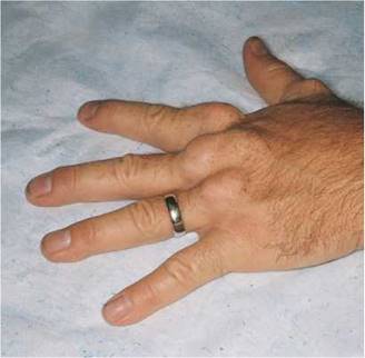
Figure 14-12 Tendon xanthomata.
The patient should be supine, and the examiner should stand on the right side of the bed. The head of the bed may be elevated slightly if the patient is more comfortable in this position.
Inspection
Evaluate General Appearance
The general inspection of the patient offers clues to cardiac diagnosis. Is the patient in acute distress? What is the patient's breathing like? Is it labored? Are accessory muscles being used?
Inspect the Skin
The skin can reveal many changes associated with cardiac disease. Inspect the skin color. Is cyanosis present? If so, does it appear central or peripheral? Is pallor present?
The temperature of the skin may reflect cardiac disease. Severe anemia, beriberi, and thyrotoxicosis tend to make the skin warmer; intermittent claudication is associated with coolness of the lower extremity in comparison with the upper extremity.
Are xanthomata present? Tendon xanthomata are stony-hard, slightly yellowish masses that are commonly found on the extensor tendons of the fingers and are pathognomonic for familial hypercholesterolemia. The Achilles tendon and plantar tendons of the soles are also common locations for tendon xanthomata. Figure 14-12 shows tendon xanthomata on the extensor surfaces of the fingers of a patient with a total serum cholesterol concentration higher than 450 mg/dL.*
*The total cholesterol concentration for an adult is normally lower than 220 mg/dL.
Figure 14-13 shows multiple tuberous xanthomata of the hand of another patient. This patient had primary biliary cirrhosis and extremely elevated cholesterol levels. Primary biliary cirrhosis is a rare, progressive, and often fatal liver disease occurring mostly in women. Pruritus is a common symptom. Xanthomata develop in approximately 15% to 20% of affected patients and are typically found on the palms, soles, knees, elbows, and hands. The serum cholesterol, usually low-density lipoprotein, is often as high as 1000 to 1500 mg/dL. Antimitochondrial antibody is present in nearly 90% of patients.
Eruptive xanthomata are seen in several familial disturbances of fat metabolism, specifically hyperlipidemia types I and IV. The chest, buttocks, abdomen, back, face, and arms are most commonly affected. Figure 14-14 shows eruptive xanthomata on the abdomen of a patient with uncontrolled diabetes mellitus and hypertriglyceridemia. Eruptive xanthomata on the face of a patient are pictured in Figure 14-15. Eruptive xanthomata result from elevations
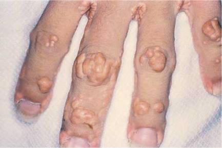
Figure 14-13 Multiple tuberous xanthomata of the hand.

Figure 14-14 Eruptive xanthomata on the abdomen.
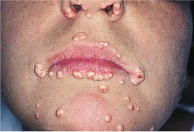
Figure 14-15 Eruptive xanthomata on the face.
in plasma triglyceride concentrations, usually to levels greater than 1500 mg/dL.* These lesions developed in this patient after excessive alcohol consumption; his serum triglyceride concentrations exceeded 2000 mg/dL. The lesions, frequently found on the abdomen, buttocks, elbows, knees, and back, are small (1 to 3 mm in diameter), yellowish papules on an erythematous base. With reduction in the level of triglycerides, the lesions may recede.
*The serum triglyceride concentration for an adult is normally lower than 200 mg/dL.
Is a rash present? The presence of erythema marginatum (erythema in which the reddened areas are disc-shaped with raised edges) in a febrile patient is suggestive of acute rheumatic fever.
Are any painful lesions on the fingers or toes present? Osier's nodes are painful lesions that occur in the tufts of the fingers and toes in patients with infective endocarditis. They are evanescent but are said to occur in 10% to 25% of patients with infective endocarditis.
Inspect the Nails
Frequently, splinter hemorrhages are visible as small, reddish-brown lines in the nail bed. These hemorrhages run from the free margin proximally and are classically associated with infective endocarditis. However, the finding is nonspecific because it is seen in many other conditions, even local trauma to the nail. A nail with splinter hemorrhages in a patient with endocarditis is shown in Figure 14-16.
Inspect the Facies
Abnormalities of the heart may also be associated with peculiarities of the face and head. Supravalvular aortic stenosis, a congenital problem, occurs in association with widely set eyes, strabismus, low-set ears, an upturned nose, and hypoplasia of the mandible. Moon facies and widely spaced eyes are suggestive of pulmonic stenosis. Expressionless facies with puffy eyelids and loss of the outer third of the eyebrow is seen in hypothyroidism. Affected individuals may have a cardiomyopathy. The earlobe crease, or Lichtstein's sign, is an oblique crease, often bilateral, seen frequently in patients older than 50 years with significant CHD. This sign is shown in Figure 14-17. Although it is a useful sign, there are too many false-positive and false-negative findings for this sign to be very reliable.
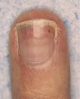
Figure 14-16 Splinter hemorrhages.
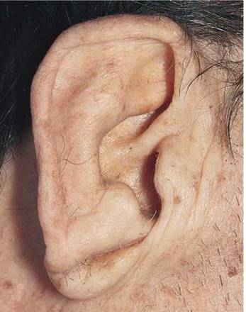
Figure 14-17 Earlobe creases.
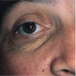
Figure 14-18 Xanthelasma.
Inspect the Eyes
The presence of yellowish plaques on the eyelids, called xanthelasma, should raise the suspicion of an underlying hyperlipoproteinemia, even though this lesion is less specific than the xanthoma. Xanthelasma in a patient with hypercholesterolemia is shown in Figure 14-18.
Examination of the eyes may reveal an arcus senilis. An arcus (see Figs. 10-53 and 10-54) seen in a patient younger than 40 years should raise the suspicion of hypercholesterolemia. Opacities in the cornea may be evidence for sarcoidosis, which may be responsible for cor pulmonale or myocardial involvement. Displacement of the lens is frequently seen in patients with Marfan's syndrome, an important cause of aortic regurgitation (see Fig. 10-69). Conjunctival hemorrhages are commonly seen in infective endocarditis. Hypertelorism, or widely set eyes, is often associated with congenital heart disease, especially pulmonic stenosis and supravalvular aortic stenosis. Retinal evaluation may furnish valuable information about diabetes (see Figs. 10-88 to 10-100), hypertension (see Figs. 10-101 to 10-105), and atherosclerosis. Roth's spots may develop in patients with infective endocarditis (see Fig. 10-109).
Inspect the Mouth
Have the patient open the mouth widely. Inspect the palate. Is the palate highly arched? A high-arched palate may be associated with congenital heart problems such as mitral valve prolapse.
Are there petechiae on the palate? Infective endocarditis is often associated with palatal petechiae, as seen in Figure 14-19.
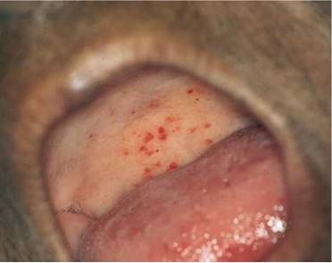
Figure 14-19 Palatal petechiae.
Inspect the Neck
Examination of the neck may reveal webbing. Webbing is seen in individuals with Turner's syndrome,* who may have coarctation of the aorta, and in patients with Noonan's syndrome. Pulmonic stenosis is the associated cardiac abnormality in this condition.
Inspect the Chest Configuration
Inspection of the chest often reveals information about the heart. Because the chest and the heart develop at about the same time during embryogenesis, it is not surprising that anything interfering with the development of the chest may interfere with the heart. A pectus excavatum, or caved-in chest, is seen in Marfan's syndrome and in mitral valve prolapse (see Fig. 13-7). Pectus carinatum, or pigeon breast, is also associated with Marfan's syndrome (see Fig. 13-8). Are there any visible cardiac motions?
Inspect the Extremities
Some congenital abnormalities of the heart are associated with abnormalities of the extremities. Patients with atrial septal defects may have an extra phalanx, an extra finger, or an extra toe. Long, slender fingers may be suggestive of Marfan's syndrome and aortic regurgitation. Short stature, cubitus valgus, and medial deviation of the extended forearm are typical of patients with Turner's syndrome.
Blood Pressure Assessment
The Principles
Blood pressure can be measured directly with an intra-arterial catheter or indirectly with a sphygmomanometer. The sphygmomanometer consists of an inflatable rubber bladder within a cloth cover, a rubber bulb to inflate the bladder, and a manometer to measure the pressure in the bladder. Indirect measurement of blood pressure involves the auscultatory detection of the appearance and disappearance of the Korotkoff sounds over the compressed artery. Korotkoff sounds are low-pitched sounds originating in the vessel that are related to turbulence produced by partially occluding an artery with a blood pressure cuff. Several phases occur in sequence as the occluding pressure drops. Phase 1 occurs when the occluding pressure falls to the systolic blood pressure. The tapping sounds are clear and gradually increase in intensity as the occluding pressure falls. Phase 2 occurs at a pressure about 10 to 15 mm Hg lower than that in phase 1 and consists of tapping sounds followed by murmurs.* Phase 3 occurs when the occluding pressure falls enough to allow a large amount of volume to cross the partially occluded artery. The sounds are similar to the sounds of phase 2, except that only the tapping sounds are heard. Phase 4 is the abrupt muffling and decreased intensity of the sounds as the pressure approaches the diastolic blood pressure. Phase 5 is the complete disappearance of the sounds. The vessel is no longer compressed by the occluding cuff. Turbulent flow is no longer present.
*Short stature, retarded sexual development, and webbed neck in a female patient, associated with an abnormality of the sex chromosomes (45,XO).
*Male Turner's syndrome (46,XY).
*A murmur is a blowing auscultatory sign produced by turbulence in blood flow. These vibrations can originate in the heart or in blood vessels as a result of hemodynamic changes.
The normal blood pressure for adults is up to 140 mm Hg systolic and up to 85 mm Hg diastolic. For the diastolic blood pressure reading, the point of disappearance of the Korotkoff sounds is probably more accurate than the point of muffling. However, if the point of disappearance is more than 10 mm Hg lower than the point of muffling, the point of muffling is probably more accurate. Recording both the point of muffling and disappearance frequently helps in communication. A blood pressure might be recorded as 125/75-65: the systolic blood pressure is 125; the point of muffling is 75; the point of disappearance is 65 (the diastolic blood pressure).
Blood pressure should be recorded only to the nearest 5 mm Hg because there is a ± 3 mm Hg limit of accuracy for all sphygmomanometers. In addition, normal blood pressure changes occur from moment to moment, and measuring to less than 5 mm Hg provides a false sense of accuracy.
The size of the cuff is important for the accurate determination of blood pressure. It is recommended that the cuff be snugly applied around the arm, with its lowest edge 1 inch above the antecubital fossa. The cuff should be approximately 20% wider than the diameter of the extremity. The bladder should overlie the artery. The use of a cuff that is too small for a large arm results in an erroneously high reading of blood pressure.
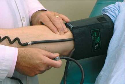
Figure 14-20 Technique for blood pressure assessment by palpation.
Another cause of falsely elevated blood pressure readings is lack of support of the patient's arm. To obtain an accurate measurement, the cuff must be at heart level. If the arm is not supported, the patient is performing isometric exercise, which raises the recorded pressure. In contrast, excessive pressure on the diaphragm of the stethoscope produces a spuriously lower reading of the diastolic blood pressure without any significant alteration of systolic pressure. If the arm is held correctly, no skin indentations should occur.
The auscultatory gap is the silence that occurs between the disappearance of the Korotkoff sounds after the initial appearance and their reappearance at a lower pressure. The auscultatory gap is present when there is a decreased blood flow to the extremities, as is found in hypertension and in aortic stenosis. Its clinical importance lies in the fact that the systolic blood pressure may be mistaken for the lower blood pressure, the point of reappearance.
Determine Blood Pressure by Palpation
Blood pressure assessment is performed with the patient lying comfortably in the supine position. The cuff bladder is centered over the right brachial artery. If the arm is obese, a large adult or thigh cuff should be used. The arm should be slightly flexed, and it should be supported at approximately the level of the heart. To determine the systolic blood pressure adequately and to exclude an error as a result of an auscultatory gap, blood pressure is first assessed by palpation. In this procedure, the right brachial or right radial artery is palpated while the cuff is inflated above the pressure required to obliterate the pulse. The adjustable screw is opened slowly for deflation. The systolic pressure is identified by the reappearance of the brachial pulse. As soon as the pulse is felt, the adjustable screw is opened for rapid deflation. This assessment is demonstrated in Figure 14-20.
Determine Blood Pressure by Auscultation
Blood pressure by auscultation is assessed in the right arm by inflating the cuff to about 20 mm Hg above the systolic pressure that was determined by palpation. The diaphragm of the stethoscope should be placed over the artery as close to the edge of the cuff as possible, preferably just under the edge. The cuff is deflated slowly while the Korotkoff sounds are evaluated. The systolic blood pressure, the point of muffling, and the point of disappearance are determined. The systolic blood pressure is the point at which the initial tapping sounds are heard. The technique of determining auscultatory blood pressure is shown in Figure 14-21. If the blood pressure is high, it is useful to measure the blood pressure again at the end of the examination, when the patient may be calmer.
Rule Out Orthostatic Hypotension
After the patient has been recumbent for at least 5 minutes, measure the baseline blood pressure and pulse. Then have the patient stand, and repeat the measurements immediately.
Orthostatic hypotension is defined as a drop in systolic blood pressure of 20 mm Hg or more, in association with the development of symptoms such as dizziness or syncope, when the patient assumes the standing position. In most affected patients, there is also an increase in heart rate.
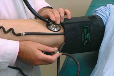
Figure 14-21 Technique for blood pressure assessment by auscultation.
Rule Out Supravalvular Aortic Stenosis
If hypertension is detected in the right arm, perform the following test: Place the cuff on the patient's left arm, and determine the auscultatory pressure. It is not necessary to remeasure the palpatory pressure or reevaluate for orthostatic changes. In supravalvular aortic stenosis, there is a difference in the blood pressures in the arms; hypertension may be detected in the right arm, whereas hypotension may be present in the left arm.
Rule Out Coarctation of the Aorta
If the blood pressure is elevated in the arms, determination of the blood pressure in the lower extremities is important for ruling out coarctation of the aorta. The patient is asked to lie on the abdomen while the thigh cuff, which is 6 cm wider than the arm cuff, is placed around the posterior aspect of the midthigh. The stethoscope is placed over the artery in the popliteal fossa. The Korotkoff sounds are determined as in the upper extremity. If a thigh cuff is not available, the regular cuff can be applied to the lower leg with the distal border just at the malleoli. The stethoscope is placed over either the posterior tibial or the dorsalis pedis artery, and the auscultatory blood pressure is taken. A leg systolic blood pressure that is lower than that in the arm should raise suspicion of coarctation of the aorta.
Rule Out Cardiac Tamponade
In the presence of low arterial blood pressure and a rapid and feeble pulse, it is necessary to rule out the presence of cardiac tamponade. A valuable clinical sign suggestive of cardiac tamponade is the presence of a marked paradoxical pulse (also known as a pulsus paradoxus), which is characterized by an exaggeration of the normal inspiratory fall in systolic pressure. There is much confusion about the definition of a normal paradoxical pulse. A normal paradoxical pulse should be defined as the normal fall (about 5 mm Hg) in systolic arterial pressure during inspiration. It is the magnitude of the phenomenon that should determine whether the pulsus paradoxus is normal or abnormal.
The technique for assessing the magnitude of a paradoxical pulse is as follows: Have the patient breathe as normally as possible. Inflate the blood pressure cuff until no sounds are heard. Gradually deflate the cuff until sounds are heard in expiration only. Note this pressure. Continue to deflate the cuff slowly until sounds are heard during inspiration. Note this pressure. If the difference in these two pressures exceeds 10 mm Hg, a marked (abnormal) pulsus paradoxus is present; cardiac tamponade may be the cause. Cardiac tamponade results when there is an increase in intrapericardial pressure that interferes with normal diastolic filling. A marked paradoxical pulse is not a specific phenomenon for tamponade because it is also seen in large pericardial effusions, in constrictive pericarditis, and in conditions associated with increased ventilatory effort, such as asthma and emphysema.
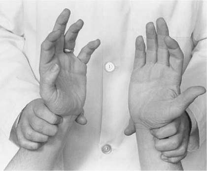
Figure 14-22 Technique for evaluating the radial artery pulses.
The Arterial Pulse
The following information is gained from palpation of the arterial pulse:
• The rate and rhythm of the heart
• The contour of the pulse
• The amplitude of the pulse
Determine the Cardiac Rate
Cardiac rate is routinely assessed by the radial pulse. The examiner should stand in front of the patient and grasp both radial arteries. The second, third, and fourth fingers should overlie the radial artery, as shown in Figure 14-22. The examiner should count the pulse for 30 seconds and multiply the number of beats by 2 to obtain the beats per minute. This method is accurate for most regular rhythms. If the patient has an irregularly irregular rhythm, as is found in atrial fibrillation, a pulse deficit may be present. In atrial fibrillation, many impulses bombard the atrioventricular node and ventricles. Owing to the varying lengths of diastolic filling periods, some of the contractions may be very weak and unable to produce an adequate pulse wave despite ventricular contraction. A pulse deficit, which is the difference between the apical (precordial) and radial pulses, occurs. In such cases, only auscultation of the heart, not the radial pulse, provides an accurate assessment of the cardiac rate.
Determine the Cardiac Rhythm
When palpating the pulse, carefully evaluate the regularity of the rhythm. The slower the rate, the longer you should palpate. If the rhythm is irregular, is there a pattern to the irregularity?
Cardiac rhythm may be described as regular, regularly irregular, or irregularly irregular. A regularly irregular rhythm is a pulse with an irregularity that occurs in a definite pattern. An irregularly irregular pulse has no pattern.
Electrocardiography is really the best medium for diagnosing the rhythm, but the physical examination may provide some clues. Premature beats may be recognized by the presence of isolated extra beats during a regular rhythm. Bigeminy is a coupled rhythm of beats in pairs. The first beat is the sinus beat, which is followed by a premature, usually ventricular, beat. If the premature beat is very early in the diastolic period, the pulse from this beat may be missed if the examiner evaluates the rhythm by palpation alone. A rhythm that is grossly irregular with no pattern is irregularly irregular and is the pulse in patients with atrial fibrillation.
Palpate the Carotid Artery
Assess the carotid artery pulse by standing at the patient's right side, with the patient lying on the back. Auscultate the patient's carotid arteries for bruits first (see Chapter 15, The Peripheral Vascular System). If a bruit is present, do not palpate the carotid artery. If a cholesterol plaque is present, palpation may produce an embolus.
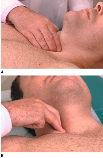
Figure 14-23 A and B, Technique for evaluating the carotid artery pulsations.
To palpate the carotid artery, place your index and third fingers on the patient's thyroid cartilage and slip them laterally between the trachea and the sternocleidomastoid muscle. You should be able to feel the carotid pulsations just medial to the sternocleidomastoid muscle. Palpation should be performed low in the neck to avoid pressure on the carotid sinus, which would cause a reflex drop in blood pressure and heart rate. Each carotid artery is evaluated separately. Never press on both carotid arteries at the same time. After the right carotid artery is evaluated, stand in the same position, and place the same fingers back on the patient's trachea and slip them laterally to the left to feel the left carotid artery. This technique is demonstrated in Figure 14-23.
Evaluate the Characteristics of the Pulse
The carotid artery is used for the assessment of the contour and amplitude of the pulse. Contour is the shape of the wave. It is frequently described as the speed of the upward slope, downward slope, and duration of the wave. Place a hand firmly against the carotid artery until maximal force is felt. At this moment, the wave form should be discernible. The pulse may be described as normal, diminished, increased, or double-peaked. The normal carotid pulse wave is smooth, with the upward stroke steeper and more rapid than the downward stroke. A diminished pulse is a small, weak pulse. The palpating finger feels a gentle pressure rise with a distinct peak. An increased pulse is a large, strong, hyperkinetic pulse. The palpating finger feels an increased rate of rise of the ascending limb of the pulse and a brisk tap at its peak. A double-peaked pulse has a prominent percussion and tidal wave with or without a dicrotic wave. Arterial pulse abnormalities are summarized in Figure 14-24.
|
Type |
Description |
Cause |
|
Anacrotic* |
Small, slow rising, delayed pulse with a notch or shoulder on the ascending limb |
Aortic stenosis |
|
Waterhammer (Corrigan's) |
Rapid and sudden systolic expansion |
Aortic regurgitation |
|
Bisferiens |
Double-peaked pulse with a midsystolic dip |
Aortic regurgitation |
|
Alternans |
Alternating amplitude of pulse pressure |
Congestive heart failure |
|
Paradoxical (marked) |
Detected by blood pressure assessment. An exaggerated drop in systolic blood pressure during inspiration |
Tamponade Constrictive pericarditis Chronic obstructive lung disease |
|
*Also known as plateau pulse or pulsus parvus et tardus. |
||

Figure 14-24 Arterial pulse abnormalities. ECG, electrocardiogram.
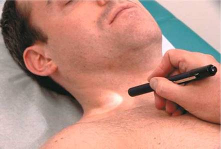
Figure 14-25 Technique for evaluating the jugular wave forms.
The Jugular Venous Pulse
The internal jugular vein provides information about the wave forms and right atrial pressure. The pulsations of the internal jugular vein are beneath the sternocleidomastoid muscle and are visible as they are transmitted through the surrounding tissue. The vein itself is not visible. Because the right internal jugular vein is straighter than the left, only the right internal jugular vein is evaluated. Measurements from the external jugular system, which is easier to visualize, are much less accurate and should not be used.
Determine the Jugular Wave Forms
To visualize the jugular wave forms, the patient should lie flat without a pillow so that his or her neck is not flexed and does not interfere with the pulsations. The patient's trunk should be at approximately 25° to the horizontal. The higher the venous pressure is, the greater the elevation that is required; the lower the pressure, the lower the elevation needed. The patient's head should be turned slightly to the right and slightly down to relax the right sternocleidomastoid muscle. Standing on the patient's right side, the examiner should place his or her right hand, holding a small pocket flashlight, on the patient's sternum and shine the light tangentially across the right side of the patient's neck. Shadows of the pulsations will be cast on the sheet behind the patient. The light and shadows magnify the wave forms. This technique is demonstrated in Figure 14-25. If no wave forms are seen, the angle of elevation of the head of the bed should be reduced. To help identify the wave forms, the examiner can time the cardiac cycle by palpating the cardiac impulse beneath his or her right hand or by feeling the left carotid impulse with his or her left hand. The descents, rather than the waves themselves, tend to be more obvious. If the neck veins are visible at the jaw margin while the patient is seated, the examiner should watch for the wave forms at the angle of the jaw with the patient seated upright.
The jugular pulse must be differentiated from the pulsation of the carotid artery. Table 14-6 lists the most important characteristic differences of these pulses.
Estimate the Jugular Venous Pressure
To assess the pressure in the right side of the heart, it is necessary to establish a reference level. The standard reference is the manubriosternal angle. At any degree of elevation, this position is used to measure the pressure in the internal jugular system. The examiner must first determine the height of the venous distention by noting the top of the wave forms in the internal jugular venous pulsations. An imaginary horizontal line is then drawn from this height to the sternal angle. The examiner should then measure the distance from the sternal angle to this imaginary line. The angle of elevation of the head of the bed is also estimated. It might be stated, ''At 45° elevation, the jugular pulse is 7 cm above the sternal angle.'' At 45°, the upper limit of normal is 4 to 5 cm above the sternal angle; if the patient is at 30°, the upper limit of normal is 6 cm. When the height of the venous column is equal to or lower than the sternal angle in the supine position, venous pressure is usually normal.
There is tremendous inaccuracy in attempting to determine the pressure in the right atrium by the jugular manometer, as just indicated. It has been demonstrated numerous times that the sensitivity and specificity of this test are low, and thus it is inaccurate in predicting elevated pressures. The only accurate statement is that right atrial pressure is high when there is neck vein distention up to the jaw margin while the patient is seated at 90°. In this situation, the right atrial pressure usually exceeds 15 mm Hg. In Figure 14-26, the neck veins are distended to the angle of the jaw while the patient is seated upright. His right atrial pressure was 21 mm Hg.
Table 14-6 Differentiation of Jugular and Carotid Pulses
|
Feature |
Internal Jugular Pulse |
Carotid Pulse |
|
Palpation |
Not palpable |
Palpable |
|
Waveforms |
Multiform: two or three components |
Single |
|
Quality |
Soft, undulating |
Vigorous |
|
Pressure* |
Wave forms obliterated |
No effect |
|
Inspiration |
Decreased height of wave forms |
No effect |
|
Sitting up |
Decreased height of wave forms |
No effect |
|
Valsalva maneuver |
Increased height of wave forms |
No effect |
|
*Light pressure on the vessel above the sternal end of clavicle. |
||
Evaluate the Hepatojugular Reflux
A useful test in assessing high jugular venous pressure is that of the hepatojugular reflux, also known as abdominal compression. By applying pressure over the liver, the examiner can grossly assess right ventricular function. Patients with right ventricular failure have dilated sinusoids in the liver. Pressure on the liver pushes blood out of these sinusoids and into the inferior vena cava and right side of the heart, causing further distention of the neck veins. The procedure is performed with the patient lying in bed, mouth open, breathing normally; this prevents a Valsalva maneuver. The examiner places his or her right hand over the patient's liver in the right upper quadrant and applies a firm, progressive pressure. Compression is maintained for 10 seconds. The normal response is for the internal and external jugular veins to show a transient increase in distention during the first few cardiac cycles, which is followed by a fall to baseline levels during the later part of the compression. In patients with right ventricular failure or elevated pulmonary artery wedge pressure, the neck veins remain distended during the entire period of compression; this distention diminishes rapidly (at least 4 cm) on sudden release of the compressing hand. If the examination is incorrectly performed with the patient's mouth closed, a Valsalva maneuver results and produces inaccurate results of the hepatojug- ular reflux test.
Like most other clinical maneuvers, the hepatojugular reflux test must be performed in a standardized manner. If performed correctly, this test can be of considerable value in the bedside assessment of the patient. The test result correlates best with the pulmonary artery wedge pressure and, as such, is a reflection of increased central blood volume. Ewy (1988) evaluated this test and showed that in the absence of right ventricular failure, a positive test result is suggestive of a pulmonary artery wedge pressure of 15 mm Hg or greater.
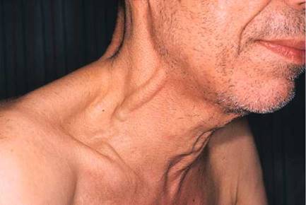
Figure 14-26 Neck vein distention.
Percussion
Percuss the Heart's Borders
The technique of percussion is discussed in Chapter 13, The Chest. Percussion of the heart is performed at the third, fourth, and fifth intercostal spaces from the left anterior axillary line to the right anterior axillary line. Normally, there is a change in the percussion note from resonance to dullness about 6 cm lateral to the left of the sternum. This dullness is attributable to the presence of the heart.
A percussion dullness distance of greater than 10.5 cm in the left fifth intercostal space has a sensitivity of 91.3% and a specificity of 30.3% for detecting increased left ventricular end- diastolic volume (LVEDV) or left ventricular mass. Percussion dullness of more than 10.5 cm in the fifth intercostal space has a sensitivity of 94.4% and a specificity of 67.2% in detecting cardiomegaly. In patients with a palpable apical impulse of greater than 3 cm in the left decubitus position, the sensitivity of detecting increased LVEDV or left ventricular mass increases to 100% and the specificity is 40%.
Palpation
Palpation is performed to evaluate the apical impulse, the right ventricle, the pulmonary artery, and the left ventricular motions. The presence or absence of thrills* is also determined by palpation. The point of maximum impulse (PMI) describes the outward motion of the cardiac apex as it rotates counterclockwise, as viewed from below, to strike the anterior chest wall during isovolumetric contraction.
Palpate the Point of Maximum Impulse
The examiner should stand on the right side of the patient, with the bed at a level comfortable for the examiner. Palpation for the PMI is most easily performed with the patient in a sitting position. Only the examiner's fingertips should be applied to the patient's chest in the fifth intercostal space, midclavicular line, because they are the most sensitive for assessing localized motion. The PMI should be noted. This technique is demonstrated in Figure 14-27. If the apical impulse is not felt, the examiner should move his or her fingertips in the area of the cardiac apex. The PMI is usually within 10 cm of the midsternal line and is no larger than 2 to 3 cm in diameter. A PMI that is laterally displaced or is felt in two interspaces during the same phase of respiration is suggestive of cardiomegaly.
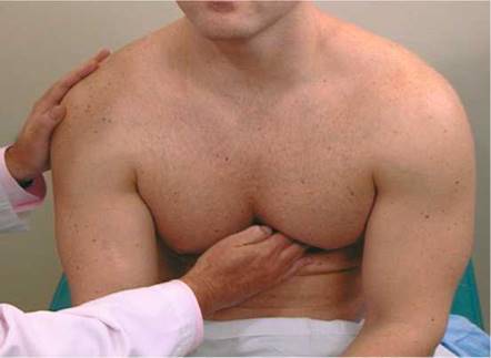
Figure 14-27 Technique for assessing point of maximum impulse.
*Low-frequency cutaneous vibrations associated with loud heart murmurs.
The PMI is felt in approximately 70% of normal individuals while they are sitting. If it cannot be felt in the sitting position, the patient should be reevaluated while supine and in the left lateral decubitus position. The position of the PMI in the left lateral decubitus position must be assessed with the understanding that the normal cardiac impulse is now shifted slightly to the left. If the patient is in the left lateral decubitus position and the PMI is not laterally displaced, the examiner can suspect that cardiomegaly is not present.
In a patient without conditions predisposing to left ventricular hypertrophy, a palpable apical impulse felt in the left lateral decubitus position that is greater than 3 cm is said to be a specific (91%) and sensitive (92%) indicator of left ventricular enlargement. An apical diameter greater than 3 cm is predictive (86%) of an increased LVEDV. In patients with an apical diameter of less than 3 cm and a normal LVEDV, the negative predictive value is 95%.
The PMI usually corresponds to the left ventricular apex, but in patients with an enlarged right ventricle, the heart is rotated clockwise, as viewed from below, and the PMI may actually be produced by the right ventricle. This rotation turns the left ventricle posteriorly and makes it difficult to palpate. The apical impulse by the right ventricle is diffuse, whereas that of the left ventricle tends to be more localized.
In patients with chronic obstructive lung disease, the overinflation of the lungs displaces the PMI downward and to the right. The PMI in such patients is felt in the epigastric area, at the lower end of the sternum. In patients with chronic obstructive lung disease, a PMI in the normal location is suggestive of cardiomegaly.
Palpate for Localized Motion
The patient should now lie down so that all four main cardiac areas can be palpated. The examiner uses his or her fingertips to assess any localized motion. This technique is demonstrated in Figure 14-28.
The presence of a systolic impulse in the second intercostal space to the left of the sternum is suspect for pulmonary hypertension. This impulse is caused by the closure of the pulmonic valve under increased pressure. The presence of this impulse is suggestive of a dilated pulmonary artery, but it may also be felt in thin individuals without pulmonary hypertension.
Palpate for Generalized Motion
After the chest has been palpated with the fingertips, the examiner uses the proximal portion of his or her hand to palpate for any large area of sustained outward motion, called a heave or lift. The examiner again palpates each of the four main cardiac areas. The technique for assessing heaves is demonstrated in Figure 14-29. The presence of a right ventricular rock, which is a sustained left parasternal impulse associated with lateral retraction, is suggestive of a large right ventricle.
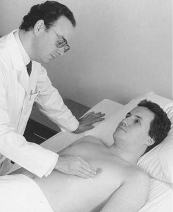
Figure 14-28 Technique for assessing localized cardiac motion.
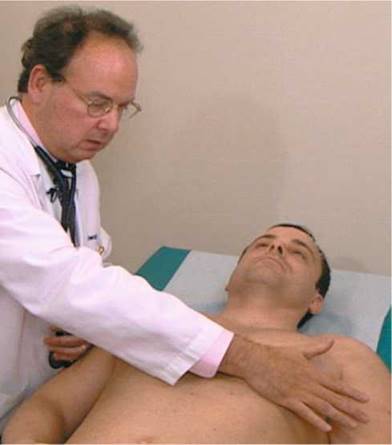
Figure 14-29 Technique for assessing generalized cardiac motion.
Any condition that increases the rate of ventricular filling during early diastole can produce a palpable impulse that occurs after the main left ventricular impulse. This second impulse in the area of the PMI is usually felt in association with an S3. Frequently, an S3 is more easily felt than heard.
The use of a tongue blade or an applicator stick can be helpful to reinforce visually what has been palpated. The tip of the stick is placed directly over the area and held in place by the examiner's finger. This acts as a fulcrum, and the motions tend to be magnified by the movement of the stick. The technique is demonstrated in Figure 14-30.
Palpate for Thrills
Thrills are the superficial vibratory sensations felt on the skin overlying an area of turbulence. The presence of a thrill indicates a loud murmur. Thrills are best felt by using the heads of your metacarpal bones rather than the fingertips and applying very gentle pressure on the skin.
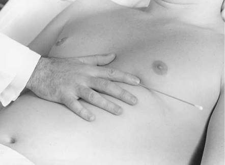
Figure 14-30 Technique for amplifying detection of cardiac movement.
If too much pressure is applied, thrills are not felt. The palpation of thrills is generally of little importance because auscultation reveals the presence of the loud murmur that has produced the thrill. Therefore, the finding of a thrill adds little to the diagnosis, but it is an interesting physical sign to alert the examiner as to what will be heard.
Auscultation The Technique
Proper auscultation requires a quiet area. Every attempt should be made to eliminate extraneous noise from radios, televisions, and so forth. The earpieces of the stethoscope are directed anteriorly or parallel to the direction of the external auditory canal. If the earpieces are put in backward, the openings of the earpieces impinge on the wall of the external canal and lower the intensity of the sounds. The earpieces should fit properly so as to be comfortable but tight enough to exclude external noises.
It is often useful for you to close your eyes when listening to the heart. Sounds that are more difficult to hear sound louder with your eyes closed, because the brain is flooded with all types of sensory input. The input from the eyes appears to be the most important. The next important sensory input is auditory, which is followed by tactile input. If you eliminate the distraction of visual stimuli, the brain concentrates more on the auditory input, and the sounds become more evident.
As indicated in Chapter 13, The Chest, the bell of the stethoscope should be applied lightly to the skin, whereas the diaphragm should be pressed tightly against the skin. High-pitched sounds, such as valve closure, systolic events, and regurgitant murmurs, are better heard with the diaphragm. Low-pitched sounds, such as gallop rhythms or the murmur of atrioventricular stenosis, are better heard with the bell.
It is common in many countries to examine patients through their clothing or a hospital gown. In the United States, however, never listen through any type of clothing.
There are several other pitfalls in auscultation. Make sure that the stethoscope is in good shape: Cracked tubing certainly interferes with good listening. Both the examiner and the patient must be comfortable for the best hearing. An examiner who is straining over the patient and is uncomfortable will want to finish the examination quickly without a proper assessment. Always inspect and palpate before auscultation. Accumulate as much information as possible before listening!
Auscultate the Cardiac Areas
The examiner should be on the right side of the patient while the patient lies flat on the back. If not already at the proper height, the bed should be adjusted so that the examiner is comfortable. The examiner should listen in the aortic, pulmonic, tricuspid, and mitral areas. However, the examiner should not limit auscultation to these areas alone. The examiner should start at any area and move the stethoscope gradually over the precordium from area to area. The areas have been established to provide some degree of standardization.
While listening at the apex and left lower sternal border with the bell, the examiner should determine whether an S3 or an S4 is present.
Cardiac murmurs may radiate widely. The examiner should determine where the sounds are loudest or best heard. There are no acoustic walls in the chest. A murmur typically heard at the apex with radiation to the axilla may be heard in the neck, if it is loud enough. The murmur in this example is probably loudest at the apex and axilla.
The Standard Auscultation Positions
The four standard positions for auscultation are shown in Figure 14-31. They are as follows:
• Supine
• Left lateral decubitus
• Upright
• Upright, leaning forward
All precordial areas are examined while the patient is supine. Using a systematic approach, the examiner starts at either the aortic area or the apex and carefully listens to the heart sounds. After all areas are examined, the patient is then instructed to turn onto the left side. The examiner should now listen at the apex for the low-pitched diastolic murmur of mitral stenosis, which is best heard with the bell of the stethoscope. Then the patient sits upright, and all areas are examined with the diaphragm of the stethoscope. Finally, the patient sits up and leans forward. The patient is asked to exhale and hold his or her breath while the examiner, using the diaphragm, listens for the high-pitched diastolic murmur of aortic regurgitation at the right and left second and third intercostal spaces.
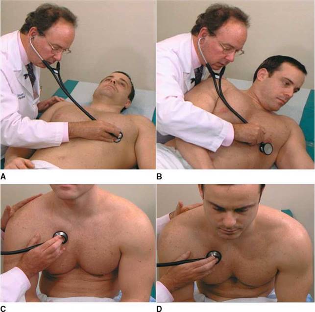
Figure 14-31 Positions for auscultation. A, The supine position, used for listening to all areas. B, The left lateral decubitus position, used for listening with the bell in the mitral area. C, The upright position, used for listening to all areas. D, The upright, leaning-forward position, used for listening with the diaphragm at the base positions.
The Influence of Breathing
The examiner should pay special attention to the influence of breathing on the intensity of heart sounds. Most murmurs or sounds originating in the right side of the heart are accentuated with inspiration. This is related to the increased return of blood that occurs with inspiration and the resultant increased right ventricular output. In addition, an S3 or an S4 originating in the right side of the heart is accentuated during inspiration.
Time the Cardiac Events
To interpret heart sounds accurately, the examiner must be able to time the events of the cardiac cycle. The most reliable way of identifying S1 and S2 is to time the sounds by palpating the carotid artery. While the examiner's right hand is positioning the stethoscope, the left hand is placed on the patient's carotid artery. This technique is demonstrated in Figure 14-32. The sound that precedes the carotid pulse is the S1. The S2 follows the pulse. The carotid, not the radial pulse, must be used. The time delay from S2 to the radial pulse is significant, and errors in timing will result.
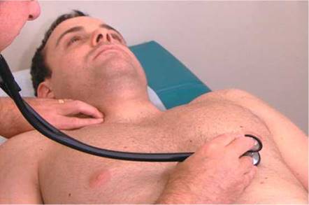
Figure 14-32 Technique for timing the heart sounds.
Approach to Careful Auscultation
Until the examiner gains expertise in cardiac examination, heart sounds should be evaluated in the manner suggested in Table 14-7. The examiner should take time in each area before continuing on to the next area. The examiner should listen to several cardiac cycles at each position to be certain of the observations made, which include respiratory effects.
Describe Any Murmurs Present
If a murmur is present, attention should be directed to the following features:
• Timing in the cardiac cycle
• Location
• Radiation
• Duration
• Intensity
• Pitch
• Quality
• Relationship to respiration
• Relationship to body position
Timing of murmurs as to systole and diastole is paramount. Does the systolic murmur begin with, or after, Si? Does it end before, with, or after S2? Does the murmur occupy the entire systolic period? Murmurs occurring throughout systole are termed holosystolic or pansystolic. These murmurs begin with S1 and end after S2. A systolic ejection murmur begins after S1 and ends before S2. Does the murmur occur only in early systole, midsystole, or late systole? Does the murmur persist throughout the entire diastolic period? Such murmurs are termed holodiastolic.
Table 14-7 Approach to Cardiac Auscultation
|
Position |
Evaluate |
|
Supine |
51 in all areas 52 in all areas Systolic murmurs or sounds in all areas |
|
Left lateral decubitus |
Diastolic events at apex with bell of stethoscope |
|
Upright |
51 in all areas 52 in all areas Systolic murmurs or sounds in all areas Diastolic murmurs or sounds in all areas |
|
Upright, leaning forward |
Diastolic events at base with diaphragm of stethoscope |
In which area is the murmur best heard?
The radiation of the murmur can provide a clue as to its cause. Does it radiate to the axilla? the neck? the back?
The intensity of a murmur is graded from I to VI, based on increasing loudness. The following grading system, although antiquated, serves as a means of communicating the intensity of the murmur:
I: Lowest intensity, often not heard by inexperienced listeners II: Low intensity, usually audible by inexperienced listeners III: Medium intensity without a thrill IV: Medium intensity with a thrill
V: Loudest murmur that is audible when the stethoscope is placed on the chest; associated with a thrill
VI: Loudest intensity: audible when stethoscope is removed from chest; associated with a thrill
Murmurs can be described, for example, as ''grade II/VI, grade IV/VI,'' or ''grade II-III/ VI.'' Any murmur associated with a thrill must be at least a grade IV/VI. A grade IV/VI murmur is louder than a grade II/VI murmur only because there is more turbulence; both or neither may have clinical significance. The ''/VI'' is used because there is another, less popular, grading system involving only four categories. An important axiom to remember is the following:
In general, the intensity of a murmur tells nothing about the severity of the clinical state.
The quality of a murmur can be described as rumbling, blowing, harsh, musical, machinery, or scratchy.
Describe Any Pericardial Rubs
Friction rubs are extracardiac sounds of short duration that have a unique quality similar to the sound of scratching on sandpaper. Rubs may result from irritation of the pleura (i.e., a pleural rub) or of the pericardium (i.e., a pericardial rub). Pericardial rubs typically have three components: one systolic and two diastolic. The systolic component occurs during ejection; the two diastolic components occur during rapid filling and atrial contraction. Pericardial rubs are best heard with the patient sitting while holding breath in expiration. Patients with pericardial rubs commonly have chest pain that is lessened by sitting forward. A rub that disappears while the patient holds the breath originates from the pleura.
The Goals of Auscultation
The goals at the end of auscultation are to be able to describe the following:
• The intensity of S1 in all areas
• The intensity of S2 in all areas
• The characterization of any systolic sounds
• The characterization of any diastolic sounds
With experience, the examiner is able to listen to all parts of the cardiac cycle in one area and compare the sounds and events with those of other areas. Normally, S1 is loudest at the apex, and S2 is loudest at the base. Splitting of S2 into A2 and P2 during inspiration is best heard at the pulmonic area with the patient lying on his or her back. This increases venous return and widens the A2-P2 split.
Examination for Edema
When peripheral venous pressure is high, as in congestive heart failure, pressure in the veins is distributed in a retrograde manner to the smaller vessels. Transudation of fluid occurs, and edema of dependent areas results. This increase in tissue fluid produces edema that ''pits.''
Test for Edema
To test for pitting edema, the examiner presses his or her fingers into a dependent area, such as the patient's shin, for 2 to 3 seconds. If pitting edema is present, the fingers sink into the tissue, and when the fingers are removed, the impression of the fingers remains. This technique is demonstrated in Figure 14-33.
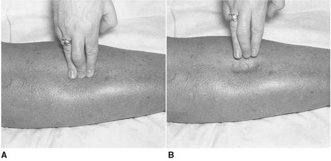
Figure 14-33 Technique for testing for pitting edema. A, The examiner presses into the patient's shin area. B, When pitting edema is present, indentation occurs after the fingers are lifted.
Pitting edema is usually quantified from 1+ to 4+, depending on how long the indentation persists. The most noticeable is 4+. In patients who are bedridden, the dependent area is usually the sacrum and not the shins. The examiner should evaluate for edema over the sacrum in these patients. Figure 14-34 illustrates 4+ sacral edema in a bedridden patient.
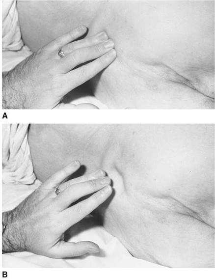
Figure 14-34 Technique for testing for pitting edema over the sacrum. A, The examiner presses into the sacrum of a bedridden patient. B, The pitting edema is evident.
Clinicopathologic Correlations
Discussion now focuses on the pathologic changes that result in the following:
• Abnormalities of S1
• Abnormalities of S2
• Systolic clicks
• Diastolic opening snaps
• Murmurs
Abnormalities of the First Heart Sound
The factors that are responsible for the intensity of S1 are as follows:
• The rate of rise of ventricular pressure
• The condition of the valve
• The position of the valve
• The distance of the heart from the chest wall
The faster the rate of rise of left ventricular pressure is, the louder the mitral component of S1 is. Increased contractility increases the intensity of S1. Decreased contractility softens S1.
When the atrioventricular valve stiffens as a result of fibrosis or calcification, its closure is louder. The pathologically deformed valve of mitral stenosis produces an accentuated or louder S1. After many years, as the valve becomes increasingly calcified, it becomes unable to move, causing S1 to soften.
The position of the valve at the time of ventricular contraction affects the intensity of S1. The arc of coaptation is the angle through which the valve closes. If the valve is in a midposition, it travels less than when it closes from a widely opened position. The more it is opened, the wider the arc of coaptation, and the louder is S1. This situation is directly related to the pressure in the left atrium at the moment that the left ventricular pressure exceeds it and closes the valve. This can occur in conditions in which there is a shortened PR interval on the electrocardiogram. The mitral valve is opened normally during diastole for ventricular filling. The P wave of the electrocardiogram corresponds to atrial contraction, which elevates left atrial pressure (the ''a'' wave of the left atrial tracing), further opening the mitral valve in late diastole. If the PR interval is short, ventricular contraction occurs so quickly after atrial contraction that the atrial pressure is still high when the left ventricular pressure exceeds it. The mitral valve stays open longer and closes later than normal, during the rapid rate of rise of pressure of the ventricle, which accentuates S1.
In general, the longer the PR interval, the softer the S1. Lengthening of the PR intervals, as is seen in Wenckebach's phenomenon,* produces an S1 that softens until the dropped beat occurs.
*Gradually increasing PR intervals until a dropped beat occurs.
Whenever the heart is farther from the chest wall, the S1 is softer than normal. In patients who are very obese or who have chronic obstructive lung disease, the intensity of the S1 is softer than normal. In patients with a large pericardial effusion, the S1 is likewise soft.
Abnormalities of the Second Heart Sound
Abnormalities of the Intensity of the Second Heart Sound
The conditions that change the intensity of S2 are the following:
• Changes in systolic pressure
• Condition of the valve
Any condition that produces an increase in the systolic pressure increases the intensity of the S2. Conversely, conditions that lower the systolic pressure soften the S2. Hypertension raises aortic systolic pressure and produces a loud A2 component of S2.
Calcification or fibrosis of the semilunar valves produces a softening of their closure, S2. Because the semilunar valves are a morphologically different type of valve, fibrosis does not cause an increased intensity, as in closure of a fibrotic atrioventricular valve.
Abnormalities of Splitting of the Second Heart Sound
Normal physiologic splitting of S2 was discussed in the section The Cardiac Cycle. This section deals with abnormalities of splitting.
Any condition that delays right ventricular systole, either electrically or mechanically, delays P2 and produces a widened splitting of S2. Right ventricular emptying is delayed by a right bundle branch block or pulmonic stenosis. The pulmonic component of S2 is delayed during both inspiration and expiration, and wide splitting of S2 occurs.
Any condition that shortens left ventricular systole allows A2 to occur earlier than normal, and wide splitting likewise occurs. Conditions such as mitral regurgitation, ventricular septal defect, and PDA shorten left ventricular systole, and the S1-A2 interval is shorter than normal. In these conditions, there is a ''double outlet'' to the left ventricle, and systole therefore is shorter. In a ventricular septal defect, with a left-to-right shunt, not only is left ventricular systole shorter but also right ventricular systole is prolonged. Both factors are crucial in producing the wide splitting of S2.
Any condition, either electrical or mechanical, that delays left ventricular emptying produces paradoxical splitting of S2. Left bundle branch block or aortic stenosis delays left ventricular emptying. These conditions delay the closure of the aortic valve after right ventricular systole and P2 have occurred. The normal sequence of A2-P2 is reversed. During inspiration, P2 moves normally away from S1 toward A2. The split is said to be narrowed. With expiration, P2 moves normally and approaches S1(- the P2-A2 split widens. This widening during expiration is paradoxical. Other conditions, such as left ventricular failure and severe hypertension, delay left ventricular ejection and cause paradoxical splitting of S2.
Fixed splitting of S2 is the auscultatory hallmark of an atrial septal defect. In this situation, the split is wide and does not change with respiration. This is because inspiratory increases in venous return to the right atrium normally raise its pressure. During expiration, the right atrial pressure is lower, but the left-to-right atrial shunt keeps the volume in the right atrium constant during respiration; therefore, normal splitting does not occur.
Normal physiologic splitting of the second heart sound and abnormalities of splitting are illustrated in Figure 14-35.
Systolic Clicks
Ejection clicks are high-pitched sounds that occur early in systole at the onset of ejection and are produced by the opening of pathologically deformed semilunar valves. Pulmonic or aortic stenosis may produce ejection clicks. The sounds are short and have the quality of a ''click.'' Pulmonic ejection clicks are best heard at the pulmonic area, and aortic ejection clicks are heard at the aortic area. As calcification progresses, the mobility of the valve decreases, and the ejection click disappears.
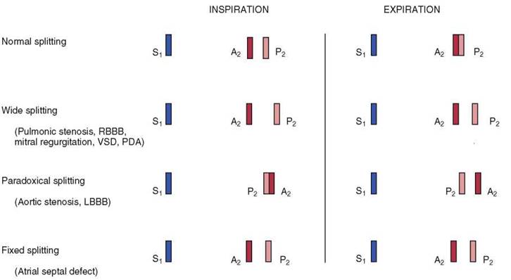
Figure 14-35 Abnormalities of splitting of the second heart sound. (RBBB, right bundle branch block; VSD, ventricular septal defect; PDA, patent ductus arteriosus; LBBB, left bundle branch block.)
Midsystolic clicks are not ejection clicks. They occur in the middle of systole. They may be single or multiple, and they may change in position during the cardiac cycle with various maneuvers that change ventricular geometry. The most common condition associated with a midsystolic click is prolapse of the mitral or tricuspid valve.
Diastolic Opening Snaps
The opening of an atrioventricular valve is normally silent and occurs about 100 msec after S2. This is about as long as it takes to say ''ma-ma'' quickly. An opening snap is a diastolic event that is the sound of the opening of a pathologically deformed atrioventricular valve. The sound is sharp and high-pitched. The mitral opening snap of mitral stenosis occurs after A2; the tricuspid opening snap of tricuspid stenosis occurs after P2.
The interval between S2 and the opening snap is termed the S2-OS interval and has specific significance as to the severity of the stenosis. As mitral stenosis worsens, the resistance and obstruction to flow increase. Pressure in the left atrium increases, and the gradient across the mitral valve increases. The mitral valve, therefore, opens earlier than normal when the left ventricular pressure falls below left atrial pressure. The S2-OS time shortens as the severity increases. Try saying ''ma-da'' as quickly as possible. This interval is about 50 to 60 msec and approximates a very short S2-OS interval, as heard in severe mitral stenosis.
Murmurs
Murmurs are produced when there is turbulent energy in the walls of the heart and blood vessels. Obstruction to flow or flow from a narrow vessel to a large-diameter vessel produces turbulence. Turbulence sets up eddies that strike the walls to produce vibrations that the examiner recognizes as a murmur. Murmurs can also be produced when there is a large volume of blood through a normal opening. In this circumstance, the normal opening is relatively stenotic for the increased volume. ''Blowing'' murmurs are produced by large gradients with variable flow volumes. ''Rumbling'' murmurs result from areas of small gradients that are dependent on flow. ''Harsh'' murmurs result from large gradients and high flow.
An ejection murmur is a murmur produced by turbulence across a semilunar valve during systole, such as in aortic stenosis or pulmonic stenosis. Ejection murmurs appear diamond-shaped and are described as ''crescendo-decrescendo.'' They begin slightly after S1 and end before S2. An ejection click from stenosis of the semilunar valve may precede the murmur. These murmurs are medium-pitched and are best heard with the diaphragm of the stethoscope. Because they are based on flow, the intensity of these murmurs does not indicate the degree of severity. Increased flow across a minimally narrowed aortic valve produces a loud murmur; decreased flow across a severely stenotic aortic valve may produce a barely audible murmur. Any increase in flow or volume may produce an ejection murmur even in the presence of a normal valve. An ejection murmur as a sign of aortic stenosis is a finding of high sensitivity but low specificity. Figure 14-36 illustrates an ejection murmur.
Regurgitant systolic murmurs are produced by retrograde flow from a higher pressure area to a lower pressure area during systole, such as in mitral or tricuspid regurgitation. These murmurs are holosystolic or pansystolic. They begin with S1 and end after S2. They extend past S2 because ventricular pressure is higher than atrial pressure, even after the closure of the semilunar valve. An S3 indicative of volume overload to the ventricle is often heard. These murmurs are high-pitched and are best heard with the diaphragm of the stethoscope. The terms regurgitation, incompetence, and insufficiency are often used synonymously for this type of murmur. The preferred term is regurgitation because it implies the retrograde direction of flow. The holosystolic murmur of atrioventricular valve regurgitation is a finding of high sensitivity. Figure 14-37 illustrates a regurgitant murmur.
Diastolic atrioventricular murmurs begin after S2 with the opening of the atrioventricular valve. Mitral stenosis and tricuspid stenosis are examples of this type of murmur. There is a pause between S2 and the beginning of the murmur. Isovolumetric relaxation is occurring during this period. The murmur is decrescendo in shape, beginning with an opening snap, if the valve is mobile. These murmurs are low-pitched and are best heard with the bell of the stethoscope, with the patient lying in the left lateral decubitus position. Because the atrioventricular valve is stenotic, rapid filling does not occur, and a gradient persists throughout diastole. If the patient is in normal sinus rhythm, atrial contraction increases the gradient at the end of diastole, or presystole, and there is an increase in the murmur at this time. The diastolic atrioventricular murmur is a sensitive and specific sign of atrioventricular valve stenosis.
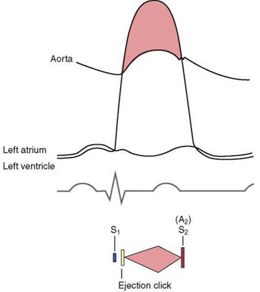
Figure 14-36 A systolic ejection murmur such as that occurring in aortic stenosis.
The first heart sound is the loudest sound. The cadence and emphasis of the sounds are best heard by saying the following mnemonic: in which OS is the mitral opening snap, and DM is the diastolic murmur. The ''aaaalve'' is the presystolic accentuation heard when a patient with mitral stenosis is in normal sinus rhythm. Figure 14-38 illustrates a diastolic atrioventricular murmur.

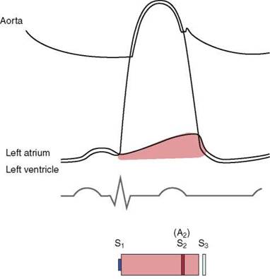
Figure 14-37 A systolic regurgitant murmur such as that occurring in mitral regurgitation.
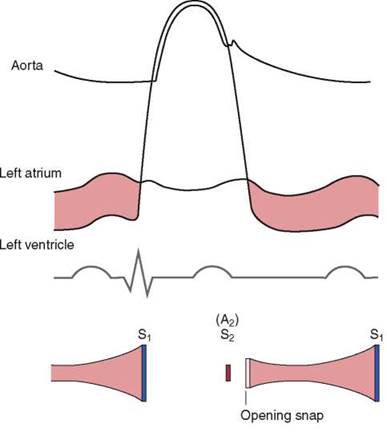
Figure 14-38 A diastolic atrioventricular murmur such as that occurring in mitral stenosis.
Diastolic semilunar murmurs begin immediately after S2, as heard in aortic or pulmonic regurgitation. In contrast to the diastolic atrioventricular murmurs, there is no delay after S2 to the beginning of the murmur. The high-pitched murmur is decrescendo in shape and is best heard with the diaphragm of the stethoscope while the patient is sitting up and leaning forward. A diastolic semilunar murmur is a sign of low sensitivity but high specificity. Figure 14-39 illustrates the pressure curves responsible for the generation of a diastolic semilunar murmur.
Table 14-8 lists important cardiac sounds according to the cardiac cycle. Figure 14-40 lists important characteristics of the systolic murmurs of aortic stenosis and mitral regurgitation. Table 14-9 summarizes the differentiation of some additional systolic murmurs. Figure 14-41 lists important characteristics of the diastolic murmurs of mitral stenosis and aortic regurgitation.

Figure 14-39 A diastolic semilunar murmur such as that occurring in aortic regurgitation. Note the systolic ejection murmur, which is related to the increased volume and flow.
Table 14-8 Cardiac Sounds
|
Cardiac Cycle |
Sound |
|
Early systolic |
Ejection click Aortic prosthetic valve opening sound* |
|
Midsystolic to late systolic |
Midsystolic click Rub |
|
Early diastolic |
Opening snap S3 Mitral prosthetic valve opening sound* Tumor plop* |
|
Mid-diastolic |
S3 Summation gallop§ |
|
Late diastolic (sometimes called presystolic) |
S4 Pacemaker sound |
|
*Opening and closure of the prosthetic aortic valve are heard with many prosthetic valves. The opening is comparable to an ejection click; the closing is a ''prosthetic'' S2. *Opening and closure of the prosthetic mitral valve are heard with many prosthetic valves. The opening is comparable to an opening snap; the closing is a ''prosthetic'' Si. *A left atrial myxoma that is pedunculated may ''plop'' in and out of the mitral annulus, simulating the auscultatory signs of mitral stenosis. §At fast heart rates, the diastolic period shortens. If an S3 and S4 are present, the sounds may be summated into a single sound called a summation gallop. |
|
|
Systolic Murmurs Differentiating As from MR |
||
|
Feature |
Aortic Stenosis |
Mitral Regurgitation |
|
Location |
Aortic area |
Apex |
|
Radiation |
Neck |
Axilla |
|
Shape |
Diamond |
Holosystolic |
|
Pitch |
Medium |
High |
|
Quality |
Harsh |
Blowing |
|
Associated signs |
Decreased A2 Ejection click S4 Narrow pulse pressure Slow rising and delayed pulse |
Decreased $1 $3 Laterally displaced diffuse PMI |
|
PMI, point of maximum impulse. |
||

Figure 14-40 Systolic murmurs. A, Pathophysiology of aortic stenosis. Note the paradoxical splitting of the second heart sound (S2), the S4, and the ejection click. B, Mitral regurgitation. Note that the murmur ends after S2, and note the presence of the S3.
Table 14-9 Differentiation of Other $ystolic Murmurs
|
Feature |
Pulmonic Stenosis |
Tricuspid Regurgitation |
Ventricular Septal Defect |
Venous Hum |
Innocent Murmur |
|
Location |
Pulmonic area |
Tricuspid area |
Tricuspid area |
Above clavicle |
Widespread |
|
Radiation |
Neck |
Right of sternum |
Right of sternum |
Right neck |
Minimal |
|
Shape |
Diamond |
Holosystolic |
Holosystolic |
Continuous |
Diamond |
|
Pitch |
Medium |
High |
High |
High |
Medium |
|
Quality |
Harsh |
Blowing |
Harsh |
Roaring; humming |
Twanging; vibratory |
*Usually between the apex and the left lower sternal border.
|
Diastolic Murmurs Differentiating M$ from AR |
||
|
Feature |
Mitral Stenosis |
Aortic Regurgitation |
|
Location |
Apex |
Aortic area |
|
Radiation |
No |
No |
|
$hape |
Decrescendo |
Decrescendo |
|
Pitch |
Low |
High |
|
Quality |
Rumbling |
Blowing |
|
Associated signs |
Increased S1 Opening snap RV rock* Presystolic accentuation |
S3 Laterally displaced PMI Wide pulse pressure* Bounding pulses Austin Flint murmur* Systolic ejection murmur* |
|
PMI, point of maximum impulse; RV, right ventricular. *Right ventricular impulse at lower left sternal border. *The wide pulse pressure is the cause of the many physical signs of aortic regurgitation: Quincke's pulse, De Musset's sign, Duroziez's sign, Corrigan's pulse, and so forth. *An apical diastolic murmur heard in association with aortic regurgitation, mimicking that of mitral stenosis. *A flow murmur across a valve that is relatively narrow for the increased blood volume as a result of aortic regurgitation. It is relatively stenotic and need not be anatomically stenotic, as in true aortic stenosis. |
||
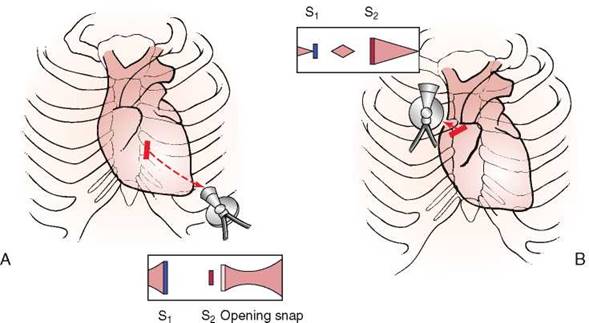
Figure 14-41 Diastolic murmurs. A, Pathophysiology of mitral stenosis. Note the intensity of S1 and the accentuation of the diastolic murmur in late diastole. B, Aortic regurgitation. Note the systolic flow murmur.
Useful Vocabulary
Listed here are the specific roots that are important for understanding the terminology related to cardiac disease.
|
Root |
Pertaining to |
Example |
Definition |
|
brady- |
slow |
bradycardia |
Slow heart rate |
|
-cardio- |
heart |
cardiomegaly |
Enlargement of the heart |
|
sphygmo- |
pulse |
sphygmomanometer |
Instrument for measuring blood pressure |
|
supra- |
above |
supraventricular |
Above the level of the ventricles |
|
tachy- |
fast |
tachycardia |
Rapid heart rate |
Writing Up the Physical Examination
Listed here are examples of the write-up for the examination of the heart.
• No abnormal jugular venous distention or abnormal jugular wave forms are present. The PMI is in the fifth intercostal space, midclavicular line. S1 and S2 are normal.* Physiologic splitting is present. No murmurs, gallops, or rubs are heard. There is no clubbing, cyanosis, or edema.
• The jugular venous pressure is elevated. There is a prominent ''v'' wave present in the neck. The jugular vein is distended 8 cm above the sternal angle at 45°. The PMI is in the sixth intercostal space, anterior axillary line. S1 is soft. S2 is widely split on inspiration and expiration. A grade III/VI, high-pitched, holosystolic murmur is heard at the apex with radiation to the axilla. A palpable S3 is present at the apex. There is 2+ pitting edema on the shins bilaterally. No cyanosis or clubbing is present.
• No abnormal jugular venous distention is seen. The PMI is in the fifth intercostal space, midclavicular line. S1 is normal. S2 is soft. An S4 is present at the apex. A grade IV/VI, harsh, medium-pitched, crescendo-decrescendo murmur, beginning slightly after S1 and ending before S2, is present at the aortic area. This murmur radiates to both carotid arteries. No clubbing, cyanosis, or edema is present.
• The jugular venous pressure is very elevated. The jugular vein is distended to the jaw margin when the patient is seated at 90°. The PMI is in the fifth intercostal space, midclavicular line. S1 is accentuated. S2 is normal. An RV* rock is present at the left lower sternal border. There is a grade II/VI, low-pitched, diastolic rumble heard at the apex, best heard in the left lateral decubitus position. A grade III/VI, high-pitched, holosystolic murmur is heard at the left lower sternal border, which increases in intensity with inspiration. A right ventricular S3 may be present at the left lower sternal border.* There is 4+ pitting sacral edema. No cyanosis or clubbing is present.
*The descriptors for S1 and S2 are normal, increased, decreased, widely split, narrowly split, fixed split, or paradoxically split. Never indicate that S1 and S2 are present.
*Right ventricular.
*Notice that in this example the examiner stated that a finding may be present.
Bibliography
American Heart Association: Heart Disease and Stroke Statistics—2005 Update. Dallas, American Heart Association, 2005.
Braunwald E, Zipes DP, Libby P: Heart Disease: A Textbook of Cardiovascular Medicine, 6th ed. Philadelphia, WB Saunders, 2001.
Chandra NC, Ziegelstein RC, Rogers WJ, et al: Observations of the treatment of women in the United States with myocardial infarction: A report from the National Registry of Myocardial Infarction—I. Arch Intern Med 158:981, 1998.
Cook DJ, Simel DL: Does this patient have abnormal central venous pressure? JAMA 275:630, 1996.
Crawford MH, DiMarco JP: Cardiology. St. Louis, Mosby, 2000.
Ducas J, Magder S, McGregor M: Validity of the hepatojugular reflux as a clinical test for congestive heart failure. Am J Cardiol 52:1299, 1983.
Eilen SD, Crawford MH, O'Rourke RA: Accuracy of precordial palpation for detecting increased left ventricular volume. Ann Intern Med 99:628, 1983.
Elliott WJ: Ear lobe crease and coronary artery disease. Am J Med 75:1024, 1983.
Ewy G: The abdominojugular test: Technique and hemodynamic correlates. Ann Intern Med 109:456, 1988.
Heckerling PS, Wiener SL, Moses VK, et al: Accuracy of precordial percussion in detecting cardiomegaly. Am J Med 91:328, 1991.
Heckerling PS, Wiener SL, Wolfkiel CJ, et al: Accuracy and reproducibility of precordial percussion and palpation for detecting increased left ventricular end-diastolic volume and mass. JAMA 270:1943, 1993.
Henkind SJ, Benis AM, Teichholz LE: The paradox of pulsus paradoxus. Am Heart J 114:198, 1987.
Johnson R, Swartz MH: A Simplified Approach to Electrocardiography. Philadelphia, WB Saunders, 1986.
Julian D, Wenger NK (eds): Women and Heart Disease. London, Martin Dunitz, 1997.
Kroenke MK: Sphygmomanometry: The correct arm position. West J Med 140:459, 1984.
Maisel AS, Atwood JE, Goldberger AL: Hepatojugular reflux: Useful in the bedside diagnosis of tricuspid regurgitation. Ann Intern Med 101:781, 1984.
Marrugat J, Sala J, Masia R, et al: Mortality differences between men and women following myocardial infarction. JAMA 280:1405, 1998.
Minmo AM, Heron MP, Murphy SL, et al: Deaths: Final Data for 2004. National Vital Statistics Report, vol 55, no. 19. Hyattsville, Md, National Center for Health Statistics, 2007.
Murphy SL: Deaths: Final Data for 1998. National Vital Statistics Report, vol 48, no. 11. Hyattsville, Md, National Center for Health Statistics, 2000.
Parikh NI, Pencina MJ, Wang TJ, et al: A risk score for predicting near-term incidence of hypertension: The Framingham Heart Study. Ann Intern Med 148:102, 2008.
Pryor DB, Shaw L, McCants CB, et al: Value of the history and physical in identifying patients at increased risk for coronary artery disease. Ann Intern Med 118:81, 1993.
Rosendorff C: Essential Cardiology: Principles and Practice. Philadelphia, WB Saunders, 2000.
Schouten VW, Bohnen AM, Bosch JLHR, et al: Erectile dysfunction prospectively associated with cardiovascular disease in the Dutch general population: Results from the Krimpen Study. Int J Impotence Res 20:92, 2008.
Silverberg DS, Shemesh E, Iaina A: The unsupported arm: A cause of falsely raised blood pressure readings. BMJ 2:1331, 1977.
Thomas JE, Schirger A, Fealey RD, et al: Orthostatic hypotension. Mayo Clin Proc 56:117, 1981.
Tsang TSM, Barnes ME, Gersh BJ, et al: Risks of coronary heart disease in women: Current understanding and evolving concepts. Mayo Clin Proc 75:1289, 2000.