
Veins which by the thickening of their tunicles in the old restrict the passage of blood, and by this lack of nourishment destroy their life without any fever, the old coming to fail little by little in slow death.
Leonardo da Vinci (1452-1519)
General Considerations
Diseases of the peripheral vascular system are common and may involve the arteries, veins, or lymphatic vessels. The arterial conditions include cerebrovascular, aortoiliac, femoropopliteal, renal, aortic occlusive, and aneurysmal diseases. The two most important diseases of the peripheral arteries are atherosclerosis of the larger arteries and microvascular disease.
The most common cause of peripheral arterial occlusive disease is atherosclerosis affecting the medium-sized and large vessels of the extremities. Narrowing of the vessel causes a decreased blood supply, resulting in ischemia. In addition, atherosclerosis may become manifest by aneurysmal dilatation. The abdominal aorta is the artery most frequently involved. The aneurysm is commonly below the renal arteries and may extend as far as the external iliac arteries. Often, this aneurysm produces few, if any, symptoms. The examiner may discover a pulsatile mass as an incidental finding. Frequently, the first manifestation is the catastrophic rupture of the aneurysm. An abdominal aortic aneurysm (AAA) larger than 5 cm in diameter carries a 20% risk of rupturing within the first year of discovery and a 50% risk of rupturing within 5 years.
Vesalius described the first AAA in the 16th century. Before the development of a surgical intervention for the process, attempts at medical management failed. The initial surgical attempts at control entailed ligation of the aorta, with poor results. In 1923, Rudolph Matas performed the first successful aortic ligation on a patient. Attempts were made to induce thrombosis by inserting intraluminal wires. In 1948, C. E. Rea wrapped reactive cellophane around the aneurysm in order to induce fibrosis and limit expansion. This technique was used on Albert Einstein in 1949, and he survived 6 years before dying of rupture. However, not until 1951 was an abdominal aneurysm surgically treated by resection and grafting. In that year, C. Dubost performed the first AAA repair with a homograft. Since then, great strides have been made in understanding the natural history of vascular disease, as well as in developing new technology to help diagnose and treat it.
In autopsy studies, the frequency rate of AAA ranges from 0.5% to 3.2%. In a large U.S. Veterans Administration screening study, the prevalence rate was 1.4%. The frequency of rupture is 4.4 cases per 100,000 persons. AAA is 5 times more common in men than in women and is 3.5 times more common in white men than in African-American men. The likelihood of development varies from 3 to 117 cases per 100,000 person-years.
Microvascular arterial disease occurs in patients with diabetes. Changes develop in the small arterioles that impair circulation to the skin or nerves, especially of the lower extremities, producing symptoms of ischemia. Peripheral neuropathy is a common sequela of microvascular disease. This neuropathy may be manifested as a defect in the sensory, motor, or autonomic system. Microvascular disease affects more than 15 million individuals in the United States.
Peripheral venous disease often progresses to venous stasis and thrombotic disorders. One of the dreaded complications of thrombotic disease is pulmonary embolism. In the United States, more than 175,000 deaths per year are attributed to acute pulmonary embolism.
Structure and Physiology
Diseases of the peripheral arterial system cause ischemia of the extremities. When the body is at rest, collateral blood vessels may be able to provide adequate circulation. During exercise, when oxygen demand increases, this circulation may not be sufficient for the actively contracting muscles, and ischemia may result.
The venous system consists of a series of low-pressure capacitance vessels. Nearly 70% of the blood volume is contained in this system. Although offering little resistance, the veins are controlled by a variety of neural and humoral stimuli that enhance venous return to the right side of the heart. In addition, valves aid in the return of blood.
When an individual is in the upright posture, the venous pressure in the lower extremity is the highest. Over many years, the veins dilate as a result of weakening of their walls. As the walls dilate, the veins are unable to close adequately, and reflux of blood occurs. In addition, the venous pump becomes less efficient in returning blood to the heart. Both of these factors are responsible for the venous stasis seen in patients with chronic venous insufficiency. Complications from venous stasis include pigmentation, dermatitis, cellulitis, ulceration, and thrombus formation.
The lymphatic system is an extensive vascular network and is responsible for returning tissue fluid (lymph) back to the venous system. The extremities are richly supplied with lymphatic tissue. Lymph nodes, many of which are located between major proximal joints, aid in filtering the lymphatic fluid before it enters the blood. The most important clinical symptoms of lymphatic obstruction are lymphedema and lymphangitis.
Review of Specific Symptoms
Many patients with peripheral vascular disease are asymptomatic. When patients are symptomatic, vascular disease causes the following:
• Pain
• Changes in skin temperature and color
• Edema
• Ulceration
• Emboli
• Stroke
• Dizziness
Pain
Pain is the principal symptom of atherosclerosis. Whenever a patient complains of pain in the calf, arch of the foot, thighs, hips, or buttocks while walking, peripheral vascular disease of the arteries must be considered. The symptom of pain in the lower extremity during exercise is called intermittent claudication. The site of the pain is always distal to the occlusive disease. As the disease progresses, pain at rest occurs. This is often severe and is aggravated by cool temperatures and elevation, especially during sleep in bed. Pain may also occur with deep vein thrombosis.
If a male patient complains of buttock or thigh pain while walking, the examiner should inquire about erectile dysfunction. Leriche's syndrome is chronic aortoiliac obstruction; the patient presents with intermittent claudication and erectile dysfunction. In this condition, the terminal aorta and iliac arteries are involved by severe atherosclerosis at the aortic bifurcation.
Patients occasionally complain of bilateral leg pain or numbness that occurs while walking, as well as while resting. This is called pseudoclaudication and is a symptom of musculoskeletal disease in the lumbar area.
Skin Changes
Skin color changes are common with vascular disease. In chronic arterial insufficiency, the affected extremity is cool and pale. In chronic venous insufficiency, the extremity is warmer than normal. The leg becomes erythematous, and erosions produced by excoriation result. With chronic insufficiency, stasis changes produce increased pigmentation, swelling, and an ''aching'' or ''heaviness'' in the legs. These changes characteristically occur in the lower third of the extremity and are more prominent medially. When venous insufficiency occurs, edema of dependent areas results.
Patients with acute deep vein thrombosis have secondary inflammation of the tissue surrounding the vein. This produces signs of inflammation: warmth, redness, and fever. Swelling is the most reliable symptom and sign associated with venous obstruction. This finding is indicative of severe deep vein obstruction because the superficial veins of the lower extremity carry only 20% of the total drainage and are not associated with swelling. The extremities should be compared, and a difference in circumference of 2 cm at the ankle or midcalf should be considered significant.
Edema
Lymphedema results from either a primary abnormality in the development of the lymphatic system or an acquired obstruction to flow. Whether the congenital or the acquired form is involved, the net result is stasis of lymph fluid in the tissues, producing a firm, nonpitting edema. Over several years, the skin takes on a rough consistency similar to that of pigskin. Because lymphedema is usually painless, the only symptom is ''heaviness'' of the extremity.
Ulceration
Persistent ischemia of a limb is associated with ischemic ulceration and gangrene. Ulceration is almost inevitable once skin has thickened and the circulation is compromised. Ulceration related to arterial insufficiency occurs as a result of trauma to the toes and heel. These ulcers are painful, have discrete edges that produce a ''punched-out'' appearance, and are often covered with crust. When infected, the tissue is erythematous.
In contrast to arterial insufficiency ulceration, venous insufficiency leads to stasis ulceration, which is painless and occurs in the ankle area or lower leg just above the medial malleolus. The classic manifestation is a diffusely reddened, thickened area over the medial malleolus. The skin has a cobblestone appearance resulting from fibrosis and venous stasis. Ulceration occurs with the slightest trauma. Rapidly developing ulcers are commonly caused by arterial insufficiency, whereas slowly developing ulceration is usually the result of venous insufficiency. Figures 15-1 and 15-2 show stasis dermatitis and ulcerations over the medial malleoli. Patients with leg ulcers should be asked the following:
''What did the ulcer look like when it first appeared?''
''What do you think started the ulcer?''
''How quickly did it develop?''
''How painful is the ulcer?''
''What kind of medications have you been taking?''
''Is there a history of any generalized diseases, such as anemia? rheumatoid arthritis?''
''Is there a family history of leg ulcers?''

Figure 15-1 Stasis dermatitis and bilateral ulceration over the medial malleoli.
Emboli
A history of emboli is important. Thrombus formation results from stasis and hypercoagulability. It appears, however, that venous stasis is the most important cause of thrombus formation. Bed rest, congestive heart failure, obesity, pregnancy, recent extended travel on airplanes, and oral contraceptives have been linked to thrombus formation and emboli.
Symptoms secondary to emboli can include shortness of breath from pulmonary emboli; abdominal pain from splenic, intestinal, or renal artery emboli; neurologic symptoms from carotid or vertebrobasilar artery emboli; and pain and paresthesias from peripheral artery emboli.
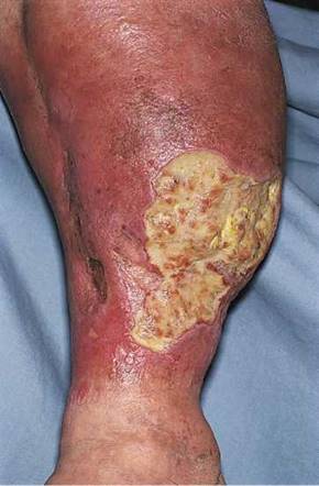
Figure 15-2 Stasis dermatitis and ulceration over the medial malleolus.
Neurologic Symptoms
Cerebrovascular occlusive disease causes many neurologic symptoms, including strokes,* dizziness, and changes in consciousness. Occlusion of the internal carotid artery produces a syndrome of contralateral hemiplegia, contralateral sensory deficits, and dysphasia. Vertebrobasilar disease is associated with diplopia, cerebellar dysfunction, changes in consciousness, and facial paresis.
Impact of Vascular Disease on the Patient
A patient with chronic arterial insufficiency has worsening pain while walking. As the condition progresses, ulceration of the toes, feet, and areas susceptible to trauma, such as the shins, develops. Pain may become excruciating. Gangrene of a toe may develop, and amputation of the toe is frequently followed by amputation of the foot and leg. In addition, the patient becomes increasingly depressed as a result of ongoing mutilation of the body.
Physical Examination
The equipment necessary for the examination of the peripheral vascular system consists of a stethoscope, a tourniquet, and a tape measure.
The physical examination of the peripheral vascular system consists of inspection, palpation of the arterial pulses, and some additional tests if disease is thought to be present. All these techniques are usually integrated with the rest of the physical examination.
The patient lies supine, with the examiner standing to the right of the bed. The evaluation of the peripheral vascular system includes the following:
Inspection
• Examination of the arterial pulses
• Examination of the lymphatic system
• Other special techniques
Inspection
Inspect for Symmetry of the Extremities
The extremities should be compared for asymmetries in size, color, temperature, and venous patterns. Figure 15-3 depicts massive lymphedema of the right upper extremity secondary to a right total mastectomy 18 years earlier.
Inspect the Lower Extremities
The lower extremities should be inspected for pigmentary abnormalities, ulcers, edema, and venous patterns. Is cyanosis present? Is edema present? If edema is present, does it pit? Bilateral color changes and swelling of the legs of the patient are pictured in Figure 15-4. The patient had chronic venous insufficiency. She died of a massive pulmonary embolus 1 day after this photograph was taken.
Assess the Skin Temperature
Evaluate the temperature by using the back of your hand. Compare similar areas of each extremity. Coolness of an extremity is common with arterial insufficiency.
Inspect for Varicosities
Ask the patient to stand, and inspect the lower extremities for varicosities. Look at the area of the proximal femoral ring, as well as in the distal portion of the legs. Varicose veins in these locations may not have been visible when the patient was lying down.
The patient in Figure 15-5 was a 37-year-old woman with severe right-sided heart failure. Notice the marked dilated and tortuous veins in the popliteal fossa. Also notice the increased pigmentation of the skin over the lower legs.
*A stroke is also known as a cerebrovascular accident.

Figure 15-3 Lymphedema.
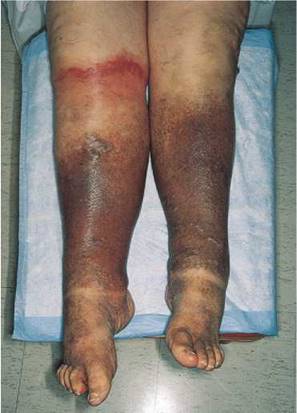
Figure 15-4 Chronic venous insufficiency.
Examination of the Arterial Pulses
The most important finding when the peripheral arterial tree is examined is a decreased or absent pulse. The radial, brachial, femoral, popliteal, dorsalis pedis, and posterior tibial pulses are routinely evaluated.
Palpate the Radial Pulse
You should stand in front of the patient. The radial pulses are evaluated by grasping both of the patient's wrists and palpating the pulses with your index, middle, and fourth fingers. Hold the patient's right wrist with your left fingers and the patient's left wrist with your right fingers, as demonstrated in Figure 14-22. The symmetry of the pulses is evaluated for timing and strength.

Figure 15-5 Marked varicosities of the popliteal fossa.

Figure 15-6 Technique for brachial artery palpation.
Palpate the Brachial Pulse
Because the brachial pulse is stronger than the digital pulses, the examiner may use his or her thumbs to palpate the patient's brachial pulses. The brachial artery can be felt medially just under the belly or tendon of the biceps muscle. With the examiner still standing in front of the patient, both brachial arteries can be palpated simultaneously. The examiner's left hand holds the patient's right arm, and the examiner's right hand holds the patient's left arm. Once the examiner's thumbs feel the brachial pulsation, the examiner should apply progressive pressure to it until the maximal systolic force is felt. This is demonstrated in Figure 15-6. The examiner should now be able to assess its wave form.
Auscultate the Carotid Artery
Auscultation for carotid bruits* is performed by placing the diaphragm of the stethoscope over the patient's carotid artery while the patient is lying supine. This is demonstrated in Figure 15-7. The patient's head should be slightly elevated on a pillow and turned slightly away from the carotid artery being evaluated. It is often helpful to ask the patient to hold his or her breath during the auscultation. Normally, either nothing or transmitted heart sounds are heard. After one carotid artery is evaluated, the other is examined. After auscultation of the carotid arteries, they are palpated (see Chapter 14, The Heart).
The presence of a murmur should be noted. This may be a bruit resulting from atherosclerotic disease of the carotid artery. On occasion, loud murmurs originating from the heart can be transmitted to the neck. With experience, the examiner is able to determine whether the disorder is local in the neck or distal in the heart.

Figure 15-7 Technique for auscultation of the carotid artery.
*A bruit is a sound or murmur heard in a vessel as a result of increased turbulence.
Table 15-1 Characteristics of Physical Signs for Detecting an Abdominal Aortic Aneurysm
|
Physical Sign |
Sensitivity (%) |
Specificity (%) |
|
Definite pulsatile mass |
28 |
97 |
|
Definite or suggestive pulsatile mass |
50 |
91 |
|
Abdominal bruit |
11 |
95 |
|
Femoral bruit |
17 |
87 |
|
Femoral pulse deficit |
22 |
91 |
Data from Lederle FA, Walker JM, Reinke DB: Selective screening for abdominal aortic aneurysms with physical examination and ultrasound. Arch Intern Med 148:1753, 1988.
Palpate the Abdominal Aorta
AAAs kill approximately 10,000 people in the United States each year. Many of these deaths could be prevented if the patients were aware of the presence of this defect. Once the defect is recognized, the rate of operative mortality for a nonruptured aneurysm is less than 5%, and after operation, the survival rate equals that of the general population. An AAA that ruptures carries a mortality rate of nearly 90%. Even among patients who reach the operating room alive, the surgical mortality rate is 50%.
The examination is performed by palpating deeply, but gently, into the midabdomen. The presence of a mass with laterally expansive pulsation suggests an AAA. Some caution is urged in making this diagnosis in thin individuals, in whom the normal pulsatile aorta can be easily palpated. The high false-positive rate with this examination should not be a problem, however, because confirmation with abdominal ultrasonography is safe and inexpensive.
Other findings associated with an AAA include an abdominal bruit, a femoral bruit, and a femoral pulse deficit. In fewer than 10% of patients with AAAs, a bruit may be present. Acute rupture of an AAA is suggested when a bruit is associated with severe pain in the abdomen or back and when the distal pulse is absent or diminished and later returns.
Table 15-1 summarizes the physical signs useful for detecting an AAA.
Rule Out Abdominal Bruits
The patient should be supine. The examiner places the diaphragm of the stethoscope in the midline of the patient's abdomen about 2 inches (5 cm) above the umbilicus and listens carefully for the presence of an aortic bruit. This technique is demonstrated in Figure 15-8.
A renal bruit may be the only clue to renal artery stenosis. Auscultation should be performed about 2 inches (5 cm) above the umbilicus and 1 to 2 (2.5 to 5 cm) inches laterally to the right and to the left of midposition.
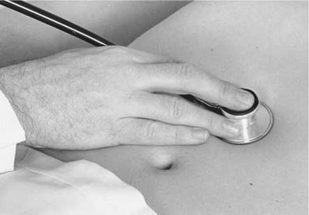
Figure 15-8 Technique for auscultation of the abdominal aorta.
Abdominal bruits that are present only during systole are frequently of little clinical value because they are found in normal individuals and in patients with essential hypertension. The presence of a systolic-diastolic abdominal bruit, however, should raise the suspicion of renovascular hypertension. Nearly 60% of all patients with renovascular hypertension have such a bruit.
The presence of a combined systolic-diastolic abdominal bruit has a sensitivity of 39% and a specificity of 99% for detecting renovascular hypertension. The presence of this type of bruit has a positive likelihood ratio (LR+) of 39 and, if absent, a negative likelihood ratio (LR—) of 0.6. In a study that evaluated the presence of any epigastric or flank bruit and its association with renovascular hypertension, the sensitivity was 63%, but the specificity dropped to 90%. The presence of any abdominal bruit confers a much lower LR+ for renovascular hypertension (i.e., 6.4).
Palpate the Femoral Pulse
The femoral pulse is evaluated with the patient lying on the back and the examiner at the patient's right side. The lateral corners of the pubic hair triangle are observed and palpated. The femoral artery should run obliquely through the corner of the pubic hair triangle inferior to the inguinal ligament at a point midway between the pubic tubercle and the anterior superior iliac spine. Both femoral pulses may be compared simultaneously. The technique is demonstrated in Figure 15-9.
If one of the femoral pulses is diminished or absent, auscultation for a bruit is necessary. The diaphragm of the stethoscope is placed over the femoral artery. The presence of a bruit may indicate obstructive aortoiliofemoral disease.
Rule Out Coarctation of the Aorta
The timing of the femoral and radial pulses is important. Normally, these pulses peak either at the same time or with the femoral pulse preceding the radial pulse. By placing one hand on the patient's femoral artery and the other on the radial artery, the examiner can determine the peaking of these pulses. This technique need be performed on one side only. Any delay in the femoral pulse should raise suspicion of coarctation of the aorta, especially in a hypertensive individual. This technique is demonstrated in Figure 15-10.
Palpate the Popliteal Pulse
The popliteal artery is often difficult to assess. Each artery is evaluated separately. While the patient is lying on the back, the examiner's thumbs are placed on the patella, and the remaining fingers of both hands are pressed in the popliteal fossa medial to the lateral biceps femoris tendon, as demonstrated in Figure 15-11. The examiner should hold the leg in a mild degree of flexion. The patient should not be asked to elevate the leg, because this tightens the muscles and makes it more difficult to feel the pulse. The examiner should squeeze both hands in the popliteal fossa. Firm pressure is usually necessary to feel the pulsation.

Figure 15-9 Technique for palpation of the femoral arteries.
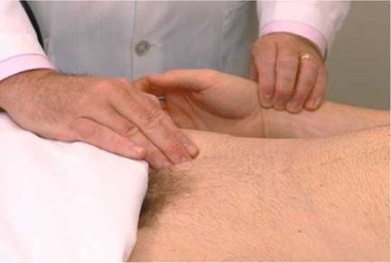
Figure 15-10 Technique for timing the femoral and radial pulses.
Palpate the Dorsalis Pedis Pulse
The dorsalis pedis pulse is best palpated when the foot is dorsiflexed. The dorsalis pedis artery passes along a line from the extensor retinaculum of the ankle to a point just lateral to the extensor tendon of the great toe. It is usually easily palpated in the groove between the extensor digitorum longus and hallucis longus tendon. The dorsalis pedis pulses may be felt simultaneously, as demonstrated in Figure 15-12.
Palpate the Posterior Tibial Pulse
The posterior tibial artery can be palpated just posterior to the medial malleoli between the tibialis posterior tendon and the flexor digitorum longus tendon. Both arteries can be evaluated simultaneously. Figure 15-13 demonstrates this procedure. Although posterior tibial pulses are absent in 15% of normal subjects, the most sensitive sign of occlusive peripheral arterial disease in patients older than 60 years of age is the absence of the posterior tibial pulse.
Grading of Pulses
The description of the amplitude of the pulse is most important. The following is the most widely accepted grading system:
0 Absent
1 Diminished
2 Normal
3 Increased
4 Bounding

Figure 15-11 Technique for palpation of the popliteal artery. A, Correct position of the hands from the front. B, View from behind the popliteal fossa.

Figure 15-12 Technique for palpation of the dorsalis pedis arteries.
It is important that the patient's socks or stockings be removed when the examiner assesses the peripheral pulses of the lower extremities. If there is confusion about whether you are feeling the patient's pulse or your own pulse, you can palpate the patient's pulse with your right hand and use your left hand to palpate your own right radial pulse. If the pulses are different, you are feeling the patient's pulse with your right hand.
Examination of the Lymphatic System
Physical signs of lymphatic system disease include the following:
• Palpable lymph nodes
• Lymphangitis
• Lymphedema
Lymph nodes should be described as painless or tender and as single or matted. Generalized lymphadenopathy and localized lymphadenopathy are suggestive of different diagnoses. Generalized lymphadenopathy is the presence of palpable lymph nodes in three or more lymph node chains. Lymphoma, leukemia, collagen vascular disorders, and systemic bacterial, viral, and protozoal infections may be responsible. Localized lymphadenopathy is usually the result of localized infection or neoplasm.

Figure 15-13 Technique for palpation of the posterior tibial arteries.
Lymphangitis is lymphatic spread manifested by thin red streaks on the skin. Obstruction to lymphatic flow produces lymphedema, which is usually indistinguishable from other types of edema. In Figure 16-8, the patient has marked lymphedema of her left arm secondary to inflammatory breast carcinoma.
The examinations for lymphadenopathy of the head, neck, and supraclavicular areas are described in Chapter 9, The Head and Neck, and in Chapter 13, The Chest. Chapter 16, The Breast, describes the examination for palpating axillary adenopathy. Chapter 18, Male Genitalia and Hernias, describes the technique for palpating inguinal lymph nodes. The only other important lymph node chain is the epitrochlear nodes, discussed in the following section.
Palpate for Epitrochlear Nodes
To palpate epitrochlear nodes, have the patient flex the elbow approximately 90°. Feel for the nodes in the fossa about 3 cm proximal to the medial epicondyle of the humerus, in the groove between the biceps and the triceps muscles. Epitrochlear nodes are rarely palpable, but if they are present, their size, consistency, and tenderness should be described. Acute infections of the ulnar aspect of the forearm and hand may be responsible for epitrochlear adenopathy. Epitrochlear nodes are also observed in non-Hodgkin's lymphoma.
Other Special Techniques
All the special tests described here are to be used in conjunction with other forms of testing. Each of these vascular techniques is associated with many false-positive and false-negative findings. The results, therefore, must be considered as part of the total evaluation.
Evaluate Arterial Supply in the Lower Extremity
The most important sign of arterial insufficiency is a decreased pulse. In patients in whom chronic arterial insufficiency of the lower extremity is suspected, another test may be useful. The amount of pallor that develops after elevation and dependency of the ischemic extremity provides an approximate guide to the extent of decreased circulation. The patient is asked to lie on the back, and the examiner elevates the patient's legs at about 60° above the bed. The patient is asked to move the ankles to help drain the blood from the venous system, making the color changes more obvious. After about 60 seconds, the feet are inspected for pallor. Normally, no pallor is present (grade 0). Definite pallor in 60 seconds is grade 1; pallor in 30 to 60 seconds is grade 2; pallor in less than 30 seconds is grade 3; and pallor without elevation is grade 4. The patient is then asked to sit dangling the feet off the side of the bed, and the examiner assesses the time for color return. Normally it takes 10 to 15 seconds for color to return and 15 seconds for the superficial veins to fill. If it takes 15 to 25 seconds for color return, moderate occlusive disease is present; if it takes more than 40 seconds for color to return, severe ischemia is present. A dusky or cyanotic color may also develop. This test is useful only if the valves of the superficial veins are competent.
Evaluate Capillary Refill Time in the Lower Extremity
The capillary refill time is usually 3 to 5 seconds. It can be determined by compressing the toe tufts until they blanch. Prolongation of the time it takes for normal coloration to return after release is synonymous with arterial vascular insufficiency.
Evaluate Arterial Supply in the Upper Extremity
Chronic arterial insufficiency of the upper extremity is much less common than that of the lower extremity. The Allen test, which determines the patency of the radial and ulnar arteries, can be used to assess whether arterial insufficiency exists in the upper extremity. (The ulnar artery is normally not palpable.) This test takes advantage of the radial-ulnar loop. The examiner first occludes the radial artery by applying firm pressure over it. The patient is asked to clench the fist tightly. The patient is then asked to open the fist, and the color of the palm is observed. The test is repeated with occlusion of the ulnar artery. Pallor of the palm during compression of one artery indicates occlusion of the other.
Test for Incompetent Saphenous Veins
It is easy to demonstrate incompetent saphenous vein valves on examination. The patient is asked to stand, and the dilated varicose vein becomes obvious. The examiner compresses the proximal end of the varicose vein with one hand while placing his or her other hand
about 15 to 20 cm below it at the distal end of the vein. When saphenous valves in the portion of the vein examined are incompetent, an impulse is transmitted to the examiner's distal fingers.
Test for Retrograde Filling
The Trendelenburg maneuver is used to assess venous valvular competency in the communicating veins, as well as in the saphenous system. A tourniquet is placed around the patient's upper thigh after it has been elevated 90° for 15 to 20 seconds. The tourniquet occludes the great saphenous vein and should not occlude the arterial pulse. The patient is then instructed to stand while the examiner watches for venous filling. The saphenous vein should fill slowly from below in about 30 seconds as the femoral artery pushes blood through the capillary bed into the venous system. Rapid filling of the superficial veins from above indicates retrograde flow through incompetent valves of the communicating veins. After 30 seconds, the tourniquet is released. Any sudden additional filling also indicates incompetent valves of the saphenous vein.
Clinicopathologic Correlations
The signs of an acute arterial occlusion are the five Ps: pain, pallor, paresthesia, paralysis, and pulselessness.
Chronic progressive small vessel disease is characteristic of diabetes mellitus. It is commonly observed that arterial pulses are present despite gangrene in the extremity. Figure 15-14 depicts dry gangrene of the toes in a diabetic patient.
Diabetes has been associated with many skin disorders. The cutaneous hallmark of diabetes is a waxy, yellow or reddish-brown, sharply demarcated, plaquelike lesion known as necrobiosis lipoidica diabeticorum. These lesions are classically found on the anterior surface of the lower legs. They are shiny and atrophic, with marked telangiectasia over their surface. The lesions have a tendency to ulcerate, and the ulcers, once present, heal very slowly. Necrobiosis lipoidica diabeticorum often predates the development of frank diabetes. The severity of the cutaneous lesion is not related to the severity of the diabetes. Figure 15-15 shows necrobiosis lipoidica diabeticorum; Figure 15-16 is a close-up photograph of the lesion in another patient with diabetes.
Deep vein thrombosis of a lower extremity is diagnosed when there is unilateral marked swelling, venous distention, erythema, pain, increased warmth, and tenderness. There is often resistance to dorsiflexion of the ankle. Calf swelling is present in most patients with femoral or popliteal venous involvement, whereas thigh swelling occurs with iliofemoral thrombosis. Figure 15-17 shows deep femoral vein thrombosis secondary to cancer. Notice the marked swelling of the left leg.
Gentle squeezing of the affected calf or slow dorsiflexion of the ankle may produce calf pain in approximately 50% of patients with femoral vein thrombosis. Pain elicited by this technique is referred to as Homans' sign. Unfortunately, owing to the low sensitivity of Homans' sign, this finding should not be used as a single criterion for diagnosing deep vein thrombophlebitis. A variety of unrelated conditions also may elicit a false-positive response.

Figure 15-14 Diabetic gangrene.

Figure 15-15 Necrobiosis lipoidica diabeticorum. Note the shiny, waxy surface.

Figure 15-16 Close-up of a necrobiosis lipoidica diabeticorum lesion.

Figure 15-17 Deep vein thrombosis, left leg.
Table 15-2 Precipitating Factors in Thromboembolism
|
Factor |
Cause |
|
Stasis |
Arrhythmia Heart failure Immobilization Obesity Varicose veins Dehydration |
|
Blood vessel injury |
Trauma Fracture |
|
Increased coagulability |
Neoplasm Oral contraceptives Pregnancy Polycythemia Previous thromboembolism |
Secondary to venous thrombosis, inflammation around the vein may result. Erythema, warmth, and fever then occur, and thrombophlebitis is present. In many cases, the examiner can palpate this tender, indurated vein in the groin or medial thigh. This is commonly referred to as a cord.
Deep vein thrombophlebitis is associated with symptomatic pulmonary embolism in approximately 10% of patients. If the embolus is large, main pulmonary artery obstruction may occur, which can result in death. It is estimated that an additional 45% of patients with thrombophlebitis have asymptomatic pulmonary emboli.
Several factors that are important in precipitating thromboembolism are outlined in Table 15-2.
An important and common peripheral vascular condition is Raynaud's disease or phenomenon. Classically, this condition is associated with three color changes of the distal fingers or toes: white (pallor), blue (cyanosis), and red (rubor). These color changes are related to arterio- spasm and decreased blood supply (pallor), increased peripheral extraction of oxygen (cyanosis), and return of blood supply (rubor). The patient may experience pain or numbness of the involved area as a result of the causes of pallor and cyanosis. During the hyperemic or rubor stage, the patient may complain of burning paresthesias. Between episodes, there may be no symptoms or signs of the condition.
Raynaud's disease, which is primary or idiopathic, must be differentiated from Raynaud's phenomenon, which is secondary. Table 15-3 lists some of the different characteristics of these conditions.
Gangrene is necrosis of the deep tissues resulting from a decreased blood supply. Features of the main vascular diseases causing gangrene of the lower extremities are summarized in Table 15-4.
Table 15-3 Differential Diagnosis of Raynaud's Disease and Raynaud's Phenomenon
|
Feature |
Raynaud's Disease |
Raynaud's Phenomenon |
|
Sex |
Female |
Female |
|
Bilaterality |
Present (often symmetric) |
± (asymmetric) |
|
Precipitated by cold |
Common |
Increases symptoms |
|
Ischemic changes |
Rare |
Common |
|
Gangrene |
Rare |
More common |
|
Disease association* |
No |
Yes |
*Such as scleroderma, systemic lupus erythematosus, dermatomyositis, or rheumatoid arthritis.
|
Feature |
Diabetes |
Atherosclerosis |
Thromboangiitis Obliterans |
Raynaud's Disease |
Arterial Embolism |
|
Age |
Any |
Older than 60 years |
Younger than 40 years |
Younger than 40 years |
Any |
|
Sex |
Either |
Either |
Male |
Female |
Either |
|
Onset |
Gradual |
Gradual |
Gradual |
Gradual |
Sudden |
|
Pain |
Moderate |
Moderate |
Severe |
Moderate |
Often severe |
|
Distal pulses |
May be absent |
May be absent |
May be absent |
Present |
Absent* |
|
*Affected artery does not have pulsation. |
|||||
Table 15-4 Differential Diagnosis of the Main Vascular Diseases Causing Gangrene
|
Listed here are the specific roots that are important terminology related to vascular disease. |
for understanding the |
||
|
Root |
Pertaining to |
Example |
Definition |
|
angi(o)- |
blood vessel |
angiography |
Radiographic visualization of blood vessels |
|
embol(o)- |
wedge; stopper |
embolism |
Sudden blocking of a vessel by a clot |
|
phleb(o)- |
veins |
phlebotomy |
Incision into a vein for blood removal |
|
thrombo- |
clot |
thromboembolism Obstruction of a blood vessel by a clot that has broken loose from its site of formation |
|
|
varico- |
twisted; swollen |
varicose |
Unnaturally swollen and twisted |
Writing Up the Physical Examination
Listed here are examples of the write-up for the examination of the vascular system.
• The extremities are normal in color, size, and temperature. All pulses in the upper and lower extremities are grade 2 and are equal. No bruits are present. No clubbing, cyanosis, or edema is present.
• There are yellow, waxy, sharply demarcated lesions on both anterior shins. Both legs appear slightly cool, the left greater than the right. There is a punched-out ulceration on the lateral aspect of the left ankle. The left big toe is blackened, with a sharp demarcation. The femoral pulses are grade 2 bilaterally. No distal pulses are felt. There is 1+ pretibial edema present bilaterally. No clubbing or cyanosis is present.
• The right lower extremity is 3 cm larger than the left, measured 4 cm below the inferior aspect of the patella. The right calf is tender, warm, and erythematous. A cord is palpated in the right groin. Both lower extremities are edematous and hyperpigmented. There is 3+ pretibial edema on the right and 2+ pretibial edema on the left. A small ulceration is present above the left medial malleolus. The arterial pulses are grade 2 at the femoral arteries and grade 1 at the popliteal arteries. No pulses are palpated distal to the popliteal arteries. No clubbing or cyanosis is present.
Bibliography
Ailawadi G, Eliason JL, Upchurch GR Jr: Current concepts in the pathogenesis of abdominal aortic aneurysm. J Vasc Surg 38:584, 2003.
Bloch MJ, Basile J: Clinical insights into the diagnosis and management of renovascular disease. An evidence-based review. Minerva Med 95:357, 2004.
Grim CE, Luft FC, Myron H, et al: Sensitivity and specificity of screening tests for renal vascular hypertension. Ann Intern Med 91:617, 1979.
Hacklander T, Mertens H, Stattaus J, et al: Evaluation of renovascular hypertension: Comparison of functional MRI and contrast-enhanced MRA with a routinely performed renal scintigraphy and DSA. J Comput Assist Tomogr 28:823, 2004.
Hirsch AT, Haskal ZJ, Hertzer NR, et al: ACC/AHA 2005 Practice Guidelines for the management of patients with peripheral arterial disease (lower extremity, renal, mesenteric, and abdominal aortic): A collaborative report from the American Association for Vascular Surgery/Society for Vascular Surgery, Society for Cardiovascular Angiography and Interventions, Society for Vascular Medicine and Biology, Society of Interventional Radiology, and the ACC/AHA Task Force on Practice Guidelines (Writing Committee to Develop Guidelines for the Management of Patients with Peripheral Arterial Disease); endorsed by the American Association of Cardiovascular and Pulmonary Rehabilitation; National Heart, Lung, and Blood Institute; Society for Vascular Nursing; TransAtlantic Inter-Society Consensus; and Vascular Disease Foundation. Circulation 113(11):e463, 2006.
Lederle FA, Johnson GR, Wilson SE, et al: Prevalence and associations of abdominal aortic aneurysm detected through screening. Aneurysm Detection and Management (ADAM) Veterans Affairs Cooperative Study Group. Ann Intern Med 126:441, 1997.
Lederle FA, Walker JM, Reinke DB: Selective screening for abdominal aortic aneurysms with physical examination and ultrasound. Arch Intern Med 148:1753, 1988.
Rutherford RB, Krupski WC: Current status of open versus endovascular stent-graft repair of abdominal aortic aneurysm. J Vasc Surg 39:1129, 2004.
Tewari A, Chung K-S, Button M, et al: Differential diagnosis, investigation, and current treatment of lower limb lymphedema. Arch Surg 138:152, 2003.
Turnbull JM: Is listening for abdominal bruits useful in the evaluation of hypertension? JAMA 274:1299, 1995.