
A good eater must be a good man; for a good eater must have good digestion, and good digestion depends upon a good conscience.
Benjamin Disraeli (1804-1881)
General Considerations
Diseases of the abdomen are common. In the United States, approximately 10% of the adult male population is affected by peptic ulcer disease. Five percent of the population older than 40 years has diverticular disease. Colorectal cancer is the third common malignant neoplasm (11% of all cancers) affecting American men and women. It is the second most common cause of cancer deaths in men (10%) and the third most common cause in women (11%). In 2007, approximately 158,410 new cases of cancer of the colon and rectum were diagnosed, and there were 52,870 deaths from colorectal cancer.
In the general American population, the lifetime probability for development of colorectal cancer is approximately 5.5%, or 1 per 18 people. The risk for this type of cancer differs widely among individuals. The highest incidence is in African Americans, who have a rate of 50.4 cases per 100,000 population. The incidence for the white population is second, at 43.9 per 100,000. The lowest incidence is in the Native American population, at 16.4 per 100,000. Some patients, such as those with congenital polyposis or ulcerative colitis, have a predisposition to the development of cancer of the colon, frequently at an early age. The lifetime risk of colonic cancer in patients with polyposis coli is 100%. The incidence of polyposis in the population of the United States varies from 1 per 7000 to 1 per 10,000 live births. The risk for development of colonic cancer in patients with ulcerative colitis is 20% per decade of life. Diet has been shown to have a relationship to the incidence of colonic cancer. Individuals on a low-fiber and high-fat diet are at higher risk. Earlier physical diagnosis has been clearly shown to lower the mortality rates for colorectal cancer
In 2007, in addition to deaths related to cancer of the colon and rectum, deaths from cancer of the liver and intrahepatic bile ducts accounted for 4% (12,540 patients) of all cancer deaths in men. There were also 37,170 new cases of pancreatic cancer and 33,370 deaths, which account for 6% of all cancer deaths in men and women in the United States. There were 15,560 new cases of esophageal cancer, with a 4:1 male-female ratio and 13,940 deaths, which makes esophageal cancer the seventh leading cause of cancer deaths in men (4% of all cancer deaths).
Structure and Physiology
For descriptive purposes, the abdominal cavity is usually divided visually into four quadrants. Two imaginary perpendicular lines cross at the umbilicus to divide the abdomen into the right upper and right lower quadrants and the left upper and left lower quadrants. One line extends from the sternum to the pubic bone through the umbilicus. The second line is at right angles to the first at the level of the umbilicus. The four quadrants formed and the abdominal organs within each quadrant are shown in Figure 17-1.
Another method of description divides the abdomen into nine areas: epigastric, umbilical, suprapubic, right and left hypochondrium, right and left lumbar, and right and left inguinal. Two imaginary lines are drawn by extending the midclavicular lines to the middle of the inguinal ligaments. These lines form the lateral extent of the rectus abdominis muscles. At right angles to these lines, two parallel lines are drawn: one at the costal margins and the other at the anterosuperior iliac spines. The nine-area system is shown in Figure 17-2.
The examiner should recognize the abdominal structures that are located in each area. Table 17-1 lists the organs present in each of the four quadrants.
Because the kidneys, duodenum, and pancreas are posterior organs, it is unlikely that abnormalities in these organs can be palpated in adults. In children, in whom the abdominal muscles are less developed, renal masses, especially on the right side, can often be palpated.
A detailed description of the pathophysiology of the gastrointestinal system is beyond the scope of this text. A brief statement regarding the basic physiology serves to integrate the signs and symptoms of abdominal disease.
As food passes into the esophagus, an obstructing lesion can produce dysphagia, or difficulty swallowing. Gastroesophageal reflux can lead to heartburn. Upon entry of partially digested food into the stomach, the stomach relaxes. A failure of this relaxation may lead to early satiety or pain. The stomach functions as a food reservoir, secreting gastric juice and providing peristaltic activity with its muscular wall. Between 2 and 3 L of gastric juice is produced daily by the stomach lining and affects the digestion of proteins. The semifluid, creamy material produced by gastric digestion of food is called chyme. Secretion of gastric juice may produce pain if a gastric ulcer is present. Intermittent emptying of the stomach occurs when intragastric pressure overcomes the resistance of the pyloric sphincter. Emptying is normally complete within 6 hours after eating. Any obstruction to gastric emptying may produce vomiting.
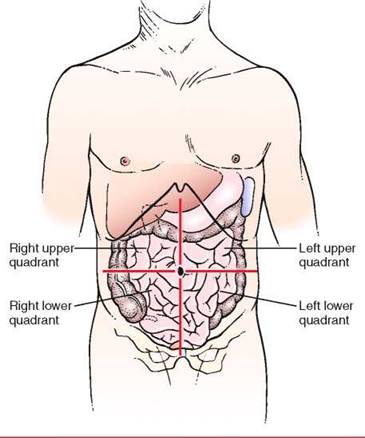
Figure 17-1 The four abdominal quadrants.
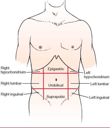
Figure 17-2 The nine abdominal areas.
Table 17-1. Abdominal Structures by Quadrants
|
Right |
Left |
|
Upper Quadrant Liver Gallbladder Pylorus Duodenum Pancreas: head Right adrenal gland Right kidney: upper pole Hepatic flexure Ascending colon: portion Transverse colon: portion |
Liver, left lobe Spleen Stomach Pancreas: body Left adrenal gland Left kidney: upper pole Splenic flexure Transverse colon: portion Descending colon: portion |
|
Lower Quadrant |
|
|
Right kidney: lower pole |
Left kidney: lower pole |
|
Cecum |
Sigmoid colon |
|
Appendix |
Descending colon: portion |
|
Ascending colon: portion |
Left ovary |
|
Right ovary |
Left fallopian tube |
|
Right fallopian tube |
Left ureter |
|
Right ureter |
Left spermatic cord |
|
Right spermatic cord |
Uterus (if enlarged) |
|
Uterus (if enlarged) |
Bladder (if enlarged) |
|
Bladder (if enlarged) |
The entry of chyme from the stomach into the duodenum stimulates the secretion of pancreatic enzymes and contraction of the gallbladder. The flow of pancreatic juice is maximal approximately 2 hours after a meal; the daily output is 1 to 2 L. The three enzymes of chyme—lipase, amylase, and trypsin—are responsible for the digestion of fats, starches, and proteins, respectively. In cases of pancreatic insufficiency, the stool is pale and bulky and has an odor that is more offensive than normal. The chyme and the neutralizing effect of these enzymes reduce the acidity of the duodenal contents and relieve the pain of peptic duodenal ulcer. The pain from an acutely inflamed gallbladder or from pancreatitis worsens at this phase of the digestive cycle.
The digested food continues its course through the small intestine, in which further digestion and absorption occur. Failure of bile production or its release from the gallbladder results in decreased digestion and absorption of fats, leading to diarrhea. Gallstones may form as a result of diet or hereditary predisposition.
The liver produces bile, detoxifies the byproducts of the digestion of food, and metabolizes proteins, lipids, and carbohydrates. The daily output of bile is about 1 L. In the absence of normal liver function, jaundice, ascites, and coma may result.
The jejunum and ileum further digest and absorb the nutrients. Bile acids and vitamin B12 are absorbed in the ileum. The dark color of stool is caused by the presence of stercobilin, a metabolite of bilirubin, that is secreted in the bile. If bile does not flow into the small intestine, the stools become pale brown to gray and are called acholic, or free from bile.
The colon functions to remove much of the remaining water and electrolytes from the chyme. Approximately 600 mL of fluid enters the colon daily, and only 200 mL of water is excreted in the stool daily. Abnormal colonic function leads to diarrhea or constipation. Aneurysmal pouches of colonic mucosa may cause bleeding; if they are infected, pain results. Colonic obstruction produces severe pain. Tumors may cause obstruction or bleeding.
Review of Specific Symptoms
The most common symptoms of abdominal disease are as follows:
• Pain
• Nausea and vomiting
• Change in bowel movements
• Rectal bleeding
• Jaundice
• Abdominal distention
• Mass
• Pruritus (itching)
Pain
Pain is probably the most important symptom of abdominal disease. Although abdominal neoplasia may be painless, most abdominal disease manifests itself with some amount of pain. Pain can result from mucosal irritation, smooth muscle spasm, peritoneal irritation, capsular swelling, or direct nerve stimulation. Abdominal pain necessitates speedy diagnosis and therapy. When a patient complains of abdominal pain, ask the following questions:
''Where is the pain?''
''Has the pain changed its location since it started?''
''Do you feel the pain in any other part of your body?''
''How long have you had the pain?''
''Have you had recurrent episodes of abdominal pain?''
‘‘Did the pain start suddenly?''
''Can you describe the pain? Is it sharp? dull? burning? cramping?''
''Is the pain continuous? Does it come in waves?''
''Has there been any change in the severity or nature of the pain since it began?''
''What makes it worse?''
''What makes it better?''
Is the pain associated with nausea? vomiting? sweating? constipation? diarrhea? bloody stools? abdominal distention? fever? chills? eating?''
Have you ever had gallstones? kidney stones?''
If the patient is a woman, ask this question:
When was your last period?''
Note the exact time at which the pain started and what the patient was doing at that time. Sudden, severe pain awakening a patient from sleep may be associated with acute perforation, inflammation, or torsion of an abdominal organ. A stone in the biliary or renal tract also causes intense pain. Note acuteness of the pain. Acute rupture of a fallopian tube by an ectopic pregnancy, perforation of a gastric ulcer, peritonitis, and acute pancreatitis cause such severe pain that fainting may result.
It is crucial to determine the location of the pain at its onset, its localization, its character, and its radiation. Commonly, when an abdominal organ ruptures, pain is felt ''all over the belly,'' without localization to a specific area. Pain arising from the small intestine is commonly felt in the umbilical or epigastric regions; for example, pain from acute appendicitis begins at the umbilicus.
After time, pain may become localized to other areas. Pain from acute appendicitis travels from the umbilicus to the right lower quadrant in about 1 to 3 hours after the initial event. Pain in the chest followed by abdominal pain should raise the suspicion of a dissecting aortic aneurysm.
Note the nature of the pain. Pain caused by a perforated gastric ulcer is often described as ''burning''; dissecting aneurysm as ''tearing''; intestinal obstruction as ''gripping''; pyelone- phritis as ''dull, aching''; and biliary or renal colic as ''crampy, constricting.''
Referred pain often provides insight as to the cause. Referred pain is a term used to describe pain originating in the internal organs but described by the patient as being located in the abdominal or chest wall, shoulder, jaw, or other areas supplied by the somatic nerves. Pain appears to originate in areas supplied by the somatic nerves entering the spinal cord at the same segment as the sensory nerves from the organ responsible for the pain. For example, right-shoulder pain may result from acute cholecystitis; testicular pain may result from renal colic or from appendicitis. The common sites for referred pain are shown in Figure 17-3. The locations of pain in abdominal disease are summarized in Table 17-2.
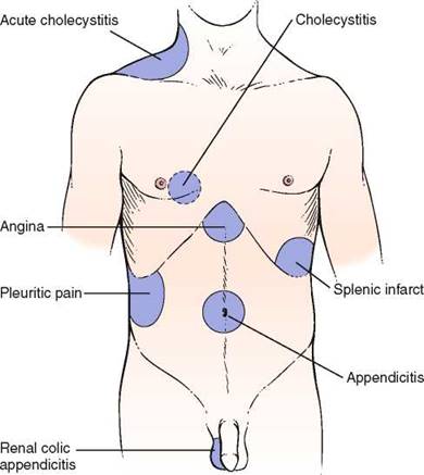
Figure 17-3 Common areas of referred pain. The dotted area is on the posterior chest.
The time of occurrence and factors that aggravate or alleviate the symptoms (e.g., meals or defecation) are particularly important. Periodic epigastric pain occurring | to 1 hour after eating is a classic symptom of gastric peptic ulcers. Patients with a duodenal peptic ulcer have pain 2 to 3 hours after eating or before the next meal. Food tends to lessen the pain, especially in duodenal ulcers. Perforation of a duodenal ulcer to the pancreas may produce backache, simulating an orthopedic problem. Nocturnal pain is a classic symptom of duodenal peptic ulcer disease. Pain after eating may also be associated with vascular disease of the abdominal viscera. Patients with this condition are older and have postprandial pain, anorexia, and weight loss. This triad is seen in abdominal angina resulting from obstructive vascular disease in the celiac axis or the superior mesenteric artery. Table 17-3 summarizes the important maneuvers for ameliorating abdominal pain.
Table 17-2. Location of Pain in Abdominal Disease
|
Area of Pain |
Affected Organ |
Clinical Example |
|
Substernal |
Esophagus |
Esophagitis |
|
Shoulder |
Diaphragm |
Subphrenic abscess |
|
Epigastric |
Stomach |
Peptic gastric ulcer |
|
Duodenum |
Peptic duodenal ulcer |
|
|
Gallbladder |
Cholecystitis |
|
|
Liver |
Hepatitis |
|
|
Bile ducts |
Cholangitis |
|
|
Pancreas |
Pancreatitis |
|
|
Right scapula |
Biliary tract |
Biliary colic |
|
Midback |
Aorta |
Aortic dissection |
|
Pancreas |
Pancreatitis |
|
|
Periumbilical |
Small intestine |
Obstruction |
|
Hypogastrium |
Colon |
Ulcerative colitis Diverticulitis |
|
Sacrum |
Rectum |
Proctitis Perirectal abscess |
Table 17-3. Maneuvers for Ameliorating Abdominal Pain
|
Maneuver |
Affected Organ |
Clinical Example |
|
Belching |
Stomach |
Gastric distention |
|
Eating |
Stomach, duodenum |
Peptic ulcer |
|
Vomiting |
Stomach, duodenum |
Pyloric obstruction |
|
Leaning forward |
Retroperitoneal structures |
Pancreatic cancer Pancreatitis |
|
Flexion of knees |
Peritoneum |
Peritonitis |
|
Flexion of right thigh |
Right psoas muscle |
Appendicitis |
|
Flexion of left thigh |
Left psoas muscle |
Diverticulitis |
Nausea and Vomiting
Vomiting may be caused by severe irritation of the peritoneum resulting from the perforation of an abdominal organ; from obstruction of the bile duct, ureter, or intestine; or from toxins. Vomiting resulting from a perforation is rarely massive. Obstruction of the bile duct or other tube produces stretching of the muscular wall, resulting in episodic vomiting that occurs at the height of the pain. Intestinal obstruction prevents the intestinal contents from passing distally; consequently, vomiting may result in the expulsion of intestinal contents. Toxins generally cause persistent vomiting. Not all abdominal emergencies cause vomiting. Intraperitoneal bleeding may occur in the absence of vomiting. Vomiting is frequently also caused by inflammation of intra-abdominal structures, as well as by extra-abdominal conditions, including drug toxicity, central nervous system disorders, myocardial infarction, and pregnancy. Ask the following questions if a patient complains of nausea, vomiting, or both:
''How long have you had nausea or vomiting?''
''What is the color of the vomit?''
''Is there any unusually foul odor to the vomitus?''
How often do you vomit?''
''Is vomiting related to eating?'' If yes, ' How soon after eating do you vomit? Do you vomit only after eating certain foods?''
''Do you have nausea without vomiting?''
''Is the nausea or vomiting associated with abdominal pain? constipation? diarrhea? a loss of appetite? a change in the color of your stools? a change in the color of your urine? fever? chest pain?''
Have you noticed a change in your hearing ability?''
Have you noticed ringing in your ears?''
If the patient is a woman, ask this question:
When was your last period?''
The relationship of the pain to vomiting is important and may help in providing the diagnosis. In acute appendicitis, pain precedes the vomiting usually by a few hours. The character of the vomitus may aid in determining its cause. Acute gastritis causes the patient to vomit stomach contents. Biliary colic produces bilious, or greenish-yellow, vomitus. Intestinal obstruction often causes the patient to expel bilious vomitus, followed by feculent-smelling fluid. Feculent vomitus is usually caused by intestinal obstruction.
Nausea without vomiting is a common symptom in patients with hepatocellular disease, pregnancy, and metastatic disease. Nausea may be associated with a hearing loss and tinnitus in patients with Meniere's disease.
Change in Bowel Movements
Take a careful history of bowel habits. A change in bowel movements necessitates further elaboration. Ask these questions of the patient with acute onset of diarrhea:
“How long have you had the diarrhea?''
“How many bowel movements do you have a day?''
‘‘Did the diarrhea start suddenly?''
''Did the diarrhea begin after a meal?'' If yes, ''What did you eat?''
‘‘Are the stools watery? bloody? malodorous?''
''Is the diarrhea associated with abdominal pain? loss of appetite? nausea? vomiting?''
The acute onset of diarrhea after a meal suggests an acute infection or toxin. Watery stools are often associated with inflammatory processes of the small bowel and colon. Shigellosis is a disease of the colon that produces bloody diarrhea. Amebiasis is also associated with bloody diarrhea.
The patient with chronic diarrhea should be asked the following:
‘‘How long have you had diarrhea?''
''Do you have periods of diarrhea alternating with constipation?''
''Are the stools watery? loose? floating? malodorous?''
‘‘Have you noticed blood in the stools? mucus? undigested food?''
''What is the color of the stools?''
‘‘How many bowel movements do you have a day?''
‘‘Does the diarrhea occur after eating?''
‘‘What happens when you fast? Do you still have diarrhea?''
‘‘Is the diarrhea associated with abdominal pain? abdominal distention? nausea? vomiting?''
''Have you noticed that the diarrhea is worse at certain times of the day?''
How is your appetite?''
Has there been any change in your weight?''
Diarrhea and constipation frequently alternate in patients with colon cancer or diverticulitis. Loose bowel movements are common in diseases of the left colon, whereas watery movements are seen in severe inflammatory bowel disease and protein-losing enteropathies. Floating stools may result from malabsorption syndromes. Patients with ulcerative colitis commonly have stool mixed with blood and mucus. Any inflammatory process of the small bowel or colon can manifest with blood mixed with stool or undigested food. Irritable bowel syndrome classically produces more diarrhea in the morning.
Patients complaining of constipation should be asked these questions:
How long have you been constipated?''
How often do you have a bowel movement?''
‘‘What is the size of your stools?''
''What is the color of your stools?''
Is the stool ever mixed with blood? mucus?''
''Have you noticed periods of constipation alternating with periods of diarrhea?'' Have you noticed a change in the caliber of the stool?''
Do you have much gas?''
How's your appetite?''
Has there been any change in your weight?''
Change in the caliber of the stool is significant. ''Pencil''-diameter stools may result from an anal or a distal rectal carcinoma. A change in the color of stools is important. As is discussed later, pale brown to gray stools indicate an absence of bile. This can result from an obstruction to bile flow from the gallbladder or from decreased production of bile. Weight changes are important with the symptom of constipation. An increase in weight may indicate decreased metabolism seen in hypothyroidism; a decrease in weight may be associated with cancer of the colon or other hypermetabolic conditions.
Rectal Bleeding
Rectal bleeding may be manifested by bright red blood, blood mixed with stool, or black, tarry stools. Bright red blood per rectum, also known as hematochezia, can occur from colonic tumors, diverticular disease, or ulcerative colitis. Blood mixed with stool can be the result of ulcerative colitis, diverticular disease, tumors, or hemorrhoids. Ask the patient who describes rectal bleeding the following questions:
How long have you noticed bright red blood in your stools?''
Is the blood mixed with the stool?''
‘‘Are there streaks of blood on the surface of the stool?''
Have you noticed a change in your bowel habits?''
''Have you noticed a persistent sensation in your rectum that you have to move your bowels, but you cannot?''
Tenesmus is the painful, continued, and ineffective straining at stool. It is caused by inflammation or a space-occupying lesion such as a tumor at the distal rectum or anus. Hemorrhoidal bleeding is a common cause of hematochezia and streaking of stool with blood.
Melena is a black, tarry stool that results from bleeding above the first section of the duodenum, with partial digestion of the hemoglobin. Inquire about the presence of melena. A useful way of questioning is to show the patient the black tubing on the stethoscope and ask, ''Have your bowel movements ever been this color?'' If asked directly whether the bowel movements have ever been black, the patient may answer in the affirmative, equating dark (normal) stools with black stools. Ask these questions of a patient who describes melena:
Have you passed more than one black, tarry stool?'' If yes, When?''
How long have you been having black, tarry stools?''
Have you noticed feeling lightheaded?''
Have you had any nausea associated with these stools? any vomiting? diarrhea? abdominal pain? sweating?''
The answers to these questions can provide some information regarding the acuteness and the amount of the hemorrhage. Lightheadedness, nausea, and diaphoresis are seen with rapid gastrointestinal bleeding and hypotension.
The presence of silver-colored stools is rare but pathognomonic of acholic stools with melena, a condition resulting from cancer of the ampulla of Vater in the duodenum. The cancer produces biliary obstruction, and the cancerous fronds are sloughed, causing melena.
Jaundice
The presence of jaundice (icterus) must alert the examiner that there is either liver parenchymal disease or an obstruction to bile flow. The presence of icterus, or jaundice, results from a decreased excretion of conjugated bilirubin into the bile. This can result from intrahepatic biliary obstruction, known as medical jaundice, or from extrahepatic biliary obstruction, known as surgical jaundice. In any patient with icterus, the examiner should search for clues by asking the following questions:
''How long have you been jaundiced?''
''Did the jaundice develop rapidly?''
‘‘Is the jaundice associated with abdominal pain? loss of appetite? nausea? vomiting? distaste for cigarettes?''
''Is the jaundice associated with chills? fever? itching? weight loss?''
''In the past year have you had any transfusions? tattooing? inoculations?''
''Do you use any recreational drugs?'' If yes, ''Do you use any drugs intravenously?''
''Do you eat raw shellfish? oysters?''
''Have you traveled abroad in the past year?'' If yes, ''Where? Were you aware that you may have consumed unclean water?''
''Have you been jaundiced before?''
''Has your urine changed color since you noticed that you were jaundiced?''
''What is the color of your stools?''
Do you have any friends or relations who are also jaundiced?''
''What type of work do you do? What other types of work have you done?''
What are your hobbies?''
Viral hepatitis is associated with nausea, vomiting, a loss of appetite, and an aversion to smoking. Hepatitis A has a fecal-oral route of transmission and an incubation period of 2 to 6 weeks. It may be linked to ingestion of raw shellfish. Hepatitis B is blood-borne and has an incubation period of 1 to 6 months. Health professionals are at increased risk for hepatitis. Any contact with an individual with viral hepatitis places a person at a higher risk of contracting viral hepatitis. Hepatitis C virus (HCV) is the most common chronic blood- borne infection in the United States. The Centers for Disease Control and Prevention has estimated that during the 1980s, as many as 230,000 new infections have occurred in this country. It is estimated that as many as 3.9 million Americans have antibody to HCV and are currently asymptomatic. They are, however, at risk for chronic liver disease, the 10th leading cause of death in the United States. About 40% of chronic liver disease is HCV related and results in 8000 to 10,000 deaths a year. End-stage liver disease secondary to HCV infection is the most common indication for liver transplantation. Because most of these individuals are younger than 50 years, it is anticipated that by 2020, there will be a marked increase in the number of patients with chronic liver disease.
Slowly developing jaundice that is accompanied by pale stools and cola-colored urine is obstructive jaundice, either intrahepatic or extrahepatic. Jaundice accompanied by fever and chills is considered cholangitis until proved otherwise. Cholangitis may result from stasis of bile in the bile duct that results from a gallstone or from cancer of the head of the pancreas. Determine whether chemicals are used in a patient's occupation or hobbies, because they may be related to the cause of the jaundice. Many industrial chemicals and drugs have been associated with liver disease. These agents may be responsible for a viral hepatitis-like illness, cholestasis, or for granulomas or hepatic tumors. Occupational exposure to carbon tetrachloride and vinyl chloride is well known to cause liver disease. Ask questions related to alcohol abuse. These are described in Chapter 1, The Interviewer's Questions.
Abdominal Distention
Abdominal distention may be related to increased gas in the gastrointestinal tract or to the presence of ascites. Increased gas can result from malabsorption, irritable colon, or air swallowing (aerophagia). Ascites can have a variety of causes, such as cirrhosis, congestive heart failure, portal hypertension, peritonitis, or neoplasia. To try to identify the cause of abdominal distention, ask these questions:
How long have you noticed your abdomen to be distended?''
Is the distention intermittent?''
Is the distention related to eating?''
Is the distention lessened by belching or by passing gas from below?''
Is the distention associated with vomiting? loss of appetite? weight loss? change in your bowel habits? shortness of breath?''
Gaseous distention related to eating is intermittent and is relieved by the passage of flatus or belching. A patient with ascites has the insidious development of increased abdominal girth, noted through a progressive increase in belt size. Loss of appetite is often associated with cirrhosis and malignancy, although end-stage congestive heart failure may produce this symptom as well. Shortness of breath and ascites may be symptoms of congestive heart failure, but the shortness of breath may be the result of a decrease in pulmonary capacity owing to ascites from another cause. Questions related to alcoholic abuse are most appropriate and are outlined in Chapter 1, The Interviewer's Questions.
Mass
An abdominal mass may be a neoplasm or a hernia. An abdominal hernia is a protrusion from the peritoneal cavity into which peritoneal contents are extruded. The contents may be omentum, intestine, or bladder wall. An abdominal hernia may be inguinal, femoral, umbilical, or internal, depending on its location. The most common complaint is swelling, which may or may not be painful. An inguinal hernia may manifest as a mass in the groin or scrotum. The major complications of a hernia are intestinal obstruction and intestinal strangulation from interference of blood supply. A hernia is termed reducible when it can be emptied of its contents by pressure or a change in posture.
The sign or symptom of a pulsatile abdominal mass should alert the examiner to the possibility of an aortic aneurysm.
Pruritus
Pruritus, or itching, is a common symptom. Generalized itching may be a symptom of a diffuse skin disorder* or a manifestation of chronic renal or hepatic disease. Intense pruritus may be associated with lymphoma or Hodgkin's disease, as well as with malignancies of the gastrointestinal tract. In older individuals, pruritus may also be caused by dry skin alone. Pruritus ani is localized itching of the anal skin. It has many causes, including fistulas, fissures, psoriasis, parasites, poor hygiene, and diabetes.
Impact of Inflammatory Bowel Disease on the Patient
Inflammatory bowel disease constitutes a group of diseases of unknown cause. The symptoms produced depend on the location, extent, and acuteness of the inflammatory lesions. The common presenting features are fever, anorexia, weight loss, abdominal discomfort, diarrhea, rectal urgency, and rectal bleeding. It is a chronic, potentially disabling illness, often resulting in the need for multiple surgeries, in fistula formation, and in cancer.
*For example, dermatitis herpetiformis, a blistering disease predominantly on the buttocks, shoulders, elbows, and knees.
Inflammatory bowel disease may lead to long absences from school or work, disruption of family life, malabsorption, malnutrition, and multiple hospitalizations. A patient can have 10 to 30 watery or bloody bowel movements each day. As a consequence, patients with inflammatory bowel disease can have many psychologic problems, particularly when they are young adults. Because of malabsorption, the prevalence of osteopenia in patients with inflammatory bowel disease ranges from 40% to 50%; osteoporosis is present in 5% to 30% of all patients. Fractures of the hip, spine, and distal radius occur. One study revealed that the incidence of fractures among persons with inflammatory bowel disease is 40% greater than in the general population.
Sexual development may be delayed as a result of malnutrition. Social development is also delayed. The necessity of constantly having to remain near a bathroom inhibits patients' abilities to develop normal dating patterns. Many of these patients are socially immature, and social introversion is common. By necessity, they remain at home. Their lives revolve around their bowel habits.
In most cases, there is a positive correlation between the severity of the physical disease and the extent of emotional disturbance. Dependency is the most reported characteristic of patients with inflammatory bowel disease. Repressed rage, suppression of feelings, and anxieties are also common. It is reported that many patients have a constant desire to rid themselves of events in their lives. This characteristic can be acted out through the diarrhea. Another characteristic of these patients is to be obsessive-compulsive. The marked obsessive character becomes even more obvious when the patient is ill. It is typical for patients to worry incessantly about what is happening in their bowels. The patients are intelligent, often having read much literature, including medical textbooks, about their disease.
Denial is usually not a prominent symptom. In contrast, these patients concentrate obsessively on the details of their bowel habits.
Sexual problems are common. Interest and participation in sexual activity tend to be at a low level. Many of these individuals prefer to be fondled like a child and largely reject any genital contact. Patients are prone to regard sexual activity in anal terms, such as ''dirty, unclean,'' or ''soiling.'' They are squeamish about body contact, odors, and secretions. The loss of libido and decreased sexual drive may be related to their fear of bowel action during intercourse, of perineal pain, or that sexual intercourse may in some way further damage the bowel.
The frequent hospitalizations cause anxiety and depression, which exacerbate the disease. The fear of cancer may be the basis of depression, which is a common response to the disease. It is well established that emotional factors are important in maintaining and prolonging an existing attack. Schoolwork deteriorates as young patients are forced to miss more and more school, further increasing their anxiety.
An often unappreciated major complication of inflammatory bowel disease is substance abuse. As a result of chronic pain, as much as 5% of patients with inflammatory bowel disease are physically addicted to oral narcotics. Many more are psychologically dependent on their pain medication.
Many patients with ulcerative colitis require an ileostomy. The fear of disfigurement, the loss of self-confidence, the potential lack of cleanliness, and the dread of unexpected spillage are common.
Time for listening and an interest in a patient's problem are important in gaining the patient's confidence. Listening may reveal and help unravel the emotional problems that may be the source of the exacerbation of the bowel disease. Talking with the patient may be more efficacious than prescribing anti-inflammatory agents or tranquilizers. Careful and thoughtful discussion of the illness strengthens the doctor-patient relationship and produces immeasurable therapeutic benefits.
Physical Examination
The equipment necessary for the examination of the abdomen and rectum consists of a stethoscope, gloves, lubricant, tissues, and occult blood testing card and Hemoccult developer.
The patient should be lying flat in bed, and the abdomen should be fully exposed from the sternum to the knees. The arms should be at the sides, and the legs flat. Frequently, patients tend to place their arms behind their heads, which tightens the abdominal muscles and makes the examination more difficult. Placing a pillow beneath the knees often aids in relaxation. The examiner should be standing on the patient's right side. A sheet or towel is placed over the genitalia, as shown in Figure 17-4.
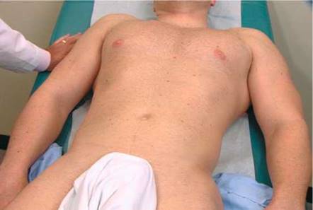
Figure 17-4 Technique for inspecting the abdomen.
If the patient has complained of abdominal pain, examine the area of pain last. If the examiner touches the area of maximal pain, the abdominal muscles tighten, and the examination is more difficult.
The physical examination of the abdomen includes the following:
• Inspection
• Auscultation
• Percussion
• Palpation
• Rectal examination
• Special techniques
Inspection
Evaluate General Appearance
The general appearance of the patient often furnishes valuable information as to the nature of the condition. Patients with renal or biliary colic writhe in bed. They squirm constantly and can find no comfortable position. In contrast, patients with peritonitis, who have intense pain on movement, characteristically remain still in bed because any slight motion worsens the pain. They may be lying in bed with their knees drawn up to help relax the abdominal muscles and reduce intra-abdominal pressure. Patients who are pale and sweating may be suffering from the initial shock of pancreatitis or a perforated gastric ulcer.
Determine Respiratory Rate
The respiratory rate is increased in patients with generalized peritonitis, intra-abdominal hemorrhage, or intestinal obstruction.
Inspect the Skin
Inspect the skin and sclera for jaundice. Whenever possible, the patient should be evaluated for jaundice in natural light because incandescent light frequently masks the existence of icterus. Jaundice becomes apparent when the serum bilirubin level exceeds 2.5 mg/dL in adults or 6 mg/dL in neonates. Figure 17-5 shows a patient with jaundice. Notice the scleral icterus as well as the yellow discoloration of the skin. Hyperbilirubinemia can also result in intense generalized pruritus.
Inspect for spider angiomas. Spider angiomas have a high degree of sensitivity in patients with alcoholic cirrhosis but are nonspecific because they also occur in pregnancy and collagen vascular disorders (see Fig. 8-75).
Figure 17-6 shows the leg of a patient with pyoderma gangrenosum. Notice the necrotic, undermined ulceration with pus. These tender ulcerations, commonly on the lower extremities, are associated with inflammatory bowel disease, especially ulcerative colitis. In general, the clinical course of pyoderma gangrenosum follows the course of the bowel disease. This condition is also seen in patients with rheumatoid arthritis, myeloid metaplasia, and chronic myelogenous leukemia.
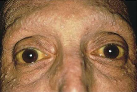
Figure 17-5 Jaundice.
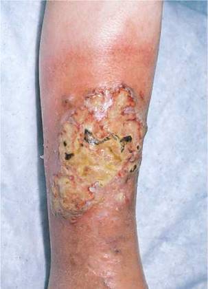
Figure 17-6 Pyoderma gangrenosum.
Inspect the Hands
Is there muscle loss in the small muscles of the hands? This is associated with wasting.
The nails are examined for changes in the nail bed, especially an increase in the size of the lunula. The fingers of a patient with cirrhosis showing ''half-and-half'' nails are shown in Figure 17-7 (see also Fig. 8-8).
Inspect the Facies
Are the eyes sunken? Is temporal wasting present? These signs indicate wasting and poor nutrition.
The skin around the mouth and oral mucosa may provide evidence of gastrointestinal disorders. Melanin deposition around and in the oral cavity, especially the buccal mucosa, suggests Peutz-Jeghers syndrome. Figure 12-12 shows the lips of a patient with the classic brown pigmentary changes of Peutz-Jeghers syndrome. This is an autosomal dominant disorder characterized by generalized gastrointestinal, hamartomatous polyposis, and mucocutaneous pigmentation. The benign polyps are most common in the jejunum and only rarely become malignant. The polyps, however, may bleed, cause intussusception, or cause obstruction. Telangiectases of the lips and tongue are suggestive of Osler-Weber-Rendu syndrome (see Fig. 12-11), in which multiple telangiectases are present throughout the gastrointestinal tract. These lesions may bleed insidiously, causing anemia. The classic oral lesions of a patient with Osler-Weber-Rendu syndrome are shown in Figure 17-8.
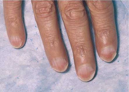
Figure 17-7 Lindsay's nails.
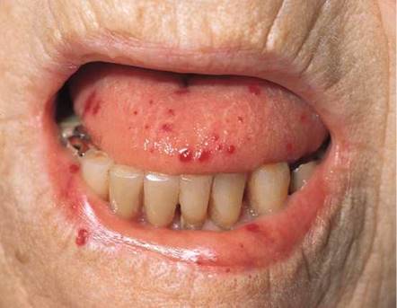
Figure 17-8 Osler-Weber- Rendu syndrome.
Hypercortisolism, or Cushing's syndrome, has a range of clinical manifestations. The most common are obesity, facial plethora, hirsutism, and hypertension. Obesity occurs in 90% of affected patients. Most patients with hypercortisolism characteristically have round, puffy, red faces called moon facies. They also have prominent fat deposits in the supraclavicular and retrocervical areas (buffalo hump). Figure 17-9 shows a patient with Cushing's syndrome and the typical moon facies.

Figure 17-9 Cushing's syndrome.
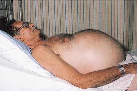
Figure 17-10 Ascites.
Inspect the Abdomen
The contour of the abdomen should be assessed. A scaphoid, or concave, abdomen may be associated with cachexia; a protuberant abdomen may result from gaseous distention of the intestines, ascites, (splenomegaly lator), or obesity. When a patient with ascites stands, the fluid sinks into the lower abdomen; when lying supine, the fluid bulges in the flanks. If a patient with ascites lies on the side, the fluid flows to the dependent lower side. A patient with a protuberant abdomen as a result of carcinomatous ascites is shown in Figure 17-10.
The examiner should focus attention on the abdomen to describe adequately the presence of any asymmetry, distention, masses, or visible peristaltic waves. The examiner should then observe the abdomen from above, looking for the same signs. Inspection of the abdomen for striae and scars may provide valuable data. Silver striae are stretch marks consistent with weight loss. Pinkish-purple striae are classic signs of adrenocortical excess. Figure 17-11 shows the characteristic purplish striae in a patient with Cushing's syndrome.
Is the umbilicus everted? An everted umbilicus is often a sign of increased abdominal pressure, usually from ascites or a large mass. An umbilical hernia may also cause an umbilicus to become everted.
Are there ecchymoses on the abdomen or on the flanks? Massive ecchymoses may occur in these areas as a result of hemorrhagic pancreatitis or strangulated bowel. This is Grey Turner's sign.
Cullen's sign is a bluish discoloration of the umbilicus resulting from hemoperitoneum of any cause.
Recognition of classic surgical scars may be helpful. Figure 17-12 shows the locations of some common surgical scars.
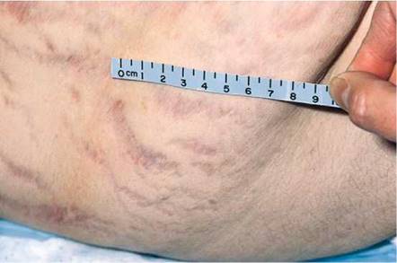
Figure 17-11 Abdominal striae.
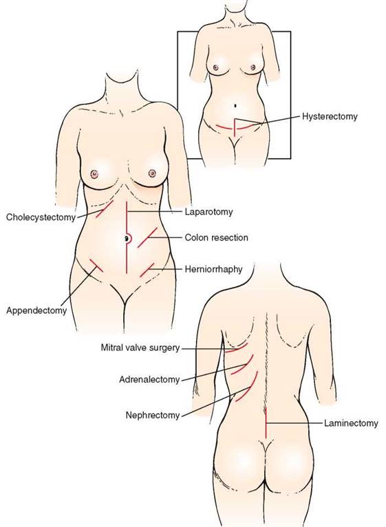
Figure 17-12 Locations of common surgical scars.
Inspect for Hernias
The patient lying in bed should be asked to cough while the examiner inspects the inguinal, umbilical, and femoral areas. This maneuver, by increasing intra-abdominal pressure, may produce a sudden bulging in these areas, which may be related to a hernia. If the patient has had surgery, coughing may reveal a bulging along the abdominal scar from the previous incision. In addition, coughing may elicit pain localized to a specific area. This technique enables the examiner to identify the area of maximal tenderness and to perform most of the abdominal examination without too much discomfort to the patient. Ascites secondary to metastatic breast carcinoma and an umbilical hernia are shown in Figure 17-13.
Inspect the Superficial Veins
The venous pattern of the abdomen is usually barely perceptible. If it is visible in the normal individual, the drainage of the lower two thirds of the abdomen is downward. In the presence of vena caval obstruction, superficial veins may dilate, and the veins drain cephalad (toward the head). In patients with portal hypertension, the dilated veins appear to radiate from the umbilicus. This is caused by backflow through the collateral veins within the falciform ligament, and this pattern is called caput medusae.
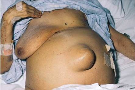
Figure 17-13 Ascites with umbilical hernia.
If the superficial veins are distended, evaluate the direction of drainage by the following technique: Place the tips of your index fingers on a vein that is oriented cephalad-caudad, not transverse, and compress it. Using continuous pressure, slide your index fingers apart for about 3 to 4 inches (7.5 to 10 cm). Remove one finger and observe the refilling in the direction of flow. Repeat this procedure, but this time remove your other finger and observe the direction of flow. Figure 17-14 shows the abdomen of a patient with intrahepatic portal hypertension. Notice the engorged paraumbilical veins; the flow was away from the umbilicus toward the caval system.
Auscultation
Auscultation of bowel sounds can provide information about the motion of air and liquid in the gastrointestinal tract. Many examiners perform auscultation of the abdomen before percussion or palpation, in contrast to the usual order. It is believed that percussion or palpation may change the intestinal motility; therefore, auscultation should be performed first to produce a more accurate assessment of the existing bowel sounds.
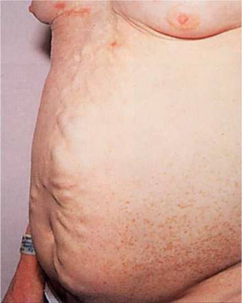
Figure 17-14 Abdominal venous pattern.
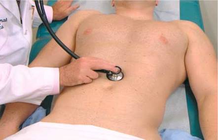
Figure 17-15 Technique for evaluating bowel sounds.
Evaluate Bowel Sounds
The patient is placed in a supine position. To perform auscultation of the abdomen, the examiner places the diaphragm of the stethoscope over the midabdomen and listens for bowel sounds. This technique is pictured in Figure 17-15.
Normal bowel sounds occur approximately every 5 to 10 seconds and have a high-pitched sound. If after 2 minutes no bowel sounds are heard, ''absent bowel sounds'' may be noted. The absence of bowel sounds suggests a paralytic ileus that results from diffuse peritoneal irritation. There may be rushes of low-pitched rumbling sounds, termed borborygmi, which are associated with hyperperistalsis. These sounds are similar to the sound of the word ''borborygmi'' itself. They are common in early acute intestinal obstruction.
Rule Out Obstructed Viscus
A succussion splash may be detected in a distended abdomen as a result of the presence of gas and fluid in an obstructed organ. The examiner applies the stethoscope over the patient's abdomen while shaking the patient's abdomen from side to side. The presence of a sloshing sound usually indicates distention of the stomach or colon. This technique is illustrated in Figure 17-16.
Rule Out Abdominal Bruits
Auscultation is also useful for determining the presence of bruits. Each quadrant should be evaluated for their presence. Bruits may result from stenosis of the renal artery or of the abdominal aorta. (See discussion of abdominal bruits in Chapter 15, The Peripheral Vascular System.)
Rule Out Peritoneal Rubs
A peritoneal friction rub, like a pleural or pericardial rub, is a sound that indicates inflammation. During respiratory motion, a friction rub may be heard in the right or left upper quadrants in the presence of hepatic or splenic disorder.
Percussion
Percussion is used to demonstrate the presence of gaseous distention and fluid or solid masses. In the normal examination, usually only the size and location of the liver and spleen can be determined. Some examiners prefer to palpate before percussion, especially if the patient complains of abdominal pain; either approach is correct. The technique of percussion is discussed in Chapter 13, The Chest.
Percuss the Abdomen
The patient lies supine. All four quadrants of the abdomen are evaluated by percussion. Tympany is the most common percussion note in the abdomen. It is caused by the presence of gas in the stomach, small bowel, and colon. The suprapubic area, when percussed, may sound dull if the urinary bladder is distended or, in a woman, if the uterus is enlarged.
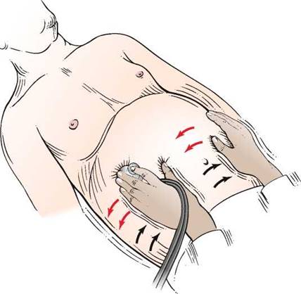
Figure 17-16 ''Succussion splash'' technique for assessing distention of abdominal viscera.
Percuss the Liver
The upper border of the liver is percussed in the right midclavicular line, starting in the midchest. As the chest is percussed downward, the resonant note of the chest becomes dull as the liver is reached. As percussion continues still further, this dull note becomes tympanic because the percussion is now over the colon. The upper and lower borders of the liver should be no more than 4 inches (10 cm) apart. The technique is illustrated in Figure 17-17.
There are several problems with predicting liver size by percussion. If ascites is present, the examiner can only speculate about the correct size of the liver. A more common cause of overestimating liver size (false-positive measurement) is some form of chronic obstructive lung disease. This makes percussion of the upper border of the liver difficult. Obesity in a patient can cause problems in both percussion and palpation. Distention of the colon may obscure the lower liver dullness. This may result in underestimating the size of the liver (false-negative measurement).
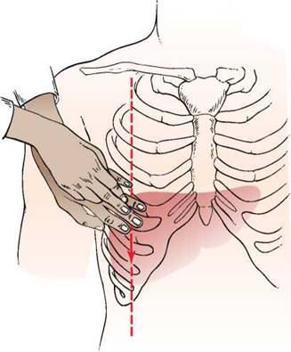
Figure 17-17 Technique for liver percussion.
Percuss the Spleen
Although the size of the spleen is often difficult to determine, evaluation of the spleen should begin with percussion of splenic size. In normal individuals, the spleen lies hidden within the rib cage against the posterolateral wall of the abdominal cavity in Traube's space. Traube's space is defined by the sixth rib superiorly, the left anterior axillary line laterally, and the costal margin inferiorly. As the spleen enlarges, it remains close to the abdominal wall, and the tip moves downward and toward the midline. Because the early enlargement is in an anteroposterior direction, considerable enlargement may occur without its becoming palpable below the costal margin. It is thought that dullness to percussion in Traube's space—the loss of tympany from the air-filled colon and stomach because of the enlarged spleen—is a useful sign for determining splenic enlargement.
Have the patient lie in the supine position. With the patient breathing normally, percuss in the lowest intercostal space in the left anterior axillary line. Normal percussion yields either a resonant or tympanic note. A positive test of splenomegaly is diagnosed when the percussion note is dull. With ultrasonography, the standard imaging technique, splenic percussion has a sensitivity of 62% and a specificity of 72%. In leaner individuals who had not eaten in the previous 2 hours, the sensitivity was 78% and the specificity was 82%.
Rule Out Ascites
On patients in whom ascites is thought to be present, a special percussion test for shifting dullness may be performed. While the patient is lying on the back, the examiner determines the borders of tympany and dullness. The area of tympany is present above the area of dullness and is caused by gas in the bowel that is floating on top of the ascites. The patient is then asked to turn on the side, and the examiner again determines the borders of the percussion notes. If ascites is present, dullness ''shifts'' to the more dependent position; the area around the umbilicus that was initially tympanic becomes dull. Shifting dullness has a sensitivity of 83% to 88% and a specificity of 56%. The test for shifting dullness is illustrated in Figure 17-18.
An additional test for ascites is the presence of a fluid wave. Another examiner's hand or the patient's own hand is placed in the middle of the patient's abdomen. Indenting the abdominal wall stops transmission of an impulse by the subcutaneous adipose tissue. The examiner then taps one flank while palpating the other side. The appearance of a fluid wave is suggestive of ascites. This technique is illustrated in Figure 17-19. The presence of a prominent fluid wave is the most specific of all physical diagnostic tests for ascites and has a specificity of 82% to 92%, according to several studies. A false-positive result may be obtained in obese individuals, and a false-negative result may be obtained when the ascites is small to moderate.
Another physical finding with ascites is the presence of bulging flanks. This change occurs when the weight of free abdominal fluid is sufficient to push the flanks outward. It has a 93% sensitivity and a 54% specificity for detecting ascites.
The most sensitive sign for ascites is the presence of shifting dullness, whereas the most specific sign is the presence of a prominent fluid wave. For an individual patient, the examiner must know the prevalence of disease or the pretest probability to apply sensitivity and specificity. Several studies have reviewed the operating characteristics of the physical examination tests for ascites. Their pooled sensitivity, specificity, and likelihood ratios for the presence of ascites are summarized in Table 17-4. The presence of a prominent fluid wave or shifting dullness is associated with the highest likelihood of the presence of ascites; the absence of bulging flanks, flank dullness, or shifting dullness decreases the likelihood.
Among the more common historical items perceived by patients, an increase in abdominal girth or recent weight gain has the highest chance of being associated with ascites. An increase in abdominal girth has a positive likelihood ratio (LR+) of 4.16; recent weight gain has an LR+ of 3.20. Conversely, the absence of the subjective increase in abdominal girth has a negative likelihood ratio (LR—) of 0.17; the absence of subjective ankle swelling carries an LR— of 0.10. With regard to the physical examination, the presence of a fluid wave or shifting dullness has the highest LR+ (9.6 or 5.76, respectively). The absence of bulging flanks or edema makes the presence of ascites least likely, with an LR— of 0.12 or 0.17, respectively.
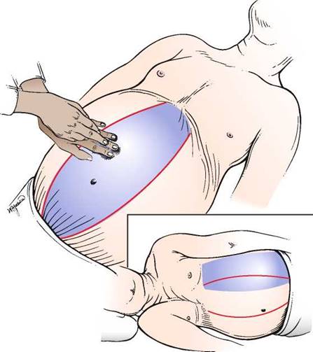
Figure 17-18 Technique for testing for shifting dullness. The shaded areas represent the areas of tympany.
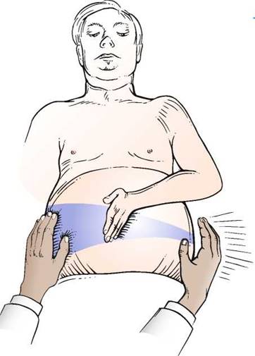
Figure 17-19 Technique for testing a fluid wave.
Table 17-4. Characteristics of Physical Signs (Pooled Data) for Detection of Ascites*
|
Physical Sign |
Sensitivity |
Specificity |
LR+ |
LR- |
||
|
% |
Range |
% |
Range |
|||
|
Bulging flanks |
81 |
69-93 |
59 |
50-68 |
2.0 |
0.3 |
|
Flank dullness |
84 |
80-94 |
59 |
47-71 |
2.0 |
0.3 |
|
Shifting dullness |
86 |
64-90 |
72 |
63-81 |
2.7 |
0.3 |
|
Prominent fluid wave |
62 |
47-77 |
90 |
84-96 |
6.0 |
0.4 |
*95% confidence interval; data pooled from Cummings et al (1985), Simel et al (1988), Cattau et al (1982), and Williams and Simel (1992).
LR+, positive likelihood ratio; LR—, negative likelihood ratio.
Palpation
Abdominal palpation is commonly divided into the following:
• Light palpation
• Deep palpation Liver palpation
• Spleen palpation
• Kidney palpation
The patient is supine during palpation. Always begin palpation in an area of the abdomen that is farthest from the location of pain.
Light Palpation
Light palpation is used to detect tenderness and areas of muscular spasm or rigidity. The examiner should systematically palpate the entire abdomen by using the flat part of his or her right hand or the pads of the fingers, not the fingertips. The fingers should be together, and sudden jabs are to be avoided. The hand should be lifted from area to area instead of sliding over the abdominal wall. Light palpation is demonstrated in Figure 17-20.
With patients who are ticklish, it may be useful to sandwich their hand between the examiner's hands, as demonstrated in Figure 17-21.
During expiration, the rectus muscles usually relax and soften. If there is little change, rigidity is said to be present. Rigidity is involuntary spasm of the abdominal muscles and is indicative of peritoneal irritation. Rigidity may be diffuse, as in diffuse peritonitis, or localized, as over an inflamed appendix or gallbladder. In patients with generalized peritonitis, the abdomen is described as ''boardlike.''
In patients who complain of abdominal pain, palpation should be performed gently. Lightly stroking the abdomen with a pin may reveal an area of increased sensation that is caused by inflammation of the visceral or parietal peritoneum. This sensation is hyperesthesia. The patient is asked to determine whether the pin feels sharper on one side of the abdomen than on the corresponding area on the other side. Although useful, the presence or absence of this finding must be considered in view of all other findings.
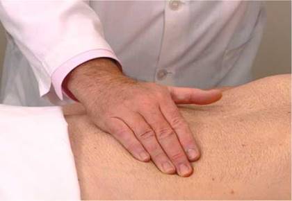
Figure 17-20 Technique for light palpation.
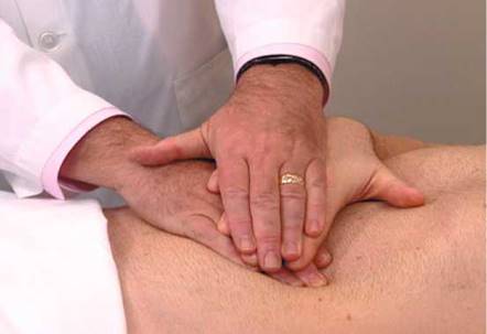
Figure 17-21 Technique used for ticklish patients. The patient's hand is sandwiched between the examiner's hands.
Deep Palpation
Deep palpation is used to determine organ size, as well as the presence of abnormal abdominal masses. In deep palpation, the examiner places the flat portion of his or her right hand on the patient's abdomen, and his or her left hand is placed over the right hand. The fingertips of the left hand exert the pressure, while the right hand should appreciate any tactile stimulation. Pressure should be applied to the abdomen gently but steadily. The technique of deep palpation is demonstrated in Figure 17-22.
During deep palpation, the patient should be instructed to breathe quietly through the mouth and to keep arms at the sides. Asking the patient to open the mouth when breathing seems to aid in generalized muscular relaxation. The palpating hands should be warm, because cold hands may produce voluntary muscular spasm called guarding. Engaging the patient in conversation often aids in relaxing the patient's abdominal musculature. Patients with well- developed rectus muscles should be instructed to flex their knees to relax the abdominal muscles. Any tender areas must be identified.
Rule Out Rebound Tenderness
In a patient with abdominal pain, it should be determined whether rebound tenderness is present. Rebound tenderness is a sign of peritoneal irritation and can be elicited by palpating deeply and slowly in an abdominal area away from the suspected area of local inflammation.
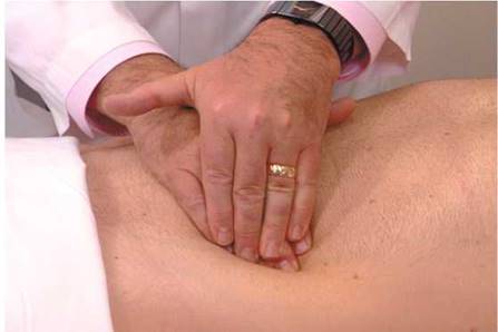
Figure 17-22 Technique for deep palpation.
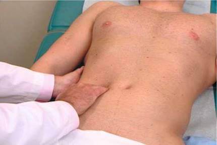
Figure 17-23 Technique for liver palpation.
The palpating hand is then quickly removed. The sensation of pain on the side of the inflammation that occurs on release of pressure is rebound tenderness. If generalized peritonitis is present, pain is felt in the area of palpation. The patient should be asked, ''Which hurts more, now (while pressing) or now (during release)?'' This is a useful test, but because generalized pain is elicited in the patient with peritonitis, the maneuver should be performed near the conclusion of the abdominal examination.
Liver Palpation
To perform palpation of the liver, place your left hand posteriorly between the patient's right 12th rib and the iliac crest, lateral to the paraspinal muscles. Place your right hand in the patient's right upper quadrant parallel and lateral to the rectus muscles and below the area of liver dullness. The patient is instructed to take a deep breath as you press inward and upward with your right hand and pull upward with your left hand. You may feel the liver edge slipping over the fingertips of your right hand as the patient breathes. Start as low as the pelvic brim and gradually work upward. If the examination does not start low, a markedly enlarged liver edge may be missed. The technique of liver palpation is demonstrated in Figure 17-23.
The normal liver edge has a firm, regular ridge, with a smooth surface. If the liver edge is not felt, repeat the maneuver after readjusting your right hand closer to the costal margin. Enlargement of the liver results from vascular congestion, hepatitis, neoplasm, or cirrhosis.
Another technique for liver palpation is the ''hooking'' method. The examiner stands near the patient's head and places both hands together below the right costal margin and the area of dullness. The examiner presses inward and upward and ''hooks'' around the liver edge while the patient inhales deeply. The technique of hooking the liver is shown in Figure 17-24.
On occasion, the liver appears to be enlarged, but the actual border is difficult to determine. The scratch test may be helpful in ascertaining the liver's edge. The examiner holds the diaphragm of the stethoscope with the left hand and places it below the patient's right costal margin over the liver. While the examiner listens through the stethoscope, his or her right index finger ''scratches'' the abdominal wall at points in a semicircle equidistant from the stethoscope. As the finger scratches over the liver's edge, there is a marked increase in the intensity of the sound. This technique is illustrated in Figure 17-25.
A palpable liver is not necessarily enlarged or diseased; however, its being palpable does increase the possibility of hepatomegaly. Being nonpalpable does not rule out hepatomegaly, but it does reduce the likelihood that the liver is enlarged. The LR+ for hepatomegaly, if the liver is palpable, is 2.5; if the liver is not palpable or if enlargement of the liver is detected by scintigraphic scanning, the LR— is 0.45.
Rule Out Hepatic Tenderness
To elicit hepatic tenderness, place the palm of your left hand over the patient's right upper quadrant and gently hit it with the ulnar surface of the fist of your right hand. Inflammatory processes involving the liver or gallbladder produce tenderness on fist palpation. Figure 17-26 demonstrates this technique.
On occasion during liver palpation, pain is elicited in the left upper quadrant during inspiration, and the patient suddenly stops inspiratory efforts. This is called Murphy's sign and is suggestive of acute cholecystitis. On inspiration, the lungs expand, pushing the diaphragm, liver, and inflamed gallbladder down into the abdominal cavity. As the inflamed gallbladder descends against the palpating hand, pain is produced, and there is sudden inspiratory arrest.
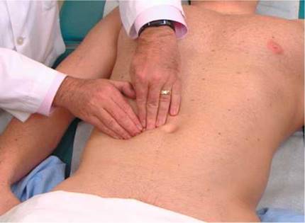
Figure 17-24 Technique for ''hooking'' the liver.
Spleen Palpation
Palpation of the spleen is more difficult than palpation of the liver. The patient lies on the back, with the examiner at the patient's right side. The examiner places his or her left hand over the patient's chest and elevates the patient's left rib cage. The examiner's right hand is placed flat below the patient's left costal margin and presses inward and upward toward the anterior axillary line. The left hand exerts an anterior force to displace the spleen anteriorly. Figure 17-27 illustrates the positions of the hands for splenic palpation.
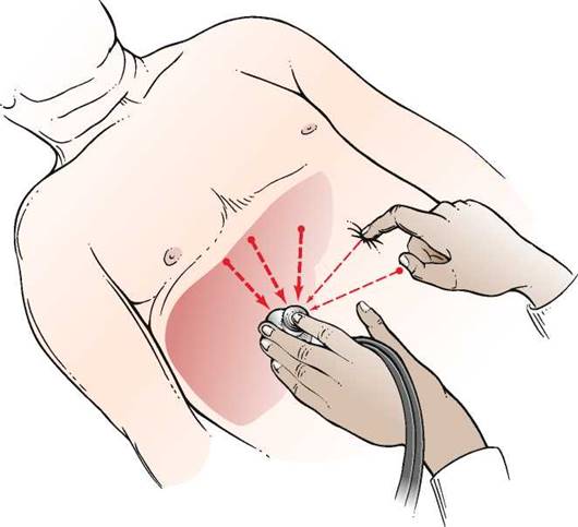
Figure 17-25 The scratch test for determining liver size.
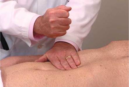
Figure 17-26 Liver tap technique for assessing liver tenderness.
The patient is instructed to take a deep breath as the examiner presses inward with the right hand. The examiner should attempt to feel the tip of the patient's spleen as it descends during inspiration. The tip of an enlarged spleen will lift the fingers of the right hand upward.
The examination of the spleen is repeated with the patient lying on the right side. This maneuver allows gravity to help bring the spleen anterior and downward into a more favorable position for palpation. The examiner places his or her left hand on the patient's left costal margin while the right hand palpates in the left upper quadrant. The technique is demonstrated in Figure 17-28.
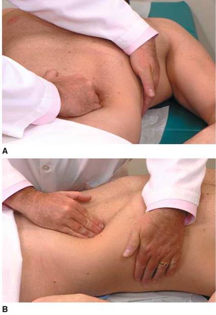
Figure 17-27 A and B, Technique for splenic palpation, with patient lying on the back.
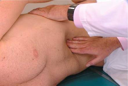
Figure 17-28 Another technique for splenic palpation, with patient lying on right side.
Because the spleen enlarges diagonally in the abdomen from the left upper quadrant toward the umbilicus, the examiner's right hand should always palpate near the umbilicus and gradually move toward the left upper quadrant. This is particularly important if the spleen is massively enlarged, because starting the palpation too high may cause the examiner to miss the splenic border.
The spleen is not palpable in normal conditions, but both techniques should be used in an attempt to palpate it. Splenic enlargement may result from hyperplasia, congestion, infection, or infiltration by tumor or myeloid elements. Massive splenomegaly in a patient with chronic myelocytic leukemia is pictured in Figure 17-29.
Kidney Palpation
More often than not, neither kidney can be palpated in the adult. The technique, however, is important to know, especially in the evaluation of a newborn.
Palpation of the right kidney is performed by deep palpation below the right costal margin. The examiner stands at the patient's right side and places his or her left hand behind the patient's right flank, between the costal margin and the iliac crest. The right hand is placed just below the patient's costal margin with the tips of the fingers pointing to the examiner's left. The method of kidney palpation is demonstrated in Figure 17-30.
Very deep palpation may reveal the lower pole of the right kidney as it descends during inspiration. The lower pole may be felt as a smooth, rounded mass.
The same procedure is used for the left kidney except that the examiner is on the patient's left side. Because the left kidney is more superior than the right, the lower pole of a normal left kidney is rarely palpable. On occasion, the spleen maybe mistaken for an enlarged left kidney. The medial notch of the spleen is helpful in differentiating it from the kidney (see Fig. 17-29).
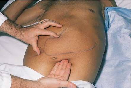
Figure 17-29 Splenomegaly. Note the splenic notch.
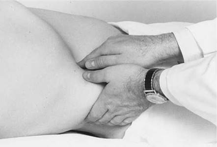
Figure 17-30 Technique for kidney palpation.
Rule Out Renal Tenderness
For this part of the examination, the patient should be seated. The examiner should make a fist and gently hit the area overlying the costovertebral angle on each side. Figure 17-31 demonstrates the technique. Patients with pyelonephritis usually have extreme pain even on slight percussion in these areas. If pyelonephritis is suspected, only digital pressure should be used. As is described in Chapter 22, Putting the Examination Together, this portion of the abdominal examination is usually performed when the posterior chest is examined.
Rectal Examination
The routine abdominal examination concludes with the digital rectal examination (DRE). Because the anterior rectum has a peritoneal surface, the DRE may reveal tenderness if peritoneal inflammation is present.
The rectal examination for the male patient is discussed here; for the female patient, it is discussed in Chapter 19, Female Genitalia.
Patient Positioning
The examination of the rectum in the male patient may be performed with the patient lying on his back, lying on the left side, or standing and bent over the examination table. The modified lithotomy position (patient on the back with knees flexed) is used when the patient has difficulty standing or when a detailed examination of the anus is not required. The examiner passes the right hand under the patient's right thigh. The index finger in the patient's rectum is used in conjunction with the examiner's left hand, which is placed on the patient's abdomen. This bimanual approach is useful and causes minimal disturbance of the sick patient.
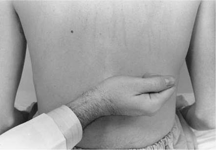
Figure 17-31 Assessing costovertebral angle tenderness (CVAT).
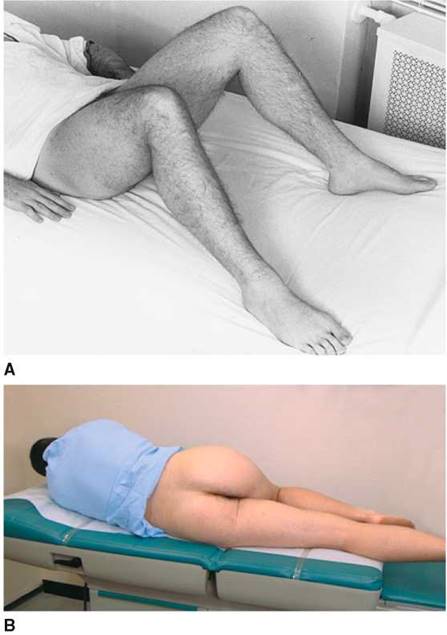
Figure 17-32 Positions for the rectal examination. A, Modified lithotomy position. B, Sims' position.
The left lateral prone position, called Sims' position, is used commonly in patients who are weak and confined to bed. In this position, the right upper leg should be flexed while the left lower leg is semiextended. The modified lithotomy and left lateral prone positions are pictured in Figure 17-32.
The standing position is the one most commonly used for men and allows for thorough inspection of the anus and palpation of the rectum. The patient is instructed to stand bent over, with shoulders and elbows supported on the bed or examination table. The examiner's gloved right hand is used to examine the anus and the tissue surrounding it while the left hand carefully spreads the buttocks. The examiner should wear gloves on both hands. The anal skin is inspected for signs of inflammation, excoriation, fissures, nodules, fistulas, scars, tumors, and hemorrhoids. Any abnormal areas should be palpated. The patient is asked to strain while the examiner inspects the anus for hemorrhoids or fissures. Figure 17-33 shows prolapsed internal hemorrhoids.
The Technique
The patient is told that a rectal examination will now be performed. The examiner should tell the patient that a lubricant that will feel cool will be used, and this will be followed by the sensation of having to move the bowels; the patient should be assured that he will not do so.
The examiner lubricates the right gloved index finger and places the left hand on the patient's buttocks. As the left hand spreads the patient's buttocks, the examiner's right index finger is gently placed on the anal verge. The sphincter should be relaxed by gentle pressure with the palmar surface of the finger, as shown in Figure 17-34. Figure 17-35A illustrates the procedure.
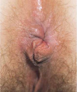
Figure 17-33 Prolapsed internal hemorrhoids.
The patient is instructed to take a deep breath, at which time the examiner's right index finger is inserted into the anal canal as the anal sphincter relaxes. The sphincter should close completely around the examining digit. The sphincter tone should be assessed. The finger should be inserted as far as possible into the rectum, although 4 inches (10 cm) is the probable limit of digital exploration. The examiner's left hand can now be moved to the patient's left buttock, while the right index finger examines the rectum. This examination is illustrated in Figure 17-35B.
Palpate the Rectal Walls
The lateral, posterior, and anterior walls of the rectum are palpated. To palpate the lateral walls, rotate your digit along the sides of the patient's rectum, as shown in Figure 17-36. The ischial spines, coccyx, and lower sacrum can be felt easily. Palpate the walls for polyps, which may be sessile (attached by a base) or pedunculated (attached by a stalk). Any irregularities or undue tenderness should be noted. The only way to examine the entire circumference of the rectal wall fully is to turn your back to the patient, which will allow you to hyperpronate your hand. Unless you do so, you will be unable to examine the portion of the rectal wall between the 12 o'clock and 3 o'clock positions. A small lesion in this quadrant may go undetected.
Intraperitoneal metastases may be palpated anterior to the rectum. These tumors are hard, and a shelflike structure projects into the rectum as a result of infiltration of Douglas' pouch with neoplastic cells. This is Blumer's shelf.
Palpate the Prostate Gland
The prostate gland lies anterior to the wall of the rectum. The size, surface, consistency, sensitivity, and shape of the prostate gland should be assessed.
The prostate is a bilobed, heart-shaped structure approximately 1.5 inches (4 cm) in diameter. It is normally smooth and firm and has the consistency of a hard rubber ball. The apex of the heart shape points toward the anus. Identify the median sulcus and the lateral lobes. Note any masses, tenderness, and nodules. Only the lower apex portion of the gland is palpable. The superior margin is usually too high to reach. The examination of the prostate is illustrated in Figure 17-37. The size of the prostate in relation to the examiner's finger is illustrated in Figure 17-38.
A hard, irregular nodule produces asymmetry of the prostate gland and is suggestive of cancer. Carcinoma of the prostate frequently involves the posterior lobe, which can easily be identified during the DRE. Carcinoma of the prostate is the third leading cause of death in men (9% of all cancer deaths) in the United States. In 2007, there were 218,890 new cases and 27,050 deaths from cancer of the prostate. Early detection is usually limited to detection of an abnormality on DRE.
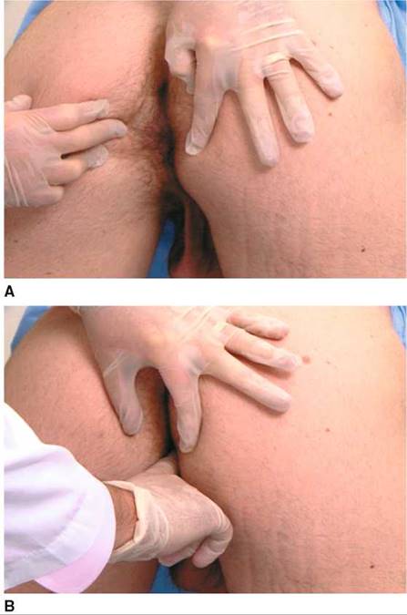
Figure 17-34 Technique of the rectal examination. A, Inspection. B, Palpation. The examiner's finger is inserted with the palm of the hand facing downward.
Benign prostatic hypertrophy produces a symmetrically enlarged, soft gland that protrudes into the rectal lumen. This diffuse enlargement is common among men older than 60 years. A boggy, fluctuant, or tender prostate may indicate acute prostatitis. The seminal vesicles lie superior to the prostate gland and are rarely palpated, unless they are enlarged.
The rectal examination is concluded by informing the patient that you are now going to withdraw your finger. Gently remove the examining finger, and give the patient tissues to wipe himself.
Fecal Occult Blood Test
The examining digit should be inspected. The color of the fecal material should be noted. The fecal material should be placed on the fecal occult blood test (FOBT) card and examined with the Hemoccult developer.
The guaiac or benzidine test detects occult blood. If blood is present, a chemical reaction results in a blue coloration on the card. The reaction is graded by the intensity of the blue color, from light blue (trace blood) to dark blue (4+ positive).
Although the examination for inguinal hernias is part of the abdominal examination, it is discussed in Chapter 18, Male Genitalia and Hernias.
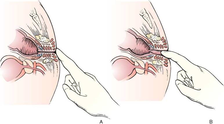
Figure 17-35 Illustration of the rectal examination. A, The sphincter is relaxed by gentle pressure with the palmar surface of the examiner's finger. B, With the examiner's left hand spreading the patient's buttocks, examination is carried out with the examiner's right index finger.
Special Techniques
Intra-abdominal inflammation may involve the psoas muscle. A special test performed when there is suspicion of intra-abdominal inflammation is the iliopsoas test. The patient is asked to lie on the unaffected side and extend the other leg at the hip against the resistance of the examiner's hand. A positive psoas sign is abdominal pain with this maneuver. Irritation of the right psoas muscle by an acutely inflamed appendix produces a right psoas sign. This test is demonstrated in Figure 17-39.
Another useful test for inflammation is the obturator test. While the patient is lying on the back, the examiner flexes the patient's thigh at the hip, with the patient's knees bent, and rotates the leg internally and externally at the hip. If there is an inflammatory process adjacent to the obturator muscle, pain is elicited. The obturator test is demonstrated in Figure 17-40.
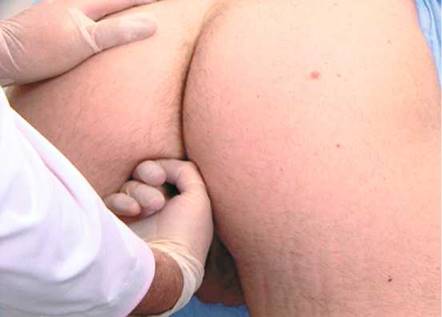
Figure 17-36 Technique for rectal examination. Note position of the examiner's left hand.
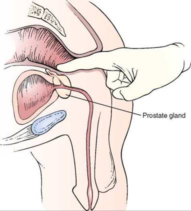
Figure 17-37 Examination of the prostate.
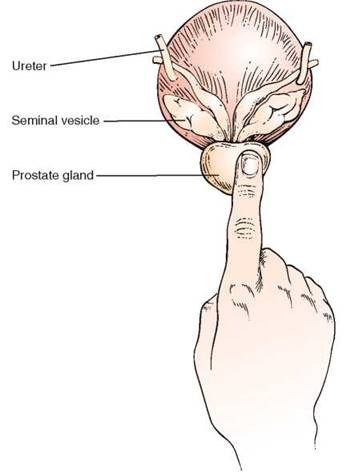
Figure 17-38 Relationship of the size of the prostate gland to the examining finger.
Clinicopathologic Correlations
There is evidence that detecting and removing polyps reduces the incidence of colorectal cancer and that detecting early cancers lowers the mortality rate from colorectal cancer. The American Cancer Society has suggested the following guidelines for the early detection of colorectal cancer in individuals without increased risk. Beginning at age 50 years, men and women should follow one of the following examination schedules:
1. FOBT every year
2. Flexible sigmoidoscopy every 5 years
3. Annual FOBT and flexible sigmoidoscopy every 5 years
4. Double-contrast barium enema testing every 5 years
5. Colonoscopy every 10 years
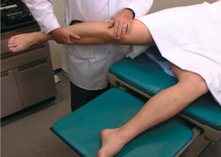
Figure 17-39 The iliopsoas test.
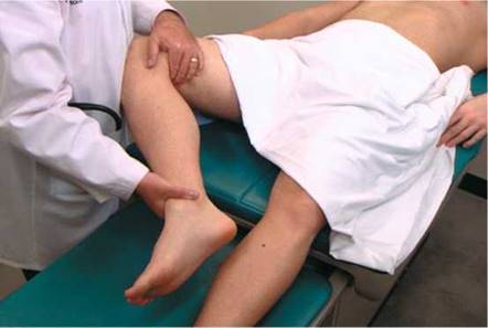
Figure 17-40 The obturator test.
The colorectal cancer mortality rate can be reduced 15% to 33% by FOBT and diagnostic evaluation and treatment for positive test results. Annual FOBT screening leads to a greater reduction in the colorectal cancer mortality rate than does biennial screening. Flexible sigmoidoscopy identifies nearly all cancers and polyps greater than 0.4 inch (1 cm) in diameter and 75% to 80% of small polyps that are located in the portion of the bowel examined. Screening with flexible sigmoidoscopy has been shown to result in a 60% to 80% reduction in risk of death from colorectal cancer in the part of the colon examined. There is indirect evidence supporting the use of double-contrast barium enema testing in screening for colorectal cancer. This test can image the entire colon and detect cancers and large polyps. Screening colonoscopy offers the potential both to identify and remove cancers and premalignant lesions throughout the colon and rectum. However, no studies that show a reduction in the mortality rate associated with screening colonoscopy have been completed to date.
Anal cancer is increasing in both men and women worldwide. Men who have sex with men, HIV-positive individuals, transplant recipients, and women with cervical neoplasia have a higher risk for anal cancer than the general population. Men who have sex with men have a 44% greater risk for anal cancer than the general population (Chin-Hong, 2008). Anal cancer, like cervical cancer, is potentially preventalbe. Anal cytology has been studied and found to be a useful screening test to detect anal cancer. It has been shown that a high proportion of men who have sex with men were infected with anal human papillomavirus (HPV) (66%).
Table 17-2 lists the classic locations of pain referred from abdominal structures. Table 17-3 summarizes the maneuvers for ameliorating abdominal pain. Table 17-4 summarizes the sensitivities and specificities of the various maneuvers used to detect ascites. Table 17-5 is a comparison of the clinical manifestations of ulcerative colitis and Crohn's disease. Table 17-6 lists the clinical features of cancer of the stomach, pancreas, and colon. Table 17-7 lists the variation of symptoms in right-sided and left-sided colon cancer and in rectal cancer. Table 17-8 compares the symptoms and signs of cirrhosis, which are numerous, as they relate to hepatocellular failure and portal hypertension.
Table 17-5. Clinical Comparison of Ulcerative Colitis and Crohn's Disease
|
Feature |
Ulcerative Colitis |
Crohn's Disease |
|
Diarrhea |
Present |
Present |
|
Hematochezia |
Common |
Rare |
|
Extraintestinal manifestations |
Common |
Common |
|
Perirectal disease |
Fissures |
Fistulas |
|
Abscesses |
||
|
Rectal disease |
Present |
Absent |
|
Anal disease |
Absent |
Present |
Table 17-6. Clinical Comparison of Cancer of the Stomach, Pancreas, and Colon
|
Cancer of Stomach |
Cancer of Pancreas |
Cancer of Colon |
|
Major Symptoms Upper abdominal pain Occult bleeding Weight loss Vomiting Anorexia Dysphagia |
Upper abdominal pain Back pain Weight loss Jaundice |
Change in bowel habits Gastrointestinal bleeding Lower abdominal pain |
|
Risk Factors |
||
|
Adenomatous polyps |
Smoking |
Adenomatous polyps |
|
Pernicious anemia |
Alcoholism (?) |
Ulcerative colitis |
|
Family history |
Familial polyposis |
|
|
Immigrants from Japan |
Gardner's syndrome Villous adenomas |
Prostate cancer is the leading cancer diagnosed among men in the United States, accounting for 33% of all cancers in men. However, racial/ethnic variations in the Surveillance, Epidemiology, and End Results (SEER) study data are striking: The incidence rate among African-American men (180.6 per 100,000) is more than seven times that among Koreans (24.2 per 100,000). Indeed, African Americans in the United States have the highest rates of this cancer in the world. Although the incidence among white persons is quite high, it is distinctly lower than that among African Americans. Asian and Native American men have the lowest rates. The very low rate in Korean men possibly reflects the fact that most of the Koreans in the SEER areas were recent immigrants from Asia, where rates are lower than in the United States.
Nearly 9 per 10 prostate cancers (86%) are diagnosed at a localized stage, when the 5-year survival rate is 100%. The incidence rates for prostate cancer, however, continue to increase, in part because of increased numbers of screening tests. Prostate cancer can often be found early by a DRE and by testing the amount of prostate-specific antigen (PSA) in a sample of blood. If a man has routine yearly examinations and either one of these test results becomes abnormal, any cancer that he may have has probably been found at an early, more treatable stage. Since the use of these early detection tests for prostate cancer became relatively common around 1990, the death rate from prostate cancer has dropped.
PSA is a substance made by the normal prostate gland. Although PSA is found mostly in semen, a small amount is also present in the blood. Most men have levels under 4 ng/mL of blood. When prostate cancer develops, the PSA level usually goes above 4 ng/mL. If the level is above 4 ng/mL but less than 10 ng/mL, the man has about a 25% chance of having prostate cancer. If it goes above 10 ng/mL, the chance of having prostate cancer is more than 67% and increases as the PSA level increases.
PSA occurs in two major forms in the blood. One is bound (attached) to blood proteins and the other circulates free (unattached). The percent-free PSA test indicates how much PSA circulates free in comparison with the total PSA level. The percentage of free PSA is lower in men who have prostate cancer than in men who do not. If the PSA results are in the borderline range (4 to 10 ng/mL), a low percent-free PSA (< 10%) means that the likelihood of having prostate cancer is about 50%, and a biopsy should be performed.
Table 17-7. Variation of Symptoms of Cancer of the Right Colon, Left Colon, and Rectum
|
Symptom |
Cancer of Right Colon |
Cancer of Left Colon |
Cancer of Rectum |
|
Pain |
Ill defined |
Colicky* |
Steady, gnawing |
|
Obstruction |
Infrequent |
Common |
Infrequent |
|
Bleeding |
Brick-red |
Red mixed with stool |
Bright red coating stool |
|
Weakness1" |
Common |
Infrequent |
Infrequent |
|
*Worse with ingestion of foods. {Secondary to anemia. |
|||
Table 17-8. Signs and Symptoms of Cirrhosis
|
Hepatocellular Failure |
Portal Hypertension |
|
Spider angiomata |
Ascites |
|
Gynecomastia |
Varices: esophageal |
|
Palmar erythema |
Hemorrhoids |
|
Ascites |
Caput medusae |
|
Jaundice Testicular atrophy Erectile dysfunction Bleeding problems Changes in mental function |
Splenomegaly |
The PSA level can be affected by many factors. It is increased with noncancerous enlargement of the prostate (i.e., benign prostatic hypertrophy), and with prostatitis. PSA levels also normally rise slowly with aging. Ejaculation can cause a temporary increase in blood PSA levels, so it is often recommended that men abstain from ejaculation for 2 days before testing.
The American Cancer Society believes that health-care professionals should offer the PSA blood test and DRE yearly, beginning at age 50 years, to men who have at least a 10-year life expectancy. Men at high risk, such as African Americans and men who have a first-degree relative (father, brother, son) in whom prostate cancer was diagnosed at an early age should begin testing at age 45 years. Men at even higher risk (because they have several first-degree relatives who had prostate cancer at an early age) could begin testing at age 40 years. Depending on the results of this initial test, further testing might not be needed until age 45 years.
Useful Vocabulary
Listed here are the specific roots that are important for understanding the terminology related to abdominal disease.
|
Root |
Pertaining to |
Example |
Definition |
|
aer(o)- |
air; gas |
aerophagia |
Air swallowing |
|
celi(o)- |
abdomen |
celiac |
Pertaining to the abdomen |
|
chol(e)- |
bile |
cholelith |
Gallstone |
|
cyst- |
sac containing liquid |
cholecystitis |
Inflammation of the gallbladder |
|
enter(o)- |
intestines |
enteritis |
Inflammation of the small intestines |
|
gastr(o)- |
stomach |
gastrectomy |
Surgical removal of the stomach |
|
lapar(o)- |
loin; flank |
laparotomy |
Surgical incision through the flank; generally, any abdominal incision |
|
-phago- |
eating |
phagocyte |
Any cell that ingests other cells or microorganisms |
|
-tripsy |
shock waves |
lithotripsy |
Noninvasive technique for breaking up stones by the use of shock waves |
Writing Up the Physical Examination
Listed here are examples of the write-up for the examination of the abdomen.
• The abdomen is scaphoid without scars. Bowel sounds are present. The liver is felt 2 fingerbreadths below the right costal margin for a total span of 10 cm [4 inches]. Neither the spleen nor any masses are felt. The kidneys are not palpable. Rectal examination reveals normal sphincter tone. The prostate is soft, without any masses. The walls of the rectum are smooth, without masses. Results of testing of the stool for blood is negative. No costovertebral angle tenderness [CVAT] is present.
• The abdomen has a scar in the right upper quadrant. Bowel sounds are absent. Marked tympany is present throughout the abdomen. Rigidity is present throughout. Marked tenderness is present in the abdomen, especially in the left lower quadrant. Examination of the rectum reveals tenderness in the same area. Stool guaiac test result is 4+. The liver, spleen, and kidneys are not felt. No CVAT is present.
• The abdomen is obese. Bowel sounds are present. Percussion notes are normal. There is an area of significant pain in the right lower quadrant, immediately above the right midposition of the inguinal ligament. Rectal examination discloses severe pain in the same area. The obturator and straight leg raising signs are positive on the right. Stool guaiac test result is negative. Right CVAT is present. No organomegaly is felt.
• The abdomen is protuberant, with a midline well-healed scar. Bowel sounds are present. Shifting dullness and a fluid wave are present. A large mass 8 x 15 cm [3 x 6 inches] is felt in the right upper quadrant. Results of examination of the rectum are unremarkable except for a trace positive stool guaiac. There is no hepatosplenomegaly.
• The abdomen is scaphoid and soft, without guarding, rigidity, or tenderness. Bowel sounds are present. The liver measures 12 cm [4.7 inches] in span in the midclavicular line. The spleen tip is felt below the left costal margin. No masses are present. Rectal examination reveals a 2-cm [0.75-inch] hard nodule in the posterior lobe of the prostate, which is nontender. Stool guaiac test result is negative.
Bibliography
Alter MJ, Kruszon-Moran D, Nainan OV, et al: The prevalence of hepatitis C virus infection in the United States, 1988 through 1994. N Engl J Med 341:556, 1999.
Barkun AN, Camus M, Green L, et al: The bedside assessment of splenic enlargement. Am J Med 91:512, 1991.
Barkun AN, Camus M, Meagher T, et al: Splenic enlargement and Traube's space: How useful is percussion? Am J Med 87:562, 1989.
Bernstein CN, Blanchard JF, Leslie W, et al: The incidence of fracture among patients with inflammatory bowel disease: A population-based cohort study. Ann Intern Med 133:795, 2000.
Cancer Facts & Figures. Atlanta, American Cancer Society, 2007.
Cancer Trends Progress Report—2007 Update. Bethesda, Md, National Cancer Institute, National Institutes of Health, Department of Health and Human Services, December 2007. Available at: http://progressreport.cancer.gov; accessed June 25, 2008.
Carter HB, Ferrucci L, Kettermann A, et al: Detection of life-threatening prostate cancer with prostate- specific antigen velocity during a window of curability. J Natl Cancer Institute 98:1521, 2006.
Castell DO: The spleen percussion sign: A useful diagnostic technique. Ann Intern Med 67:1265, 1967.
Cattau EL Jr, Benjamin SB, Knoff TE, et al: The accuracy of the physical examination in the diagnosis of suspected ascites. JAMA 247:1164, 1982.
Chin-Hong PV, Berry M, Cheng SC, et al: Comparison of patient- and clinician-collected anal cytology samples to screen for human papillamovirus-associated anal intraepithelial neoplasia in men who have sex with men. Ann Intern Med 149:300, 2008.
Cotterchio M, Manno M, Klar N, et al: Colorectal screening is associated with reduced colorectal cancer risk: A case-control study within the population-based Ontario Familial Colorectal Cancer Registry. Cancer Causes Control 16:865, 2005.
Cummings S, Papadakis M, Melnick J, et al: The predictive value of physical examination for ascites. West J Med 142:633, 1985.
Espey DK, Wu XC, Swan J, et al: Annual report to the nation on the status of cancer, 1975-2004, featuring cancer in American Indians and Alaska Natives. Cancer 110:2119, 2007.
Fowler FJ Jr, Barry MJ, Walker-Corkery B, et al: The impact of a suspicious prostate biopsy on patients' psychological, socio-behavioral, and medical care outcomes. J Gen Intern Med 21:715, 2006.
Grover SA, Barkun AN, Sackett DL: Does this patient have splenomegaly? JAMA 270:2218, 1993.
Halpern S, Coel M, Ashburn W, et al: Correlation of liver and spleen size: Determinations by nuclear medicine studies and physical examination. Arch Intern Med 134:123, 1974.
Harris R, Lohr KN: Screening for prostate cancer: An update of the evidence for the U.S. Preventive Services Task Force. Ann Intern Med 137:917, 2002.
Helzer JE, Chammas S, Norland CC, et al: A study of the association between Crohn's disease and psychiatric illness. Gastroenterology 86:324, 1984.
Imperiale TF, Ransohoff DF, Itzkowitz SH, et al: Fecal DNA versus fecal occult blood for colorectal-cancer screening in an average-risk population. N Engl J Med 351:2704, 2004.
Johnson LG, Madeleine MM, Newcomer LM, et al: Anal cancer incidence and survival: the surveillance, epidemiology, and end results experience, 1973-2000. Cancer 101:281, 2004.
Leitzmann MF, Platz EA, Meir J, et al: Ejaculation frequency and subsequent risk of prostate cancer. JAMA 291:1578, 2004.
Levin B, Brooks D, Smith RA, et al: Emerging technologies in screening for colorectal cancer: CT colonog- raphy, immunochemical fecal occult blood tests, and stool screening using molecular markers. CA Cancer J Clin 53:44, 2003.
Markowitz SD, Dawson DM, Willis J, et al: Focus on colon cancer. Cancer 1:233, 2002.
Meidl EJ, Ende J: Evaluation of liver size by physical examination. J Gen Intern Med 8:635, 1993.
Naylor CD: Physical examination of the liver. JAMA 271:1859, 1994.
Ries LAG, Melbert D, Krapcho M, et al: SEER Cancer Statistics Review, 1975-2004 [based on November 2006 Surveillance Epidemiology and End Results (SEER) data submission]. Bethesda, Md: National Cancer Institute, 2007. Available at: http://seer.cancer.gov/csr/1975_2004/; accessed June 9, 2008.
Robertson DJ, Greenberg ER, Beach M, et al: Colorectal cancer in patients under close colonoscopic surveillance. Gastroenterology 129:34, 2005.
Schiller LR: Chronic diarrhea. Gastroenterology 127:287, 2004.
Seeff L, Shapiro J, Nadel M: Are we doing enough to screen for colorectal cancer? Findings from the 1999 Behavioral Risk Factor Surveillance System. J Fam Pract 51:761, 2003.
Simel DL, Halvorsen RA, Feussner JR: Quantitating bedside diagnosis: Clinical evaluation of ascites. J Gen Intern Med 3:423, 1988.
Smith RA, Cokkinides V, Eyre HJ: American Cancer Society guidelines for the early detection of cancer, 2004. CA Cancer J Clin 54:41, 2004.
Surveillance, Epidemiology, and End Results (SEER) Program: SEER Stat Database: Incidence—SEER 9 Regs Public-Use, November 2002 Sub (1973-2000). Bethesda, Md, National Cancer Institute, DCCPS, Surveillance Research Program, Cancer Statistics Branch. Released April 2003, based on the November 2002 submission. Available at www.seer.cancer/gov; accessed June 23, 2008.
Thompson IM, Pauler DK, Goodman PJ, et al: Prevalence of prostate cancer among men with a prostate- specific antigen level < or =4.0 ng per milliliter. New Engl J Med 350:2239, 2004.
Williams JW, Simel DL: Does this patient have ascites? How to divine fluid in the abdomen. JAMA 267:2645, 1992.