
The shape of the breast is like a gourd. They are round for holding blood to be changed into milk. ... They have teats, that the new born child may suck therefrom.
Mondino De' Luzzi (1275-1326)
General Considerations
In the United States, the National Cancer Institute estimates that one of every eight women (approximately 12.5%) will develop breast cancer during her lifetime. Among the malignant diseases in women, breast cancer is the most common to develop and is the second most common cancer cause of death. In 2007, it accounted for 31% of new cancer cases in American women, and 15% of cancer deaths; there were 180,510 new cases of invasive breast cancer (stages I to IV) and 40,910 related deaths in the United States (40,460 women and 450 men). Breast carcinoma in situ, a very early form of the disease, was diagnosed in another 62,030 women.
The incidence of cancer of the breast is higher in the United States than in European or Asian countries. It has been well established that women in underdeveloped nations have lower rates of breast cancer than do women from more affluent societies. Among racial/ethnic groups, white and African-American women have the highest incidence rates of breast cancer (113.2 and 99.3 per 100,000 population, respectively). Asian and Pacific Islander women and Latino women have a lower risk (72.6 and 69.4 per 100,000 population, respectively). The risk is lowest among Native Americans (33.9 per 100,000 population). The rate of mortality from breast cancer is highest in African-American women (31.4 per 100,000); the mortality rate in white women is 25.7 per 100,000. The lowest rate of mortality from breast cancer is in Asian and Pacific Islander women (11.4 per 100,000). The incidence of breast cancer continues to increase, partly because of increased screening through mammography.
Once breast cancer has occurred in a family, the risk that other women in the same family will have breast cancer is significantly higher. First-degree relatives, such as sisters or daughters, have more than twice the risk for development of breast cancer if the original patient developed cancer in one breast after menopause. Women with a family history of premenopausal breast cancer in one breast have three times the risk. If the original patient had postmenopausal cancer in both breasts, the first-degree relatives have more than four times the risk. First-degree relatives of patients with cancer in both breasts before menopause have nearly nine times the risk.
The age at onset of menarche and the reproductive cycle seems to play some role in the development of breast cancer. In women with menarche before the age of 12 years, the incidence of breast cancer appears to be higher. Women who had their first child at the age of 30 years or older have three times the risk of those who had their first child at a younger age.
Most breast cancers are detected as painless masses, noticed by either the patient or the examiner during a routine physical examination. The earlier the diagnosis is made, the better the prognosis is. Screening for breast cancer is best accomplished by a thorough clinical breast examination, breast self-examination, and mammography. Mammography is the most sensitive method for the detection of breast cancer and has been demonstrated to reduce the breast cancer mortality rate.
Structure and Physiology
The mammary glands are the distinguishing feature of all mammals. Human breasts are conical in form and are often unequal in size. The breast extends from the level of the second or third rib to the level of the sixth or seventh rib, from the sternal edge to the anterior axillary line. The ''tail'' of the breast extends into the axilla and tends to be thicker than the other breast areas. This upper outer quadrant contains the greatest bulk of mammary tissue and is frequently the site of neoplasia. Figure 16-1 illustrates the normal breast.
The normal breast consists of glandular tissue, ducts, supporting muscular tissue, fat, blood vessels, nerves, and lymphatic vessels. The glandular tissue consists of 15 to 25 lobes, each of which drains into a separate excretory duct that terminates in the nipple. Each duct dilates as it enters the base of the nipple to form a milk sinus. This serves as a reservoir for milk during lactation. Each lobe is subdivided into 50 to 75 lobules, which drain into a duct that empties into the excretory duct of the lobe.

Figure 16-1 Anatomy of a normal breast.
Both the nipple and areola contain smooth muscle that serves to contract the areola and compress the nipple. Contraction of the smooth muscle makes the nipple erect and firm, thereby facilitating the emptying of the milk sinuses.
The skin of the nipple is deeply pigmented and hairless. The dermal papillae contain many sebaceous glands, which are grouped near the openings of the milk sinuses. The skin of the areola is also deeply pigmented but, unlike the skin of the nipple, contains occasional hair follicles. Its sebaceous glands are commonly seen as small nodules on the areolar surface and are termed Montgomery's tubercles.
Cooper's ligaments are projections of the breast tissue that fuse with the outer layers of the superficial fascia and serve as suspensory structures.
The blood supply to the breast is carried by the internal mammary artery. The breast has an extensive network of venous and lymphatic drainage. Most of the lymphatic drainage empties into the nodes in the axilla. Other nodes lie beneath the lateral margin of the pectoralis major muscle, along the medial side of the axilla, and in the subclavicular region. The main lymph node chains and lymphatic drainage of the breast are illustrated in Figure 16-2.
Several physiologic changes occur in the breast. These changes are a result of the following factors:
• Growth and aging
• The menstrual cycle
• Pregnancy
At birth, the breasts contain a branching system of ducts emptying into a developed nipple. There is elevation of only the nipple at this stage. Shortly after birth, there is a slight secretion of milky material. After 5 to 7 days, this secretory activity stops. Before puberty, there is elevation of the breast and nipple, called the breast bud stage. The areola has increased in size. At the onset of puberty, the areola enlarges further and darkens. A distinct mass of glandular tissue begins to develop beneath the areola. By the onset of menstruation, the breasts are well developed, and there is forward projection of the areola and nipple at the apex of the breast. One to 2 years later, when the breast has reached maturity, only the nipple projects forward; the areola has receded to the general contour of the breast. The stages of breast development from birth to adulthood are illustrated in Figure 16-3. Figure 24-46 (Chapter 24, The Pediatric Patient) further illustrates and describes the breast developmental stages.

Figure 16-2 Lymphatic drainage of the breast.

Figure 16-3 Stages of breast development.
The nodularity, density, and fullness of the adult breast depend on several factors. Most important is the presence of excess adipose tissue. Because the mammary gland consists mainly of adipose tissue, women who are overweight have larger breasts. Pregnancy and nursing also alter the character of the breasts. Often, women who have nursed have softer, less nodular breasts. However, because the glandular tissue is approximately equal in all women, the size of the breast is unrelated to nursing. With menopause, the breasts decrease in size and become less dense. There is an associated decrease in elastic tissue as women age.
The major physiologic change related to the menstrual cycle is engorgement, occurring 3 to 5 days before menstruation. This is an increase in the size, density, and nodularity of the breasts. There is also an increased sensitivity of the breasts at this time. Because the nodularity of the breasts increases, the examiner should not attempt to diagnose a breast mass at this time. The patient should be reevaluated during the midperiod of the next cycle.
With pregnancy, the breasts become fuller and firmer. The areola darkens, and the nipples become erect as they enlarge. As the woman approaches the third trimester, a thin, yellowish secretion, called colostrum, may be noted. After the birth of the child, if the mother begins nursing within 24 hours, the secretion of colostrum stops, and the secretion of milk begins. During nursing, the breasts become markedly engorged. After the woman has stopped nursing, lactation continues for a short time.
The neuroendocrine control of the breasts can be outlined as follows. Suckling produces nerve impulses that travel to the hypothalamus. The hypothalamus stimulates the anterior pituitary to secrete prolactin, which acts on the glandular tissue of the breast to produce milk. The hypothalamus also stimulates the posterior pituitary to produce oxytocin, which stimulates the muscle cells surrounding the glandular tissue to contract and force the milk into the ductular system.
Many abnormalities of the breast are related to its embryology. An epithelial ridge, called the milk line, forms along each side of the body from the axilla to the inguinal region. Along this milk line are multiple rudiments for future breast development. In humans, only one rudimentary pair in the pectoral region persists and eventually develops into normal breasts. Accessory breasts or nipples are present in as many as 2% of white women. Accessory breasts may exist as glandular tissue, nipple, or only the areola. The axilla is the most common site for these anomalous structures, followed by a site just below the normal breast. In more than 50% of all patients with accessory breast tissue, the anomalies are bilateral. In general, accessory breast tissue is of little clinical significance. It usually has no physiologic function and is rarely associated with disease. Figure 16-4 illustrates the milk line. Figure 16-5 shows an accessory nipple.
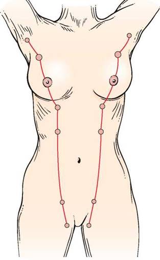
Figure 16-4 The milk line.
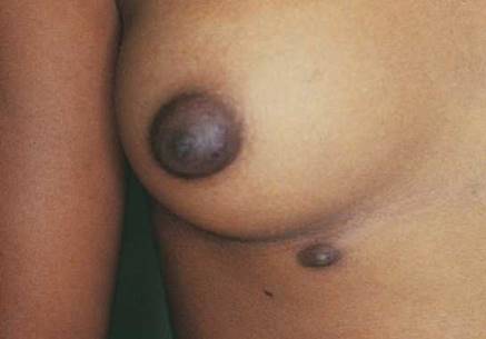
Figure 16-5 Accessory nipple.
Review of Specific Symptoms
The most important symptoms of breast disease are the following:
• Mass or swelling
• Pain
• Nipple discharge
• Change in skin over breast
Mass or Swelling
During self-examination, a patient may discover a breast mass. Ask the following questions: ''When did you first notice the lump?"
''Have you noticed that the mass changes in size during your menstrual periods?''
''Is the mass tender?''
‘‘Have you ever noticed a mass in your breast before?''
''Have you noticed any skin changes on the breast?''

Figure 16-6 Large breast mass found on selfexamination.
''Have you had any recent injury to the breast?''
‘‘Is there any nipple discharge? nipple retraction?''
''Do you have breast implants?'' If yes, ''What are they made of?''
If the lump enlarges during the premenstrual and menstrual stages of the cycle, it is likely that the woman is detecting only physiologic nodularity. The association of nipple discharge, nipple inversion, or skin changes overlying the mass is strongly suggestive of neoplasm. Figure 16-6 shows a large breast mass found on self-examination.
Pain
Breast pain or tenderness is a common symptom. Most often, these symptoms are attributable to the normal physiologic cycle. Ask the following questions of any patient with breast pain:
''Can you describe the pain?''
''When did you first experience the pain?''
''Are there any changes in the pain with your menstrual cycle?''
''Do you have pain in both breasts?''
''Have you had any injury to the breast?''
''Is the pain associated with a mass in the breast? nipple discharge? nipple retraction?''
''Has there been a change in your bra size?''
Rapidly enlarging cysts may be painful. Cystic disease of the breasts is usually painless. Although breast pain is a relatively uncommon manifestation of breast cancer, its presence does not exclude the diagnosis. Never delay evaluation of a painful breast mass.
Nipple Discharge
Nipple discharge is not a common symptom, but it should always raise the suspicion of breast disease, especially if the discharge occurs spontaneously. Any patient who describes a nipple discharge should be asked the following questions:
What is the color ofthe discharge?''
Do you have a discharge from both breasts?''
When did you first notice the discharge?''
Is the discharge related to your menstrual cycle?''
When was your last menstrual cycle?''
Is the discharge associated with nipple retraction? a breast mass? breast tenderness?''
Do you have headaches?''
''Are you taking any medications?''
Are you using oral contraceptives?''
If the woman has recently delivered a child, ask
Were there any problems during the delivery ofyour last child?''
The most common types of discharge are serous and bloody. A serous discharge is thin and watery and may appear as a yellowish stain on the patient's garments. This commonly results from an intraductal papilloma in one of the large subareolar ducts. Women taking oral contraceptives may complain of bilateral serous discharge. A serous discharge can also occur in women with breast carcinoma.
A bloody discharge is associated with an intraductal papilloma, which is common among pregnant and menstruating women. It may, however, be associated with a malignant intraductal papillary carcinoma. The presence of any nipple discharge is more important than its character because both types of discharge are associated with benign or malignant disease.
A milky discharge is usually milk. It is common for women to continue to secrete milk for a few months after they stop nursing. In rare instances, the secretion may continue for a year. Persistent lactation, also known as galactorrhea, can be a result of massive hemorrhage occurring during childbirth and producing pituitary necrosis. Abnormal lactation may also result from a pituitary tumor that interferes with the normal hypothalamic-pituitary feedback loop or from the use of certain tranquilizing medications. Mechanical stimulation or suckling may produce physiologic stimulation.
Change in Skin over Breast
A change in the color or texture of the skin of the breast or areola is an important symptom of breast carcinoma. The presence of dimpling, puckering, or scaliness warrants further investigation. The presence of unusually prominent pores, indicative of edema of the skin, is an important sign of malignancy. This clinical sign is called peau d'orange because of its orange-peel appearance. During the early stages of breast carcinoma, the lymphatic vessels of the breast are dilated and contain occasional emboli of carcinoma cells. Limited peau d'orange over the lower half of the areola is present. As the disease progresses, more lymphatic vessels become filled with carcinoma cells that block them, creating more generalized edema. The classic appearance of peau d'orange is pictured in Figure 16-7.

Figure 16-7 Peau d'orange.
General Suggestions
The interviewer should pay special attention to the family history of any woman presenting with symptoms of breast disease. As indicated earlier, breast cancer may be a familial disorder. The occurrence of breast disease in a close relative and the age at which it developed are relevant to the patient's disease. Ask the patient the following questions:
‘‘Have you had a mammogram?'' If yes, ‘‘When and what was the result?''
''Have you had breast cancer?''
''Have you had breast cancer without the removal of your breast?''
''Do you have breast implants?''
''Have you had any breast biopsies or breast surgery?''
Have you ever had radiation treatments to your breasts?''
‘‘Did your birth mother have breast cancer?'' If yes, ‘‘Premenopausal or postmenopausal? At what age was her diagnosis made?''
''Do you have a sister or daughter with breast cancer?'' If yes, ‘‘Premenopausal or postmenopausal? At what age was her diagnosis made?''
Do you use birth control pills?''
Do you take estrogen replacement therapy?''
Impact of Breast Disease on the Patient
The psychosocial problems resulting from breast cancer are far-reaching. Although the loss of an extremity is more disabling in everyday life, the loss of a breast produces intense feelings of loss of the feminine identity. Many women who lose a breast become depressed because they feel their symbol of femininity has been removed. They are afraid that they will no longer be considered ''whole'' women because their bodies have been maimed. They fear that they can no longer be loved normally and that they can no longer experience sexual satisfaction. They fear looking at themselves in a mirror and perceive themselves as ugly. The asymmetry is often described as ''mutilation'' or ''a bomb crater.'' After mastectomy, women often suffer from sexual inhibition and sexual frustration.
Once a woman has discovered a mass in her breast, she becomes intensely fearful. The fear of breast cancer is twofold: It is a cancer, often with a bad prognosis, and it is associated with disfigurement. For these reasons, the patient commonly denies the presence of the mass and delays seeking medical attention. It is not uncommon for a patient to seek medical assistance for the first time with a large tumor mass the size of an orange that has eroded through the skin and has become infected. When asked how long the mass has been present, the woman might answer that she ''discovered it yesterday.''
After a mastectomy, the patient will probably suffer from depression and low self-esteem. The patient should be supported, and counseled if necessary. Open communication and sharing of feelings among the patient, husband, significant other, physician, and family are important factors in the psychologic rehabilitation of the woman.
The patient pictured in Figure 16-8 had inflammatory carcinoma of her left breast, with massive lymphedema of the left arm. The patient had noticed that her arm had been swelling for the past few months, and she now needed support to raise it. She presented to the clinic complaining only about the heaviness of her arm. When examination revealed the breast lesion, she stated that she had noticed the breast changes ''only a few days ago.'' This is another example of denial of illness (see also the patient shown in Fig. 2-1).
Most commonly, women with invasive breast cancer and axillary lymph node involvement require chemotherapy for periods up to 6 or 7 months after mastectomy. Many of the agents used to treat the patient have significant side effects, including nausea, vomiting, and alopecia. There are medicines available to help control the nausea and vomiting, but the alopecia presents another special problem. Although the woman recognizes that she will lose her hair and knows that it will grow back, she suffers further from low self-esteem. In our society, hair is a woman's ''persona'': a bald man is acceptable; a bald woman is not. Finally, at the conclusion of chemotherapy, the woman may face further depression. While on the chemotherapeutic protocol, she feels reassured that no further cancer can develop, but what does the future hold when these medications are stopped?

Figure 16-8 Inflammatory breast carcinoma.
Physical Examination
No special equipment is necessary for the examination of the breast.
The examination of the breast consists of the following:
Inspection
Axillary examination Ф Palpation
The examination of the breast is in two parts. The first is performed with the patient sitting up. Inspection of the breasts and palpation of the lymph nodes are done in this position. The second is performed with the patient lying down. The examiner systematically palpates the entire breast by using firm, gentle pressure exerted by the pulp of his or her finger rather than the fingertips.
To facilitate communication, the breast is pictorially divided into four quadrants. Two imaginary lines run through the nipple at right angles to each other. By visualizing the breast as a face of a clock, one line is the ''12 o'clock-6 o'clock'' line, and the other is the ''3 o'clock-9 o'clock'' line. The resulting four quadrants are the upper outer, upper inner, lower outer, and lower inner. The ''tail'' is an extension of the upper outer quadrant. These regions are illustrated in Figure 16-9.
Inspection
The woman should be seated on the edge of the examination table, facing the examiner. The examiner should ask the woman to remove her gown to her waist.
Inspect the Breasts
Inspection is first accomplished with the patient's arms at her side, as shown in Figure 16-10. Tell the patient, ''I am inspecting the breasts for any changes in the skin, contour, or symmetry.'' The breasts are inspected for size, shape, symmetry, contour, color, and edema. The nipples are inspected as to size, shape, inversion, eversion, or discharge. The nipples should be symmetric. Is any abnormal bulging present?
The skin of the breast is observed for edema. Edema of the skin of the breast that overlies a malignancy may manifest as peau d'orange.

Figure 16-9 The four breast quadrants.

Figure 16-10 Position of patient for inspection of the breasts.

Figure 16-11 Erythema of the breast.

Figure 16-12 Breast carcinoma. Note dimpling of the breast and bloody nipple discharge.
Is erythema present? Erythema is associated with infection and with inflammatory carcinoma of the breast. Figure 16-11 shows marked erythema of the breast secondary to inflammatory breast carcinoma. The scar above the areola is from a previous biopsy of a benign breast mass.
Is dimpling present? The examiner must inspect the breasts for the presence of retraction phenomena. Dimpling is a sign of retraction phenomena that are caused by an underlying neoplasm and its fibrotic response. Skin retraction is commonly associated with malignancy that causes an abnormal traction on Cooper's ligaments. The shortening of the larger mammary ducts by cancer produces flattening or inversion of the nipple. A change in the position of the nipple is important because many women have a congenitally inverted nipple on one or both sides. The dimpling of the breast in Figure 16-12 is associated with a bloody nipple discharge; both are secondary to carcinoma.
Is there a red, scaling, crusting plaque around one nipple, areola, or surrounding skin? Paget's disease of the breast is a surface manifestation invariably associated with an underlying invasive or intraductal carcinoma. The lesion appears eczematous, but unlike eczema, it is unilateral. The skin may also weep and be eroded. A much less common form is extramammary Paget's disease, which is seen around the anus or genitalia and is usually associated with malignant disease of the adnexa, bowel, or genitourinary tract. Figure 16-13 shows Paget's disease of the breast; an underlying ductal adenocarcinoma was present.
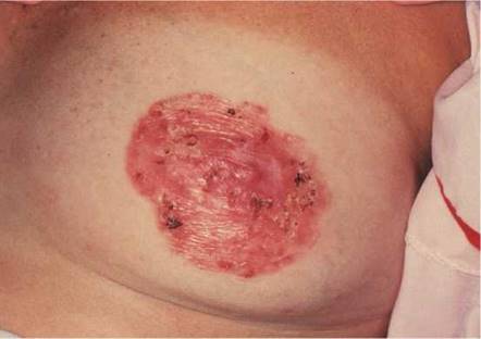
Figure 16-13 Paget's disease of the breast.

Figure 16-14 Technique for tensing the pectoralis muscles.
Inspect the Breasts in Various Postures
Inspection is next performed while the woman assumes several postures that may bring out signs of retraction that were less evident previously. Ask the woman to press her arms against her hips. This maneuver tenses the pectoralis muscles, which may bring out dimpling caused by fixation of the breast to the underlying muscles. This technique is shown in Figure 16-14. If a malignancy is present, the abnormal attachment of the tumor to the fascia and pectoralis muscle pulls on the skin and may produce skin dimpling. Any bulging may also indicate an underlying mass.
Another maneuver, which is useful for a woman with pendulous breasts, involves her bending at the waist and allowing her breasts to hang free from the chest wall. This technique is demonstrated in Figure 16-15. A carcinoma causing fibrosis in one breast produces a change in the contour of that breast.
Axillary Examination
The axillary examination is performed with the patient seated facing the examiner. Examination of the axilla is best accomplished by relaxing the pectoral muscles. To examine the right axilla, the patient's right forearm is supported by the examiner's right hand. The tips of the fingers of the examiner's left hand start low in the axilla, and, as the patient's right arm is drawn medially, the examiner advances the left hand higher into the axilla. This technique is demonstrated in Figure 16-16. The supraclavicular, subclavian, and axillary regions are palpated.
The technique of using small, circular motions of the fingers riding over the ribs is used for detecting adenopathy. Freely mobile nodes 3 to 5 mm in diameter are common and are usually indicative of lymphadenitis secondary to minor trauma of the hand and arm. After one axilla is examined, the other is evaluated by the examiner's opposite hand.

Figure 16-15 Position of patient for inspecting the breasts.
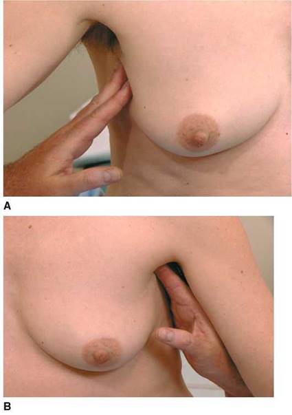
Figure 16-16 Technique for axillary examination. A, Low axilla (right side). B, High axilla (left side).
Palpation
The woman is asked to lie down and is told that the breast will next be palpated. The examiner stands at the right side of the patient's bed. Although the examiner can usually palpate each breast from the patient's right side, it is often better with large-breasted women to examine the left breast from the left side.
The breast is best palpated by allowing it to lie evenly distributed over the chest wall. Small-breasted women may lie with their arms at their sides; larger breasted women should be instructed to place their hands behind their head. A pillow placed beneath the shoulder on the side being examined facilitates the examination.
Palpate the Breast
In palpation of the breast, the examiner should use both the flat of the hand and the fingertips, as demonstrated in Figure 16-17. Palpation should be performed by the ''spokes of a wheel,'' the concentric circle, or the vertical strip method. The ''spokes of a wheel'' method starts at the nipple (see Fig. 16-17A). The examiner should start the palpation by moving outward from the nipple to the 12 o'clock position. The examiner then should return to the nipple and move along the 1 o'clock position and continue the palpation around the breasts. The concentric circles approach (see Fig. 16-17B) also starts at the nipple, but the examiner moves from the nipple in a continuous circular manner around the breast. Any lesion found by either technique is described as being a certain distance from the nipple in clock time: for example, ''3 cm from the nipple along the 1 o'clock line.'' These techniques are illustrated in Figure 16-18.
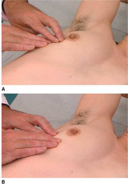
Figure 16-17. Technique for breast palpation. A, ''Spokes of a wheel.'' B, Concentric circles.
Another method is the vertical strip, or grid, technique. The breast is divided into eight or nine vertical strips, each approximately one finger's width. The examiner's three middle fingers are held together and slightly bowed to ensure contact with the skin. The pads, not the tips, of the fingers must be used for palpation. Using dime-sized circles, the examiner evaluates the breast at each of three different levels of pressure: light, medium, and deep. Each strip consists of 9 or 10 areas of palpation, slightly overlapping the previous area, and each vertical strip is evaluated with the three pressures. Although this method has been shown to be superior to the other traditional types of breast palpation, it is more time consuming and may be best used by women for breast self-examination.
The examiner should be careful when evaluating the inframammary fold. This fold is commonly observed in older women and is the area where the mammary tissue is bound tightly to the chest wall. Often, this ridge is mistaken for a breast disorder.
Describe the Findings
If a mass is palpated, the following characteristics should be described:
1. The size of the mass in centimeters and its position.
2. The shape of the mass.
3. The delimitation, referring to the borders of the mass. Is it well delimited, as with a cyst? Are the edges diffuse, as with a carcinoma?
4. The consistency, describing the ''hardness'' of the mass. A carcinoma is often stony hard. A cyst has some elastic qualities.
5. The mobility of the lesion. Is the lesion movable in the tissue that surrounds it? Benign tumors and cysts are freely mobile. Carcinomas are usually fixed to the skin, underlying muscle, or chest wall.

Figure 16-18 Method of breast palpation. A, ''Spokes of a wheel.'' B, Concentric circles.
Evaluate for Retraction Phenomenon
If a mass is detected, molding of the skin may be useful to determine whether the retraction phenomenon is present. The examiner should elevate the breast around the mass. Dimpling may occur if a carcinoma is present. Figure 16-19 demonstrates the technique of molding and its result in a patient with carcinoma of the breast. Notice the marked dimpling of the breast. This patient also had metastatic lesions of breast carcinoma on her arm, together with lymphedema. She presented to the clinic stating that she had discovered the swollen arm the day before.
Palpate the Subareolar Area
The subareolar area, the area directly under the areola, should be palpated while the patient is lying supine. In the subareolar area, the breast tissue is less dense. An abscess of Montgomery's glands in the areola may cause a tender mass in this area.
Examine the Nipple
Examination of the nipple concludes the examination of the breast. Inspect for nipple retraction, fissures, and scaling. To examine for discharge, place each hand on either side of the nipple and gently compress the nipple, noting the character of any discharge. This technique is demonstrated in Figure 16-20. Ask the woman whether she would prefer to perform this part of the examination herself.

Figure 16-19 Breast dimpling. Note the satellite skin lesions of metastatic breast cancer on the upper arm.
Examination of the Male Breast
Examination of the breast should be performed on all men. The nipples should be inspected for swelling, discharge, and ulceration. The areola and the subareolar tissue should be palpated for any masses. The axillary examination is as indicated for women.
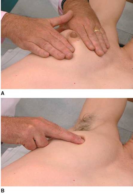
Figure 16-20 Technique for nipple examination. A, Examination for discharge. B, Examination for masses below the nipple.
Breast Self-Examination*
Women should be encouraged to perform breast self-examination monthly starting at age 20 years. It is important for a woman to learn what is normal and about the changes in her breasts that occur with her menstrual cycle. Familiarity with breast tissue makes it easier for the woman to notice any changes in the breast from month to month.
Advise the woman that the best time to perform breast self-examination is 2 to 3 days to a week after the end of her menstrual period. At this time the breasts are less tender or swollen. Women taking oral contraceptives are encouraged to do their breast self-examination each month on the day they begin their new package of pills. A woman past menopause should be advised to pick a particular day of the month and then perform the examination monthly.
Advise the patient that if she discovers anything unusual, such as a lump, discharge from the nipple, or dimpling of the skin, she should seek medical attention. Inform her that 8 of 10 breast lumps are not cancer.
The techniques of breast self-examination are included here for the purpose of patient education:
1. Stand with your arms at your sides in front of a mirror. Inspect both breasts for anything unusual, such as dimpling, puckering, discharge from the nipples, or scaling of the skin.
2. Raise your arms and clasp your hands behind your head. Press your hands forward. Look in the mirror for any changes in the breast tissue.
3. Put your hands down and place them on your hips. Bend slightly toward the mirror as you pull your shoulders and elbows forward.
4. Raise your right arm; using the pads of your left fingers, press into your right breast firmly, carefully, and thoroughly. Start at the upper outer edge and move in small circles, moving the circles slowly around the breast. Pay special attention to the breast tissue between the breast and the underarm. Use a massaging motion.
5. Gently squeeze your nipple and look for any discharge.
6. Repeat steps 4 and 5 while lying down with a small pillow under your right shoulder. Place your right arm over your head.
7. Repeat the examination on your left breast.
There are several errors that patients make with regard to breast self-examination. The first is failure to perform self-examination. The second is failure to examine the breasts both in the sitting (or standing) position and when lying down. The third is failure to use a pillow under the side that is being examined. The fourth is failure to raise the arm (on the side being examined) above the head and relax it.
The Male Breast
Gynecomastia is the enlargement of one or both breasts in a man. It often occurs at puberty, occurs with aging, or is drug-related. Figure 16-21 shows a 90-year-old man who was treated with diethylstilbestrol for carcinoma of the prostate and developed gynecomastia.
Carcinoma of the breast affects approximately 1000 men per year in the United States. More than 300 men per year die of metastatic breast cancer. The average age at the time of diagnosis is 59 years. The most common clinical manifestation, occurring in 75% of cases, is a painless, firm, subareolar mass or a mass in the upper outer quadrant of the breast.
The incidence of breast carcinoma in men, as well as in women, is highest in North America and in the British Isles; it is lowest in Japan and Finland. As in women, breast carcinoma in men most commonly metastasizes to the bone, lung, liver, pleura, lymph nodes, skin, and other visceral sites.
*The importance of breast self-examination has been recognized for more than 60 years. However, to date, the efficacy of this examination is not definitive. A large, randomized, controlled trial was conducted in 1997 by Thomas and associates in Shanghai and included more than 267,040 women, half of whom were given intensive training in breast self-examination; the other half were asked to attend training sessions on the prevention of low back pain. All women were observed for 5 years for the development of breast diseases. The study showed neither a significant difference in mortality rate in the two groups nor an earlier identification of breast disease in the group trained in breast self-examination. The authors' conclusion was that ''there was insufficient evidence to recommend for or against the teaching of breast selfexamination.''

Figure 16-21. Gynecomastia.
Clinicopathologic Correlations
Cancer of the Breast
There is mounting evidence that defects in DNA repair may be significant factors for the development of breast cancer. Two genes, BRCA1 and BRCA2, have been identified as tumor suppressor genes or breast cancer susceptibility genes and appear to be involved with gene repair. Tumor suppressor genes keep cellular growth in control but can cause cancer when their function is blocked. More than 2000 mutations have been described for these tumor suppressor genes. When mutations or deletions occur in these suppressor genes, the incidence of neoplastic transformation is much greater. BRCA1 and BRCA2 are associated with proteins, such as p53 and RAD51, which are involved with DNA repair and transcriptional activation. Carriers of germline mutations in BRCA1, located on chromosome 17, or BRCA2, located on chromosome 13, have an increased incidence of early-onset, familial breast, or ovarian cancer. Mutant BRCA1 and BRCA2 genes account for 50% of hereditable breast cancers. Mutations in these tumor suppressor genes have been found in only 5% to 10% of women with early-onset breast cancers. If a mutation in the BRCA1 gene is present in a woman, however, it is estimated that her lifetime risk for development of breast cancer is approximately 60% to 80%, and she has a 33% chance for development of ovarian cancer. A man with a mutant allele of the gene has an increased incidence of prostate cancer. BRCA2 is associated with an increased incidence of breast cancer in men and women. Researchers have discovered that 2.5% of women of Ashkenazi Jewish descent may carry one of the BRCA mutations—an occurrence that is about five times greater than that of the general population.
The finding of a breast mass on palpation, even in the presence of a normal mammogram, necessitates a biopsy. However, breast palpation has a much lower true-positive rate (sensitivity) than mammography. There are many false-negative findings in breast palpation. This error is related to the difficulty in palpating a small mass in large breasts, the inherent properties of breast tissue, and poor technique. The limitations of physical examination and mammography are shown in Table 16-1.
The physical examination is of great importance in determining the probability that a mass is cancerous. Any lump detected by either the patient or the examiner carries a 20% risk of cancer. As indicated in Table 16-2, benign lesions usually are freely mobile, have well- delimited borders, and feel soft or cystic. However, of all breast cancers, 60% are freely mobile, 40% have well-delimited borders, and 50% feel soft or cystic. A fixed lesion has a 50% chance of being malignant. If this lesion has irregular borders, the likelihood of being malignant rises to 60%. The sensitivity and specificity of certain physical findings in evaluating a breast mass for malignancy are shown in Table 16-3.
Table 16-1. Limitations of Physical Examination and Mammography
|
Operating Characteristic |
Physical Examination |
Mammography |
|
Sensitivity (%) |
24 |
62 |
|
Specificity (%) |
95 |
90 |
Data from Bond WH: The Treatment of Carcinoma of the Breast. In Jarrett AS (ed): Proceedings of a Symposium on the Treatment of Carcinoma of the Breast. Amsterdam, Excerpta Medica, 1968.
Screening Guidelines for Early Detection of Cancer of the Breast
1. The National Cancer Institute recommends that women begin receiving screening mammograms every 1 to 2 years starting at 40 years of age and every year once they reach 50 years of age, continuing for as long as a woman is in good health. Screening mammography involves taking low-dose radiographs from two views of each breast, typically from above (craniocaudal view) and from an oblique or angled position (mediolateral-oblique view). Mammography can detect approximately 85% of breast cancers. If the mammogram indicates an abnormality, the woman will most likely be urged to undergo further breast imaging (i.e., with spot-view mammography, ultrasonography, or other imaging tests). If further imaging confirms or reveals an abnormality, the woman may be referred for a biopsy to determine whether she has breast cancer. Screening mammography can miss 10% to 15% of breast cancers. These tumors can be missed (1) if the tumor is very small; (2) if the tumor is in an area not easily imaged (e.g., in the axilla); or (3) if the tumor is obscured by other shadows.
2. A clinical breast examination should be part of a periodic health examination, about every 3 years for women in their 20s and 30s and every year for women 40 years of age and older.
3. Women should know how their breasts normally feel. They should report any changes immediately to their health-care provider. Breast self-examination should be taught to women in their 20s.
4. Women at increased risk (e.g., family history, genetic tendency, past history of breast cancer) should speak to their health-care provider about the benefits and limitations of starting mammography screening earlier, having additional tests (e.g., breast ultrasonography, magnetic resonance imaging), or having more frequent examinations.
Table 16-2. Differentiation of Breast Masses
|
Characteristic |
Cystic Disease |
Benign Adenoma |
Malignant Tumor |
|
Patient age |
25-60 years |
10-55 years |
25-85 years |
|
Number |
One or more |
One |
One |
|
Shape |
Round |
Round |
Irregular |
|
Consistency |
Elastic, soft to hard |
Firm |
Stony hard |
|
Delimitation |
Well delimited |
Well delimited |
Poorly delimited |
|
Mobility |
Mobile |
Mobile |
Fixed |
|
Tenderness |
Present |
Absent |
Absent |
|
Skin retraction |
Absent |
Absent |
Present |
Table 16-3. Characteristics of Breast Masses Suspect for Cancer
|
Characteristic |
Sensitivity (%) |
Specificity* (%) |
|
Fixed mass |
40 |
90 |
|
Poorly delimited mass |
60 |
90 |
|
Hard mass |
62 |
90 |
*Based on the assumption that nonmalignant breast masses have benign characteristics. Data from Venet L, Strax P, Venet W, et al: Adequacies and inadequacies of breast examination by physicians in mass screening. Cancer 28:1546, 1971.
Useful Vocabulary
Listed here are the specific roots that are important for understanding the terminology related to breast disease.
|
Root |
Pertaining to |
Example |
Definition |
|
gyne(co)- |
woman |
gynecomastia |
Excessive development of the male breast |
|
lact(o)- |
milk |
lactation |
Secretion of milk |
|
mammo- |
breast |
mammography |
Radiographic visualization of the breast |
|
mast(o)- |
breast |
mastitis |
Inflammation of the breast |
Writing Up the Physical Examination
Listed here are examples of the write-up for the examination of the breast.
• The breasts are symmetric, with both nipples pointing outward. The overlying skin is normal. No dimpling is present. There are no masses or discharge. Axillary examination reveals no lymphadenopathy.
• The left breast is slightly larger than the right. There is a serosanguineous discharge from the left nipple. When the patient presses her arms against her hips, a dimple is seen 4 cm from the nipple at the 2 o'clock position of the left breast. Palpation reveals a 2 x 3 cm stony mass under the area of dimpling. The mass appears fixed to the underlying muscle and overlying skin. Examination of the left axilla reveals numerous hard, fixed lymph nodes.
• The right breast is larger than the left. The skin is erythematous and warm to the touch, especially around the areola. The right nipple is inverted. No masses are felt. Dimpling cannot be appreciated. No axillary adenopathy is present.
• The breasts are pendulous and symmetric. Both nipples are everted. There are multiple round, freely mobile masses in both breasts, more on the right. The masses are 2 to 3 cm in diameter, elastic in consistency, and somewhat tender. Skin retraction is absent. No discharge is present. No axillary adenopathy is present.
Bibliography
Bickell NA, Aufses AH Jr, Chassin MR: The quality of early-stage breast cancer care. Ann Surg 232:220, 2000.
Bland KI, Copeland EM III (eds): The Breast: Comprehensive Management of Benign and Malignant Diseases, 2nd ed. Philadelphia, WB Saunders, 1998.
Burnet K: Holistic Breast Care. Philadelphia, WB Saunders, 2001.
Campbell HS, Fletcher SW, Lin S: Improving physicians' and nurses' clinical breast examination: A randomized controlled trial. Am J Prev Med 7:1, 1991.
Cancer Facts & Figures. Atlanta, American Cancer Society, 2007.
Collins FS: BRCA1: Lots of mutations, lots of dilemmas. N Engl J Med 334:186, 1996.
Couch FJ, DeShano ML, Blackwood A, et al: BRCA1 mutations in women attending clinics that evaluate the risk of breast cancer. N Engl J Med 336:1409, 1997.
Dixon JM, Morrow M: Breast Disease: A Problem-Based Approach. Philadelphia, WB Saunders, 1999.
Domchek SM, Antoniou A: Cancer risk models: Translating family history into clinical management. Ann Intern Med 147:515, 2007.
Easton DF, Ford D, Bishop DT, et al: Breast and ovarian cancer incidence in BRCA1-mutation carriers. Am J Hum Genet 56:265, 1995.
Feig SA, Schwartz GF, Nerlinger R, et al: Prognostic factors of breast neoplasms detected on screening by mammography and physical examination. Radiology 133:577, 1979.
Feuer EJ, Wun LM: DEVCAN: Probability of Developing or Dying of Cancer Software, Version 4.0. Bethesda, MD, National Cancer Institute, 1999.
Fletcher SW, O'Malley MS, Earp JL, et al: How best to teach women breast self-examination: A randomized controlled trial. Ann Intern Med 112:772, 1990.
Ford D, Easton DF: The genetics of breast and ovarian cancer. Br J Cancer 72:805, 1995.
Ghafoor A, Jemal A, Cokkinides V, et al: Cancer statistics for African Americans. CA Cancer J Clin 52:326, 2002.
Ghafoor A, Jemal A, Ward E, et al: Trends in breast cancer by race and ethnicity. CA Cancer J Clin 53:342, 2003.
Healy B: BRCA genes: Bookmaking, fortunetelling, and medical care. N Engl J Med 336:1448, 1997.
Hicks MJ, Davis JR, Layton JM, et al: Sensitivity of mammography and physical examination of the breast for detecting breast cancer. JAMA 242:2080, 1979.
Hughes LE, Mansel RE, Webster DJT: Benign Disorders and Diseases of the Breast, 2nd ed. Philadelphia, WB Saunders, 1999.
Jaiyesimi IA, Buzdar AU, Sahin AA, et al: Carcinoma of the male breast. Ann Intern Med 117:771, 1992.
Jemel A, Tiwari R, Murray T, et al: Cancer Statistics, 2004. CA Cancer J Clin 54:8, 2004.
Krainer M, Silva-Arrieta S, FitzGerald MG, et al: Differential contributions of BRCA1 and BRCA2 to early-onset breast cancer. N Engl J Med 336:1416, 1997.
Malumbres M, Baracid M: Timeline: RAS oncogenes: The first 30 years. Nat Rev Cancer 3:459, 2003.
Parmigiani G, Chen S, Iversen ES, et al: Validity of models for predicting BRCA1 and BRCA2 mutations. Ann Intern Med 147:441, 2007.
Pennypacker HS, Pilgrim CA: Achieving competence in clinical breast examination. Nurse Pract 4:85, 1993.
Rebbeck TR, Lynch HT, Neuhausen SL, et al: Prophylactic oophorectomy in carriers of BRCA1 and BRCA2 mutations. N Engl J Med 346:1616, 2002.
Roses DF: Breast Cancer. Philadelphia, WB Saunders, 1999.
Schulman JD, Stern HJ: Genetic predisposition testing for breast cancer. Cancer J Sci Am 2:244, 1996.
Smith IE, Dowsett M: Aromatase inhibitors in breast cancer. N Engl J Med 348:2431, 2003.
Smith RA, Cokkinides V, Eyre HJ: American Cancer Society guidelines for the early detection of cancer, 2004. CA Cancer J Clin 54:41, 2004.
Smith RA, Saslow D, Sawyer KA, et al: American Cancer Society guidelines for breast cancer screening: Update 2003. CA Cancer J Clin 53:141, 2003.
Struewing JP, Hartge P, Wacholder S, et al: The risk of developing cancer associated with specific mutations of BRCA1 and BRCA2 among Ashkenazi Jews. N Engl J Med 336:1401, 1997.
Surveillance, Epidemiology, and End Results (SEER) Program: SEER Stat Database: Incidence—SEER 9 Regs Public-Use, November 2002 Sub (1973-2000). Bethesda, Md, National Cancer Institute, Division of Cancer Control and Population Sciences, Surveillance Research Program, Cancer Statistics Branch. Released April 2003, based on the November 2002 submission. Available at www.seer.cancer/gov; accessed June 23, 2008.
Thomas DB, Gao DL, Self SG, et al: Randomized trial of breast self-examination in Shanghai: Methodology and preliminary results. J Natl Cancer Inst 89:355, 1997.
U.S. Mortality 1973-1996, National Center for Health Statistics, Centers for Disease Control and Prevention 1999, Surveillance, Epidemiology, and End Results Program. Bethesda, Md, National Cancer Institute, Division of Cancer Control and Population Sciences, 2000.
U.S. Preventive Services Task Force: Genetic risk assessment and BRCA mutation testing for breast and ovarian cancer susceptibility: U.S. Preventive Services Task Force recommendations (clinical guidelines). Ann Intern Med 143:355, 2005.
Venet L, Strax P, Venet W, et al: Adequacies and inadequacies of breast examination by physicians in mass screening. Cancer 28:1546, 1971.
Venkitaraman AR: Cancer susceptibility and the functions of BRCA1 and BRCA2. Cell 108:171, 2002.