
Ali Abdel Raheem1 , Ahmed Zoaier2 and Ahmed El-Sherbiny1
(1)
Department of Urology, Tanta University Hospital, 27 Omar Ben Khatab Street, Tanta, El Gharbia, 31111, Egypt
(2)
Department of Urology, Tanta University Hospital, Kafr El Zayat, El Gharbia, Egypt
Ali Abdel Raheem
Email: Aliraheem82@yahoo.com
Abstract
Effects of childbirth on women’s health are often multiple, including changes in the mechanisms of urinary continence and pelvic floor support. Women after delivery may suffer from a wide range of lower urinary tract dysfunction such as voiding difficulty, urinary retention, urinary incontinence and acute or chronic injury to the urinary tract. Hormonal changes during pregnancy, regional anaesthesia and prolonged labour or instrumental delivery may predispose to postpartum urinary retention. Proper bladder care during and after labour is essential to prevent undiagnosed bladder overdistension and subsequent long-term bladder dysfunction. Stress urinary incontinence is the most common type associated with pregnancy and puerperium which results from levator ani damage, urethral and bladder neck hypermobility and/or ischaemic urethral injury. There is evidence that pelvic floor muscle training during antenatal and postnatal period can prevent urinary incontinence, while, the role of caesarean section in the prevention of incontinence is still debatable. Although intraoperative bladder and ureteric injury is uncommon, it is a potentially serious complication of caesarean delivery if unrecognised. Special attention in high risk patients is essential to avoid such injuries. Obstetric fistula is a devastating public health problem in the developing countries.
Keywords
ChildbirthLower urinary tract complicationsUrinary retentionUrinary incontinenceBladder injuryBladder ruptureUreteric injuryObstetric fistulaCaesarean sectionPelvic floor muscle trainingTreatment and prevention
Introduction
Childbirth effects on women’s health are often multiple, including changes in the mechanisms of urinary continence and pelvic floor support. Some of these changes take much longer to resolve, and some may never fully revert to the prepregnant state. Up to one-third of premenopausal women and about one-half of postmenopausal women experience some type of pelvic floor disorder during their life, including urinary or anal incontinence or pelvic organ prolapse (POP) [1].
During pregnancy, the urinary system undergoes different anatomical and functional changes due to the hormonal effects of pregnancy, the increased mechanical pressure on the bladder from the gravid uterus, and the increased urine production from increased glomerular filtration rates. At the end of pregnancy, the effect of the delivery process on the pelvic floor function has been well documented. The urinary symptoms that women experience may be related to either pregnancy or delivery or both [2, 3]. Other symptoms may be iatrogenic such as urinary retention due to epidural anaesthesia and postpartum urinary tract infection (UTI) after urethral catheterization.
There are anatomical alterations in the pelvic floor muscle (PFM) following pregnancy and vaginal delivery; however, the long-term effects of pregnancy and their relationship with the mode of delivery and development of lower urinary tract (LUT) symptoms remain controversial. Despite this, the association between childbirth and postpartum urinary symptoms is well documented [4–7]. It is still unclear whether primary elective caesarean section (CS) prevents these symptoms in the long-term or not. Primiparous women delivered vaginally have a twofold incidence of incontinence than those delivered by CS. However, the protective effect of caesarean deliveries on urinary incontinence (UI) decreases with age [8].
In this chapter, we aim to describe the different postpartum LUT complications associated with vaginal and operative childbirth, present the incidence of each complication and the risk factors and mechanism for its occurrence. Moreover, it is important to appraise the early detection and appropriate management in the postpartum period to avoid long-term sequelae.
Classification of Childbirth-Related LUT Complications
Women after delivery may suffer from a wide range of LUT dysfunction and complications, which could be classified as follows:
|
1. Postpartum voiding difficulty and urinary retention (PPUR). |
|
Overt urinary retention |
|
Covert urinary retention |
|
2. Postpartum urinary incontinence (PPUI). |
|
Stress urinary incontinence (SUI) |
|
Urgency urinary incontinence (UUI) |
|
Mixed urinary incontinence (MUI) |
|
3. Urinary tract injury: |
|
Acute complications: |
|
Iatrogenic bladder injury/laceration |
|
Iatrogenic ureteric injury/laceration |
|
Bladder rupture |
|
Chronic complications: genitourinary fistulas |
Postpartum Voiding Difficulty and Urinary Retention (PPUR)
Voiding difficulty and urinary retention are relatively common puerperal conditions among women in the immediate postpartum period. To ensure a rapid recovery and return to normal bladder function after delivery, it is important to accurately diagnose and properly manage this potentially serious condition [9].
Definition
PPUR is defined as inability to void, with a painful (usually), palpable or percussable bladder and need for catheterization to provide relief [6]. In the postpartum period, there are two main types of urinary retention:
· Overt (acute, painful or symptomatic) urinary retention: it refers to sudden onset of inability to pass urine spontaneously within 6 h of vaginal birth. Typically it is characterized by suprapubic pain, palpable or percussable bladder and patients usually need catheterization.
· Covert (chronic, painless or asymptomatic) urinary retention: it refers to increased post void residual volumes (PVR) of more than 150 ml measured by ultrasound or catheterization and no symptoms of urinary retention [10–12]. However, ultrasound measurement of postpartum bladder volumes can be inaccurate. Sometimes patients with covert urinary retention may present with frequency and feeling of incomplete emptying. However, it is usually a self-limiting condition, which often resolves within a few days [13].
Persistent PPUR may be defined as the inability to void spontaneously despite the use of an indwelling catheter for 3 days [6].
Incidence
The exact incidence of PPUR is uncertain and its range varies widely. Estimated PPUR incidences range from 0.05 to 37 % (Table 11.1) [6, 14]. This could be explained by the inconsistency in PPUR definitions and method of diagnosis. Moreover, most studies’ figures are different depending on whether these studies comment either on overt or covert urinary retention or both types.
Table 11.1
The incidence of PPUR among women
|
Authors |
Number of patients in study |
Number of patients with PPUR |
Incidence (%) |
|
Groutz et al. (2001) [6] |
8402 |
4 |
0.05 |
|
Kekre et al. (2011) [12] |
771 |
82 |
10.6 |
|
Yip et al. (1997) [13] |
164 |
19 |
11 |
|
Ismail and Emery (2008) [14] |
100 |
37 |
37 |
|
Andolf et al. (1994) [15] |
539 |
8 |
1.5 |
|
Liang et al. (2002) [16] |
605 |
101 |
16.7 |
|
Hee et al. (1992) [17] |
51 |
23 |
45 |
|
Demaria et al. (2004) [18] |
154 |
55 |
36 |
|
Yip et al. (2005) [19] |
691 |
101 |
14.6 |
|
Saultz et al. (1991) [20] |
– |
– |
1.7–17.9 |
Although in most patients urinary function improves with time, a small number (0.05 % [6] up to 1 % [15]) continue to have persistent PPUR, beyond the early puerperium. These women, if not recognized and adequately treated in the early postnatal period, may develop acute prolonged bladder overdistension (ApBO) and risk of significant long-term bladder dysfunction.
Mechanism and Pathophysiology
Several mechanisms have been suggested to explain the pathophysiology of PPUR.
Hormonal Effect on the Bladder
In the postpartum period the bladder is hypotonic, and this is a result of the physiological hormonal changes such as elevated progesterone levels during normal pregnancy and puerperium, affecting the normal bladder function, as it is a hormone-responsive organ [21]. This reduction in smooth muscle tone usually starts after the third month of pregnancy with the bladder gradually increasing in capacity as pregnancy progresses and remaining so for a number of days following birth [22]. In a small number of patients these changes may persist for days or longer especially in those patients with risk of developing PPUR (see risk factors below).
Regional Anaesthesia
Spinal, epidural and pudendal nerve block are common causes of PPUR. This may be due to suppression of the sensory stimuli from the bladder to the pontine micturition centre after afferent neural blockade, followed by inhibition of the reflex micturition mechanism, which may result in reduction of bladder contractility and urinary retention [9]. In a retrospective case controlled study of 11,322 women, 51 women (0.46 %) had overt signs of PPUR following vaginal delivery and 98 % of these were associated with epidural anaesthesia [11].
Acute Prolonged Bladder Overdistension (ApBO)
Sometimes in a woman with an epidural or following spinal anaesthesia the discomfort associated with bladder overdistension is masked and if not recognized and treated properly, ApBO occurs. One episode of overdistension is considered enough to cause bladder damage [23]. ApBO pathogenesis likely consists of 2 consecutive stages. A primary temporary neurogenic dysfunction leads to acute urinary retention; if neglected and not treated properly postnatally, it will be followed by secondary myogenic detrusor damage. Recovery depends on whether reversible or irreversible damage has occurred [24]. With irreversible damage, the detrusor becomes atonic and unable to respond; this leads to long term voiding difficulties [25].
Prolonged Labour and Instrumental Delivery
Prolonged labout and delivery may lead to PPUR by two mechanisms:
· Denervation pudendal nerve injury: The pudendal nerve (S2–4) travels along the posterior wall of the pelvis and exits the pelvis to innervate the external genitalia. Due to its length and position, it becomes more vulnerable to compression, pelvic floor tissue overstretching and damage when the fetal head is compressed against the pelvic floor for a long time during vaginal delivery. Pudendal nerve damage results in diminished reflex and voluntary mechanism required for normal voiding [11, 13]. Some studies described the status of pudendal nerve injury after vaginal delivery using electrophysiological tests and showed a significant increase in pudendal nerve terminal motor latencies, which may take a few months to recover post-delivery [26–28].
· Mechanical obstruction: Another possible mechanism that may lead to transient PPUR is extensive tissue oedema around the urogenital area which results from prolonged compression of the fetal presenting part onto the birth canal or instrumental/assisted delivery or extensive vaginal and perineal laceration. As tissue oedema resolves within few days of delivery, PPUR gradually improves.
In a prospective observational study of 2866 women, instrumental delivery accounted for 16.7 % of the retention group compared with 4.7 % of the control group [4].
Risk Factors for PPUR
It is difficult to accurately predict the group of women who are more at risk of PPUR. However, in a large systematic review and meta-analysis including 23 observational studies with original data Mulder et al. concluded that “instrumental delivery, epidural analgesia, primiparity and episiotomy” can be identified as the main four clinical risk factors for development of overt or covert PPUR [29].
Therefore, special attention and careful follow-up should be offered to this group of high-risk women. They must be educated to seek a review and advice if they develop any symptoms suggestive of voiding difficulties in the early postpartum period.
Clinical Presentation of PPUR
Symptoms of PPUR differ according to its type. In patients with overt PPUR symptoms are much more obvious than in those with covert type, as they are not able to void and they usually experience associated suprapubic bladder pain.
Symptoms of overt “acute” PPUR include:
· Pain (should not be misdiagnosed as caesarean wound pain)
· Hesitancy
· Slow or intermittent stream
· Straining to void
· Sensation of incomplete bladder emptying
Symptoms of covert urinary retention include:
· Lack of pain or other symptoms of overt urinary retention
· No urgency to void – reduced bladder sensation
· Overflow incontinence [19]
Symptoms may be masked and patients may be asymptomatic especially if they had epidural anaesthesia. On the other hand, some patients may present with overflow UI owing to overdistension of the bladder and be misdiagnosed with SUI.
Diagnosis of PPUR
The diagnosis of PPUR can be difficult especially if a woman is asymptomatic. It is important to have a high level of suspicion if a patient develops any symptoms of overt or covert retention. Sometimes absence or presence of symptoms may be misleading and make the diagnosis challenging. This necessitates targeted clinical investigations and further evaluation. In a study by Groutz et al. up to 45 % of women complain of significant voiding symptoms in early puerperium, however, of these patients, only 22 % will have low flow rates. Pain was the most common symptom (63 %) followed by weak stream, intermittent stream and hesitancy (44, 38, and 33 %, respectively). In this study, the symptomatic group had significantly longer 2nd stage of labour and more instrumental deliveries (ventouse) [30]. Ramsay et al. showed that 43 % of patients in the immediate post-partum period had abnormal flow rates, although, the majority of them were asymptomatic [31].
On examination some patients may have suprapubic tenderness and bulge and a palpable and percussable bladder. Clinically, the diagnosis of overt PPUR could be made if the woman is unable to void in the first 6 h after delivery and requires urethral catheterization to drain >600 ml [32]. On the other hand, covert PPUR diagnosis could be made if the patient have PVR >150 ml and is unable to void adequate amount of urine (>50 % of normal bladder capacity) [13].
Postvoid residual (PVR) volume measurements could be carried out by two methods, either by catheterization or bladder ultrasound. Catheterization is more accurate than bladder scan; however, it potentially carries increased risks of UTI. Bladder scanning is a popular non-invasive method of measuring PVR and although its accuracy has been debated, some authors believe that ultrasound assessment is highly accurate even in the postpartum period as the bladder maintains an ellipsoid shape [33].
Management and Prevention of PPUR
The bladder management during the early postpartum period is aimed to:
· Maintain the normal bladder function
· Minimize the risk of damage to the urethra and bladder
· Provide adequate management strategies for women who have problems with bladder emptying
· Prevent long-term complications associated with bladder emptying
Postpartum Bladder Care
In the first days after birth the production of urine is increased, as extracellular fluid is excreted. The following instructions are recommended in the early postpartum period to prevent the development of PPUR [20, 23, 25, 34–36]:
· The timing and volume of the first voided urine should be monitored.
· Timed voiding should be encouraged every 3–4 h.
· PVR should be measured.
· Frequency volume charts should be filled with strict follow-up for urinary input and output to ensure adequate fluid balance in the first 24 h post-delivery.
· Women should be counseled to report any of the following problems if occur: insentience episode, lack of awareness or desire to void urine, hesitancy, inability to empty her bladder fully; urinary frequency; pain on voiding; any other related concerns.
· If urine has not been passed within 6 h after birth, efforts to assist urination should be advised, such as taking a warm bath or shower.
· If bladder emptying has still not occurred, then either the bladder must be emptied by catheterization and the volume of urine recorded, or bladder volume estimated by ultrasound, followed by catheterization as appropriate.
What to Do if PPUR Is Suspected or Confirmed?
If the voided volume is <150 ml or PVR is >150 ml by ultrasound, the patient should be managed with in/out catheterization and PVR should be accurately measured. PVR of at least 150 ml may imply a degree of bladder dysfunction. At this stage a fluid balance chart is mandatory [32].
If Still Unable to Void after a Further 6 h, Insert an Indwelling Catheter
If urinary drainage is >500 ml in 1 h, leave the catheter for 24 h. If urinary drainage is >1000 ml in 1 h, leave the catheter for 48 h. Some authors advocate for the catheter to remain in situ for 1 week [32]. We should rule out UTI by sending a midstream or catheter urine sample for culture and if UTI is documented prescribe appropriate antibiotics [37]. Intermittent self-catheterization or indwelling catheter is recommended if a trial without catheter failed or there is persistent high PVR in the absence of infection. The urine volume drained initially can be used as a predictor for repeat catheterization. In a study by Burkhart et al. it was found that if the initial volume of PVR was <700 ml, patients did not require repeat catheterization, however, if PVR was >1000 ml, 20 % of patients required repeat catheterization [38].
Discharge Planning
After delivery all women should void of ≥400 ml before discharge. Women with risk factors and who develop PPUR must have three documented voids with normal voiding parameters post removal of catheter before discharge. Women who require bladder retraining and management should be counselled and referred to a health professional with appropriate training and expertise in the treatment options for urinary retention.
Postpartum Urinary Incontinence (PPUI)
Urinary incontinence (UI) is a common problem among adult women. It is more frequent during pregnancy and the postnatal period, which may be the first time that many women experience UI. The presence of incontinence during pregnancy may be predictive of PPUI [39]. Moreover, PPUI in the short term may be predictive of longer-term problems. Women with persistent postpartum SUI at 3 months have a 92 % risk of having stress urinary incontinence at 5 years [40].
Definitions
There is no exact definition for the term PPUI; however, various definitions of UI types are standardized according to the International Continence Society (ICS) and International Urogynecological Association (IUGA). In general UI is defined as “any complaint of involuntary leakage of urine” [41].
There are three main types of UI:
· Stress urinary incontinence (SUI) is defined as “the complaint of involuntary leakage on effort or exertion or on sneezing or coughing.”
· Urgency urinary incontinence (UUI) is defined as “the complaint of involuntary leakage preceded by urgency.”
· Mixed urinary incontinence (MUI), combination of SUI and UUI [41]
Of these types of UI, SUI is the most common type associated with pregnancy and puerperium [42–44].
Epidemiology
The prevalence of UI in adult women over 20 years is 25 % and increases with age [45]. Pregnancy and parity are considered the main risk factors for developing UI in women. Both could have a significant impact on the pelvic floor, leading to damage of the pelvic floor support (muscles, ligaments, fascias and peripheral nerves) and result in pelvic floor dysfunction with undesired sequelae regarding urogenital tract.
The prevalence of UI in women seems to increase during pregnancy and decrease following delivery; however, the prevalence of UI during the postpartum period is still higher than before pregnancy [46, 47]. The prevalence of UI during pregnancy and postpartum varies, and reports show a wide range of 7–64 % during pregnancy [48–50], while PPUI ranges from 3 to 40 % [46, 51, 52]. It is important to estimate the prevalence of PPUI to accurately assess the public health burden of this problem and also to calculate sample sizes when designing research studies.
A Norwegian large population-based study of 12,679 primiparous women found the prevalence of PPUI at 6 months to be as high as 31 %. Incontinence was most common among women with an instrumental delivery (36 %) or spontaneous vaginal delivery (34 %) and lowest among women with emergency CS (17 %) or elective CS (13 %). The prevalence of SUI was twice that of UUI in this study (Tables 11.2and 11.3) [51]. In 2009, another large population-based cross-sectional study from the United States (Boyles et al.) estimated the incidence of PPUI among 5599 primiparous women. The incidence of PPUI at 6 months after delivery was 10 % (see Tables 11.2 and 11.3) [53].
Table 11.2
The incidence of PPUI among primiparous and multiparous women
|
Author |
Number of patients |
Postpartum assessment time (month) |
Parity |
|
Wilson et al. (1996) [44] |
1505 |
3 |
12 % Primiparous 21 % Multiparous |
|
Wesnes et al. (2009) [51] |
12,679 |
6 |
21 % Primiparous |
|
Boyles et al. (2009) [53] |
5599 |
6 |
10 % Primiparous |
|
Eliasson et al. (2005) [54] |
665 |
12 |
21 % Primiparous |
|
Farrell et al. (2001) [55] |
595 |
6 |
26 % Primiparous |
|
Glazener et al. (2006) [56] |
3405 |
3 |
15 % Primiparous |
|
Solans-Domenech et al. (2010) [57] |
1128 |
2 |
5 % Primiparous |
|
Burgio et al. (2003) [58] |
523 |
3 |
10 % Multiparous |
|
Foldspang et al. (2004) [39] |
1232 |
>12 |
14 % Multiparous |
|
Iosif (1981) [59] |
1411 |
6–12 |
19 % SUI Multiparous |
|
Dimpfl et al. (1992) [60] |
350 |
3 |
4 % SUI Primiparous 4 % Multiparous |
Table 11.3
The prevalence of PPUR among primiparous and multiparous women
|
Author |
Number of patients |
Postpartum assessment time (month) |
Parity |
|
Wesnes et al. (2009) [51] |
12,679 |
6 |
31 % Primiparous |
|
Boyles et al. (2009) [53] |
5599 |
6 |
17 % Primiparous |
|
Ekstrom et al. (2008) [61] |
389 |
3 |
13 % SUI, 4 % UUI Primiparous |
|
Eliasson et al. (2005) [54] |
665 |
12 |
49 % Primiparous |
|
Foldspang et al. (2004) [39] |
1232 |
>12 |
26 % Primiparous |
|
Sampselle et al. (1996) [62] |
59 |
6 |
67 % SUI Primiparous |
|
Thomason et al. (2007) [63] |
121 |
6 |
45 % Primiparous |
|
Yang et al. (2010) [64] |
1889 |
6 |
10 % Multiparous |
|
Ege et al. (2008) [65] |
1749 |
12 |
20 % Multiparous |
|
Ewings et al. (2005) [66] |
723 |
6 |
45 % Multiparous |
|
Hvidman et al. (2003) [78] |
642 |
3 |
3 % Multiparous |
|
Mason et al. (1999) [68] |
717 |
3 |
10 % Primiparous 31 % SUI Multiparous |
|
Raza-Khan et al. (2006) [69] |
113 |
– |
46 % Primiparous 43 % Multiparous |
|
Schytt et al. (2004) [70] |
2390 |
12 |
18 % SUI Primiparous 24 % SUI Multiparous |
In a systematic review and meta-analysis of 33 population-based studies, Thom and Rortveit reported that the pooled prevalence of PPUI is 33 % in all women during the first 3 months postpartum. The mean prevalence of weekly and daily incontinence was 12 and 3 %, respectively. The mean prevalence was double in the vaginal delivery group (31 %) compared to the CS group (15 %) [7].
Mechanism and Pathogenesis
Stress Urinary Incontinence (SUI)
Most studies reported that the main underlying mechanism for development of SUI in women is the presence of intrinsic sphincter deficiency and/or bladder neck and urethral hypermobility with evidence that both pathologies are present together in most of patients with SUI. Pregnancy and childbirth are the main risk factors that predispose to both pathologies based on the hypothesis that pelvic organ support can be impaired by vaginal childbirth [71]. Peschers et al. demonstrated an impaired strength of PFM immediately after vaginal birth, which improves gradually with time [72]. Persistent PPUI could be the result of partial irreversible functional and anatomical changes of the PFM, which have a more serious prognosis than SUI developing during pregnancy [43].
The main underlying changes that occur after vaginal delivery and have a role in the development of PPUI are levator ani trauma, bladder neck and urethral hypermobility, and urethral sphincter injury.
Levator Ani Trauma
The PFM (the pubococcygeus–puborectalis complex) insert on the pelvic sidewall from the pubic rami to the ischial spine and form a V-shaped or U-shaped sling around the anorectal junction. The levator hiatus is the space bordered by this sling which contains the urethra anteriorly, the vagina centrally, and the anorectum posteriorly [73]. In nulliparous women, the levator hiatus varies from 6 to 36 cm2 on valsalva manoeuvre [74]. During labour, the average fetal head measures 60–90 cm2 in the plane of minimal diameters, requiring marked distension and deformation of the levator complex. Lien et al. showed that the most inferior and medial parts of the levator complex may have to increase in length by a factor of 3 or more during crowning of the fetal head [75], which explain that 36 % of women after vaginal delivery have a levator ani avulsion [73, 74]. Hoyte et al. found a significant decrease in levator muscle volume and levator hiatus widening in women with SUI [76]. An MRI study showed that nulliparous women do not have defects of the levator ani musculature, while up to 20 % of primiparous women have levator ani defects after vaginal birth [77].
Bladder Neck and Urethral Hypermobility
Both have been assessed as predictors of postpartum SUI. King and Freeman found that increased bladder neck mobility antenatally was predictive of postpartum SUI and they suggest that collagen susceptibility to changes during pregnancy, measured by changes in bladder neck mobility, might predict PPUI [78]. DeLancey et al. reported that low maximum urethral closure pressure was the most common factor associated with SUI in primiparous women followed by bladder neck hypermobility [79]. Toozs-Hobson and colleagues stated that bladder neck mobility was significantly greater in women who delivered vaginally compared to women with CS [80].
Urethral Sphincter Injury
The normal urethral sphincter function may be adversely affected after prolonged labour or instrumental vaginal delivery because of pressure-induced ischaemic injury [81]. In addition, in multiparous women there may be repeated urethral injury and greater urethral dysfunction following each vaginal birth [82]. Cannon et al. observed histologically in rats that prolonged vaginal distension results in extensive disruption and marked thinning of the urethral skeletal muscle fibres and regarding urethral sphincter function, there was associated lower leak point pressure with increased severity of SUI [83]. These findings suggest that ischaemic injury to the urethral sphincter is important in the development of SUI after prolonged vaginal distension.
Risk Factors
Several risk factors are found to be associated with the development of postpartum SUI and the persistence of symptoms such as:
· Preconception UI, chronic cough and smoking [84]
· Prolonged labour, second stage >1 h and large birth weight [61, 85]
· Weak pelvic floor collagen, which might be relevant during pregnancy where connective tissue is weaker than in the non-pregnant [86]
· Joint hypermobility has been proposed as a marker for connective tissue weakness and subsequent development of SUI and prolapse [87]. However, in other studies no differences were found except for elbow hyperextension [88].
· Obesity is known to be a possible risk factor for postpartum SUI [89].
Urgency Urinary Incontinence
Effects of childbirth on UUI are less well described. Postpartum UUI may occur as commonly as SUI affecting 30 % of women [90]. Some risk factors may be associated with increased symptoms of UUI such as forceps delivery, episiotomy [90], and fetal macrosomia [42].
Diagnosis of urinary incontinence (UI)
History: For diagnosis a full history should be taken to evaluate patients’ pattern of voiding and leakage, which may determine the type of incontinence. Obstetric history including difficult deliveries, grand multiparity, forceps use, obstetric lacerations, and large babies, history of pelvic surgery especially prior incontinence procedures, hysterectomy, or pelvic floor reconstructive procedures should be asked about. In addition, the patient should be asked if she takes medications that affect LUT function.
Physical examination: Adequate physical examination is essential for the diagnosis of women with UI. If possible, it is optimal to examine the patient with a comfortably full bladder. This may aid confirmation of leakage of urine with a cough test during examination (cough-stress test). Pelvic examination by vaginal speculum and bimanual examination may reveal vaginal atrophy, vulvo-vaginitis, POP or pelvic masses. In addition, weakness of the PFM could be assessed. Spinal reflex activity of L5–S5 is assessed by bulbocavernosus reflex testing (squeezing of the clitoris induces anal sphincter contraction) and spinal reflex activity of S4–S5 nerve roots by anal reflex testing.
Urinalysis and culture: A urine dipstick assessment should be performed to exclude a urinary tract infection (UTI) and a specimen sent for microscopy and culture if abnormal. UTI should be excluded and treated first before shifting to other test.
Non-invasive urodynamic testing:
· Bladder diary: the patient should keep a bladder diary form (3–7 days), record the times of urination, voided volumes, daily fluid intake, episodes of urine leakage, and estimated amounts of leakage.
· Uroflowmetry and PVR measurement: provides information about the voiding pattern “continuous, fluctuating or interrupted” and numerical values (maximum flow rate “Qmax,” average flow rate, voided volume and PVR).
Invasive urodynamic studies: These studies include many tests such as cystometry, pressure flow study, urethral pressure profilometry, videourodynamics. These tests evaluate the bladder dynamics during the filling and voiding phases. They may be indicated for diagnosis if MUI is suspected, before surgical intervention, refractory cases, failed previous surgery or in the setting of research work.
Treatment and Prevention of Urinary Incontinence (UI)
Successful treatment of UI must be tailored to the specific type of incontinence and its cause. Patient symptoms, age and any underlying medical problems may also affect the treatment outcomes. A wide range of treatment options has been successfully used in the treatment of UI (SUI, UUI and MUI) and many guidelines and recommendations in the literature have described extensively the appropriate line of therapy for each type according to patient clinical presentation and diagnosis [91].
The following treatments could be considered for the treatment of UI:
1. 1.
2. 2.
3. 3.
Role of PFMT During Pregnancy and Puerperium
Physiotherapy such as pelvic floor muscle training (PFMT) is more appropriate during pregnancy and puerperium than drugs or surgery for the treatment of UI. Some drugs are contraindicated or best avoided during pregnancy and breastfeeding, while surgery is not recommended until a woman has completed her family [92].
PFMT was first popularized by Arnold Kegel for the treatment of UI [93], and is recommended as the first treatment of choice for UI [91]. The rationale for PFMT is based on two functions of the PFM; support of the pelvic organs and a contribution to the sphincteric closure mechanism of the urethra [94, 95].
In a Cochrane Database Systematic Review Hay-Smith et al. determined the effect of PFMT for the prevention and treatment of UI in antenatal and postnatal women. They found that pregnant women without prior UI who were randomized to intensive antenatal PFMT were less likely to report UI than women randomized to no PFMT or usual antenatal care in late pregnancy (about 56 % less) and up to 6 months postpartum (about 30 % less). Postnatal women with persistent UI 3 months after delivery and who received PFMT were less likely to report UI than women who did not receive treatment or received usual postnatal care (about 20 % less) 12 months after delivery. The authors concluded that there is some evidence that PFMT in women having their first baby can prevent UI in late pregnancy and postpartum and that PFMT is an appropriate treatment for women with persistent PPUI [92].
Does CS Reduce PPUI?
The impact of delivery mode and controversy over CS versus vaginal delivery with regard to pelvic floor trauma and subsequent development of UI has been discussed and debated. Damage to important pelvic floor muscles and nerves is primarily attributable to vaginal delivery [96]. Many reviews conclude that CS may be more protective against PPUI. However, these commonly held perceptions have been challenged in recent years [42]. The protective role of CS against injury to the pelvic floor cannot be justified because pregnancy itself may increase the prevalence of UI as a result of mechanical compression on the pelvic floor [97–99]. On the other hand, some studies found that there was no significant difference between CS and vaginal delivery in the protection against SUI suggesting that the labour process, rather than vaginal birth itself, may be implicated in pelvic floor damage [100, 101].
A systematic review by Press et al. compared the prevalence of postpartum UI after CS and vaginal birth after exclusion of instrumental delivery. They reported that CS reduced the risk of postpartum SUI from 16 to 9.8 % in 6 cross sectional studies, and from 22 to 10 % in 12 cohort studies. They concluded that the short-term occurrence of any degree of postpartum SUI is reduced with CS; however the risk of severe SUI and UUI did not differ by mode of birth [96].
With the trend towards elective CS, women who request an elective CS appear to be motivated by the desire to prevent pelvic floor damage, including UI. These women should be offered detailed information and proper counseling which should include the overall risks associated with a caesarean section in order to make an informed decision.
Urinary Tract Injury During Childbirth
Childbirth either vaginal or caesarean delivery might carry a risk for urinary tract injuries, which are divided into two categories:
1. 1.
2. 2.
Acute Complications
Intraoperative urological injury is an infrequent but potentially serious complication of CS. It is important to identify risk factors that place patients at higher risk for urological injury. Identification of high-risk patients preoperatively allows for careful planning by the surgeon and greater caution during surgical dissection. Immediate recognition of bladder or ureteric injury ensures that further sequelae can be minimized [102].
Iatrogenic Bladder Injury
Although uncommon, iatrogenic bladder injury is the most common urological injury occurring at the time of CS [103, 104]. Delayed recognition of the injury can be associated with serious short-term and long-term complications. Potential complications include prolonged operative time, UTI, prolonged indwelling catheterization, urinary ascites and fistula formation [105–108].
Incidence
The incidence of bladder injuries has been reported with a range from 0.08 % up to 0.81 % [109–115]. For primary caesarean deliveries it is estimated to be 0.2 % [115] and its incidence increases with repeat cesarean deliveries to reach 0.43–0.81 % [113, 114].
Mechanism of Bladder Injury at CS
1. 1.
2. 2.
Risk Factors for Caesarean-Related Bladder Injury
Many risk factors increase the likelihood of bladder injury during CS have been described in several studies:
1. 1.
2. 2.
3. 3.
Treatment and Prevention
Most bladder injuries could be detected easily intra-operatively. A high index of suspicion for bladder injury should be noted in high-risk patients. Signs of bladder injury include:
· Leakage of urine into the operative field
· Visualization of the Foley catheter balloon
· The presence of blood-stained urine in the catheter bag
During caesarean delivery, the commonest site of bladder injury occurs in the dome, away from the trigone and ureters [102]. The prognosis of a bladder injury that is diagnosed immediately and repaired adequately is very good and without serious sequelae, especially if the injury does not involve the trigone [118].
Treatment is carried out by identification of the extent of injury and surgical repair through bladder wall closure in two layers (water tight closure) using absorbable suture (3/0). The urethral catheter should be left in situ for 7–10 days postoperatively for adequate bladder drainage. Some prefer to perform retrograde cystogram before urethral catheter removal to ensure complete healing of the bladder.
To avoid injury to the urinary tract during childbirth, the obstetrician must have an accurate understanding of the pelvic anatomy, use a meticulous surgical technique and maintain a constant high degree of vigilance during delivery, especially in difficult cases, where the risk of injury is increased. The risk of bladder injury during CS can be reduced if the following tips are considered:
· Enter the peritoneal cavity at the most superior segment of the abdominal incision.
· Reflect the bladder using sharp dissection to separate a densely adherent bladder from the uterus.
· In cases of a large fetus and a prolonged second stage of labour that requires CS, a low vertical uterine incision may be preferable to reduce the risk of extensions into the broad ligament [102].
Since prior CS is the most common risk factor for bladder injury, it is very important to counsel women requesting primary elective caesarean deliveries about the potential for significant surgical complications in repeat CS when discussing the indications for a primary elective caesarean delivery [110–112].
Iatrogenic Ureteric Injury
At the time of CS ureteric injury is less common than bladder injury, with a reported incidence of 0.10–0.27 % [115, 119].
Causes of Ureteric Injury
Most ureteric injuries at the time of CS occur in patients who have no identifiable risk factors and injury can occur as a result of the following causes: [120]
· During repair of an extension of the hysterotomy.
· After sutures have been placed to control bleeding in the broad ligament.
· During internal iliac artery ligation performed to control postpartum haemorrhage.
The most common sites of ureteric injury during hysterectomy are along the pelvic wall lateral to the uterine artery, the area of ureterovesical junction, and the base of infundibulopelvic ligament [121, 122].
Treatment and Prevention
During the course of a difficult CS, it is important to avoid compromising adequate blood supply to the ureter and prevent its devascularization by carefully dissecting the ureter from the pelvic sidewall while preserving its attachment medially to the peritoneum [120].
If a ureteric injury is suspected at the time of a difficult CS, cystoscopy should be performed intraoperatively and intravenous administration of indigo carmine or methylene blue dye to visualize efflux from the ureteric orifices. Patients with an occult ureteric injury during CS may present later with elevated creatinine levels, fever, flank pain, or symptoms of ileus. If these symptoms are noted, the patient should undergo renal ultrasonography or intravenous urography to assess for ureteric injury [123].
After ureteric injury diagnosis is confirmed, immediate surgical repair by releasing the suture ligation or ureteric stenting is recommended.
Bladder Rupture
Bladder rupture during childbirth is an extremely rare complication and only present in the literature as a few case reports [124–127]. Usually bladder rupture occurs during attempt of vaginal delivery in a woman who has undergone a prior CS as it occurs simultaneously and as a result of uterine rupture. If the injury is severe, it may extend into the urethra [126]. It may be complicated with fistula formation between the bladder and uterus (VUF) [125].
Both urologists and obstetricians should keep bladder injuries in mind, as potentially serious complications during labour, which can become life threatening. Surgical treatment is the preferred management approach in most cases.
Chronic Complications (Obstetric Fistulas)
Obstetric fistula (OF) is a devastating complication. Even with the recent developments in medical technologies and therapeutic advances, it is still one of the major public health problem with negative impact on women’s quality of life especially in developing countries of some parts of Africa and Asia, where unattended obstructed labour is common and maternal mortality rate is high. Obstetric fistula is discussed more extensively in another chapter of this book. We will present some salient points on the management of OF in this chapter.
Definition
OF is an abnormal opening between the genital and the urinary system or the gastrointestinal system, resulting in uncontrollable leakage of urine and/or faeces. OF may be one of the following:
· A vesico-vaginal fistula (VVF) occurs between the bladder and the vagina.
· A vesico-uterine fistula (VUF) occurs between the bladder and the uterus.
· A urethro-vaginal fistula (UVF) occurs between the urethra and the vagina.
· A recto-vaginal fistula (RVF) occurs between the rectum and the vagina.
· A uretero-vaginal fistula (UTVF) occurs between the ureter and the vagina.
· A double fistula occurs between the vagina, the bladder and the rectum.
· An iatrogenic fistula occurs inadvertently, for example, during CS.
Epidemiology
Obstetric fistulas are most common in the developing countries of sub-Saharan Africa and South Asia. More than two millions young women live with untreated OF in these regions. Each year between 50,000 and 100,000 new women worldwide develop OF [128]. However, the global prevalence and incidence of OF are largely unknown because many patients with OF are unable to access medical care. Indeed, one study found that one million women are affected by OF in Nigeria alone, and another one suggests that 70,000 new cases occur annually in Bangladesh [129]. On the other hand, OF is extremely rare in the industrialized countries as a result of skilled attendance in labour and comprehensive emergency obstetric and neonatal care.
The most common type of OF is VVF and accounts for 79–100 % of cases, while RVF accounts for 1–8 %, and combined VVF and RVF have been reported in 1–23 % of cases [130]. Other types of OF are very rare.
Mechanism and Pathophysiology
The main underling aetiology is prolonged obstructed labour, which accounts for 76–97 % of cases. Usually this condition starts when the descending fetus is unable to pass through the mother’s pelvis due to cephalopelvic disproportion. This will lead to ischaemic damage of the surrounding soft tissues. Usually the mother loses her child and the necrotic tissue sloughs off leaving a hole between the vagina and bladder (Fig. 11.1) or vagina and rectum (RVF), through which urine and/or stool leak. Patients can be affected by multiple devastating medical and psychosocial sequelae [131]. The second most common cause of OF is the direct tearing of the soft tissue during precipitous delivery or obstetric maneuvers. A least common cause is elective abortion [132, 133]. Some reports showed that sexual violence and abuse may be a cause.
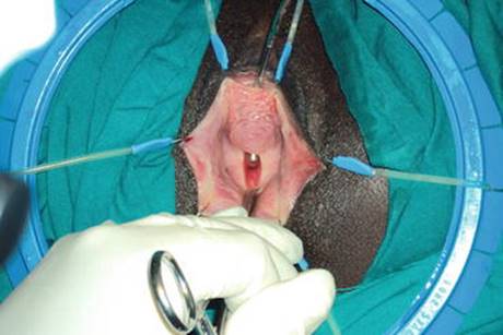
Fig. 11.1
Obstetric VVF during surgical repair
Risk Factors
Risk factors implicated in the pathogenesis of OF in developing countries are the following:
· Young age at marriage [134, 135]
· Short stature with small-sized foot [136, 137]
· Illiteracy and lower socioeconomic status [136]
· Primiparity [136]
· Lack of prenatal care (lack of skilled attendance at birth, lack of emergency obstetric care) [133, 138]
· Rural location and lack of transportation [133]
Classification
Several methods have been used to classify OF. The WHO classification divides OF into two main groups according to the anticipated difficulty of repair (simple or complex fistula) and most surgeons nowadays use this classification.
1. 1.
2. 2.
Presentation
Women with OF typically complain of persistent leakage of urine through the vagina (VVF), or leakage of flatus and stool through the vagina (RVF) or both. Both VVF and RVF are associated with persistent offensive odour leading to isolation from family and many women are divorced [138, 139].
Patients with a VUF most often present with complaints of urinary leakage or haematuria, and may also experience cyclic haematuria (menouria), amenorrhea, and UTI [140, 141].
Obstructed labour causes injury that may involve different pelvic organs, and patients may have other chronic debilitating comorbidities in addition to urinary or fecal incontinence such as:
· Footdrop from lumbo-sacral nerve damage
· Vaginal stenosis and infertility
· Chronic pelvic pain
· Chronic vulvar dermatitis and skin excoriation from chronic irritation of the skin by urine and feces
· Psychological disorders: depression, social stigma, and posttraumatic stress disorder. Some women may attempt suicide [142, 143].
Diagnosis
Diagnosis is established by careful history taking and meticulous clinical examination. The physician could identify the fistula easily, however, it is important not to miss a small fistula or another associated pathology, such as ureteric involvement because this will increase the probability of failure of the surgical repair.
Women typically give a history of obstructed labour for days with fetal death followed later by leakage of urine and/or stool. A history of emergency CS or sexual violence may also be presented. Pelvic and vaginal examination using bivalved vaginal speculum aims to assess the fistula and its surrounding tissues and also to evaluate for prolapse or UI, which may alter the surgical plan.
Dye tests (methylene blue test) may be used to detect the site of leakage and if it is a single or multiple fistulas. If the methylene blue dye test is negative, the patient can be given pyridium orally and have a tampon placed in the vagina. A subsequent yellow stained tampon following a negative bladder fill is indicative of UTVF.
Retrograde cystogram may show a communication between the bladder and the vagina or pooling of the dye in the vagina after bladder emptying. Intravenous urography may show hydroureteronephrosis if UTVF present.
Other diagnostic procedures such as cystoscopy, contrast- enhanced computed tomography and magnetic resonance imaging may confirm the presence and location of fistula, identify any associated lesions and add more information to the diagnosis.
Treatment of OF
Conservative treatment of a fresh fistula by bladder drainage through 18 F urethral catheter may be an option in some patients with small fistulas (<1 cm) [144–147], but if healing has not taken place within a few weeks, surgical repair is required.
The treatment of choice for OF is surgery with the majority of reports showing success rates over 80 % for simple fistulas at the time of first operation [148–154].
The best chance for successful fistula closure is at the first operation and closure rates tend to diminish with each subsequent attempt of operative repair [155].
Surgical procedures for OF may be undertaken through vaginal, abdominal, combined vaginal and abdominal or laparoscopic approach.
Surgical principles for the treatment of simple OF include:
· Wide exposure and dissection of the fistula with ureters’ protection by stents.
· Adequate tissue mobilization to ensure tension-free closure.
· Closure of the bladder or rectum in one or two layers and vagina separately.
· Adequate hemostasis after closure.
Other special considerations for the surgical repair of complex OF:
· The patient should be referred to a fistula center.
· Advanced training and surgical skills are prerequisites for treating this type of fistula.
· Proper selection of the most suitable approach as the patient may require urethral reconstruction or bladder augmentation during reconstruction and rarely some intractable cases may require urinary diversion.
· Relaxing posterolateral episiotomies may be needed.
· Well-vascularized inter-positioning tissue flap may be beneficial under certain circumstances (complex fistula, recurrent fistula and fistula associated with a difficult closure).
Postoperative Care
Adequate continuous bladder drainage for 2–3 weeks through urethral or suprapubic catheter. Antibiotics and anticholinergics are commonly used. Sexual abstinence for at least 3 months is recommended by many surgeons.
Prevention of OF
To reduce OF rates, effective preventive programmes that include family planning, prenatal care, safe labour, delivery and postpartum care are needed. Moreover, adequate support is required for interventions that focus on improving access to maternal health care, emergency obstetric care, and facilities for cesarean delivery when this is indicated [156].
References
1.
Brown JS, Posner SF, Stewart AL. Urge incontinence: new health-related quality of life measures. J Am Geriatr Soc. 1999;47(8):980–8.PubMed
2.
MacLennan AH, Taylor AW, Wilson DH, Wilson PD. The prevalence of pelvic floor disorders and their relationship to gender, age, parity and mode of delivery. Br J Obstet Gynaecol. 2000;107:1460–70.
3.
Rortveit G, Daltveit AK, Hannestad YS, Steinar H. Urinary incontinence after vaginal delivery or cesarean section. N Engl J Med. 2003;348:900–7.PubMed
4.
Ching-Chung L, Shuenn-Dhy C, Ling-Hong T, Ching-Chang H, Chao-Lun C, Po-Jen C. Postpartum urinary retention: assessment of contributing factors and longterm clinical impact. Aust N Z J Obstet Gynecol. 2002;42:365–8.
5.
Glavind K, Bjørk J. Incidence and treatment of urinary retention postpartum. Int Urogynecol J Pelvic Floor Dysfunct. 2003;14:119–21.PubMed
6.
Groutz A, Gordon D, Wolman I, Jaffa A, Kupferminc MJ, Lessing JB. Persistent postpartum urinary retention in contemporary obstetric practice. Definition, prevalence and clinical implications. J Reprod Med. 2001;46:44–8.PubMed
7.
Thom D, Rortveit G. Prevalence of postpartum urinary incontinence: a systematic review. Acta Obstet Gynecol Scand. 2010;89(12):1511–22.PubMed
8.
Nygaard I. Urinary incontinence: is cesarean delivery protective? Semin Perinatol. 2006;30(5):267–71.PubMed
9.
Teo R, Punter J, Abrams K, Mayne C, Tincello D. Clinically overt postpartum urinary retention after vaginal delivery: a retrospective case-control study. Int Urogynecol J Pelvic Floor Dysfunct. 2007;18(5):521–4.PubMed
10.
Rizvi RM, Khan ZS, Khan Z. Diagnosis and management of postpartum urinary retention. Int J Gynecol Obstet. 2005;91:71–2.
11.
Carley ME, Carley JM, Vasdev G, Lesnick TG, Webb MJ, Ramin KD, et al. Factors that are associated with clinically overt postpartum urinary retention after vaginal delivery. Am J Obstet Gynecol. 2002;187:430–3.PubMed
12.
Kekre AN, Vijayanand S, Dasgupta R, Kekre N. Postpartum urinary retention after vaginal delivery. Int J Gynaecol Obstet. 2011;112:112–5.PubMed
13.
Yip SK, Brieger G, Hin LY, Chung T. Urinary retention in the post-partum period. The relationship between obstetric factors and the post-partum post-void residual bladder volume. Acta Obstet Gynecol Scand. 1997;76:667–72.PubMed
14.
Ismail SI, Emery SJ. The prevalence of silent postpartum retention of urine in a heterogeneous cohort. J Obstet Gynaecol. 2008;28:504–7.PubMed
15.
Andolf E, Iosif CS, Jörgensen C, Rydhström H. Insidious urinary retention after vaginal delivery: prevalence and symptoms at follow-up in a population-based study. Gynecol Obstet Invest. 1994;38(1):51–3.PubMed
16.
Liang CC, Wong SY, Tsay PT, Chang SD, Tseng LH, Wang MF, et al. The effect of epidural analgesia on postpartum urinary retention in women who deliver vaginally. Int J Obstet Anesth. 2002;11:164–9.PubMed
17.
Hee P, Lose G, Beier-Holgersen R, Engdahl E, Falkenlove P. Postpartum voiding in the primiparous after vaginal delivery. Int Urogynecol J. 1992;3:95–9.
18.
Demaria F, Amar N, Biau D, Fritel X, Porcher R, Amarenco G, et al. Prospective 3D ultrasonographic evaluation of immediate postpartum urine retention volume in 100 women who delivered vaginally. Int Urogynecol J Pelvic Floor Dysfunct. 2004;15:281–5.PubMed
19.
Yip SK, Sahota D, Pang MW, Chang A. Screening test model using duration of labor for the detection of postpartum urinary retention. Neurourol Urodyn. 2005;24(3):248–53.PubMed
20.
Saultz JW, Toffler WL, Shackles JY. Postpartum urinary retention. J Am Board Fam Ract. 1991;4:341–4.
21.
Yip SK, Sahota D, Pang MW, Chang A. Postpartum urinary retention. Acta Obstet Gynecol Scand. 2004;83(10):881–91.PubMed
22.
Iosif S, Ingemarsson I, Ulmsten U. Urodynamic studies in normal pregnancy and in puerperium. Am J Obstet Gynecol. 1980;137(6):696–700.PubMed
23.
Kearney R, Cutner A. Postpartum voiding dysfunction. Obstetr Gynecol. 2008;10(2):71–4.
24.
Madersbacher H, Cardozo L, Chapple C, Abrams P, Toozs-Hobson P, Young JS, et al. What are the causes and consequences of bladder overdistension?: ICI-RS 2011. Neurourol Urodyn. 2012;31:317–21.PubMed
25.
Zaki MM, Pandit M, Jackson S. National survey for intrapartum and postpartum bladder care: assessing the needs for guidelines. BJOG. 2004;111:874–6.PubMed
26.
Sultan AH, Kamm MA, Hudson CN. Pudendal nerve damage during labour: prospective study before and after childbirth. Br J Obstet Gynaecol. 1994;101(1):22–8.PubMed
27.
Tetzschner T, Sorensen M, Lose G, Christiansen J. Pudendal nerve recovery after a non-instrumented vaginal delivery. Int Urogynecol J Pelvic Floor Dysfunct. 1996;7(2):102–4.PubMed
28.
Tetzschner T, Sorensen M, Lose G, Christiansen J. Pudendal nerve function during pregnancy and after delivery. Int Urogynecol J Pelvic Floor Dysfunct. 1997;8(2):66–8.PubMed
29.
Mulder FEM, Schoffelmeer MA, Hakvoort RA, Limpens J, MolBW J, Rooversa JPWR, et al. Risk factors for postpartum urinary retention: a systematic review and meta-analysis. BJOG. 2012. doi:10.1111/j.1471-0528.2012.03459.PubMed
30.
Groutz A, Hadi E, Wolf Y, Maslovitz S, Gold R, Lessing JB, et al. Early postpartum voiding dysfunction: incidence and correlation with obstetric parameters. J Reprod Med. 2004;49:960–4.PubMed
31.
Ramsay LN, Torbet TE. Incidence: of abnormal voiding parameters in the immediate postpartum period. Neurourol Urodynamics. 1993;12:119–83.
32.
Lim JL. Post-partum voiding dysfunction and urinary retention. Aust N Z J Obstet Gynecol. 2010;50:502–5.
33.
Yip SK, Sahota D, Chang AM. Determining the reliability of ultrasound measurements and the validity of the formulae for ultrasound estimation of postvoid residual bladder volume in postpartum women. Neurourol Urodynamics. 2003;22(3):255–60.
34.
Kerr-Wilson RH, Thompson SW, Orr JW, Davis RO, Cloud GA. Effect of labor on the postpartum bladder. BJOG. 1984;64:115–8.
35.
Jeffery TJ, Thyer B, Tsokos N, Taylor JD. Chronic urinary retention postpartum. Aust N Z J Obstet Gynaecol. 1990;30:364–6.PubMed
36.
NICE. Urinary incontinence: the management of urinary incontinence in women. London: NICE; (2006).
37.
Harris RE. Postpartum urinary retention: role of antimicrobial therapy. Am J Obstet Gynecol. 1979;133(2):174–5.PubMed
38.
Burkhart FL, Porges RF, Gibbs CE. Bladder capacity postpartum and catheterisation. Obstet Gynecol. 1965;26:176–9.PubMed
39.
Foldspang A, Hvidman L, Mommsen S, Nielsen JB. Risk of postpartum urinary incontinence associated with pregnancy and mode of delivery. Acta Obstet Gynecol Scand. 2004;83(10):923–7.PubMed
40.
Viktrup L, Lose G. The risk of stress incontinence 5 years after first delivery. Am J Obstet Gynecol. 2001;185:82–7.PubMed
41.
Haylen BT, de Ridder D, Freeman RM, Swift SE, Berghmans B, Lee J, et al. An International Urogynecological Association (IUGA)/International Continence Society (ICS) joint report on the terminology for female pelvic floor dysfunction. Neurourol Urodyn. 2010;29(1):4–20.PubMed
42.
Rortveit G, Daltveit AK, Hannestad YS, Hunskaar S. Vaginal delivery parameters and urinary incontinence: the Norwegian EPICONT study. Am J Obstet Gynecol. 2003;189(5):1269–74.
43.
Viktrup L, Lose G, Rolff M, Barfoed K. The frequency of urological symptoms during pregnancy and delivery in primiparae. Int Urogynecol J. 1993;4:27–30.
44.
Wilson PD, Herbison RM, Herbison GP. Obstetric practice and the prevalence of urinary incontinence three months after delivery. Br J Obstet Gynaecol. 1996;103:154–61.PubMed
45.
Hannestad YS, Rortveit G, Sandvik H, Hunskaar S. A community-based epidemiological survey of female urinary incontinence: the Norwegian EPINCONT study. Epidemiology of incontinence in the County of Nord-Trondelag. J Clin Epidemiol. 2000;53(11):1150–7.PubMed
46.
Viktrup L, Lose G, Rolff M, Barfoed K. The symptom of stress incontinence caused by pregnancy or delivery in primiparas. Obstet Gynecol. 1992;79:945–9.PubMed
47.
Viktrup L, Lose G. Lower urinary tract symptoms 5 years after the first delivery. Int Urogynecol J. 2000;11:336–40.
48.
Hojberg KE, Salvig JD, Winslow NA, Lose G, Secher NJ. Urinary incontinence: prevalence and risk factors at 16 weeks of gestation. Br J Obstet Gynaecol. 1999;106(8):842–50.PubMed
49.
Chiarelli P, Campbell E. Incontinence during pregnancy. Prevalence and opportunities for continence promotion. Aust N Z J Obstet Gynaecol. 1997;37(1):66–73.PubMed
50.
Hansen BB, Svare J, Viktrup L, Jorgensen T, Lose G. Urinary incontinence during pregnancy and 1 year after delivery in primiparous women compared with a control group of nulliparous women. Neurourol Urodyn. 2012;31(4):475–80.PubMed
51.
Wesnes SL, Hunskaar S, Bo K, Rortveit G. The effect of urinary incontinence status during pregnancy and delivery mode on incontinence postpartum. A cohort study. BJOG. 2009;116:700–7.PubMedPubMedCentral
52.
Morkved S, Bo K. Prevalence of urinary incontinence during pregnancy and postpartum. Int Urogynecol J Pelvic Floor Dysfunct. 1999;10:394–8.PubMed
53.
Boyles SH, Li H, Mori T, Osterweil P, Guise JM. Effect of mode of delivery on the incidence of urinary incontinence in primiparous women. Obstet Gynecol. 2009;113(1):134–41.PubMed
54.
Eliasson K, Nordlander I, Larson B, Hammarstrom M, Mattsson E. Influence of physical activity on urinary leakage in primiparous women. Scand J Med Sci Sports. 2005;15(2):87–94.PubMed
55.
Farrell SA, Allen VM, Baskett TF. Parturition and urinary incontinence in primiparas. Obstet Gynecol. 2001;97(3):350–6.PubMed
56.
Glazener CM, Herbison GP, MacArthur C, Lancashire R, McGee MA, Grant AM, et al. New postnatal urinary incontinence: obstetric and other risk factors in primiparae. BJOG. 2006;113(2):208–17.PubMed
57.
Solans-Domenech M, Sanchez E, Espuna-Pons M. Urinary and anal incontinence during pregnancy and postpartum: incidence, severity, and risk factors. Obstet Gynecol. 2010;115(3):618–28.PubMed
58.
Burgio KL, Zyczynski H, Locher JL, Richter HE, Redden DT, Wright KC. Urinary incontinence in the 12-month postpartum period. Obstet Gynecol. 2003;102(6):1291–8.PubMed
59.
Iosif S. Stress incontinence during pregnancy and in puerperium. Int J Gynaecol Obstet. 1981;19:13–20.PubMed
60.
Dimpfl T, Hesse U, Schussler B. Incidence and cause of postpartum urinary stress incontinence. Eur J Obstet Gynecol Reprod Biol. 1992;43(1):29–33.PubMed
61.
Ekstrom A, Altman D, Wiklund I, Larsson C, Andolf E. Planned cesarean section versus planned vaginal delivery: comparison of lower urinary tract symptoms. Int Urogynecol J Pelvic Floor Dysfunct. 2008;19(4):459–65.PubMed
62.
Sampselle CM, DeLancey JOL, Ashton-Miller J. Urinary incontinence in pregnancy and postpartum. Neurourol Urodyn. 1996;15(4):329–30.
63.
Thomason AD, Miller JM, Delancey JO. Urinary incontinence symptoms during and after pregnancy in continent and incontinent primiparas. Int Urogynecol J Pelvic Floor Dysfunct. 2007;18:147–51.PubMed
64.
Yang X, Zhang HX, Yu HY, Gao XL, Yang HX, Dong Y. The prevalence of fecal incontinence and urinary incontinence in primiparous postpartum Chinese women. Eur J Obstet Gynecol Reprod Biol. 2010;152(2):214–7.PubMed
65.
Ege E, Akin B, Altuntug K, Benli S, Arioz A. Prevalence of urinary incontinence in the 12-month postpartum period and related risk factors in Turkey. Urol Int. 2008;80(4):355–61.PubMed
66.
Ewings P, Spencer S, Marsh H, O’Sullivan M. Obstetric risk factors for urinary incontinence and preventative pelvic floor exercises: cohort study and nested randomized controlled trial. J Obstet Gynaecol. 2005;25(6):558–64.PubMed
67.
Hvidman L, Foldspang A, Mommsen S, Nielsen JB. Postpartum urinary incontinence. Acta Obstet Gynecol Scand. 2003;82(6):556–63.PubMed
68.
Mason L, Glenn S, Walton I, Appleton C. The prevalence of stress incontinence during pregnancy and following delivery. Midwifery. 1999;15(2):120–8.PubMed
69.
Raza-Khan F, Graziano S, Kenton K, Shott S, Brubaker L. Peripartum urinary incontinence in a racially diverse obstetrical population. Int Urogynecol J Pelvic Floor Dysfunct. 2006;17(5):525–30.PubMed
70.
Schytt E, Lindmark G, Waldenstrom U. Symptoms of stress incontinence 1 year after childbirth: prevalence and predictors in a national Swedish sample. Acta Obstet Gynecol Scand. 2004;83(10):928–36.PubMed
71.
Smith ARB, Hosker GL, Warell DW. The role of pudendal nerve damage in the aetiology of genuine stress incontinence in women. Br J Obstet Gynaecol. 1989;96:29–32.PubMed
72.
Peschers UM, Schaer GN, DeLancey JO, Schuessler B. Levator ani function before and after childbirth. Br J Obstet Gynaecol. 1997;104:1004–8.PubMed
73.
Dietz H, Lanzarone V. Levator trauma after vaginal delivery. Obstet Gynecol. 2005;106:707–12.PubMed
74.
Dietz H, Shek K, Clarke B. Biometry of the pubo visceral muscle and levator hiatus by three-dimensional pelvic floor ultrasound. Ultrasound Obstet Gynecol. 2005;25:580–5.PubMed
75.
Lien KC, Mooney B, DeLancey JO, Ashton-Miller JA. Levatorani muscle stretch induced by simulated vaginal birth. Obstet Gynecol. 2004;103:31–40.PubMedPubMedCentral
76.
Hoyte L, Schierlitz L, Zou K, Flesh G, Fielding JR. Two and 3-dimensinoal MRI comparison of levator ani structure, volume and integrity in women with SUI and prolapse. Am J Obstet Gynecol. 2001;185:11–9.PubMed
77.
DeLancey JOL, Kearney R, Chou Q, Speights S, Binno S. The appearance of levator ani muscle abnormalities in magnetic resonance images after vaginal delivery. Obstet Gynecol. 2003;101:46–53.PubMedPubMedCentral
78.
King JK, Freeman RM. Is antenatal bladder neck mobility a risk factor for postpartum stress incontinence? Br J Obstet Gynaecol. 1998;105:1300–7.PubMed
79.
DeLancey JO, Miller JM, Kearney R, Howard D, Reddy P, Umek W, et al. Vaginal birth and de novo stress incontinence: relative contributions of urethral dysfunction and mobility. Obstet Gynecol. 2007;110:354–62.PubMedPubMedCentral
80.
Toozs-Hobson P, Balmforth J, Cardozo L, Khullar V, Athanasiou S. The effect of mode of delivery on pelvic floor functional anatomy. Int Urogynecol J Pelvic Floor Dysfunct. 2008;19(3):407–16.PubMed
81.
Mikhail M, Anyaegbunam A. Lower urinary tract dysfunction in pregnancy: a review. Obstet Gynecol Surv. 1995;5:675–83.
82.
Tapp A. The effect of vaginal delivery on the urethral sphincter. Br J Obstet Gynaecol. 1988;95:142–6.PubMed
83.
Cannon TM, Wojcik EM, Ferguson CL, Saraga S, Thomas C, Damaser MS. Effects of vaginal distension on urethral anatomy and function. BJU Int. 2002;90:403–7.PubMed
84.
Torrisi G, Sampugnaro EG, Pappalardo EM, D’Urso E, Vecchio M, Mazza A. Postpartum urinary stress incontinence: analysis of the associated risk factors and neurophysiologicaltests. Minerva Ginecol. 2007;59:491–8.PubMed
85.
Serati M, Salvatore S, Khullar V, Uccella S, Bertelli E, Ghezzi F, et al. Prospective study to assess risk factors for pelvic floor dysfunction after delivery. Acta Obstet Gynecol Scand. 2008;87:313–8.PubMed
86.
Landon CR, Crofts CE, Smith ARB, Trowbridge A. Mechanical properties of fascia during pregnancy: a possible factor in the development of stress incontinence of urine. Contemp Rev Obstet Gynaecol. 1990;2:40–6.
87.
Norton PA, Baker JE, Sharp HC, Warenski JC. Genitourinary prolapse and joint hypermobility in women. Obstet Gynecol. 1995;85:225–8.PubMed
88.
Chaliha C, Kalia V, Stanton SL, Monga A, Sultan AH. Antenatal prediction of postpartum urinary and faecal incontinence. Obstet Gynecol. 1999;94:689–93.PubMed
89.
Rasmussen KL, Krue S, Johansson LE, Knudsen HJ, Agger AO. Obesity as a predictor of postpartum urinary symptoms. Acta Obstet Gynecol Scand. 1997;76:359–62.PubMed
90.
Casey BM, Schaffer JI, Bloom SL, Heartwell SF, McIntire DD, Leveno KJ. Obstetric antecedents for postpartum pelvic floor dysfunction. Am J Obstet Gynecol. 2005;192:1655–62.PubMed
91.
Abrams P, Andersson KE, Birder L, Brubaker L, Cardozo L, Chapple C, et al. Fourth international consultation on incontinence recommendations of the International Scientific Committee: evaluation and treatment of urinary incontinence, pelvic organ prolapse, and fecal incontinence. Neurourol Urodyn. 2010;29(1):213–40.PubMed
92.
Hay-Smith J, Mørkved S, Fairbrother KA, Herbison GP. Pelvic floor muscle training for prevention and treatment of urinary and faecal incontinence in antenatal and postnatal women. Cochrane Database Syst Rev. 2008;(4):CD007471.
93.
Kegel AH. Progressive resistance exercise in the functional restoration of the perineal muscles. Am J Obstet Gynecol. 1948;56:238–48.PubMed
94.
Price N, Dawood R, Jackson SR. Pelvic floor exercise for urinary incontinence: a systematic literature review. Maturitas. 2010;67(4):309–15.PubMed
95.
Hay-Smith EJC, Dumoulin C. Pelvic floor muscle training versus no treatment, or inactive control treatments, for urinary incontinence in women. Cochrane Database Syst Rev 2006;(1):CD005654.
96.
Press JZ, Klein MC, Kaczorowski J, Liston RM, von Dadelszen P. Does cesarean section reduce postpartum urinary incontinence? A Systematic Review. Birth. 2007;34:3.
97.
Foldspang A, Mommsen S, Djurhuus JC. Prevalent urinary incontinence as a correlate of pregnancy, vaginal childbirth, and obstetric techniques. Am J Public Health. 1999;89:209–12.PubMedPubMedCentral
98.
Mant J, Painter R, Vessey M. Epidemiology of genital prolapse: observations from the Oxford Family Planning Association Study. Br J Obstet Gynaecol. 1997;104:579–85.PubMed
99.
Faundes A, Guarisi T, Pinto-Neto AM. The risk of urinary incontinence of parous women who delivered only by cesarean section. Int J Gynecol Obstet. 2001;72:41–6.
100.
Demirci F, Ozden Z, Alpay E, Demirci ET, Ayas S. The effects of vaginal delivery and cesarean section on bladder neck mobility and stress urinary incontinence. Int Urogynecol J. 2001;12:129–33.
101.
Groutz A, Rimon E, Peled S, Gold R, Pauzner D, Lessing JB, et al. Cesarean section: does it really prevent the development of postpartum stress urinary incontinence? A prospective study of 363 women one year after their first delivery. Neurourol Urodynam. 2004;23:2–6.
102.
Barbieri RL. How to repair bladder injury at the time of cesarean delivery. OBG Manag. 2011;23(12):6–9.
103.
Yossepowitch O, Baniel J, Livne PM. Urological injuries during cesarean section: intraoperative diagnosis and management. J Urol. 2004;172(1):196–9.PubMed
104.
Silver RM, Landon MB, Rouse DJ, Leveno KJ, Spong CY, Thom EA, et al. Maternal morbidity associated with multiple repeat cesarean deliveries. Obstet Gynecol. 2006;107(6):1226–32.PubMed
105.
Vu KK, Brittain PC, Fontenot JP, Harlass FE, Hawley- Bowland CG, Diaz-Ball F. Vesicouterine fistula after cesarean section: a case report. J Reprod Med. 1995;40:221–2.PubMed
106.
Buckspan MB, Simha S, Klotz PG. Vesicouterine fistula: a rare complication of cesarean section. Obstet Gynecol. 1983;62(Suppl):64–6.
107.
Iloabachie GC, Njoku O. Vesico-uterine fistula. Br J Urol. 1985;57:438–9.PubMed
108.
Williams CH, Heaney JA, Young W. Respiratory distress following cesarean section: cryptic presentation of bladder injury. Urology. 1994;44(3):441–3.PubMed
109.
Lee JS, Choe JH, Lee HS, Seo JT. Urologic complications following obstetric and gynecologic surgery. Korean J Urol. 2012;53:795–9.PubMedPubMedCentral
110.
Maureen GP, Bryan W, Jeffrey L, Sherry W, Deborah L. Risk factors for bladder injury during cesarean delivery. Obstet Gynecol. 2005;105(1):156–60.
111.
Alcocer Urueta J, Bonilla Mares M, Gorbea Chávez V, Velázquez VB. Risk factors for bladder injuries during cesarean section. Actas Urol Esp. 2009;33(7):806–10.PubMed
112.
Gungorduk K, Asicioglu O, Celikkol O, Sudolmus S, Ark C. Iatrogenic bladder injuries during caesarean delivery: a case control study. J Obstet Gynaecol. 2010;30(7):667–70.PubMed
113.
Rahman MS, Gasem T, Al Suleiman SA, Al Jama FE, Burshaid S, Rahman J. Bladder injuries during cesarean section in a University Hospital: a 25-year review. Arch Gynecol Obstet. 2009;279(3):349–52.PubMed
114.
Cahill AG, Stout MJ, Stamilio DM, Odibo AO, Peipert JF, Macones GA. Risk factors for bladder injury in patients with a prior hysterotomy. Obstet Gynecol. 2008;112(1):116–20.PubMedPubMedCentral
115.
Eisenkop SM, Richman R, Platt LD, Paul RH. Urinary tract injury during cesarean section. Obstet Gynecol. 1982;60(5):591–6.PubMed
116.
Lucci JA. Lesiones Urológicas y Gastrointestinales. In: Gilstrap LC, Cunningham FG, Van Dorsten JP, editors. Urgencias en salas de partos y obstetricia quirúrgica. 2nd ed. Madrid: Panamericana; 2004. p. 443–52.
117.
Davis JD. Management of injuries to the urinary and gastrointestinal tract during cesarean section. Obstet Gynecol Clin North Am. 1999;26(3):469–81.PubMed
118.
Onuora VC, Ariyan AL, Koko AH, Wahab A, Al Jawini N. Major injuries to de urinary tract in association with childbirth. East Afr Med J. 1997;74(8):523–8.PubMed
119.
Rajasekar D, Hall M. Urinary tract injuries during obstetric intervention. Br J Obstet Gynaecol. 1997;104(6):731–4.PubMed
120.
Chan JK, Morrow J, Manetta A. Prevention of ureteral injuries in gynecologic surgery. Am J Obstet Gynecol. 2003;188(5):1273–7.PubMed
121.
Liapis A, Bakas P, Giannopoulos V, Creatsas G. Ureteral injuries during gynecological surgery. Int Urogynecol J Pelvic Floor Dysfunct. 2001;12:391–3.PubMed
122.
Neuman M, Eidelman A, Langer R, Golan A, Bukovsky I, Caspi E. Iatrogenic injuries to the ureter during gynecologic and obstetric operations. Surg Gynecol Obstet. 1991;173:268–72.PubMed
123.
Hammil SL, Rogers RG. Urologic injury at the time of cesarean delivery. Female Patient. 2009;34:23–5.
124.
Yang B. Bladder rupture associated with uterine rupture at delivery. Int Urogynecol J. 2011;22(5):625–7.PubMed
125.
Forsnes EV, Browning JE, Gherman RB. Bladder rupture associated with uterine rupture. A report of two cases occurring during vaginal birth after cesarean. J Reprod Med. 2000;45(3):240–2.PubMed
126.
Zeteroğlu S, Bayrakli H, Yilmaz Y, Sahin HG. Combined bladder, urethral and uterine rupture after a normal, spontaneous vaginal delivery at home: a case report. J Reprod Med. 2005;50(11):874–6.PubMed
127.
Okoro O, Onwere S, Chigbu B. Bladder and uterine rupture at vaginal birth after cesarean section. AJOL. 2008;5:1.
128.
WHO. 10 facts on obstetric fistula. 2011. Available on line at website: http://www.who.int/features/factfiles/obstetric_fistula/en/.
129.
Lewis G, de Bernis L, editors. Obstetric fistula. Guiding principles for clinical management and programme development. Geneva: World Health Organization; 2006.
130.
Tebeu PM, Fomulu JN, Khaddaj S, Bernis LD, Delvaux T, Rochat CH. Risk factors for obstetric fistula: a clinical review. Int Urogynecol J. 2012;23:387–94.PubMed
131.
Semere L, Nour NM. Obstetric fistula: living with incontinence and shame. Rev Obstet Gynecol. 2008;1(4):193–7.PubMedPubMedCentral
132.
Tebeu PM, de Bernis L, Doh AS, Rochat CH, Delvaux T. Risk factors for obstetric fistula in the Far North Province of Cameroon. Int J Gynaecol Obstet. 2009;107(1):12–5.PubMed
133.
Melah GS, Massa AA, Yahaya UR, Bukar M, Kizaya DD, El Nafaty AU. Risk factors for obstetric fistulae in northeastern Nigeria. J Obstet Gynaecol. 2007;27(8):819–23.PubMed
134.
Ampofo EK, Omotara BA, Otu T, Uchebo G. Risk factors of vesico-vaginal fistulae in Maiduguri, Nigeria: a case-control study. Trop Doct. 1990;20(3):138–9.PubMed
135.
Ampofo K, Otu T, Uchebo G. Epidemiology of vesico-vaginal fistulae in northern Nigeria. West Afr J Med. 1990;9(2):98–102.PubMed
136.
Ojanuga Onolemhemhen D, Ekwempu CC. An investigation of sociomedical risk factors associated with vaginal fistula in northern Nigeria. Women Health. 1999;28(3):103–16.PubMed
137.
Islam AI, Begum A. A psycho-social study on genito-urinary fistula. Bangladesh Med Res Counc Bull. 1992;18(2):82–94.PubMed
138.
Cook R, Dickens M, Syed S. Obstetric fistula: the challenge to human rights. Int J Gynecol Obstet. 2004;87:72–7.
139.
Bangser M. Obstetric fistula and stigma. Lancet. 2006;367(9509):535–6.PubMed
140.
Lenkovsky Z, Pode D, Shapiro A, Caine M. Vesicouterine fistula: a rare complication of cesarean section. J Urol. 1988;139(1):123–5.PubMed
141.
Mercader VP, McGuckin Jr JF, Caroline DF, Chatwani A, Seidmon EJ. CT of vesicocorporeal fistula with menouria: a complication of uterine biopsy. J Comput Assist Tomogr. 1995;19(2):324–6.PubMed
142.
Wall LL. Obstetric vesicovaginal fistula as an international public health problem. Lancet. 2006;368:1201–9.PubMed
143.
Arrowsmith S, Hamlin EC, Wall LL. Obstructed labor injury complex: obstetric fistula formation and the multifaceted morbidity of maternal birth trauma in the developing world. Obstet Gynecol Surv. 1996;51:568–74.PubMed
144.
Waaldijk K. The immediate management of fresh obstetric fistulas. Am J Obstet Gynecol. 2004;191(3):795–9.PubMed
145.
Waaldijk K. Immediate indwelling bladder catheterization at postpartum urine leakage—personal experience of 1200 patients. Trop Doct. 1997;27(4):227–8.PubMed
146.
Waaldijk K. The immediate surgical management of fresh obstetric fistulas with catheter and/or early closure. Int J Gynaecol Obstet. 1994;45(1):11–6.PubMed
147.
Bazi T. Spontaneous closure of vesicovaginal fistulas after bladder drainage alone: review of the evidence. Int Urogynecol J. 2007;18:4.
148.
Goh JT. Genital tract fistula repair on 116 women. Aust N Z J Obstet Gynaecol. 1998;38(2):158–61.PubMed
149.
Gharoro EP, Abedi HO. Vesico-vaginal fistula in Benin City. Niger Int J Gynaecol Obstet. 1999;64(3):313–4.
150.
Browning A. Prevention of residual urinary incontinence following successful repair of obstetric vesico-vaginal fistula using a fibro-muscular sling. BJOG. 2004;111(4):357–61.PubMed
151.
Raassen TJ, Verdaasdonk EG, Vierhout ME. Prospective results after first-time surgery for obstetric fistulas in East African women. Int Urogynecol J Pelvic Floor Dysfunct. 2007;19(1):73–9.PubMed
152.
Nardos R, Browning A, Member B. Duration of bladder catheterization after surgery for obstetric fistula. Int J Gynaecol Obstet. 2008;103(1):30–2.PubMed
153.
Rangnekar NP, Imdad AN, Kaul SA, Pathak HR. Role of the martius procedure in the management of urinary-vaginal fistulas. J Am Coll Surg. 2000;191(3):259–63.PubMed
154.
Browning A. Lack of value of the Martius fibrofatty graft in obstetric fistula repair. Int J Gynaecol Obstet. 2006;93(1):33–7.PubMed
155.
Hilton P, Ward A. Epidemiological and surgical aspects of urogenital fistulae: a review of 25 years’ experience in southeast Nigeria. Int Urogynecol J Pelvic Floor Dysfunct. 1998;9(4):189–94.PubMed
156.
Creanga A, Ahmed S, Genadry R, Stanton C. Prevention and treatment of obstetric fistula: identifying research needs and public health priorities. Int J Gynecol Obstet. 2007;99:S151.