
Ghazaleh Rostami Nia1 and S. Abbas Shobeiri2
(1)
Inova Fairfax Hospital, 3300 Gallows Rd, Falls Church, VA 22042, USA
(2)
Gynecologic subspecialties, Inova Fairfax Hospital, 3300 Gallows Rd, Falls Church, VA 22042, USA
S. Abbas ShobeiriProfessor, Vice Chairman
Email: Abbas.Shobeiri@inova.org
Abstract
The purpose of this chapter is to describe the 3D ultrasonography’s utility in the assessment of pelvic floor trauma associated with vaginal delivery. These pathologies are associated with urinary incontinence and cystocele. 3D ultrasonography has been overlooked in favor of MR imaging. However, this technique can be a powerful tool in the hands of the obstetricians who suspect levator ani trauma.
Keywords
UltrasoundTraumaPelvic floor
Introduction
Pregnancy and childbirth change the anatomy and function of the pelvic floor. Physicians who provide care to women can easily distinguish a multiparous from a nulliparous on pelvic examination. MRI-based computer modelling of the levator ani muscles have shown increase in length of the pubococcygeus fibers by a factor of 3 or more during crowning of the fetal head [1, 2]. The area of the minimal levator hiatus in young nulliparous women varies from 6 to 36 cm2 on Valsalva maneuver [3]. The area of the average fetal head in the plane of minimal diameters measures 70–90 cm2 (equivalent to a head circumference of 300–350 mm), requiring marked distension and deformation of the levator complex. Widened genital hiatus, less vaginal epithelium rugeation in addition to decreased muscle tone are most significant signs of multiparity during pelvic examination. There is ample scientific evidence that the vaginal childbirth process is associated with neuromuscular and soft tissue injuries to the pelvic floor. The pelvic organ support system is multifaceted and includes the endopelvic fascia, the perineal membrane, and the levator ani muscle that are controlled by nervous system. All these structures are at risk of injury in pregnancy and during vaginal delivery. These injuries can lead to short- and long-term pelvic floor structural changes. Pelvic organ prolapse, urinary and fecal incontinence, and chronic pelvic pain are considered inevitable sequelae for some women who experience injuries during birth. There is little ongoing debate about these findings and the focus has been on risk factor assessment and injury reducing interventions, however; this goal cannot be achieved without detailed understanding of structural and functional changes after delivery. Ultrasonography is taking an increasingly central role in defining birth related changes in pelvic floor support system. Levator ani muscle injury, levator hiatus enlargement, levator plate descent, widened anorectal angle and anal sphincter complex defects are examples of birth related traumas that can be diagnosed mostly by ultrasound techniques. Physical examinations even in very experienced hands are not precise enough for detection of these defects and are quite simply inadequate. The aim of this chapter is to review the role of ultrasound in evaluating the pelvic floor after pregnancy and labor.
Ultrasound
In recent years, with advances in magnetic resonance imaging (MRI) and three-dimensional (3D) ultrasound, we know that pelvic floor trauma goes beyond perineal, vaginal and anal sphincter lacerations which we can identify in the labor room. It has become evident that levator ani injury forms an underappreciated component of pelvic floor trauma. The levator ani muscle consists of three functional parts: the puboperineal and puboanal portions, the puborectalis, and iliococcygeus and pubococcygeus muscles which form the pubovisceralis complex [4]. Mechanisms of levator muscle injury became more clear with continuous works of Ashton-Miller and DeLancey on the biomechanics of vaginal birth [5]. Based on a geometric model they suggested that some muscle damage during the second stage of labor may come from overstretching [1]. The pubococcygeus portion of pubovisceralis muscle was the portion that underwent the greatest degree of stretch, and the second area of observed injury was iliococcygeus portion of the pubovisceralis.
Endoanal ultrasound was utilized by Sultan in 1993 using a series of elegant correlative histologic studies to document anal sphincter injury during labor [6, 7]. The same authors performed endovaginal ultrasound in 1994 to visualize the anal sphincter complex and incidentally visualized the levator ani muscles but the significance of these muscles at that point was unknown and they went largely unnoticed [8]. Shortly thereafter, the same group acknowledged the pelvic floor trauma during labor and suggested strategies to protect the pelvic floor, perineum, and the anal sphincter during labor [9, 10]. Strohben reported on visualization of levator ani muscles using MRI with anatomic correlation in 1996 [11] and in 1999 Tunn reported on levator ani recovery after vaginal birth in 14 women [12]. In this group of patients, for the first time they reported injury to the levator ani muscle of one patient. Transperineal imaging was reported for visualization of the levator ani muscle activity in 2001 and concluded that this modality could be used to teach pelvic floor biofeedback (Fig. 10.1). The use of transperineal ultrasound for anal sphincter imaging was reported in 2002 [13] and concluded that digital examination could discriminate between an adequate and inadequate anal sphincter laceration repair. In 2005, Dietz et al. reported on the use of 3D transperineal ultrasound (TPUS) to document a 36 % incidence of levator ani avulsion which is the total separation of the levator ani from the pubic bone [14]. Unfortunately, TPUS had not been authenticated by cadaveric and histologic correlation and the authors’ terminology named “puborectalis” as the injured muscle. Later on, levator ani muscle avulsion by TPUS was defined as an obvious detachment of the puborectalis muscle from the pelvic sidewall and if an abnormality was defined as defect seen in three or more slices [15] (Figs. 10.2 and 10.3). Different scoring systems with TPUS has been used to show the severity of muscle injury [16–18]. Zhuang et al. used the same terminology and defined full avulsion diagnosed if the “puborectalis”-to-ipsilateral sidewall attachment was not seen on any of the three central slices. Partial avulsion was diagnosed when the “puborectalis” attachment to the ipsilateral sidewall is not seen on at least one slide [19]. More recent studies have shown that the minimal levator hiatus is mostly lined with pubococcygeus [20] which is consistent with 3D modelling studies [1].
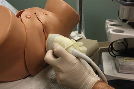
Fig. 10.1
A GE probe applied transperineally to obtain 3D/4D data volumes

Fig. 10.2
Tomographic ultrasound images obtained using transperineal ultrasound of (a) a typical intact levator ani muscle (LAM) in a nulliparous woman and (b) a bilateral LAM avulsion in a multiparous woman. Slices were obtained at 2.5-mm intervals below and above the level of minimal hiatal dimension (*). LAM defects are indicated in (b) by * (Reprinted from Schwertner-Tiepelmann et al. [63]; with permission from John Wiley and Sons

Fig. 10.3
Three-dimensional rendered volumes obtained on transperineal ultrasound showing: (a) an intact levator ani muscle (LAM) displayed in an oblique axial plane in a nulliparous woman and (b) a bilateral avulsion injury. LAM defects are indicated in (b) by (*). IR inferior ramus os pubis, L levator ani muscle, R rectum, U urethra, V vagina (Reprinted from Schwertner-Tiepelmann et al. [63]; with permission from John Wiley and Sons)
The description of the levator ani muscle subdivisions by 3D endovaginal ultrasonography (EVUS) was reported in 2008 and published in 2009 [21]. EVUS methodology and description of muscles was authenticated in a systematic manner. First the anatomic correlation [21] and subsequently histologic correlations were made in nulliparous women [22]. Interrater and interdisciplinary reliability of EVUS were subsequently described in nullipara [23]. Interrater reliability assessments of these measurements during pregnancy and postpartum have been performed.
Compared with other imaging modalities, ultrasound imaging is widely available, easy to perform, and familiar to many medical specialties. These advantages have made it feasible for many researchers to study early postpartum pelvic floor injuries and structural changes and further correlate them with obstetric factors.
Levator Ani Muscle Trauma
During vaginal delivery, overstretching of levator ani muscle can predispose muscle to disconnect from its insertion on the inferior pubic ramus and pelvic side wall. Based on studies using 3D/4D pelvic floor ultrasound, the prevalence of levator ani muscle trauma after vaginal delivery is 13–40 % [14, 24, 25]. In a study of 114 postpartum women with EVUS, one third of primiparous women delivering vaginally developed levator ani muscle hematomas within hours of delivery diagnosed using high frequency EVUS. When hematoma was located in the attachment zone of the levator ani muscle to the pubic bone, levator ani muscle detachment from the pubic symphysis was almost always identified three months postpartum. When hematoma was located away from the attachment zone, a defect was most often not seen three months postpartum [26]. The mode of delivery has an impact on levator ani muscle injury. In a study on 157 postpartum women, the risk of levator defect after vaginal delivery was more than seven times higher than after cesarean section [27]. Instrumental delivery by forceps was one the most important risk factors as levator injury was detected in 60–64 % of women who had been delivered by forceps [28, 29]. Another study reported a prevalence of 18 % of levator lesions among women who had a non-instrumental vaginal delivery, 14 % among women who delivered by vacuum and 40 % among women who delivered by forceps, but reported no levator lesions in the cesarean delivery group [30]. Chan et al. showed similar findings in Chinese women after first vaginal delivery. The rate of muscle injury for spontaneous vaginal delivery, ventouse extraction and forceps delivery were 15.4 %, 33.3 %, and 71.4 %, respectively. There was no levator muscle injury in cesarean section groups [31].
EVUS Technique for Visualization of Levator Ani Muscle
3D endovaginal ultrasound can give us high-quality images of levator ani muscle subdivisions. It is not known if the EVUS can detect LA subdivisions better than MRI. Imaging is obtained using the BK Medical Flexfocus (Peabody, MA, USA) and a 2052/8838 transducer (Fig. 10.4). All ultrasound scans are performed in the office setting, with the patient in dorsal lithotomy position, with hips flexed and abducted. No preparation is required and the patient is recommended to have a comfortable volume of urine in the bladder. No rectal or vaginal contrast is used. To avoid excessive pressure on surrounding structures that might distort the anatomy, the probe is inserted into the vagina in a neutral position. It has been shown that endovaginal probe does not have any adverse effect on anatomy comparing to transperineal ultrasound [32]. Three hundred axial images over a distance of 6 cm are taken in 60 s; 360° EVUS volumes are digitally stored for further analysis.
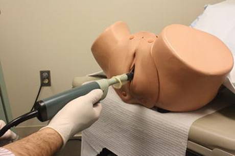
Fig. 10.4
A BK probe applied endovaginally to obtain 3D volume
The approach to 3-dimensional endovaginal ultrasound takes into account certain, easily recognizable anatomic landmarks [21]. We delineated three ascending levels with level 1 being the most caudal and level 3 the most cephalad (Fig. 10.5). This categorization was utilized for interrater reliability validation. Level 1 contained muscles that insert into the perineal body, namely the superficial transverse perinei, puboperinealis and puboanalis. The superficial transverse perinei served as a reference point (Fig. 10.6a, b). Level 2 contained the attachment of the pubovaginalis, puboperinealis, puboanalis and puborectalis, and iliococcygeus to the pubic bone (see Fig. 10.6c–f). Level 3 contained subdivisions visible cephalad to the inferior pubic ramus, namely the pubococcygeus and iliococcygeus, which winged out toward the ischial spine (see Fig. 10.6g–j). This standardized approach of endovaginal ultrasound assessment of levator ani muscle subdivisions has been verified with good to excellent interobserver and interdisciplinary reliability, with kappa values of 0.6–1 [21].

Fig. 10.5
The relative position of levator ani subdivisions during ultrasound imaging. Levels 1–3 are identified below the figure. The A-J markings on top of the figure correspond to the ultrasound images shown in Fig. 10.4. IC iliococcygeus, PP puboperinealis, STP superficial transverse perinea, PA puboanalis (Reprinted with permission from Shobeiri et al. [21])
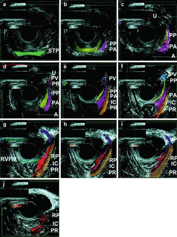
Fig. 10.6
Levator ani subdivisions seen at different levels. Midline structures are identified in lateral views with corresponding colors in the picture inserts at the upper left corner of the ultrasound images at each level. The green vertical line in the insert corresponds to the relative position in the vagina where the image was obtained. (a) Level 1A. At 0 cm, the first muscle seen is the superficial transverse perinei (green) with mixed echogenicity. (b) Level 1B. Immediately cephalad to the superficial transverse perinei is the puboperinealis (yellow), which can be traced to PB with manipulation of the three-dimensional cube. It comes in at a 45° angle as a mixed echoic band to join the perineal body. Lateral to it, the puboanalis is seen as a hypoechoic triangle (pink). (c) Level 2A marks the attachment of the muscles to the pubic arch. The external urethral meatus is visible (dark red). Puboperinealis and puboanalis insertions are highlighted. (d) Level 2B. Pubovaginalis (blue) and puborectalis (mustard) insertions come into view. The urethra and the bladder are outlined (red) in the lateral view. (e) Level 2C. The heart-shaped vaginal sulcus (outlined in red) marks the pubovaginalis insertion. Iliococcygeus fibers (red) come into view. The perineal body is outlined in the lateral view. (f) Level 2D. The puboanalis is starting to thin out. The puborectalis is seen in the lateral view. (g) Level 3A. The puboperinealis and puboanalis become obscure. Anatomically, the puboanalis becomes a thick, fibromuscular layer forming a tendineus sheet-the rectal pillar (RP). The perivesical venous plexus is prominent (purple). The rectovaginal fibromuscularis is shown (green) in the sagittal view as a continuous, mixed, echogenic structure approaching the perineal body and laterally attaching to the RP. (h) Level 3B. The RP (orange) is seen easily. The iliococcygeus becomes prominent and widens. (i) Level 3C. The iliococcygeus widens further and inserts into the arcus tendineus fascia pelvis. (j) Level 3D. The puborectalis and iliococcygeus fade out of view. The puborectalis (mustard) and iliococcygeus (red) are outlined in the lateral view, showing their entire course (Reprinted with permission from Shobeiri et al. [21])
Levator Ani Deficiency
The majority of patients with pelvic floor disorders remote from delivery did not have evidence of birth-related injury at the level of the pubic symphysis, rather they had global atrophy and deficiency of the muscle. Based on functional anatomy of subdivisions of the levator ani muscle a scoring system for evaluation the severity of levator ani deficiency (LAD) was described for EVUS [33]. By EVUS, levator ani muscle subdivisions were evaluated in their specific axial plane where the full length of muscle could have been visualized and were scored (0 = no defect, 1 = minimal defect with <50 % muscle loss, 2 = major defect with >50 % muscle loss, 3 = total absence of the muscle) on each side based on thickness and detachment from the pubic bone as previously used in the MRI studies [34]. Each muscle pair score ranged from 0, indicating no defects, to maximum score of 6, indicating total muscle absence. For the entire levator ani muscle group, a cumulative levator ani deficiency (LAD) score that ranged between 0 and 18 was possible [33] (Fig. 10.7a–c). Although patients with normal support can have severe LAD, no patient with advanced prolapse demonstrates normal musculature.

Fig. 10.7
(a) The axial view of pelvic floor muscles with no LA muscle deficiency. A anus, LA levator ani, PA puboperinealis/puboanalis, PR puborectalis, PS pubic symphysis, PV pubovisceralis, Vvagina. * denotes a missing muscle. Numbers are muscle scores. (b) The axial view of pelvic floor muscles with moderate LA muscle deficiency. * denotes a missing muscle and numbers are muscle scores. A anus, LA levator ani, PA puboperinealis/puboanalis, PR puborectalis, PS pubic symphysis, PV pubovisceralis, V vagina.* denotes a missing muscle. Numbers are muscle scores. (c) The axial view of pelvic floor muscles with severe LA muscle deficiency. * denotes a missing muscle and numbers are muscle scores. A anus, LA levator ani, PApuboperinealis/puboanalis, PR puborectalis, PS pubic symphysis, PV pubovisceralis, Vvagina. * denotes a missing muscle. Numbers are muscle scores (Reprinted with permission from Rostaminia et al. [33])
Pelvic Floor Biometry
One of more recent applications of cross-sectional imaging of the female pelvic floor is pelvic floor biometry. Minimal levator hiatus dimensions, levator plate mobility, bladder neck mobility, urethral sphincter volume, anorectal angle, and pelvic organ mobility are the most common measurements that have been used in this area [20, 30, 35–38]. Researchers work on structural changes of the pelvic floor by using these measurements and try to find associations between these changes and pelvic floor dysfunction symptoms as they believe distorted anatomy can lead to malfunction.
3D ultrasound has made it feasible to visualize morphological changes of the pelvic floor after delivery. Levator hiatus distensibility, urethral sphincter volume and bladder neck mobility were assessed using ultrasound imaging in pregnancy, 6 weeks and 6 months after delivery in 156 women. It was shown that vaginal delivery is strongly associated with a larger, more distensible levator hiatus and a greater degree of bladder neck mobility both antenatally and postnatally [39]. Using 3D perineal ultrasound in 130 primiparous on second day postpartum, women with vaginal or operative vaginal delivery had a significantly larger hiatal area and transverse diameter than women who delivered by caesarean section [40]. Shek et al. have shown that both hiatal dimensions and urethral mobility were markedly higher in late pregnancy and at 4 months after labour compared to nulliparous controls [41]. It was also shown that vaginal childbirth results in enlargement of the levator hiatus, especially after an avulsion. However, even without major levator trauma, there may be increased distensibility of the hiatus, which may be another mechanism leading to enlargement of the hiatus and pelvic organ prolapse [30]. Using 3-4D TPUS, levator hiatal area was significantly higher after forceps delivery [29]. Perineal body and anorectal junction mobility can be assessed by 3D pelvic floor ultrasound and it was shown than vaginal delivery increased the mobility of the perineal body and the anorectal junction [42].
3D EVUS Technique for Pelvic Floor Biometry
EVUS has been reliably used for pelvic floor biometry values such as minimal levator hiatus (MLH) and anorectal angle (ARA) with good to excellent interobserver and intradisciplinary reliability, with kappa values of 0.6–0.9 [23, 43].
Minimal Levator Hiatus Dimensions and Area
3D EVUS volumes obtained by 360° endovaginal probe has been used for this measurement. The mid-sagittal plane is used to identify the minimal distance between hyperechoic posterior aspect of the symphysis pubis and the hyperechogenic anterior border of the levator plate (Fig. 10.8). The shortest line between the levator plate and pubic symphysis corresponding to the anterior-posterior line or height of the minimal levator hiatus is drawn (Fig. 10.9). Ultrasound volume can be tilted and axial plane at the level of this line is used for minimal levator hiatus dimensions measurements (Fig. 10.10).
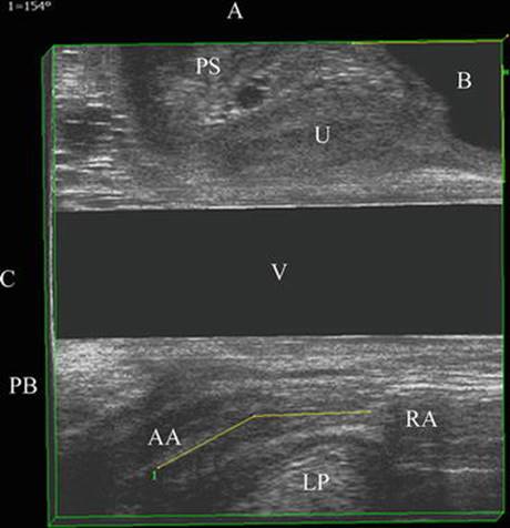
Fig. 10.8
Anorectal angle in mid-sagittal view by transvaginal 360° ultrasound. Anal axis (AA) and the rectal axis (RA) lines form the anorectal angle. A anterior, B bladder, C caudad, LP levator plate, PS pubic symphysis, U urethra, V vagina (Reprinted with permission from Shobeiri et al. [20])

Fig. 10.9
Levator plate descent angle in mid-sagittal view by transvaginal 360° ultrasound. A anterior, AP antero-posterior line of minimal levator hiatus (blue line), B bladder, C caudad, LP levator plate, LPDAlevator plate descent angle, PLURAL Pubic Levator Ultrasound Reference Assessment Line (green line), PS pubic symphysis, U urethra, V vagina (Reprinted with permission from Shobeiri et al. [20])

Fig. 10.10
Minimal levator hiatus area in axial plane by transvaginal 360° ultrasound. AP line is in blue. Puborectalis pubococcygeus border is delineated with small arrows. A anterior, AP antero-posterior line of minimal levator hiatus (blue line), LR left-right axis of minimal levator hiatus, PC pubococcygeus, PR puborectalis, PS pubic symphysis, U urethra, V vagina (Reprinted with permission from Shobeiri et al. [20])
Puborectalis Hiatus
Puborectalis hiatus is not a term frequently used. However, it is important to emphasize that the points of attachment and direction of some of the puborectalis fibers are different from that of the minimal levator hiatus (Fig. 10.11). Anteriorly, minimal levator hiatus is comprised of pubococcygeus fibers, although laterally some puborectalis fibers exist as well (Fig. 10.12). Puborectalis contributes significantly to the mid and distal edge of the levator plate.

Fig. 10.11
Puborectalis hiatus in the same patient in axial view by transvaginal 360° ultrasound. AP antero-posterior line of minimal levator hiatus, LR left-right axis of minimal levator hiatus, PRpuborectalis, PSpubic symphysis (Reprinted with permission from Shobeiri et al. [20])
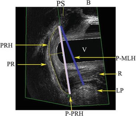
Fig. 10.12
Ultrasound showing plane of MLH and PRH in right sagittal view. AP antero-posterior line of minimal levator hiatus (blue line), B bladder, LP levator plate, LR left-right axis of minimal levator hiatus, P-MLH plane of minimal levator hiatus (blue line), MLH minimal levator hiatus, P-PRH plane of puborectalis hiatus (purple line), PR puborectalis, PS pubic symphysis, Rrectum, U urethra, V vagina (Reprinted with permission from Shobeiri et al. [20])
Levator Pubic Gap
The plane of minimal levator hiatus can be used to assess levator ani muscle attachment to the pubic bone. In case of a levator ani detachment at the level of the pubic bone, the levator pubic gap will be measured as the distance between the remnants if the levator muscle to the original insertion on the pubic bone (Fig. 10.13).
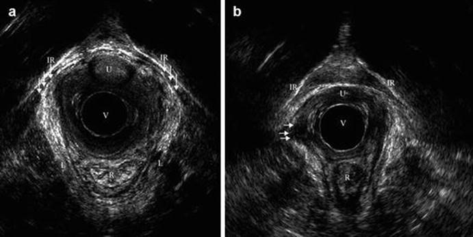
Fig. 10.13
Images obtained by EVUS in (a) a nulliparous woman with intact levator ani muscle (LAM) at level 3 and (b) a primiparous woman after forceps delivery involving a right mediolateral episiotomy and a third-degree tear with unilateral avulsion injury (axial plane at level 3). In (a) the LAM attachment is indicated by arrows. In (b) arrows indicate missing LAM muscle on patient’s right side indicating the levator ani-pubic gap. IR inferior rami os pubis, L levator ani muscle, R rectum, U urethra, V vagina (with endovaginal probe) (Reprinted from Schwertner-Tiepelmann et al. [63]; with permission from John Wiley and Sons
Levator Muscle Thickness
The plane of minimal levator hiatus can be used to assess muscle thickness at 3 and 9 o’clock in coronal plane. However, interrater and intrarater reliability of this measure has not been good in unpublished studies.
Levator Plate Descent Angle
3D-ultrasound cubes obtained by 360° endovaginal probe are used for this measurement. Midsagittal plane is the measurement plane. The shortest line between the levator plate and pubic symphysis corresponding to the anterior-posterior line or height of the minimal levator hiatus is drawn. By drawing a reference vertical mid-symphysial line in the midsagittal plane, we can calculate the angle between the two lines as a measure of the relative location of the levator plate in relation to the pubic symphysis in the resting position, the reference vertical line is referred to as the Pubic Levator plate Ultrasound Reference Assessment Line, or PLURAL (see Fig. 10.9).
Anorectal Angle
3D-ultrasound volumes obtained by 360° endovaginal probe can be used for this measurement. In the mid-sagittal plane one can measure the anorectal angle (ARA) as an angle between the axis of the anal canal and axis of the rectum (see Fig. 10.8). Caution should be used reporting these values as the ARA values obtained by EVUS are different from those obtained by MRI. Generally, EVUS ARA values >170° are associated with severe LAD.
Urethral Thickness
3D-ultrasound volumes obtained by 360° endovaginal probe can be used for this measurement. Midsagittal plane should be used to identify urethral lumen length and longitudinal axis. The midpoint of urethra will be marked. Using axial plane at the level of mid urethra, image should be tilted to become perpendicular to the longitudinal axis of the urethral lumen, visualized in the midsagittal plane. The urethral thickness can be measured in this plane. As with levator ani thickness, urethral thickness measurements are up for debate. That is because just as with muscles the thickness of a person’s muscle or the urethra are determined by genetics and do not necessarily indicate pathology nor does it have correlation with continence status [36].
Obstetric Factors
Antepartum prediction of levator muscle trauma is difficult or maybe impossible [25] but there are reports of obstetric risk factors associated with pelvic floor trauma which can suggest modification of obstetric practice. Levator trauma at the time of first delivery has been associated with vaginal delivery, forceps and a longer second stage. Epidural pain relief may exert a protective effect [17]. It is likely that birth weight, length of second stage, size of fetal head and forceps delivery increase the probability of muscle injury [24, 27–29]. Valsky et al. showed an OR of 2.27 for muscle injury detected by ultrasound when the second stage was more than 110 min and OR of 3.34 for muscle injury when fetal head circumference was more than 35.5 cm [24]. Falkert et al. also found a positive correlation with weight and head circumference of baby and area of levator hiatus [40]. There are controversies regarding maternal age at first delivery. In some studies increased maternal age contributes to muscle injury [44]; however, this has not been reproduced by other studies [24, 25]. Shek et al. found that women with lower BMI were at higher risk of sustaining muscle injury, but this finding’s clinical significance is questionable as BMI thresholds identified were 27.85 kg/m2 vs 30.01 kg/m2 [25] and this association was not confirmed by another study [40]. TPUS technique identified the interpubic gap and the infrapubic arc with a high interrater agreement, however; it was shown that infrapubic angle is not associated with length of second stage of labour and the occurrence of levator defects [45].
Birth related detachment of the levator ani muscle from the pubic bone seems to persist, whereas. levator ani stretch injury in the body of the muscle may resolve over time [26]. Although some earlier studies placed the rate at about 36 % [14], more recent MRI, and EVUS studies place the true overall rate closer to 13 % [26, 46]. Breaking down this rate by risk factors, in a recent MRI study, major defect rates were: 42 % for forceps and short second stage; 63 % for forceps and second stage arrest; and 6 % for spontaneous delivery. The odds ratios for major injury were: 11.0 for forceps and short second stage compared with spontaneous delivery; 25.9 for forceps and second stage arrest compared with spontaneous delivery; and 2.3 for forceps and second stage arrest compared with short second stage (P = 0.07) [47].
It has been established that levator ani muscle injury increases the risk of pelvic organ prolapse (POP), and specifically cystocele formation initially [33, 48]. Observational studies have shown that there is a direct correlation between the size of the muscle defect and the symptoms or/and signs of prolapse [49] and women with bilateral avulsions are more likely to suffer from uterine prolapse [50]. The relationship between levator muscle injury and urinary incontinence is controversial [14, 51–54]. Although periurethral smooth and striated muscles have been implicated as potential mechanisms by which childbirth might affect urinary continence [36], using EVUS their presence or absence is not associated with continence status [36]. Associations between anal incontinence and birth related changes in pelvic floor support system have been investigated [55–57].
Conclusion
Imaging studies have improved our understanding of the pathophysiology of pelvic floor trauma in labor [58]. From this information, we can devise population based prevention strategies, and pelvic floor trauma repair strategies (Fig. 10.14). The information about the relationship between forceps delivery and pelvic floor trauma has trickled to obstetricians in the United States, and forceps deliveries have declined significantly. Other efforts have been aimed at obstetrician education to repair pelvic floor trauma. Anal sphincter repair workshops (OASIS) help obstetricians to repair anal sphincter injury in an evidence based and reproducible manner [59]. Despite preliminary attempts at repair of levator ani trauma [60–62], the results are not applicable to the general population.

Fig. 10.14
J-hook needle in puborectalis muscle by endovaginal 360° ultrasound. N needle, R rectum, U urethra (With kind permission from Springer Science + Business Media: Rostaminia et al. [61])
EVUS provides a reliable vehicle for investigating pathophysiology of pelvic floor trauma associated with labor as it provides detailed anatomy of levator ani subdivisions and can be deployed in large scale for population-based studies. Additionally, now the endovaginal 3D ultrasonography has surpassed investigational use and is used routinely by more clinicians for evaluation of the levator ani muscles, evaluation of defecatory dysfunction, vaginal mesh, vaginal cysts and masses just to name a few of the indications.
References
1.
Lien KC, Mooney B, DeLancey JO, Ashton-Miller JA. Levator ani muscle stretch induced by simulated vaginal birth. Obstet Gynecol. 2004;103(1):31–40.PubMedPubMedCentral
2.
Lien K-C, DeLancey JOL, Ashton-Miller JA. Biomechanical analyses of the efficacy of patterns of maternal effort on second-stage progress. Obstet Gynecol. 2009;113(4):873–80.PubMedPubMedCentral
3.
Dietz HP, Shek C, Clarke B. Biometry of the pubovisceral muscle and levator hiatus by three-dimensional pelvic floor ultrasound. Ultrasound Obstet Gynecol. 2005;25(6):580–5.PubMed
4.
Shobeiri SA, Rostaminia G, Quiroz LH. Relative contributions of the levator ani subdivisions to levator ani movement. Neurourol urodyn. 2013;32(6):710–11.
5.
Ashton-Miller JA, Delancey JOL. On the biomechanics of vaginal birth and common sequelae. Ann Rev Biomed Eng. 2009;11:163–76.
6.
Sultan AH, Nicholls RJ, Kamm MA, Hudson CN, Beynon J, Bartram CI. Anal endosonography and correlation with in vitro and in vivo anatomy. Br J Surg. 1993;80(4):508–11.PubMed
7.
Sultan AH, Kamm MA, Talbot IC, Nicholls RJ, Bartram CI. Anal endosonography for identifying external sphincter defects confirmed histologically. Br J Surg. 1994;81(3):463–5.PubMed
8.
Sultan AH, Loder PB, Bartram CI, Kamm MA, Hudson CN. Vaginal endosonography. New approach to image the undisturbed anal sphincter. Dis Colon Rectum. 1994;37(12):1296–9.PubMed
9.
Sultan AH, Monga AK, Stanton SL. The pelvic floor sequelae of childbirth. Br J Hosp Med. 1996;55(9):575–9.PubMed
10.
Sultan AH, Stanton SL. Preserving the pelvic floor and perineum during childbirth–elective caesarean section? Br J Obstet Gynaecol. 1996;103(8):731–4.PubMed
11.
Strohbehn K, Ellis JH, Strohbehn JA, DeLancey JO. Magnetic resonance imaging of the levator ani with anatomic correlation. Obstet Gynecol. 1996;87(2):277–85.PubMed
12.
Tunn R, DeLancey JO, Howard D, Thorp JM, Ashton-Miller JA, Quint LE. MR imaging of levator ani muscle recovery following vaginal delivery. Int Urogynecol J. 1999;10(5):300–7.
13.
Shobeiri SA, Nolan TE, Yordan-Jovet R, Echols KT, Chesson RR. Digital examination compared to trans-perineal ultrasound for the evaluation of anal sphincter repair. Int J Gynaecol Obstet. 2002;78(1):31–6.PubMed
14.
Dietz HP, Lanzarone V. Levator trauma after vaginal delivery. Obstet Gynecol. 2005;106(4):707–12.PubMed
15.
Dietz HP, Steensma AB. The prevalence of major abnormalities of the levator ani in urogynaecological patients. BJOG. 2006;113(2):225–30.PubMed
16.
Weinstein MM, Pretorius DH, Jung SA, Nager CW, Mittal RK. Transperineal three-dimensional ultrasound imaging for detection of anatomic defects in the anal sphincter complex muscles. Clin Gastroenterol Hepatol. 2009;7(2):205–11.PubMed
17.
Shek KL, Dietz HP. Intrapartum risk factors for levator trauma. BJOG. 2010;117(12):1485–92.PubMed
18.
Dietz HP, Abbu A, Shek KL. The levator-urethra gap measurement: a more objective means of determining levator avulsion? Ultrasound Obstet Gynecol. 2008;32(7):941–5.PubMed
19.
Zhuang RR, Song YF, Chen ZQ, Ma M, Huang HJ, Chen JH, et al. Levator avulsion using a tomographic ultrasound and magnetic resonance-based model. Am J Obstet Gynecol. 2011;205(3):232.e1–8.
20.
Shobeiri SA, Rostaminia G, White DE, Quiroz LH. The determinants of minimal levator hiatus and their relationship to the puborectalis muscle and the levator plate. BJOG. 2013;120(2):205–11.PubMed
21.
Shobeiri SA, Leclaire E, Nihira MA, Quiroz LH, O’Donoghue D. Appearance of the levator ani muscle subdivisions in endovaginal three-dimensional ultrasonography. Obstet Gynecol. 2009;114:66–72.PubMed
22.
Shobeiri SA, White D, Quiroz LH, Nihira MA. Anterior and posterior compartment 3D endovaginal ultrasound anatomy based on direct histologic comparison. Int Urogynecol J. 2012;23(8):1047–53.PubMed
23.
Santoro GA, Wieczorek AP, Shobeiri SA, Mueller ER, Pilat J, Stankiewicz A, et al. Interobserver and interdisciplinary reproducibility of 3D endovaginal ultrasound assessment of pelvic floor anatomy. Int Urogynecol J Pelvic Floor Dysfunct. 2011;22:53–9.
24.
Valsky DV, Lipschuetz M, Bord A, Eldar I, Messing B, Hochner-Celnikier D, et al. Fetal head circumference and length of second stage of labor are risk factors for levator ani muscle injury, diagnosed by 3-dimensional transperineal ultrasound in primiparous women. Am J Obstet Gynecol. 2009;201(1):91.e1–7.
25.
Shek KL, Dietz HP. Can levator avulsion be predicted antenatally? Am J Obstet Gynecol. 2010;202(6):586.e1–6.
26.
van Delft K, Thakar R, Shobeiri SA, Sultan AH. Levator haematoma at the attachment zone as an early marker for levator ani muscle avulsion. Ultrasound Obstet Gynecol. 2014;43(2):210–7.PubMed
27.
Albrich SB, Laterza RM, Skala C, Salvatore S, Koelbl H, Naumann G. Impact of mode of delivery on levator morphology: a prospective observational study with three-dimensional ultrasound early in the postpartum period. BJOG. 2012;119(1):51–60.PubMed
28.
Krofta L, Otcenasek M, Kasikova E, Feyereisl J. Pubococcygeus-puborectalis trauma after forceps delivery: evaluation of the levator ani muscle with 3D/4D ultrasound. Int Urogynecol J Pelvic Floor Dysfunct. 2009;20(10):1175–81.PubMed
29.
Cassado Garriga J, Pessarrodona Isern A, Espuna Pons M, Duran Retamal M, Felgueroso Fabregas A, Rodriguez-Carballeira M. Tridimensional sonographic anatomical changes on pelvic floor muscle according to the type of delivery. Int Urogynecol J. 2011;22(8):1011–8.PubMed
30.
Shek KL, Dietz HP. The effect of childbirth on hiatal dimensions. Obstet Gynecol. 2009;113(6):1272–8.PubMed
31.
Chan SS, Cheung RY, Yiu AK, Lee LL, Pang AW, Choy KW, et al. Prevalence of levator ani muscle injury in Chinese women after first delivery. Ultrasound Obstet Gynecol. 2012;39(6):704–9.PubMed
32.
Stankiewicz A, Wieczorek A, Wozniak M. Comparison of accuracy of functional measurements of the urethra in transperineal vs. endovaginal ultrasound in incontinent women. Peliperineology. 2008;27:145–7.
33.
Rostaminia G, White D, Hegde A, Quiroz LH, Davila GW, Shobeiri SA. Levator ani deficiency and pelvic organ prolapse severity. Obstet Gynecol. 2013;121(5):1017–24.PubMed
34.
Morgan DM, Umek W, Stein T, Hsu Y, Guire K, DeLancey JOL. Interrater reliability of assessing levator ani muscle defects with magnetic resonance images. Int Urogynecol J. 2007;18(7):773–8.
35.
White D, Rostaminia G, Quiroz L, Shobeiri S. Sonographic predictors of obstructive defecatory dysfunction. Neurourol Urodyn. 2013;32(6):707–9.
36.
Rostaminia G, White DE, Quiroz LH, Shobeiri SA. Visualization of periurethral structures by 3D endovaginal ultrasonography in midsagittal plane is not associated with stress urinary incontinence status. Int Urogynecol J. 2013;24(7):1145–50.PubMed
37.
Rostaminia G, White D, Quiroz L, Shobeiri S. Levator plate descent correlates with levator ani muscle deficiency. Int Urogynecol J. 2012;23 Suppl 2:S43–244. doi:10.1007/s00192-012-1875-z.
38.
Volloyhaug I, Wong V, Shek KL, Dietz HP. Does levator avulsion cause distension of the genital hiatus and perineal body? Int Urogynecol J. 2012;27:27.
39.
Toozs-Hobson P, Balmforth J, Cardozo L, Khullar V, Athanasiou S. The effect of mode of delivery on pelvic floor functional anatomy. Int Urogynecol J Pelvic Floor Dysfunct. 2008;19(3):407–16.PubMed
40.
Falkert A, Endress E, Weigl M, Seelbach-Gobel B. Three-dimensional ultrasound of the pelvic floor 2 days after first delivery: influence of constitutional and obstetric factors. Ultrasound Obstet Gynecol. 2010;35(5):583–8.PubMed
41.
Shek KL, Kruger J, Dietz HP. The effect of pregnancy on hiatal dimensions and urethral mobility: an observational study. Int Urogynecol J. 2012;23(11):1561–7.PubMed
42.
Chantarasorn V, Shek KL, Dietz HP. Mobility of the perineal body and anorectal junction before and after childbirth. Int Urogynecol J. 2012;23(6):729–33.PubMed
43.
van Delft K, Shobeiri SA, Thakar R, Schwertner-Tiepelmann N, Sultan AH. Intra- and inter-rater reliability of levator ani muscle biometry and avulsion using three-dimensional endovaginal sonography. Ultrasound Obstet Gynecol. 2014;43(2):202–9.PubMed
44.
Dietz HP, Simpson JM. Does delayed child-bearing increase the risk of levator injury in labour? Aust N Z J Obstet Gynaecol. 2007;47(6):491–5.PubMed
45.
Albrich S, Laterza RM, Merinsky A, Skala C, Koelbl H, Naumann G. Measurement of the infrapubic angle using 3D perineal ultrasound and its relationship to obstetrical parameters. Ultraschall in der Medizin (Stuttgart, Germany, 1980). 2012;33(7):E95–100.
46.
Ashton-Miller JA, Delancey JOL. Functional anatomy of the female pelvic floor. Ann N Y Acad Sci. 2007;1101(1):266–96.PubMed
47.
Kearney R, Fitzpatrick M, Brennan S, Behan M, Miller J, Keane D, et al. Levator ani injury in primiparous women with forceps delivery for fetal distress, forceps for second stage arrest, and spontaneous delivery. Int J Gynaecol Obstet. 2010;111(1):19–22.PubMedPubMedCentral
48.
DeLancey JO, Morgan DM, Fenner DE, Kearney R, Guire K, Miller JM, et al. Comparison of levator ani muscle defects and function in women with and without pelvic organ prolapse. Obstet Gynecol. 2007;109(2 Pt 1):295–302.PubMed
49.
Dietz HP. Quantification of major morphological abnormalities of the levator ani. Ultrasound Obstet Gynecol. 2007;29(3):329–34.PubMed
50.
Dietz HP, Simpson JM. Levator trauma is associated with pelvic organ prolapse. BJOG. 2008;115(8):979–84.PubMed
51.
Dietz HP, Kirby A, Shek KL, Bedwell PJ. Does avulsion of the puborectalis muscle affect bladder function? Int Urogynecol J Pelvic Floor Dysfunct. 2009;20(8):967–72.PubMed
52.
Morgan DM, Cardoza P, Guire K, Fenner DE, DeLancey JO. Levator ani defect status and lower urinary tract symptoms in women with pelvic organ prolapse. Int Urogynecol J. 2010;21(1):47–52.PubMed
53.
DeLancey JO, Kearney R, Chou Q, Speights S, Binno S. The appearance of levator ani muscle abnormalities in magnetic resonance images after vaginal delivery. Obstet Gynecol. 2003;101(1):46–53.PubMedPubMedCentral
54.
DeLancey JOL, Trowbridge ER, Miller JM, Morgan DM, Guire K, Fenner DE, et al. Stress urinary incontinence: relative importance of urethral support and urethral closure pressure. J Urol. 2008;179(6):2286–90; discussion 90.PubMedPubMedCentral
55.
Zufferey G, Perneger T, Robert-Yap J, Rubay R, Lkhagvabayar B, Roche B. Measure of the voluntary contraction of the puborectal sling as a predictor of successful sphincter repair in the treatment of anal incontinence. Dis Colon Rectum. 2009;52(4):704–10.PubMed
56.
Chantarasorn V, Shek KL, Dietz HP. Sonographic detection of puborectalis muscle avulsion is not associated with anal incontinence. Aust N Z J Obstet Gynaecol. 2011;51(2):130–5.PubMed
57.
Bharucha AE, Fletcher JG, Melton 3rd LJ, Zinsmeister AR. Obstetric trauma, pelvic floor injury and fecal incontinence: a population-based case-control study. Am J Gastroenterol. 2012;107(6):902–11.PubMedPubMedCentral
58.
Shobeiri SA. The practical value of the levator ani muscle subdivisions in endovaginal three-dimensional ultrasonography. Obstet Gynecol. 2009;114:1145–6.
59.
Fernando RJ, Sultan AH, Radley S, Jones PW, Johanson RB. Management of obstetric anal sphincter injury: a systematic review and national practice survey. BMC Health Serv Res. 2002;2(9):1–10.
60.
Dietz HP, Gillespie AVL, Phadke P. Avulsion of the pubovisceral muscle associated with large vaginal tear after normal vaginal delivery at term. Aust N Z J Obstet Gynaecol. 2007;47(4):341–4.PubMed
61.
Rostaminia G, Shobeiri SA, Quiroz LH. Surgical repair of bilateral levator ani muscles with ultrasound guidance. Int Urogynecol J. 2013;24(7):1237–9.PubMed
62.
Shobeiri SA, Chimpiri AR, Allen A, Nihira MA, Quiroz LH. Surgical reconstitution of a unilaterally avulsed symptomatic puborectalis muscle using autologous fascia lata. Obstet Gynecol. 2009;114(2 Pt 2):480–2.PubMed
63.
Schwertner-Tiepelmann N, Thakar R, Sultan AH, Tunn R. Obstetric levator ani muscle injuries: current status. Ultrasound Obstet Gynecol. 2012;39(4):372–83.PubMed