
Jorge Milhem Haddad1 , Lilian R. Fiorelli2 and Thais V. Peterson1
(1)
Section of Urogynecology and PFD, University of São Paulo, 255 Dr. Eneas de Carvalho Aguiar Av., Sao Paulo, 05403-000, Brazil
(2)
Gynecology Division, Section of Urogynecology and PFD, University of São Paulo, Sao Paulo, Brazil
Jorge Milhem Haddad (Corresponding author)
Email: jorge_milhem@uol.com.br
Lilian R. Fiorelli
Email: lilianfiorelli@gmail.com
Thais V. Peterson
Email: thaispeterson@gmail.com
Abstract
Pelvic floor dysfunction has a high incidence mainly in aging women. Parity and vaginal childbirth are strongly associated with pelvic organ prolapse and stress urinary incontinence. Muscle injury, neurovascular injury and connective tissue remodeling may explain this association. In this chapter, the main mechanisms of injury are discussed, as well as the role of related risk factors, such as episiotomy, operative delivery and prolonged second stage of labor.
Keywords
Pelvic floorChildbirthDeliveryUrinary incontinencePelvic organ prolapseFecal incontinenceRisk factorsForcepsMenopauseEpisiotomy
Introduction
The incidence of pelvic floor dysfunction, such as pelvic organ prolapse and urinary and fecal incontinence increased by 45 % over the last 40 years. Increased life expectancy has significantly contributed since this occurrence increases over the woman’s lifetime [1, 2].
Main risk factors for pelvic floor dysfunction (PFD) include factors associated with increased intra-abdominal pressure such as obesity, multiparity, chronic cough, factors associated with the breakdown of collagen, such as smoking, deficiencies of collagen, menopause, and factors associated with local trauma, such as birth trauma, including vaginal delivery with or without the use of forceps [3]. Figure 7.1shows the incidence of pelvic floor disorders associated with vaginal delivery [4].
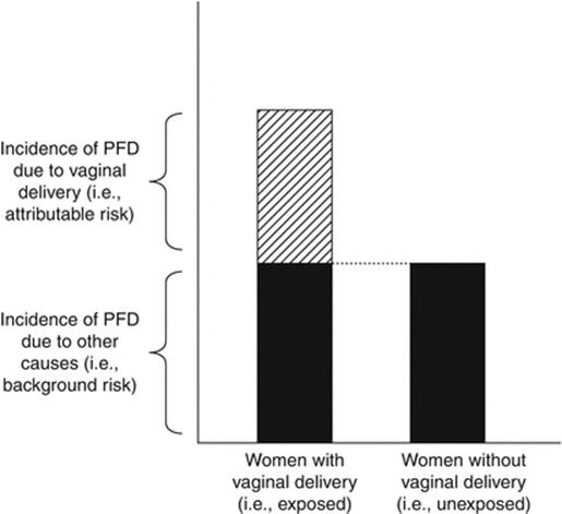
Fig. 7.1
Pelvic floor dysfunction in women with or without vaginal delivery (Reprinted from Patel et al. [4], Copyright 2006, with permission from Elsevier)
Vaginal delivery increases the incidence of genital prolapse 4–11 times [1] and the incidence of urinary incontinence 2.7 times [5]. Important genes in inflammation, collagen breakdown, and smooth muscle inhibition are upregulated in patients who had vaginal delivery and develop incontinence and genital prolapse [6].
Types of Injury in Vaginal Delivery
Intact neuromuscular function and pelvic support are crucial to pelvic stability. Pregnancy and delivery contribute to pelvic floor disorders due to compression, stretching, or tear of nerve, muscle and connective tissue. We describe below the main mechanisms of obstetrical injury.
Mechanical Injury
Levator ani muscle complex integrity is very important to pelvic floor support. Comprising puborectalis, pubococcygeus and ileococcygeus, this muscle provides support to urethra, distal vagina and rectum. During labor, stretching and damage of these muscles can occur, particularly to the pubococcygeus, since it is the shortest and most medial muscle of the complex [7–9].
Levator trauma can lead to widening of the genital hiatus and thus be a risk factor for pelvic organ prolapse. It can also be associated with urinary incontinence.
Imaging studies may help in elucidating the types of injuries of levator ani muscle, especially transvaginal or transperinaeal ultrasonography and magnetic resonance imaging of the pelvis with or without three-dimensional reconstruction [7–9].
In three-dimensional reconstruction, it is possible to observe the distinct anatomical difference of the levator ani when comparing nulliparous, asymptomatic multiparous, symptomatic multiparous and elderly, as shown in Fig. 7.2[10].
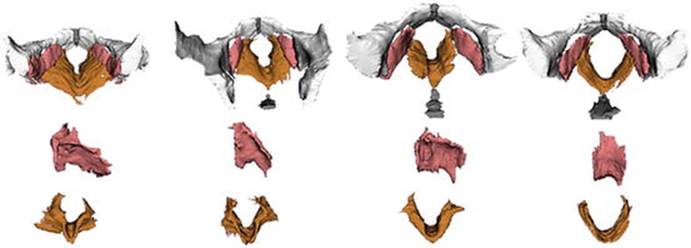
Fig. 7.2
Three-dimensional reconstruction of MRI of levator ani in nuliparous, asymptomatic multiparous, symptomatic multiparous and elderly (Reprinted from Singh et al. [10], Copyright 2003, with permission from Elsevier)
Nerve Injury
Injury to pudendal nerve can also be associated with pelvic floor disorders, especially urinary and fecal incontinence. The pudendal nerve innervates the external urethral and anal sphincters. During labor, nerve compression and stretching can occur, leading to incontinence. This process is reversible in most cases, with complete return to continence in the postpartum period. Severe cases of injury can lead to persistent incontinence [11, 12].
Connective Tissue Remodeling
During pregnancy, collagen and elastin, components of connective tissue, experience some modifications in order to increase vaginal distensiblity [13].
During labor, extensive stretching promotes collagen degradation. The endopelvic fascia and other connective tissue elements are at risk of stretch and detachment from their bony attachments during childbirth [13]. Pubic bone edema and subcortical fracture are common, and magnetic resonance shows that they persist until 7 weeks after delivery without clinical findings [14]. In rare cases, pubic symphysis rupture can occur [15].
After delivery, there is a substantial remodeling of the connective tissue components. However, this new tissue is not as strong as the original [13].
Injuries at level I of DeLancey are responsible for the appearance of prolapse of the uterus. The increase in intra-abdominal pressure generated by the pregnancy itself can cause rupture or stretching of these structures, especially if it is a twin pregnancy, macrosomic fetus or increased amniotic fluid as in gestational diabetes.
At level I, proximal transverse defect may occur with a detachment of the rectovaginal fascia from the pericervical ring, leading to the descent of the small bowel, omentum or sigmoid through the vaginal canal, called enterocele or protrusion of the rectum, called high rectocele [3, 16].
Structures of level II of DeLancey in labor are molded to hold the fetus. However, especially in cases of large fetuses or prolonged labor or even accelerated labor, rectovaginal fascia can break or stretch or it can detach from arcus tendineus. These injuries may result in the appearance of rectoceles in varied degrees and types, associated with central or transverse rupture of fascia or side rupture of fascia [17].
At level II, if there is a lesion of pubocervical fascia, the anterior vaginal wall may protrude into the vagina. When this prolapse involves the protrusion of the bladder it is called cystocele. Rarely, enterocele may occur via anterior vaginal wall, but is anatomically classified as apical prolapse; it originates from the detachment of the pubocervical fascia from the pericervical ring [7, 17]. Moreover, vaginal childbirth is associated with loss of tenting of the vaginal fornices, independent of levator trauma, and also with impaired anterior vaginal wall support. The existence of paravaginal defects may imply a role for such defects in the causation of anterior vaginal wall prolapse [18].
The expulsion phase of labor can cause injuries to level III of DeLancey as pubocervical fascia and the urethra can prolapse into the vaginal lumen, called urethrocele. In some cases it can cause hypermobility of the bladder neck and stress urinary incontinence (usually during medium or large efforts). Still, if there is injury to the urethral sphincter during the expulsion phase, it will decrease the intra-urethral pressure and, therefore, also cause stress urinary incontinence (usually during minimal efforts) [7, 17].
The second stage of labor can also be associated with level III lesions in the posterior compartment. Lesions in the rectovaginal fascia at this level cause rectoceles. Lesions in the perineal body and perineal muscles can cause perineal rupture. In these cases the patient complains of “a large or gaping vagina.” If the anal sphincter is affected, the patient may develop fecal incontinence [19].
Table 7.1 summarizes the time of pregnancy or childbirth, the types of injuries that can occur according to DeLancey levels and their clinical consequences.
Table 7.1
Types of injuries during labor
|
Pregnancy or childbirth |
Levels of De Lancey |
Place of injury |
Diagnosis by ICS/IUGA(2011) and by region [16] |
|
Pregnancy and primary stage of labor |
Level I |
Uterosacral ligament Cardinal ligament Pericervical ring |
Apical prolapse: Uterine prolapse After total hysterectomy: vaginal vault prolapse After subtotal hysterectomy: cervix prolapse |
|
Pubocervical fascia |
Anterior prolapse: Cystocele Enterocele (rare) |
||
|
Rectovaginal fascia |
Enterocele High rectocele |
||
|
Second stage of labor: active phase |
Level II |
Pubocervical fascia |
Urethrocele Cystocele |
|
Rectovaginal fascia |
Rectocele anal |
||
|
Second stage of labor: expulsive phase |
Level III |
Pubocervical fascia |
Urethrocele Stress urinary incontinence by hypermobility of bladder neck |
|
Urethral sphincter |
Stress urinary incontinence by intrinsic sphincter deficiency |
||
|
Rectovaginal fascia |
Rectocele anal |
||
|
Perineal body |
Perineal rupture (if there is lesion of sphincter can cause fecal incontinence) |
It is important to note that in some cases urinary urgency and urgency incontinence/overactive bladder can be caused by anterior vaginal prolapse. Vesical receptors present in the base of the bladder in contact with the vaginal epithelium are activated during bladder filling. However, in most cases, the etiology of overactive bladder is unknown [20].
Obstetric and Maternal Factors in Pelvic Floor Disorders
The use of forceps appears to increase the risk of pelvic organ prolapse and anal sphincter laceration, which increases the risk of fecal incontinence. Forceps delivery can affect the muscles of the levator ani (OR = 14.7 and 95 % CI: 4.9–44.3). When there is injury of these muscles, a concomitant injury of the external anal sphincter may occur (OR = 8.1 and 95 % CI: 3.3–19.5). Women with levator injury were 3.5 years older in a study by Kearney et al. and had a 78-min longer second stage of labor [7].
Episiotomy is discussed extensively elsewhere in this book. The role of episiotomy on pelvic floor disorders is unclear. The routine use of episiotomy is decreasing, and there is no evidence that this procedure prevents pelvic floor dysfunction.
Levator tear during labor is associated with levator weakness and posterior-vaginal wall descent [14].
Third-degree perineal tear is when the external anal sphincter is affected; fourth-degree tear is when external and internal anal sphincter and rectal mucosa are affected. Risk factors for these lesions are primiparity (OR = 1.8 and 95 % CI: 1.65–1.95), Asian ethnicity (OR = 1.1 and 95 % CI: 1,09–1,23), use of forceps delivery (OR = 1.8 and 95 % CI: 1.65–1.95) and male fetus (OR = 1.3 and 95 % CI: 1.27–1.34) [19].
Both third- and fourth-degree perineal tears and vaginal sidewall tears are independently associated with levator avulsion (p = 0.004 and 0.012, respectively) and consequently future pelvic floor disorder [21]. Meta-analysis of non-randomised studies showed a significant reduction in the risk of obstetric anal sphincter injuries with manual perineal support [22].
Increasing maternal age and birth weight are associated with PFD [23].
A prolonged second stage may increase soft tissue injury and neuromuscular damage to the pelvic floor. Childbirth injuries to the pelvic floor are also found in experimental rat models. After the simulation of labor with vaginal balloon, rats can develop urinary incontinence. The damage to the urethral sphincter is shown in Fig. 7.3, and the levator ani muscle is shown in Fig. 7.4 [24].
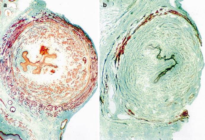
Fig. 7.3
Rat urethra after simulated birth trauma. Cross-section of the midurethra from (a) a continent rat and (b) an incontinent rat. The first one shows abundant smooth and striated muscle (a); the last one, a marked decrease (b). Trichrome stain, original magnification ×40 (Reprinted from Lin et al. [24], Copyright 1998, with permission from Elsevier)

Fig. 7.4
Rat urethra after simulated birth trauma. ATPase stain of the levator muscle from (a) a continent rat and (b) an incontinent rat. In the latter the amount of slow-twitch fiber (lighter stain) is increased (Reprinted from Lin et al. [24], Copyright 1998, with permission from Elsevier)
It is noteworthy that the lesions mentioned do not occur in all deliveries. Moreover, even after a cesarean section, the woman may develop genital prolapse or incontinence because these conditions occur after a combination of risk factors. Cesarean section has its own indications and cannot be indicated just for the prevention of pelvic floor injuries. Gestational urinary incontinence can be one of the predictors of urinary incontinence immediately after delivery and up to 2 years after birth. Weight gain during pregnancy is a risk factor for pelvic floor muscle dysfunction [25, 26].
In most cases it is observed that the genital prolapse or incontinence does not immediately appear post-partum. Other risk factors like obesity, chronic cough, constipation, smoking, collagen diseases, and especially menopause need to be present too. At menopause there is a decrease of estrogen resulting in breakdown of collagen fibers. The structures already weakened, such as fascia, ligaments and muscles, no longer support the pelvic organs, which then herniate through the vaginal canal. In addition, genital atrophy increases the occurrence of urinary incontinence [27].
Prophylactic pelvic floor muscle exercises performed during pregnancy help to decrease the short-term risk of urinary incontinence, but there is limited evidence on its long-term benefits [26]. It is important to note that there are no significant changes in sexual function after childbirth trauma with levator avulsion [28].
Knowledge of the anatomical parameters of the pelvic floor is important for the understanding of urogenital disorders such as pelvic organ prolapse and urinary incontinence that can be secondary to childbirth trauma in order to try to prevent these pathologies or propose the most appropriate treatment.
References
1.
Mant J, Painter R, Vessey M. Epidemiology of genital prolapse: observations from the Oxford Family Planning Association Study. Br J Obstet Gynaecol. 1997;104:579–85. PubMed: 9166201.PubMed
2.
Sze EH, Jones WP, Ferguson JL, Barker CD, Dolezal JM. Prevalence of urinary incontinence symptoms among black, white, and Hispanic women. Obstet Gynecol. 2002;99(4):572–5. PubMed.PubMed
3.
Rodrigues AM, de Oliveira LM, Martins Kde F, Del Roy CA, Sartori MG, Girão MJ, et al. Risk factors for genital prolapse in a Brazilian population. Rev Bras Ginecol Obstet. 2009;31(1):17–21. Portuguese. PubMed.PubMed
4.
Patel DA, Xu X, Thomason AD, Ransom SB, Ivy JS, DeLancey JO. Childbirth and pelvic floor dysfunction: an epidemiologic approach to the assessment of prevention opportunities at delivery. Am J Obstet Gynecol. 2006;195(1):23–8. Epub 2006 Mar 30. PubMed PMID: 16579934; PubMed Central PMCID: PMC1486798.PubMedPubMedCentral
5.
Rortveit G, Hannestad YS, Daltveit AK, Hunskaar S. Age- and type-dependent effects of parity on urinary incontinence: the Norwegian EPINCONT study. Obstet Gynecol. 2001;98:1004–10. PubMed: 11755545.PubMed
6.
Lin G, Shindel AW, Banie L, Deng D, Wang G, Hayashi N, et al. Molecular mechanisms related to parturition-induced stress urinary incontinence. Eur Urol. 2009;55(5):1213–22. doi:10.1016/j.eururo.2008.02.027. Epub 2008 Mar 18. PubMed PMID: 18372098; PubMed Central PMCID: PMC3001389.PubMed
7.
Kearney R, Miller JM, Ashton-Miller JA, DeLancey JO. Obstetric factors associated with levator ani muscle injury after vaginal birth. Obstet Gynecol. 2006;107(1):144–9. PubMed PMID: 16394052; PubMed Central PMCID: PMC2841321.PubMedPubMedCentral
8.
Singh K, Reid WM, Berger LA. Magnetic resonance imaging of normal levator ani anatomy and function. Obstet Gynecol. 2002;99(3):433–8. PubMed.PubMed
9.
Wisser J, Schär G, Kurmanavicius J, Huch R, Huch A. Use of 3D ultrasound as a new approach to assess obstetrical trauma to the pelvic floor. Ultraschall Med. 1999;20(1):15–8. PubMed.PubMed
10.
Singh K, Jakab M, Reid WM, Berger LA, Hoyte L. Three-dimensional magnetic resonance imaging assessment of levator ani morphologic features in different grades of prolapse. Am J Obstet Gynecol. 2003;188(4):910–5. PubMed.PubMed
11.
Fitzpatrick M, O’Brien C, O’Connell PR, O’Herlihy C. Patterns of abnormal pudendal nerve function that are associated with postpartum fecal incontinence. Am J Obstet Gynecol. 2003;189:730–5.PubMed
12.
Lin YH, Liu G, Li M, Xiao N, Daneshgari F. Recovery of continence function following simulated birth trauma involves repair of muscle and nerves in the urethra in the female mouse. Eur Urol. 2010;57(3):506–12.PubMed
13.
Memon HU, Handa V. Vaginal childbirth and pelvic floor disorders. Women’s Health. 2013;9(3). doi:10.2217/whe.13.17.
14.
Miller JM, Low LK, Zielinski R, Smith AR, DeLancey JO, Brandon C. Evaluating maternal recovery from labor and delivery: bone and levator ani injuries. Am J Obstet Gynecol. 2015. pii: S0002-9378(15)00450-0. doi:10.1016/j.ajog.2015.05.001. [Epub ahead of print] PubMed.
15.
Gräf C, Sellei RM, Schrading S, Bauerschlag DO. Treatment of parturition-induced rupture of pubic symphysis after spontaneous vaginal delivery. Case Rep Obstet Gynecol. 2014;2014:485916. doi:10.1155/2014/485916. Epub 2014 Jan 15. PubMed PMID: 24551465; PubMed Central PMCID: PMC3914324.PubMedPubMedCentral
16.
DeLancey JO. Anatomic aspects of vaginal eversion after hysterectomy. Am J Obstet Gynecol. 1992;166:1717–24.PubMed
17.
Li X, Kruger JA, Nash MP, Nielsen PM. Effects of nonlinear muscle elasticity on pelvic floor mechanics during vaginal childbirth. J Biomech Eng. 2010;132(11):111010. doi:10.1115/1.4002558. PubMed.PubMed
18.
Cassadó-Garriga J, Wong V, Shek K, Dietz HP. Can we identify changes infascial paravaginal supports after childbirth? Aust N Z J Obstet Gynaecol. 2015;55(1):70–5. doi:10.1111/ajo.12261. Epub 2014 Oct 11. PubMed.PubMed
19.
Dahlen H, Priddis H, Schmied V, Sneddon A, Kettle C, Brown C, et al. Trends and risk factors for severe perineal trauma during childbirth in New South Wales between 2000 and 2008: a population-based data study. BMJ Open. 2013;3(5). pii: e002824. doi:10.1136/bmjopen-2013-002824. PubMed PMID: 23793688; PubMed Central PMCID: PMC 3657654.
20.
de Boer TA, Slieker-ten Hove MC, Burger CW, Vierhout ME. The prevalence and risk factors of overactive bladder symptoms and its relation to pelvic organ prolapse symptoms in a general female population. Int Urogynecol J. 2011;22(5):569–75. doi:10.1007/s00192-010-1323-x. Epub 2010 Nov 23. PubMed PMID: 21104400; PubMed Central PMCID: PMC3072516.PubMed
21.
Shek KL, Green K, Hall J, Guzman-Rojas R, Dietz HP. Perineal and vaginal tears are clinical markers for occult levator ani trauma. A retrospective observational study. Ultrasound Obstet Gynecol. 2015. doi:10.1002/uog.14856. [Epub ahead of print] PubMed.
22.
Bulchandani S, Watts E, Sucharitha A, Yates D, Ismail KM. Manual perineal support at the time of childbirth: a systematic review and meta-analysis. BJOG. 2015. doi:10.1111/1471-0528.13431. [Epub ahead of print] PubMed.
23.
Handa VL, Blomquist JL, McDermott KC, Friedman S, Muñoz A. Pelvic floor disorders after vaginal birth: effect of episiotomy, perineal laceration, and operative birth. Obstet Gynecol. 2012;119(2 Pt 1):233–9. PubMed PMID: 22227639; PubMed Central PMCID:PMC3266992.PubMedPubMedCentral
24.
Lin AS, Carrier S, Morgan DM, Lue TF. Effect of simulated birth trauma on the urinary continence mechanism in the rat. Urology. 1998;52(1):143–51. PubMed.PubMed
25.
Barbosa AM, Marini G, Piculo F, Rudge CV, Calderon IM, Rudge MV. Prevalence of urinary incontinence and pelvic floor muscle dysfunction in primiparae two years after cesarean section: cross-sectional study. Sao Paulo Med J. 2013;131(2):95–9. PubMed.PubMed
26.
Boyle R, Hay-Smith EJ, Cody JD, Mørkved S. Pelvic floor muscle training for prevention and treatment of urinary and faecal incontinence in antenatal and postnatal women. Cochrane Database Syst Rev. 2012;(10):CD007471. doi:10.1002/14651858.CD007471.pub2. Review. PubMed.
27.
Versi E, Harvey MA, Cardozo L, Brincat M, Studd JW. Urogenital prolapse and atrophy at menopause: a prevalence study. Int Urogynecol J Pelvic Floor Dysfunct. 2001;12(2):107–10. PubMed.PubMed
28.
Thibault-Gagnon S, Yusuf S, Langer S, Wong V, Shek KL, Martin A, et al. Do women notice the impact of childbirth-related levator trauma on pelvic floor and sexual function? Results of an observational ultrasound study. Int Urogynecol J. 2014;25(10):1389–98. doi:10.1007/s00192-014-2331-z. Epub 2014 May 23. PubMedPMID: 24853113.PubMed