
Maya Basu1
(1)
Obstetrics and Gynaecology, Medway NHS Foundation Trust, Windmill Road, Gillingham, Kent, ME7 5NY, UK
Maya Basu
Email: mayabasu@aol.com
Abstract
Perineal trauma is the most common type of maternal morbidity encountered by healthcare professionals, and it may result in significant effects on quality of life. An understanding of relevant anatomy is necessary for a full evaluation of the underlying trauma, and the extent of a tear will be classified according to the structures involved. Assessment requires good lighting, good analgesia and good positioning of the patient to ensure adequate visualization. The available evidence suggests that structured and multi-professional training enhances detection of severe perineal trauma. Limited evidence suggests that ultrasound imaging of the anal sphincter may also improve detection, but this is an area that will require further study to more precisely delineate its role in management.
Keywords
PerineumTraumaPostpartumAnal sphincterAssessmentDiagnosisEndoanal ultrasoundTraining
Introduction
Childbirth injury affects millions of women worldwide, and is the most common form of maternal morbidity encountered by obstetric and maternity healthcare professionals. Although childbirth injury has conventionally been taken to refer to perineal and vaginal trauma following delivery, this term can also be taken to include trauma to the levator ani muscles, which is a more recent concept. Recent population studies have reported that the incidence of perineal trauma is over 91 % in nulliparous women and over 70 % in multiparous women [1]. A clinical diagnosis of obstetric anal sphincter injury (OASIS) is made in between 1 and 11 % of women following vaginal delivery [2, 3]. There is evidence that increased awareness and training with regards to OASIS is associated with an increase in the reported incidence [2].
A good working knowledge of the assessment of childbirth trauma, both in the acute delivery room setting and in the later postnatal setting, is essential for any clinicians involved in obstetric care. Inadequate assessment may lead to incorrect diagnosis, with consequent inappropriate management. Both short- and long-term symptoms following repair of childbirth trauma can have a significant effect on daily functioning, psychological well-being and sexual function.
Assessment of Childbirth Trauma in the Delivery Room Setting
In the acute setting, injury to any segment of the female genital tract may be encountered. A systematic approach to assessment is necessary prior to any repair, in order to establish the type of repair needed, who should carry out the repair and where it should take place. Injury to the uterus and its lateral anatomical relations is outside the scope of this chapter and will not be discussed.
Certain women will be at higher risk of severe lower genital tract trauma, e.g., obstetric anal sphincter injury, and this should be considered in the assessment of women in the delivery room. The following are risk factors for spontaneous lower genital tract trauma [1]:
· Nulliparity
· Forceps delivery
· Duration of second stage
· Episiotomy
· Labour dystocia
· Macrosomia
Although these risk factors have been consistently identified in retrospective studies, most cannot be feasibly used to prevent or predict the occurrence of an obstetric anal sphincter injury [4].
Relevant Anatomy
The muscles of the pelvic floor, perineum or anal sphincter may be disrupted by childbirth trauma. The perineal body is a fibromuscular structure situated at the centre of the perineum, which acts as an insertion point for several different muscles. Fibres from muscles including the bulbospongiosus, external anal sphincter and superficial transverse perineal are incorporated into the perineal body. The most superficial of the perineal muscles are the superficial transverse perineal muscle, bulbospongiosus and ischiocavernosus. The superficial transverse perineal muscle arises from the ischial tuberosity and inserts into the perineal body. The bulbospongiosus muscle runs either side of the introitus. The ischiocavernosus is situated on either side on the medial aspect of the ischiopubic ramus (Fig. 8.1).

Fig. 8.1
Schematic of the perineal muscles (With kind permission from Springer Science + Business Media: Thakar and Fenner [12], p. 1–12)
Obstetric anal sphincter injury involves the muscles of the anal triangle. The anal canal is 3–4 cm long and is lined by an epithelial cell layer with the anal sphincter complex being situated externally. The anal sphincter complex is separated into external (EAS) and internal (IAS) components by a layer of fibromuscular and connective tissue. The EAS consists of striated muscle fibres and permits voluntary squeeze (via the pudendal nerve) as well as reflex contractions. The IAS consists of circular smooth muscle under autonomic control, and is responsible for the majority of the resting tone of the sphincter complex. The anatomical configuration of the anal sphincter complex is illustrated in Fig. 8.2.
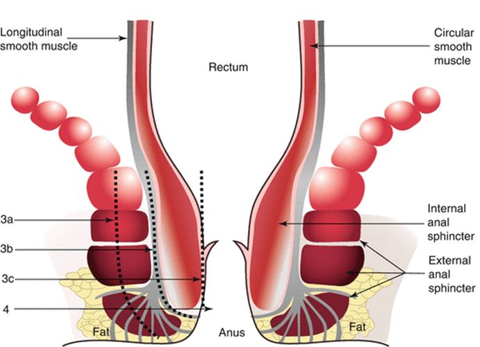
Fig. 8.2
The anal sphincter complex (With kind permission from Springer Science + Business Media: Sultan and Kettle [13], p. 13–19)
The term “pelvic floor” is used to refer to a muscular layer that spans the pelvic outlet and is comprised mainly of the paired levator ani muscles, which are found deep to the muscles of the perineum and anal sphincter complex. These muscles arise from the arcus tendineous fascia pelvis on each side and are subdivided according to their bony attachments into three main portions- iliococcygeus, pubococcygeus and ischiococcygeus. Medial fibres from the pubococcygeus are arranged to form a hammock-like configuration around the rectum, and are designated the puborectalis.
Perineal trauma may occur spontaneously with vaginal delivery, or it may be iatrogenic, i.e., an episiotomy. An episiotomy may also extend to involve other structures. Although perineal trauma is conventionally thought of as incorporating trauma to the posterior vaginal wall and the muscles of the perineum and anal sphincter complex, tears may also be seen which involve the anterior vaginal wall, urethra, clitoris or labia. Tears can sometimes be highly complex, involving multiple compartments of the vagina in a “spiral” fashion, or with complete detachment of the vaginal epithelium from underlying structures.
The following classification has been recommended for use in the assessment of perineal trauma [2]:
· First degree: Injury to the vaginal epithelium or perineal skin only
· Second degree: Involvement of the superficial perineal muscles (bulbospongiosus, transverse perineal) and sometimes the pubococcygeus muscle, but with no involvement of the anal sphincter.
· Third degree: Involvement of the anal sphincter complex (Fig. 8.3). Can be further subdivided into:

Fig. 8.3
External view of a third-degree tear
|
3A |
Less than 50 % thickness of external anal sphincter torn |
|
3B |
More than 50 % thickness of external anal sphincter torn |
|
3C |
External and internal anal sphincters torn |
· Fourth Degree: A third-degree tear with additional involvement of the anorectal mucosa
A buttonhole tear refers to an isolated tear of the rectal mucosa into the vagina, without involvement of the anal sphincters. These sit outside of the classification above as they do not involve the muscles of the perineum. They may be difficult to detect without a thorough assessment including a digital rectal examination. Detection is essential in order to avoid debilitating consequences such as rectovaginal fistula, although there is no association between buttonhole tears and continence outcomes.
Prior to assessment for genital tract trauma, it is recommended that the examining clinician ensures the following criteria are met [5]:
1. 1.
2. 2.
3. 3.
4. 4.
Once these criteria are met, a vaginal examination should be performed to evaluate the extent of the vaginal tear, plus visualization of the perineum.
· Although parting the labia is usually adequate to visualize the tear, complex or deep tears may necessitate the use of a Sims speculum to identify the apex of the tear(s).
· A Sims speculum and two sponge-holding (Rampley’s) forceps will be necessary for evaluation if a cervical tear is suspected; the sponge-holding forceps should be used to gently grasp the cervix in quadrants in order to systematically inspect for any disruption.
· Following a full vaginal examination, a digital rectal examination should be performed to evaluate the integrity of the anal sphincter complex, and to exclude buttonhole tears of the anal epithelium.
· Clear visualization is necessary to diagnose a third- or fourth-degree tear.
· The EAS is striated muscle that is red in appearance; the IAS is smooth muscle and is paler in appearance.
· The examiner’s index finger should be inserted into the rectum and the labia parted with the other hand to inspect for any tears to the EAS and IAS (remembering that anal sphincter damage can still be present with an intact perineum).
· The woman can be asked to contract her anal sphincter around the examiners finger; a defect will be felt anteriorly if there is anal sphincter damage.
· If there is any doubt about whether a tear involves the anal sphincter complex, an assessment should be undertaken by a more experienced professional.
· If adequate assessment cannot be undertaken to exclude anal sphincter trauma due to inadequate analgesia, it may sometimes be necessary to consider additional measures such as pudendal or regional blockade.
Once a full assessment has been undertaken by a suitably experienced individual as outlined above, the tear can be classified and arrangements for repair made. Repair should be undertaken by a trained healthcare professional in a suitable environment. The exact requirements will depend on the classification of the tear and the comfort and analgesia requirements of the patient.
Ultrasound as an Assessment Method in the Acute Setting
Although clinical assessment remains the most commonly used modality to detect and correcty classify perineal trauma in the immediate postpartum period, other methods, such as ultrasound, have been explored. Figure 8.4 shows the normal ultrasonographic appearance of the anal sphincter complex.

Fig. 8.4
Endoanal ultrasound images showing normal appearance of the internal (IAS) and external (EAS) anal sphincters
An evaluation of endoanal ultrasound performed postpartum in 150 primiparous women revealed clinically undiagnosed anal sphincter defects to be present in 28 % [6]. The sensitivity of anal endosonography was 68 % and the positive predictive value 37 %. A subsequent randomized trial evaluating endoanal ultrasonography against standard clinical assessment showed postpartum ultrasound examination to be associated with a significant improvement in diagnosis of anal sphincter tears, together with a lower incidence of faecal incontinence at 3 months [7]. A further study of 154 primiparous women who underwent a transperineal ultrasound scan 6–24 h after their first delivery found ultrasound evidence of anal sphincter damage to be associated with a higher risk of anal incontinence symptoms up to 6 months after delivery [8].
The limited available evidence does seem to suggest that ultrasound evaluation of the anal sphincter complex improves detection of anal sphincter injuries in the immediate postnatal period, however widespread use of this tool is influenced by resource and training limitations. Therefore drives to improve care of women with perineal trauma tend to focus on optimizing clinical assessment.
Improving Clinical Assessment of Perineal Trauma
Over recent years, the focus has been on improving detection of severe perineal trauma (i.e., injury to the anal sphincter complex) by training in standardized assessment methods. The rationale behind this is that improved detection will ensure adequate repair. An evaluation of clinicians who attended a structured training course in the detection and management of obstetric anal sphincter injury reported improved identification and classification of tears after the course, together with a shift towards more evidence-based practice [9]. An analysis of third-degree tears repaired by doctors assessing and repairing tears after a structured training programme showed the incidence of persistent sphincter defects to be lower than that reported in the literature, with no significant deterioration in anal incontinence symptoms [10].
A randomized controlled trial of an enhanced cascaded multiprofessional training programme focusing on evidence based perineal management was conducted amongst 3681 women who sustained a second-degree tear. The primary outcome measure was perineal pain on sitting and walking at 10–12 days post partum. Although there was no difference between the intervention and no intervention for the primary outcome measure, implementation of the training programme was associated with significant improvements in evidence based practice; this again supports the use of structured training for perineal assessment [11].
References
1.
Smith L, Price N, Simonite V, Burns E. Incidence of and risk factors for perineal trauma: a prospective observational study. BMC Pregnancy Childbirth. 2013;13:59.CrossRefPubMedPubMedCentral
2.
Royal College of Obstetricians and Gynaecologists. The Management of Third- and Fourth-Degree Perineal Tears, Green-top Guideline No. 29. RCOG, London, June 2015.
3.
Dudding T, Vaizey C, Kamm M. Obstetric anal sphincter injury: incidence, risk factors and management. Ann Surg. 2008;247:224–37.CrossRefPubMed
4.
Williams A, Tincello D, White S, Adams E, Alfirevic Z, Richmond D. Risk scoring system for prediction of obstetric anal sphincter injury. BJOG. 2005;112(8):1066–9.CrossRefPubMed
5.
National Institute for Health and Care Excellence. Intrapartum care: care of healthy women and their babies during childbirth. NICE Clinical Guideline. 2014. p. 190.
6.
Faltin D, Boulvain M, Irion O, Bretones S, Stan C, Weil A. Diagnosis of anal sphincter tears by postpartum endosonography to predict faecal incontinence. Obstet Gynecol. 2000;95(5):643–7.PubMed
7.
Faltin D, Boulvain M, Floris L, Irion O. Diagnosis of anal sphincter tears to prevent fecal incontinence: a randomised controlled trial. Obstet Gynecol. 2005;106(1):6–13.CrossRefPubMed
8.
Maslovitz S, Jaffa A, Levin I, Almog B, Lessing J, Wolman I. The clinical significance of postpartum transperineal ultrasound of the anal sphincter. Eur J Obstet Gynecol Reprod Biol. 2007;134(1):115–9.CrossRefPubMed
9.
Andrews V, Thakar R, Sultan A. Structured hands-on training in repair of obstetric anal sphincter injuries (OASIS): an audit of clinical practice. Int Urogynecol J Pelvic Floor Dysfunc. 2009;20(2):193–9.CrossRef
10.
Andrews V, Thakar R, Sultan A. Outcome of obstetric anal sphincter injuries (OASIS)- role of structured management. Int Urogynecol J Pelvic Floor Dysfunc. 2009;20(8):973–8.CrossRef
11.
Ismail K, Kettle C, MacDonald S, Tohill S, Thomas B, Bick D. Perineal assessment and repair longitudinal study (PEARLS): a matched pair cluster randomised trial. BMC Med. 2013;11:209.CrossRefPubMedPubMedCentral
12.
Thakar R, Fenner DE. Anatomy of the Perineum and the Anal Sphincter. In: Perineal and anal sphincter trauma. Sultan AH, Thakar R and Fenner DE (Eds) Springer-Verlag London Limited 2007. p.1-12
13.
Sultan AH, Kettle C. Diagnosis of Perineal Trauma. In: Perineal and anal sphincter trauma. Sultan AH, Thakar R and Fenner DE (Eds) Springer-Verlag London Limited 2007. p.13–9.