
Ashish Khandelwal1 and Alampady Shanbhogue2
(1)
Department of Radiology, Brigham and Women’s Hospital, Harvard Medical School, 75 Francis Street, Boston, MA 02115, USA
(2)
Department of Radiology, NYU Langone Medical Center, 660 First Avenue, 3rd Floor, New York, NY 10018, USA
Ashish Khandelwal
Email: drashish83@gmail.com
Keywords
Obstetric emergencyPlacentaUterine ruptureDehiscencePostpartum complicationsMR imagingEctopic pregnancy
19.1 Introduction
According to World Health Organization data of year 2013, every day about 800 women died due to complications of pregnancy and childbirth. The mortality and morbidity can be related to direct obstetric causes or medical condition unrelated to pregnancy, labor, and puerperium. The risk factors associated with increased maternal complications during pregnancy include advanced maternal age, African-American race, history of prior complication during pregnancy, and lack of prenatal care [1]. Third trimester poses the greatest risk for maternal obstetric complications with majority of complications occurring within 24 h of delivery [2]. The complications can affect different organ systems often requiring emergent treatment. Radiologists play vital role in making accurate diagnosis in a particular clinical scenario using varying imaging modalities. Imaging protocol with maximum diagnostic benefit and minimal justifiable risk is required. Ultrasonography (USG) should be the initial modality for evaluation of the pregnant patient, with other modalities used in cases in which USG is limited. USG is inexpensive, fast, and free of radiation. However, USG is operator dependent and may be limited by patient body habitus or bowel gas. Magnetic resonance imaging (MRI) is the next best investigation for evaluation of pregnancy-related obstetric complications with indeterminate ultrasonography findings. MR has excellent soft tissue contrast, provides large field of view assessment, and has multiplanar capabilities. In this chapter, a comprehensive update is provided about the role of MRI imaging in wide spectrum of pregnancy-related obstetric complications.
19.2 MRI
19.2.1 Patient Preparation and Positioning
MR imaging during pregnancy is challenging due to image degradation with fetal motion and maternal breathing difficulties. Protocols should be modified to answer the specific clinical question in the shortest amount of time and with the least amount of maternal discomfort. MR imaging is avoided in first trimester unless potential benefits outweigh the theoretical risks. Patient is usually placed in the supine position. However, a left lateral decubitus position might be preferred in the third trimester for improved patient comfort and reduced risk of impaired venous return. Multichannel phased-array surface coils are usually used to ensure maximum signal. However, with advanced pregnancy, a body coil may be necessary. The scans are usually performed with distended bladder. This strategy allows accurate detection of conditions such as placenta percreta [3].
19.2.2 MR Safety
The radiofrequency radiation used in MR imaging is nonionizing. However, radiofrequency pulses result in energy deposition and can potentially result in tissue heating. The amount of energy deposited in a patient as a result of an MR imaging examination is referred to as the specific absorption rate (SAR), with units of watts per kilogram. The SAR increases with the static magnetic field strength, flip angle, and number and spacing of radiofrequency pulses. Typically, single-shot fast spin-echo sequences cause increased specific absorption rate, in comparison with gradient-recalled echo sequences, which do not depend on radiofrequency refocusing, and are associated with a relatively low SAR [4].
MR imaging, with its varying magnetic fields and pulsed radiofrequency gradients, may theoretically increase the risk of hazardous biological effects and miscarriage [5, 6]. Present data have not conclusively documented any deleterious effects of MR imaging exposure on the developing fetus [7–9]. The American College of Radiology (ACR) approves imaging of pregnant patients in any trimester, although discourages unnecessary MR use [10]. In MR exposures up to 1 h, the total body exposure should be limited to a total energy deposition of 120 Wmin/kg in order not to overload the thermoregulatory system in the normal individual [10]. For exposures to pregnant women, a reduction of these values by a factor of 2 is recommended.
19.2.3 MR Protocol
After acquisition of localizing images, usually nonenhanced T1-weighted, fat-suppressed T1-weighted, fat-suppressed T2-weighted, and heavily T2-weighted images using turbo spin-echo sequences or gradient- and spin-echo sequences are obtained. Fat-suppressed T1- weighted images are useful in the differentiation of blood from fat, including fat within a neoplasm, such as a teratoma. Fat-suppression techniques can also increase conspicuity of inflammatory lesions. In addition, ascites and complex fluid can be easily identified on T2- weighted images. T2*-weighted images are useful for the identification of hemorrhage and air bubbles. Furthermore, contrast-enhanced imaging is useful in the evaluation of lesion vascularity and location in relation to pelvic vessels, as well as for the presence and site of active bleeding. However, if an intrauterine gestation is a diagnostic possibility, gadolinium-based contrast agents should be avoided.
19.2.4 IV Contrast
Gadolinium is classified as a category C drug by the United States Food and Drug Administration [11]. The 2010 American College of Radiology guideline for safe MRI practices recommends that intravenous gadolinium be avoided during pregnancy and used only if it is judged absolutely essential. Gadolinium agents administered intravenously cross the placenta and may enter the fetal circulation to be excreted into amniotic fluid from the fetal bladder. Despite animal data and concerns about the use of gadolinium in pregnancy, there have been no reported adverse human fetal effects [12]. Obtaining informed consent from the pregnant patient based on current evidence is recommended.
Use of MR contrast agent during breast-feeding is also controversial. Less than 0.04 % of the dose administered to a mother will appear in her milk [13]. Also, of that tiny amount excreted into the milk, only 0.8 % is actually absorbed by the baby [14]. According to American College of Radiology, Committee on Drugs and Contrast Media 2010, it is safe for the mother and infant to continue breast-feeding after receiving MRI contrast agent although informed decision to temporarily stop breast-feeding should be left up to the mother after these facts are communicated.
19.3 Obstetric Diseases
Wide spectrum of diseases can affect maternal reproductive tract during the course of pregnancy and postpartum period. MR can provide important information not only by diagnosing these complex diseases but also help guide management by providing information required to stratify the patient into surgical and nonsurgical management categories. These diseases are briefly discussed below.
19.3.1 Placental Complications
The normal disk-shaped placenta attaches to the anterior or posterior uterus. The umbilical cord typically inserts centrally, but eccentric and velamentous insertions can also occur. The overall appearance of the placenta changes during the course of pregnancy with the midportion of the placenta typically measuring between 2 and 4 cm. Pregnancy may be complicated by abnormalities of placental formation, position, or implantation.
19.3.1.1 Abruptio Placentae
Abruptio placentae refers to premature separation of a normally implanted placenta and occurs in an estimated 0.4–1 % of childbirths [15, 16]. Risk factors for abruption include prior abruption, smoking, trauma, cocaine use, multifetal gestation, hypertension, preeclampsia, thrombophilias, advanced maternal age, preterm premature rupture of the membranes, intrauterine infections, and hydramnios [15, 17]. The incidence is highest at 24–26 weeks of gestation and decreases with advancing gestation [18]. Placental abruption occurring in trauma affects 5–6 % of patients sustaining minor abdominal trauma and up to 20–50 % of patients sustaining major abdominal trauma [19, 20].
Placental abruption can be of three subtypes based on the location of the blood: retroplacental (when the bleeding was behind the placenta) (Fig. 19.1), marginal or subchorionic (when only the margin of the placenta was separated), and preplacental (bleeding was anterior to the placenta and limited by the umbilical cord). Clinically, most placental abruptions present with vaginal bleeding, pain, and uterine tenderness. Placental abruption can result in hypovolemic shock, DIC, renal failure, and even maternal death. Worse fetal outcome is associated with preterm birth, low birth weight, and fetal distress as a result of disruption.

Fig. 19.1
Placental abruption. Axial T1-weighted (a) and T2-weighted (b) MR images show linear low T2-weighted signal intensity signal in the retroplacental region, with corresponding mild increased T1 signal (arrows) consistent with retroplacental hemorrhage. Also seen is the high T1-weighted signal intensity within the uterine cavity consistent with intra-amniotic hemorrhage (*)
USG is often the first modality to begin assessment of vaginal bleeding, and it can show preplacental fluid collection between the placenta and amniotic fluid; jellylike movement of the chorionic plate induced by fetal activity; presence of marginal, subchorionic, or intra-amniotic hematomas; and increased heterogeneous placental thickness (>5 cm in a perpendicular plane) in patients with placental abruption [21]. However, USG has been shown to have low sensitivity (24–53 %) and high specificity (85–96 %) [22, 23]. In patients with a high index of suspicion for abruption and a negative USG evaluation, MR imaging is the imaging modality of choice for further evaluation if necessary. MR imaging, especially with a combination of T1-weighted and diffusion-weighted imaging, is shown to be 100 % sensitive for establishing the diagnosis [22]. Hematomas are seen as well-marginated intrauterine blood collection (Fig. 19.2); the signal intensity of which varies according to chronicity over time. By considering the signal intensity changes on T1-, T2-, and diffusion-weighted images, with special reference to the paramagnetic effects of methemoglobin, it is possible to estimate the age of the bleeding and intrauterine hematomas which can be accurately classified as follows: hyperacute (first few hours, intracellular oxyhemoglobin), acute (1–3 days, intracellular deoxyhemoglobin), early subacute (3–7 days, intracellular methemoglobin), late subacute (>14 days, extracellular methemoglobin), and chronic (>4 weeks, intracellular hemosiderin and ferritin). Further hyperacute/acute hematomas and higher hematoma volume are shown to be associated with clinical deterioration requiring urgent treatment [22].

Fig. 19.2
Subchorionic hematoma. Axial T1-weighted (a) and T2-weighted (b) MR images show focal T1 hyperintense, T2 mildly hypointense hematoma in the retroplacental region (arrows) in a pregnant patient presenting with blunt abdominal trauma consistent with subchorionic hematoma
Management of placental abruption depends on the clinical presentation, the gestational age, and the presence or absence of maternal and fetal compromise.
19.3.1.2 Placental Implantation Abnormalities
Placenta previa denotes to placental implantation in the lower uterine segment and can be of complete/central or partial subtypes [24]. The incidence of placenta previa is approximately 0.3–1 % of deliveries [25]. Placenta previa is more frequently seen with multiparity, multiple gestations, advanced maternal age (>40 years), previous cesarean delivery (1.5- to 5-fold risk), previous abortions, and previous placenta previa [26]. Placenta previa is risk factor for placental abruption, hemorrhage, fetal malpresentation, fetal malformations, fetal growth restriction, preterm births, cervical outlet obstruction, and need for cesarean delivery or hysterectomy. Abnormal cord insertion, circumvallate placenta, and placenta accreta (occurs in 3–5 % of patients with placenta previa) also frequently coexist with placenta previa. Clinically, painless bright red vaginal bleeding in the second or third trimester is the hallmark clinical presentation of placenta previa (seen in 70 % cases). Overall, placenta previa can increase perinatal mortality by up to 3–4 times compared with that of normal pregnancy.
On imaging, the diagnosis is established by showing placenta partially or completely covering the internal os (Fig. 19.3). USG is usually sufficient in showing placental position well. MR imaging can serve as a problem-solving tool, because it accurately depicts the abnormality, including a posterior placenta previa, and depicts coexistent abnormalities, including placenta accreta, percreta, or placental abruption which can be challenging on USG (Figs. 19.4and 19.5). It is important to consider the gestational age at the time of diagnosis because most (>90 %) placenta previas diagnosed at 20 weeks of gestation resolve by term.

Fig. 19.3
Complete placenta previa. Sagittal single-shot fast spin-echo image of a 30-year-old woman shows the placenta completely covering the internal cervical os (arrow)
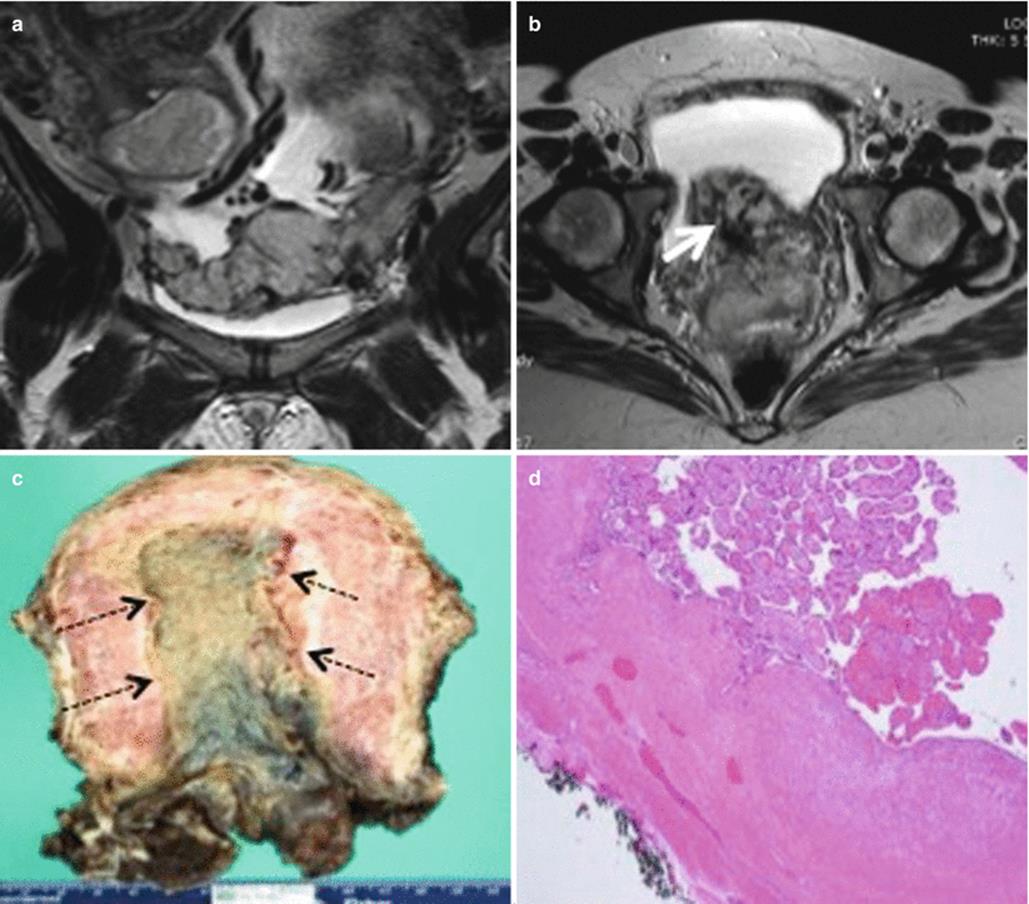
Fig. 19.4
Placenta previa with placenta increta. Coronal (a) and axial (b) T2-weighted MR images show loss of normal pear shape of the uterus with randomly distributed dark intraplacental bands anteroinferiorly in low-lying placenta (thick white arrow), raising the suspicion for placenta increta. (c) Gross pathology specimen shows adherent placental tissue (black dotted arrows) to the myometrium. (d) Histopathology slide of the same confirms third trimester villi at the endometrial surface invading the myometrium without intervening decidua
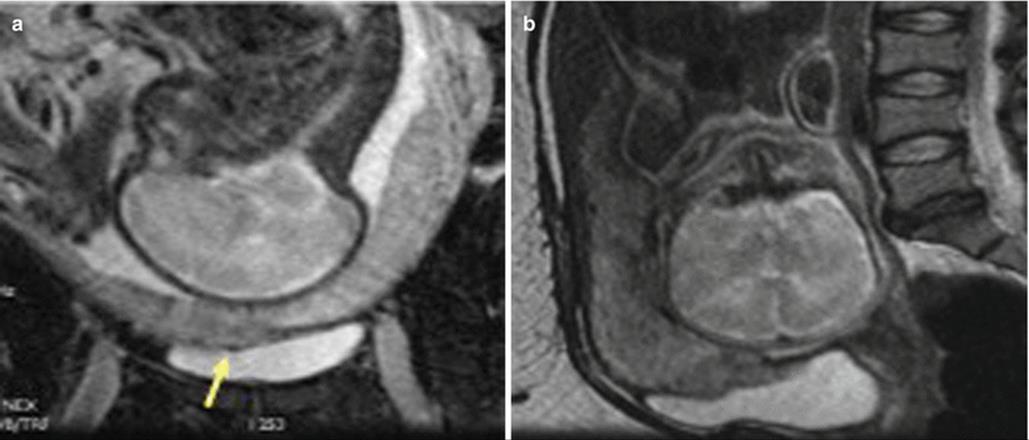
Fig. 19.5
Placenta previa with placenta percreta. Coronal (a) and sagittal (b) T2-weighted MR images show invasion of the placenta beyond the contour of the myometrium into the urinary bladder (arrows) consistent with placenta percreta. There is loss of normal T2 hypointense signal of the dome of the urinary bladder at the site of placental invasion (arrowhead)
Management of placenta previa involves careful maternal and fetal monitoring until term or fetal maturity and an elective cesarean delivery. Vaginal delivery is advocated in placenta previa only if the os-placental distance is more than 2 cm at 35 weeks of gestation.
19.3.1.3 Placental Adhesive Disorders
Placental adhesive disorder results from abnormality in decidua basalis with resultant direct invasion of chorionic villi to myometrium. Depending on extent of placental adherence and myometrial penetration, it can be further categorized as placenta accreta (only adhesion, no myometrial invasion), increta (penetrates myometrium without breaching serosa), and percreta (penetrates serosa and adjacent structures) (Fig. 19.6). Abnormal placentation occurs from 1 in 2500 to 1 in 500 pregnancies [27, 28]. Prior history of cesarean delivery, uterine surgery, and placenta previa are risk factors for abnormal placentation [28, 29].
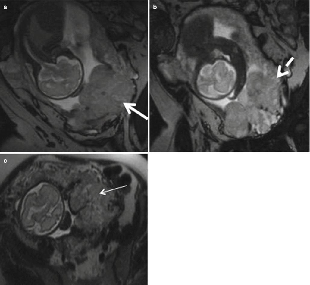
Fig. 19.6
Placenta previa with percreta. (a, b) Coronal T2-weighted MR images showing complete placenta previa (thick white arrow), thickened placenta (white dotted arrow), and bulging of lower uterine segment. (c) Axial T2-weighted MR image shows marked heterogeneity (thin white arrow) of the placenta
Imaging can play vital role in early recognition of these placental disorders that can result in postpartum hemorrhage and/or retained products of conception. Both USG and MRI are complimentary to each other in diagnosing placental implantation abnormalities with reported sensitivity and specificity of MRI in literature ranging from 72–90 % and 81–94 % [30–32]. The normal myometrium has a trilayered appearance on T2-weighted images. The middle layer is a heterogeneously hyperintense vascular layer, with thinner low-signal-intensity muscle layers on either side. Variety of imaging findings can suggest abnormal placentation, which includes myometrial thinning, interruption of the normal layered appearance of the myometrium, lobulated myometrial-placental margin, dark intraplacental bands, abnormal placental vascularity, outward bulging of the placenta with deformity or disruption of the smooth outer uterine contour (Fig. 19.7), and widening of lower uterine segment with hourglass uterine configuration [22, 24, 33, 34]. Treatment is cesarean section and possible cesarean hysterectomy.

Fig. 19.7
Placenta percreta. Sagittal T2-weighted MR image showing small focus of extra-myometrial-placental extension at the superior and anterior aspect of myometrial wall (arrow)
19.3.1.4 Placental Neoplasms
Placental origin tumors mostly comprise of those arising from trophoblastic tissue. Discussion of other rare placental neoplasm is beyond the scope of this chapter.
Gestational trophoblastic disease (GTD) embraces gamut of disease arising from uninhibited growth of placental trophoblastic tissue, with a spectrum of severity extending from premalignant hydatidiform mole to malignant invasive mole, choriocarcinoma, placental-site trophoblastic tumor, and epithelioid trophoblastic tumor. Hydatidiform mole, usually referred to as molar pregnancy, accounts for 80 % of all GTDs [35]. Hydatidiform mole is estimated to occur in 0.6–1.1 per 1000 pregnancies in North America [36]. Choriocarcinoma is rare with an estimated incidence of 1 in 20,000–40,000 pregnancies [36]. Approximately 50 % of choriocarcinomas arise from molar pregnancies, 25 % from term or preterm pregnancies, and the remainder from pregnancy termination [37]. Risk factors include Asian race, history of oral contraceptive use, prior molar pregnancy, and maternal age (both <20 and >40 years) [38, 39].
Hydatidiform Mole
Hydatidiform moles are classified into two subtypes: complete and partial with distinctive histologic, clinical, imaging, and genetic features [35]. Complete moles result from fertilization of an empty ovum with subsequent duplication of the paternal chromosomes. Gross pathological appearance of the trophoblastic tissue has been classically described as “cluster of grapes” which on USG gives “snowstorm appearance” [40]. No identifiable fetal tissue is formed. Hyperstimulation of the ovaries due to excessive production of β-hCG by abnormal trophoblastic tissue results in formation of theca lutein cysts, which can be present in 20–46 % cases [41].
Partial hydatidiform moles are less common and form due to fertilization of a normal ovum by two sperms with resultant chromosomal composition of 69,XXX or 69,XXY [42]. Fetal tissue may be present which can help in differentiating it from complete mole.
USG is often the first imaging modality to assess a patient presenting with amenorrhea and routinely used in first trimester. Most molar pregnancies now present with findings of early pregnancy failure rather than the classic “cluster of grapes” appearance [43]. Ultrasonography is not very sensitive or specific for detection of GTD and identifies less than 50 % of all hydatidiform moles [43, 44]. Overall, the accuracy of ultrasonography in the detection of complete mole is better than that for partial mole and increases after the 16th week of gestation [42]. MR imaging is archetypally not used in routine assessment of hydatidiform moles but can be used to define if there is extension of molar tissue to the myometrium or outside the uterus. MR imaging findings are nonspecific and can imitate the features of RPOC. Moles appear as heterogeneous tissue expanding the endometrial cavity, with mostly low signal intensity on T1-weighted images, high signal intensity on T2-weighted images, and avid enhancement on postcontrast images (Fig. 19.8). Focal areas of hemorrhage and cystic spaces may also be seen [45]. The intact normal myometrium, appearing as hypointense layer surrounding the molar tissue, helps in differentiation from invasive disease [45].

Fig. 19.8
Invasive mole. Coronal T2-weighted (a) and axial T1-weighted gadolinium-enhanced (b) MR images demonstrate a heterogeneous lesion seen in the uterine fundus invading the myometrium, with multiple T2 hyperintense cystic foci, heterogeneous enhancing septae, and multiple nonenhancing cystic spaces Image courtesy- Dr Christine O. Menias (thin arrow). Fetal parts are identified on coronal image
Invasive Mole and Choriocarcinoma
Invasive moles indicate growth of the trophoblastic tissue into and beyond the myometrium. Invasive moles are considered locally invasive neoplasms. Choriocarcinomas can invade locally but are also capable of metastasizing, frequently manifesting with hematogenous metastases. The lung is the most common site (76–87 %) followed by the liver (10 %), brain (10 %), kidney, gastrointestinal tract, and spleen [42, 46, 47]. The vagina is the second most common site of metastasis (30 %), but this occurs through contiguous spread [22, 48].
Invasive moles and choriocarcinomas are mostly similar appearing at imaging. Patients with low b-hCG levels (<500 mIU/mL) often have normal MR imaging findings. Choriocarcinomas tend to show more areas of necrosis, hemorrhage, and solid-enhancing components within the tumor [4]. Choriocarcinoma staging is often performed with CT as it allows detection of distant metastases. MR imaging can have a role in demonstrating myometrial and parametrial invasion and thus aids in the anatomic staging of disease. Choriocarcinoma is often seen as an intrauterine mass with heterogeneous high signal intensity on T2-weighted images and marked enhancement on postcontrast images, findings that reflect the high vascularity of the tumor. Tumor vascularity can be demonstrated by focal signal voids on T1- and T2-weighted images. Myometrial invasion is visible as high-signal-intensity foci within the myometrium causing junctional zone disruption and demonstrate enhancement on postcontrast images. Enhancing parametrial soft tissue is characteristic of local spread. MR imaging can also help detect metastatic disease, particularly within the pelvic organs and lymph nodes. GTDs are treated with primary removal of tumor surgically with or without chemoradiation depending upon stage of disease.
19.3.2 Uterine Complications
19.3.2.1 Rupture/Scar Dehiscence
Uterine rupture accounts for up to 20 % of maternal deaths with estimated overall uterine rupture rate of 1 in 1146 pregnancies (0.07 %). Most uterine ruptures (90 %) are related with a previous cesarean delivery (Fig. 19.9); incidence of uterine rupture with low transverse incision is approximately 0.2–1.5 % and with the classic incision is 4–9 % [49, 50]. Other risk factors for uterine rupture include previous uterine surgeries (Fig. 19.10), multiple gestation, grand multiparity, polyhydramnios, manual removal of the placenta, and other invasive procedures such as dilatation and curettage [3].

Fig. 19.9
Uterine rupture at the incision site, post-lower segment cesarean section. Sagittal T2-weighted MR image, in a patient with history of previous cesarean delivery who presented with acute pelvic pain and vaginal bleeding, shows a large defect in the lower anterior uterine segment consistent with uterine rupture (arrow)

Fig. 19.10
Uterine rupture at the fundus region post-dilatation and curettage. Sagittal T1 pre- (a) and post- (b) contrast images as well as sagittal T2-weighted MR (c) images show discontinuity in the uterine fundal region (arrow) suggestive of uterine rupture. This MRI was performed after dilatation and curettage of uterine cavity for retained products of conception
The diagnosis is made on clinical symptoms of abdominal pain and vaginal bleeding with shock. Fetal monitoring shows fetal heart rate variations, and tocometer often shows lack of uterine contractions or hyperstimulation without progression of labor raising suspicion of uterine rupture.
Transvaginal USG is often the first imaging study performed which may show intraperitoneal or extraperitoneal hematoma in relation to the uterus, fetal parts in an extrauterine location, intra-amniotic hemorrhage, and focal bulging of membranes through the site of dehiscence [51]. MR imaging may act as a problem-solving tool, showing the presence and extent of rupture in indolent cases with atypical presentation. The difference between the scar dehiscence and uterine rupture is that in scar dehiscence there is separation of an old myometrial scar without penetration of the uterine serosa (Fig. 19.11). The normal appearance of the post-cesarean section incision site on MRI demonstrates signal intensity consistent with subacute hematoma within the myometrium at the presumed incision site; however, overlying endometrial and serosal layers are continuous and no myometrial defects are seen. The primary MR features of uterine dehiscence consist of absence of coaptation of the endometrial or serosal layer with either fluid or blood between the layers of the incision site or low-signal-intensity foci through the incision site suggestive of emphysematous infection.
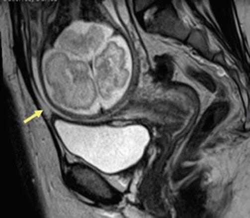
Fig. 19.11
Scar dehiscence. Sagittal T2-weighted MR image in patient with prior history of cesarean section shows thinning of the myometrium in the lower uterine segment without any disruption of serosa (arrow) suggestive of scar dehiscence
Uterine rupture often requires emergent surgery with operative delivery and uterine repair.
Bladder flap hematoma refers to postpartum hematomas that occur beneath the surgically incised and reapproximated peritoneal fold adjacent to the lower uterine segment cesarean delivery incision. Although hematomas in the bladder flap area may be seen normally in the post-cesarean section patient, they are unlikely to exceed 5 cm in size [52]. On MRI and CT, bladder flap hematomas manifest as heterogeneous lesions of varying intensity/density and size extending between the urinary bladder and the lower segment of the uterus (vesicouterine space) [53]. The sagittal images are most helpful when localizing the site of dehiscence and evaluating involvement of adjacent structures such as the bladder. To best demonstrate the myometrial gap, an imaging plane perpendicular to the incision is recommended [52]. If gas is seen within the collection, superimposed infection is suggested. This often requires surgical evacuation if causing symptoms due to mass effect or if it gets infected.
19.3.2.2 Acute Fibroid Degeneration
Leiomyoma are benign smooth muscle neoplasm occurring with prevalence of 11 % during pregnancy [54]. They may be completely asymptomatic even when increased in size due to hormonal changes during pregnancy. Uterine fibroids can be symptomatic in pregnancy because of rapid growth, torsion, or acute red degeneration. Other complications of leiomyomas during pregnancy include pain, bleeding, spontaneous abortion, placental abruption, fetal malposition, and mechanical obstruction of the cervix.
Red degeneration is a hemorrhagic infarction caused by either venous thrombosis or rupture of intratumoral arteries and commonly occurs in the second half of pregnancy or in the puerperium. On MRI, they are seen as sharply marginated, low-signal-intensity masses in submucosal, intramural, or subserosal location. Leiomyomas larger than 3 cm often are heterogeneous due to varying degrees of degeneration. Volume increase >200 cm3 in fibroid is associated with increased rate of complications [55]. Red degeneration shows diffuse or peripheral high signal intensity at T1-weighted images with variable signal intensity at T2-weighted images depending on the degree of intralesional hemorrhage (Fig. 19.12) [10]. These findings correspond with numerous dilated vessels filled with red blood cells at the periphery of the lesion. Management of red degeneration is conservative and involves bed rest, sedation, and analgesia with resolution of symptoms in 7–10 days.

Fig. 19.12
Red degeneration of fibroid. Axial Fat suppressed T1 (a) and T2 (b) images show rounded subserosal fibroid with intrinsic T1 prolongation and T2 shortening in keeping with internal hemorrhage in patient presenting with acute left hemipelvic pain at 20 weeks
19.3.2.3 Retained Product of Conception (RPOC)
RPOC most frequently occurs after second trimester delivery or termination and present with pain, fever, and vaginal bleeding. Ultrasound with color Doppler is the modality of choice, which shows thickened endometrial echo complex (>10 mm) with increased color flow on color Doppler imaging and is suggestive of RPOC. However, there is a high false-positive rate on USG ranging from 17–51 % with blood clot being the most common mimicker of RPOC [56]. Other hypervascular lesions such as uterine arteriovenous malformation (AVM), endometrial polyp, submucosal fibroid, and invasive mole can be difficult to distinguish from RPOC [57]. MRI can be useful tool where RPOC can be seen as variable signal characteristic mass on T2-weighted and precontrast T1-weighted images depending on the degree of hemorrhage and tissue necrosis. It can be seen as variably enhancing endometrial mass with associated uterine disruption making it indistinguishable from gestation trophoblastic disease (Fig. 19.13). However, knowledge about β-hCG levels helps in clinically differentiating these two entities. MR imaging may also be helpful in showing anatomic variants that hinder successful instrumentation of the uterine cavity in patients with suspected retained products of conception [58]. Treatment options for RPOC include uterotonic medications, methotrexate, transarterial embolization, dilation and curettage, and hysteroscopic removal.
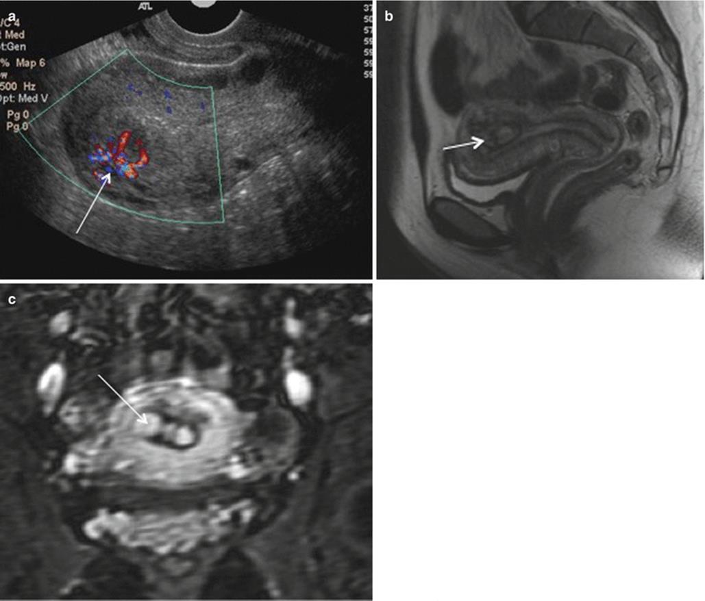
Fig. 19.13
RPOC. Color Doppler (a) ultrasonography images of the uterus show a heterogeneously echogenic endometrial mass with increased vascular flow (arrow). Sagittal T2-weighted MRI (b) and coronal gadolinium-enhanced MR images (c) show heterogeneously enhancing endometrial contents (arrow) consistent with RPOC
19.3.2.4 Endometritis/Wound Infection
Postpartum endometritis denotes to the infection of endometrium or decidua with or without extension into the myometrium and parametrial tissues. It is often polymicrobial (75 %) and arises from ascending infection from vaginal flora. Estimated incidence of endometritis after vaginal delivery is 1–3 %, and the incidence after cesarean delivery is about 20 % [59]. Cesarean delivery, invasive procedure during labor, premature rupture of membranes, prolonged labor, and retained placenta are risk factors for infection during postpartum period. Postpartum endometritis is classified as early (occurring within 48 h) or late (3 days–6 weeks after delivery). If untreated, endometritis may progress to myometritis, pelvic abscess, or septic thrombophlebitis. Endometritis is the clinical diagnosis and may present with fever, lower abdominal pain, foul-smelling vaginal discharge, vaginal bleeding, tachycardia, and leukocytosis.
MR imaging shows fluid and air within the endometrial cavity; thick, abnormally enhancing, and heterogeneous endometrium (Fig. 19.14); parametrial stranding; pelvic abscesses; and coexistent septic pelvic or ovarian thrombophlebitis. Associate superficial wound infection may be identified as fat stranding and abscess formation (Fig. 19.15). Pelvic abscess is seen as mass with thick, irregular, enhancing wall that contains fluid with low to intermediate signal intensity on T1-weighted images and hyperintense signal on T2-weighted images. Management of endometritis involves use of broad-spectrum antibiotics and evacuation of abscess, if any.
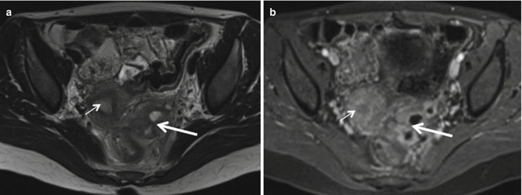
Fig. 19.14
Acute endometritis. Axial T2 (a) and postcontrast T1-weighted MR (b) images show fluid within endometrial canal with endometrial lining enhancement (thin arrow). Also note associated extensive inflammatory changes in the parametrial and adnexal region (thick arrow)

Fig. 19.15
Uterocutaneous fistula resulting from chronic endomyometritis. Transverse gray scale USG (a) and sagittal postcontrast MR (b) showing direct fistulous communication (arrow) between the skin and endometrial cavity due to chronic endomyometritis
19.3.2.5 Postpartum Hemorrhage (PPH)
Postpartum hemorrhage is defined as blood loss exceeding 500 mL after vaginal delivery and 1000 mL after cesarean delivery [60]. It can be due to uterine atony, RPOC (Fig. 19.16), or birth trauma (vascular and nonvascular injury) often occurring within 24 h and are classified as primary PPH. Delayed bleeding occurring between 24 h and 6 weeks is labeled as secondary PPH and can be related to subinvolution of the placenta, endometritis, or choriocarcinoma.

Fig. 19.16
Postpartum hemorrhage. Coronal T2- (a) and postcontrast T1-weighted MRI shows retained products of conception (thick arrow, b) with prolonged PPH treated with balloon tamponade (thin arrow)
Ultrasonography is the imaging investigation of choice for initial evaluation of PPH. MR/CT imaging can be performed in doubtful cases to evaluate for occult disease such as uterine injury or infection as well as to evaluate for vascular causes such as pseudoaneurysms and AVMs [61]. Patients present with pain, decreased hemoglobin, and in severe cases, hemodynamic instability. CT angiography is preferred due to its rapidity and ease of performance for delineating vascular causes. On MR images, the normal postpartum uterus often appears as an enlarged uterus and then gradually returns to a nongravid size within 6–11 weeks. The uterine cavity may have a small amount of blood or fluid. However, there is no evidence of intravenous contrast material extravasation in the uterine cavity on the contrast-enhanced MR.
MRI when performed shows uterine AVMs as serpiginous signal voids on spin-echo sequences with prominent parametrial vessels (Fig. 19.17) and focal disruption of the junctional zone, whereas uterine artery aneurysm is seen as focal contrast-filled outpouching on MR angiography. Uterine artery embolization can be performed in the hemodynamically stable patients with up to 95 % success rate. It is often the second choice in hemodynamically unstable patients when surgery can’t be performed as the first choice.

Fig. 19.17
Post-cesarean arteriovenous fistula. Coronal (a) and sagittal (b) postcontrast MR images show direct fistulous communication (arrow) between left internal iliac artery and vein, which was confirmed with catheter angiogram (c). Note multiple collaterals in the pelvis and dilated IVC. Patient was treated with endovascular stent
19.3.3 Ectopic/Heterotopic Pregnancy
Implantation of blastocyst at site other than endometrium of uterus is called ectopic pregnancy. Ectopic pregnancy has an incidence of approximately 1–2 % of all reported pregnancies and accounts for 9 % of all pregnancy-related deaths [62]. Major risk factors for ectopic pregnancy include a history of ectopic pregnancy, pelvic inflammatory disease, use of in vitro fertilization, intrauterine device, congenital uterine anomalies, endometriosis, and gynecologic surgery [3]. Clinical manifestations of ectopic pregnancy typically appear 6–8 weeks after the menstrual period. The classic triad of signs and symptoms of ectopic pregnancy consists of abdominal or pelvic pain, vaginal bleeding, and a tender adnexal mass [63]. Patients with a ruptured ectopic pregnancy often present with signs of shock, such as hypotension, tachycardia, rebound tenderness, and decreasing hematocrit [64].
USG is often the first modality sought after to confirm the intrauterine pregnancy in patients with positive pregnancy test. Nonvisualization of an intrauterine pregnancy in a patient with a positive serum β-HCG test is suggestive of a very early IUP, a failed pregnancy, or an ectopic pregnancy, which may initially be sonographically occult [65]. However, sonography can be limited due to body habitus, poor acoustic window and operator experience [63]. It is also limited in characterizing the adnexal masses that are not typical for ectopic pregnancy. In such cases, MRI is extremely helpful as problem-solving tool. MRI should be held in reserve for circumstances in which patients are hemodynamically and clinically stable, and additional information is required to direct patient care decisions and cannot be obtained using ultrasound.
Ectopic pregnancy can occur at different sites in the abdomen and pelvis. Ninety-five percent of ectopic pregnancies are tubal occurring most often in the ampulla of the fallopian tube (Fig. 19.18). Other sites affected include interstitium/cornua (2–4 %), ovary (3 %), cervix (<1 %), abdominal cavity (1.4 %), or cesarean scar region (0.15 %). MRI features may vary depending upon site affected. The MRI features of tubal ectopic pregnancy include hematosalpinx with or without tubal dilatation, a hemorrhagic or heterogeneous mass, ascites/hemoperitoneum, and wall enhancement [66]. The most specific finding of ectopic pregnancy is identification of the extrauterine gestational sac, which is typically seen as a cystic saclike structure surrounded by a thick wall, corresponding to the tubal ring sign seen on sonography. The wall of the saclike structure typically exhibits high signal intensity on T2-weighted images, with areas of hemorrhage showing distinct low T2 signal intensity and intermediate or high signal intensity on T1-weighted images. Enhancing solid components representing fetoplacental tissues within a hemorrhagic mass may also be seen. T1 hyperintense fluid in pelvis can be identified and represents hemorrhage. Ten percent of cases may show fluid within endometrial canal representing pseudogestational sac. Ill-defined complex adnexal mass with free fluid/hemorrhage in the pelvis should raise suspicion of a ruptured ectopic pregnancy (Fig. 19.19) [67]. Disruption of tubal wall enhancement with surrounding acute hematoma suggests rupture. Other differential for complex hemorrhagic mass on MRI includes corpus luteal cyst, abscess, or neoplastic mass [68]. The diagnosis of interstitial or cornual pregnancy may be confirmed on MRI by the presence of an uninterrupted junctional zone separating the gestational sac from the endometrium [69]. Ovarian ectopic pregnancy can be seen as a gestational saclike structure on or within the ovary, often containing acute hematoma with distinct low intensity on T2-weighted images [65]. The cervical pregnancy (Fig. 19.20) may have the appearance of a lobulated mass with heterogeneous mixed signal intensity and a partial or complete dark rim on T2-weighted images [70].
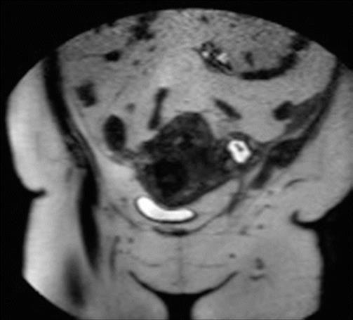
Fig. 19.18
Tubal ectopic pregnancy. Coronal T2-weighted MR image showing left tubal ectopic pregnancy. USG confirmed the presence of fetal heart activity (not shown)
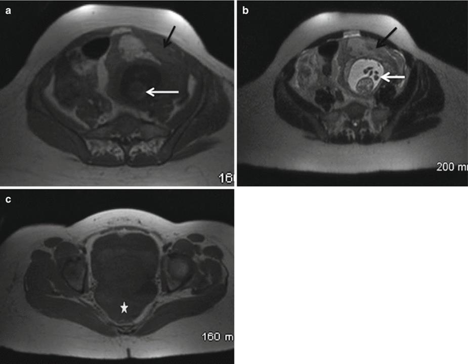
Fig. 19.19
Ruptured cornual ectopic pregnancy. Axial T1- (a, c) and T2- (b) weighted MR images show extrauterine ill-defined soft tissue in keeping with hematoma (black arrow) resulting from ruptured ectopic pregnancy. Also noted is cornual fetal pole (white arrow) and pelvic hemorrhage (c, *)
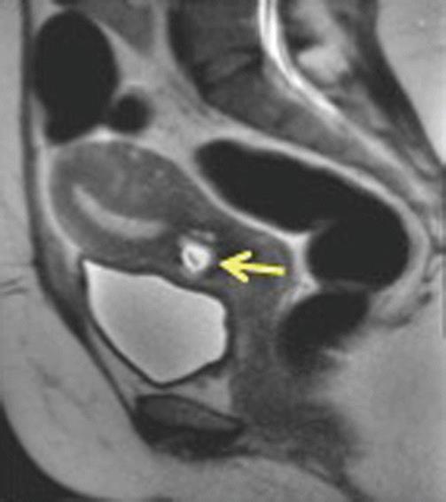
Fig. 19.20
Cervical ectopic pregnancy. Sagittal T2-weighted MR image shows empty endometrium and gestational sac in the cervix (arrow)
In case of abdominal pregnancy (Fig. 19.21), MRI can confirm the location of abdominal pregnancy at various intraperitoneal locations such as the uterine serosa, omentum, pelvic sidewall, broad ligament, pelvic cul-de-sac, large pelvic vessels, abdominal organs, and the diaphragm. MRI is useful in the accurate localization of the placenta, detection of arterial feeders, and assessment of placental adherence to surrounding organs [71]. A cesarean scar pregnancy (Fig. 19.22) occurs when there is implantation of a pregnancy within the scar of a previous cesarean section, resulting in a gestational sac that is surrounded by myometrium and fibrous tissue. The gestational sac may be visualized within the anterior wall of the lower uterine segment. The diagnosis can be made by visualizing enlargement of the cesarean scar and a mixed mass or clear gestational sac at the site of the scar [72]. A very thin layer of myometrium should be seen separating the maternal urinary bladder wall and the gestational sac. Implantation and trophoblastic invasion into cesarean scar tissue may result in uterine rupture and hemorrhage, which can be life threatening. Heterotopic pregnancy (Fig. 19.23) refers to the presence of simultaneous pregnancies at two different implantation sites, generally a combination of intrauterine and ectopic pregnancies [73]. Though extremely rare, this condition is seen more commonly among women who have undergone assisted reproductive therapy [74].
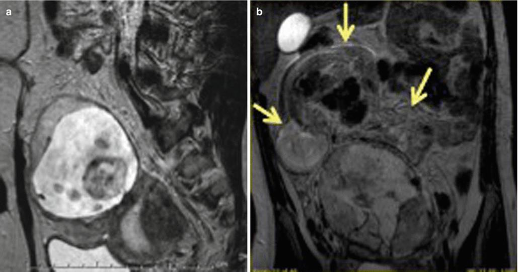
Fig. 19.21
Abdominal pregnancy. Sagittal T2 MR image (a) done at the time of initial presentation due to amenorrhea shows fetus and placenta lying outside the uterine cavity (U). Patient was lost to follow up and returned after 3 months with abdominal pain when the repeat MRI (b) showed free-floating fetus in the peritoneal cavity (arrows) suggestive of abdominal pregnancy
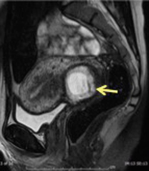
Fig. 19.22
Cesarean ectopic pregnancy. Sagittal T2-weighted MR image scans show gestational sac within anterior lower uterine segment, with adjacent myometrial thinning. Pregnancy is centered at level of cesarean section scar indicating that it represents cesarean scar pregnancy

Fig. 19.23
Heterotopic pregnancy. Axial T2- (a) and T1- (b) weighted MR images show intrauterine gestational sac (thin white arrow). Extrauterine complex cystic lesions representing multiple ectopic pregnancies (thick white arrows) are seen in right adnexal region. T1 hyperintense free fluid (black arrows) represents pelvic hemorrhage due to rupture
19.3.4 Ovarian Complications
19.3.4.1 Ovarian Torsion
Ovarian torsion occurs around 7 % of ovarian masses in pregnancy [75]. The prevalence of ovarian torsion during pregnancy is 1 in 1800 [76]. Ovarian torsion occurs most often in the first trimester. Mature cystic teratoma is the most common underlying cause of torsion (Fig. 19.24), followed by corpus luteum cysts. Clinically, patients often present with acute-onset abdominal or pelvic pain. If initial USG doesn’t point out diagnosis, then MRI may be performed. Classically on MRI, the ovary is enlarged, displaced in midline, and edematous, with small peripheral follicles. Ovarian stroma is seen as intermediate signal on T2-weighted MR images. Late torsion demonstrates increased signal intensity on T2-weighted images secondary to necrosis with periovarian fat stranding. Another specific imaging finding includes identification of a thickened, twisted fallopian tube [10]. On T1-weighted images, the signal intensity of the torted ovary varies according to the presence of internal blood products. Identification of intrauterine pregnancy is helpful in differentiating ovarian torsion from ectopic pregnancy. Management of ovarian torsion includes emergent detorsion with removal of tumor if present.

Fig. 19.24
Torsion of an ovarian teratoma in a pregnant patient presenting with acute right lower quadrant pain. Transverse color Doppler (a) and sagittal MR (b) images show predominantly solid fat-containing mass seen in the right midabdomen (arrows), and color Doppler shows lack of internal flow. Surgical pathology (c) confirmed the diagnosis of torsion of an ovarian teratoma
19.3.4.2 Ovarian Vein Thrombosis
One in 600 term pregnancies are complicated by ovarian vein thrombosis, mostly on right side [77]. Bland thrombus can get infected in patient with puerperal sepsis. Clinically, presence of fever, increased white blood cell count, and acute flank pain in the immediate postpartum period with absence of source of infection should prompt the radiologist to evaluate ovarian veins for the presence of thrombosis. A well-timed diagnosis is essential to institute anticoagulant treatment and prevent extension of the clot into the inferior vena cava and consequent pulmonary thromboembolism. MRI is the most reliable investigation with sensitivity as well as specificity of 100 % [78]. On contrast-enhanced CT or MR imaging, filling defects can be readily identified within the ovarian veins, with or without extension into the IVC. Presence of enhancement of the wall of the ovarian vein with intraluminal thrombus and perivenous stranding supports the diagnosis of thrombophlebitis (Fig. 19.25). MR can help in differentiating between acute and subacute thrombus with T1/T2 hyperintense signal in subacute clot [78]. Treatment essentially involves antibiotic and anticoagulant therapy.

Fig. 19.25
Ovarian vein thrombosis. Axial (a) and coronal (b) T1-weighted fat suppressed gradient-echo MR images show filling defect in the right ovarian (arrow in a, b) in keeping with ovarian vein thrombosis
19.3.5 Trauma
Trauma can affect 5–7 % of pregnancies with motor vehicle collisions being most common reason for traumatic injuries. While any organ can be injured, pregnancy-specific injuries may result in abruption, uterine rupture, premature rupture of membranes, and spontaneous abortions [20]. CT is the first imaging modality of choice in traumatic patients, as MR requires longer time of examination. Details about imaging appearance of trauma-specific injuries are discussed elsewhere in this book and beyond the scope of this chapter.
In conclusion, MRI is a valuable diagnostic tool which when used judiciously can provide accurate diagnosis in appropriate clinical setting and help in management of this peculiar obstetric population.
References
1.
Berg CJ, Chang J, Callaghan WM, Whitehead SJ (2003) Pregnancy-related mortality in the United States, 1991–1997. Obstet Gynecol 101(2):289–296, PubMed Epub 2003/02/11.engPubMed
2.
Rigo J Jr, Csakany G, Laky M, Nagy B, Horvath E, Joo JG (2014) Trends in maternal mortality in Hungary between 1978 and 2010. Eur J Obstet Gynecol Reprod Biol 173:29–33, PubMed Epub 2013/11/28.engPubMed
3.
Shanbhogue AK, Menias CO, Lalwani N, Lall C, Khandelwal A, Nagar A (2013) Obstetric (nonfetal) complications. Radiol Clin North Am 51(6):983–1004, PubMed Epub 2013/11/12.engPubMed
4.
Leyendecker JR, Gorengaut V, Brown JJ (2004) MR imaging of maternal diseases of the abdomen and pelvis during pregnancy and the immediate postpartum period. Radiographics Rev Publ Radiol Soc N Am Inc 24(5):1301–1316, PubMed Epub 2004/09/17.eng
5.
De Wilde JP, Rivers AW, Price DL (2005) A review of the current use of magnetic resonance imaging in pregnancy and safety implications for the fetus. Prog Biophys Mol Biol 87(2–3):335–353, PubMed Epub 2004/11/24.engPubMed
6.
Gowland PA, De Wilde J (2008) Temperature increase in the fetus due to radio frequency exposure during magnetic resonance scanning. Phys Med Biol 53(21):L15–L18, PubMed Epub 2008/10/10.engPubMed
7.
Kanal E, Barkovich AJ, Bell C, Borgstede JP, Bradley WG Jr, Froelich JW et al (2013) ACR guidance document on MR safe practices: 2013. J Magn Reson Imaging 37(3):501–530, PubMed PMID: 23345200. Epub 2013/01/25.engPubMed
8.
Strizek B, Jani JC, Mucyo E, De Keyzer F, Pauwels I, Ziane S et al (2015) Safety of MR imaging at 1.5 T in fetuses: a retrospective case-control study of birth weights and the effects of acoustic noise. Radiology 275(2):530–537, PubMed PMID: 25575119. Epub 2015/01/13.engPubMed
9.
Patenaude Y, Pugash D, Lim K, Morin L, Lim K, Bly S et al (2014) The use of magnetic resonance imaging in the obstetric patient. J Obstet Gynaecol Can 36(4):349–363, PubMed PMID: 24798674. Epub 2014/05/07.engfrePubMed
10.
Khandelwal A, Fasih N, Kielar A (2013) Imaging of acute abdomen in pregnancy. Radiol Clin North Am 51(6):1005–1022, PubMed PMID: 24210441. Epub 2013/11/12.engPubMed
11.
Chen MM, Coakley FV, Kaimal A, Laros RK Jr (2008) Guidelines for computed tomography and magnetic resonance imaging use during pregnancy and lactation. Obstet Gynecol 112(2 Pt 1):333–340, PMID: 18669732. Epub 2008/08/02.engPubMed
12.
Garcia-Bournissen F, Shrim A, Koren G (2006) Safety of gadolinium during pregnancy. Can Fam Physician 52:309–310, PubMed Pubmed Central PMCID: PMC1479713. Epub 2006/04/01.engPubMedCentralPubMed
13.
Kubik-Huch RA, Gottstein-Aalame NM, Frenzel T, Seifert B, Puchert E, Wittek S et al (2000) Gadopentetate dimeglumine excretion into human breast milk during lactation. Radiology 216(2):555–558, PubMed PMID: 10924585. Epub 2000/08/05.engPubMed
14.
Rofsky NM, Weinreb JC, Litt AW (1993) Quantitative analysis of gadopentetate dimeglumine excreted in breast milk. J Magn Reson Imaging 3(1):131–132, PubMed PMID: 8428080. Epub 1993/01/01.engPubMed
15.
Tikkanen M (2011) Placental abruption: epidemiology, risk factors and consequences. Acta Obstet Gynecol Scand 90(2):140–149, PubMed PMID: 21241259. Epub 2011/01/19.engPubMed
16.
Ananth CV, Berkowitz GS, Savitz DA, Lapinski RH (1999) Placental abruption and adverse perinatal outcomes. JAMA 282(17):1646–1651, PubMed PMID: 10553791. Epub 1999/11/30.engPubMed
17.
Oyelese Y, Ananth CV (2006) Placental abruption. Obstet Gynecol 108(4):1005–1016, PubMed PMID: 17012465. Epub 2006/10/03.engPubMed
18.
Rasmussen S, Irgens LM, Bergsjo P, Dalaker K (1996) The occurrence of placental abruption in Norway 1967–1991. Acta Obstet Gynecol Scand 75(3):222–228, PubMed PMID: 2360584. Epub 1990/06/01.engPubMed
19.
Pearlman MD, Tintinallli JE, Lorenz RP (1990) A prospective controlled study of outcome after trauma during pregnancy. Am J Obstet Gynecol 162(6):1502–1507; discussion 7–10. PubMed PMID: 2360584. Epub 1990/06/01.engPubMed
20.
Raptis CA, Mellnick VM, Raptis DA, Kitchin D, Fowler KJ, Lubner M et al (2014) Imaging of trauma in the pregnant patient. Radiographics Rev Publ Radiol Soc N Am Inc 34(3):748–763, PubMed PMID: 24819793. Epub 2014/05/14.eng
21.
Nyberg DA, Cyr DR, Mack LA, Wilson DA, Shuman WP (1987) Sonographic spectrum of placental abruption. AJR Am J Roentgenol 148(1):161–164, PubMed PMID: 3538831. Epub 1987/01/01.engPubMed
22.
Masselli G, Brunelli R, Di Tola M, Anceschi M, Gualdi G (2011) MR imaging in the evaluation of placental abruption: correlation with sonographic findings. Radiology 259(1):222–230, PubMed PMID: 21330568. Epub 2011/02/19.engPubMed
23.
Glantz C, Purnell L (2002) Clinical utility of sonography in the diagnosis and treatment of placental abruption. J Ultrasound Med Official J Am I Ultrasound Med 21(8):837–840, PubMed PMID: 12164566. Epub 2002/08/08.eng
24.
Allen BC, Leyendecker JR (2013) Placental evaluation with magnetic resonance. Radiol Clin North Am 51(6):955–966, PubMed PMID: 24210438. Epub 2013/11/12.engPubMed
25.
Harper LM, Odibo AO, Macones GA, Crane JP, Cahill AG (2010) Effect of placenta previa on fetal growth. Am J Obstet Gynecol 203(4):330.e1–330.e5, PubMed Pubmed Central PMCID: PMC3128804. Epub 2010/07/06.eng
26.
Oyelese Y, Smulian JC (2006) Placenta previa, placenta accreta, and vasa previa. Obstet Gynecol 107(4):927–941, PubMed PMID: 16582134. Epub 2006/04/04.engPubMed
27.
Wu S, Kocherginsky M, Hibbard JU (2005) Abnormal placentation: twenty-year analysis. Am J Obstet Gynecol 192(5):1458–1461, PubMed PMID: 15902137. Epub 2005/05/20.engPubMed
28.
Miller DA, Chollet JA, Goodwin TM (1997) Clinical risk factors for placenta previa-placenta accreta. Am J Obstet Gynecol 177(1):210–214, PubMed PMID: 9240608. Epub 1997/07/01.engPubMed
29.
Clark SL, Koonings PP, Phelan JP (1985) Placenta previa/accreta and prior cesarean section. Obstet Gynecol 66(1):89–92, PubMed PMID: 4011075. Epub 1985/07/01.engPubMed
30.
Warshak CR, Eskander R, Hull AD, Scioscia AL, Mattrey RF, Benirschke K et al (2006) Accuracy of ultrasonography and magnetic resonance imaging in the diagnosis of placenta accreta. Obstet Gynecol 108(3 Pt 1):573–581, PubMed PMID: 16946217. Epub 2006/09/02.engPubMed
31.
Dwyer BK, Belogolovkin V, Tran L, Rao A, Carroll I, Barth R et al (2008) Prenatal diagnosis of placenta accreta: sonography or magnetic resonance imaging? J Ultrasound Med Off J Am I Ultrasound Med 27(9):1275–1281, PubMed Pubmed Central PMCID: PMC2743470. Epub 2008/08/22.eng
32.
Meng X, Xie L, Song W (2013) Comparing the diagnostic value of ultrasound and magnetic resonance imaging for placenta accreta: a systematic review and meta-analysis. Ultrasound Med Biol 39(11):1958–1965, PubMed PMID: 23972487. Epub 2013/08/27.engPubMed
33.
Alamo L, Anaye A, Rey J, Denys A, Bongartz G, Terraz S et al (2013) Detection of suspected placental invasion by MRI: do the results depend on observer’ experience? Eur J Radiol 82(2):e51–e57, PMID: 23020968. Epub 2012/10/02.engPubMed
34.
Leyendecker JR, DuBose M, Hosseinzadeh K, Stone R, Gianini J, Childs DD et al (2012) MRI of pregnancy-related issues: abnormal placentation. AJR Am J Roentgenol 198(2):311–320, PubMed PMID: 22268173. Epub 2012/01/24.engPubMed
35.
Sebire NJ, Foskett M, Fisher RA, Rees H, Seckl M, Newlands E (2002) Risk of partial and complete hydatidiform molar pregnancy in relation to maternal age. BJOG 109(1):99–102, PubMed PMID: 11843379. Epub 2002/02/15.engPubMed
36.
Semer DA, Macfee MS (1995) Gestational trophoblastic disease: epidemiology. Semin Oncol 22(2):109–112, PubMed PMID: 7740310. Epub 1995/04/01.engPubMed
37.
Soper JT, Mutch DG, Schink JC (2004) Diagnosis and treatment of gestational trophoblastic disease: ACOG Practice Bulletin No. 53. Gynecol Oncol 93(3):575–585, PubMed PMID: 15196847. Epub 2004/06/16.engPubMed
38.
Palmer JR (1994) Advances in the epidemiology of gestational trophoblastic disease. J Reprod Med 39(3):155–162, PMID: 8035370. Epub 1994/03/01.engPubMed
39.
Buckley JD, Henderson BE, Morrow CP, Hammond CB, Kohorn EI, Austin DF (1988) Case-control study of gestational choriocarcinoma. Cancer Res 48(4):1004–1010, PubMed PMID: 3338071. Epub 1988/02/15.engPubMed
40.
Lazarus E, Hulka C, Siewert B, Levine D (1999) Sonographic appearance of early complete molar pregnancies. J Ultrasound Med Off J Am I Ultrasound Med 18(9):589–594; quiz 95–96. PubMed PMID: 10478967. Epub 1999/09/09.eng
41.
Fatima M, Kasi PM, Baloch SN, Kassi M, Marri SM, Kassi M (2011) Incidence, management, and outcome of molar pregnancies at a tertiary care hospital in Quetta, Pakistan. ISRN Obstet Gynecol 2011:925316, PubMed Pubmed Central PMCID: PMC3195536. Epub 2011/10/27.engPubMedCentralPubMed
42.
Shanbhogue AK, Lalwani N, Menias CO (2013) Gestational trophoblastic disease. Radiol Clin North Am 51(6):1023–1034, PubMed PMID: 24210442. Epub 2013/11/12.engPubMed
43.
Fowler DJ, Lindsay I, Seckl MJ, Sebire NJ (2006) Routine pre-evacuation ultrasound diagnosis of hydatidiform mole: experience of more than 1000 cases from a regional referral center. Ultrasound Obstet Gynecol Off J Int Soc Ultrasound Obstet Gynecol 27(1):56–60, PubMed Epub 2005/11/08.eng
44.
Kirk E, Papageorghiou AT, Condous G, Bottomley C, Bourne T (2007) The accuracy of first trimester ultrasound in the diagnosis of hydatidiform mole. Ultrasound Obstet Gynecol Off J Int Soc Ultrasound Obstet Gynecol 29(1):70–75, PubMed Epub 2007/01/04.eng
45.
Barton JW, McCarthy SM, Kohorn EI, Scoutt LM, Lange RC (1993) Pelvic MR imaging findings in gestational trophoblastic disease, incomplete abortion, and ectopic pregnancy: are they specific? Radiology 186(1):163–168, PubMed PMID: 7677973. Epub 1993/01/01.engPubMed
46.
Allen SD, Lim AK, Seckl MJ, Blunt DM, Mitchell AW (2006) Radiology of gestational trophoblastic neoplasia. Clin Radiol 61(4):301–313, PubMed PMID: 16546459. Epub 2006/03/21.engPubMed
47.
Kumar J, Ilancheran A, Ratnam SS (1988) Pulmonary metastases in gestational trophoblastic disease: a review of 97 cases. Br J Obstet Gynaecol 95(1):70–74, PubMed PMID: 2829961. Epub 1988/01/01.engPubMed
48.
Berkowitz RS, Goldstein DP (1981) Pathogenesis of gestational trophoblastic neoplasms. Pathobiol Annu 11:391–411, PubMed PMID: 6276846. Epub 1981/01/01.engPubMed
49.
Neuhaus W, Bauerschmitz G, Gohring U, Schmidt T, Bolte A (2001) [Risk of uterine rupture after cesarean section--analysis of 1,086 births]. Zentralbl Gynakol 123(3):148–152, PubMed Epub 2001/05/09. Das Risiko der Uterusruptur nach vorausgegangenem Kaiserschnitt--eine Analyse von 1,086 Geburten.gerPubMed
50.
Miller DA, Goodwin TM, Gherman RB, Paul RH (1997) Intrapartum rupture of the unscarred uterus. Obstet Gynecol 89(5 Pt 1):671–673, PubMed PMID: 9166298. Epub 1997/05/01.engPubMed
51.
Di Salvo DN (2003) Sonographic imaging of maternal complications of pregnancy. J Ultrasound Med Off J Am I Ultrasound Med 22(1):69–89, PubMed PMID: 12523613. Epub 2003/01/14.eng
52.
Maldjian C, Milestone B, Schnall M, Smith R (1998) MR appearance of uterine dehiscence in the post-cesarean section patient. J Comput Assist Tomogr 22(5):738–741, PubMed PMID: 9754109. Epub 1998/10/01.engPubMed
53.
Laifer-Narin SL, Kwak E, Kim H, Hecht EM, Newhouse JH (2014) Multimodality imaging of the postpartum or posttermination uterus: evaluation using ultrasound, computed tomography, and magnetic resonance imaging. Curr Probl Diagn Radiol 43(6):374–385, PubMed PMID: 25041975. Epub 2014/07/22.engPubMed
54.
Laughlin SK, Baird DD, Savitz DA, Herring AH, Hartmann KE (2009) Prevalence of uterine leiomyomas in the first trimester of pregnancy: an ultrasound-screening study. Obstet Gynecol 113(3):630–635, PubMed Pubmed Central PMCID: PMC3384531. Epub 2009/03/21.engPubMedCentralPubMed
55.
Rosati P, Exacoustos C, Mancuso S (1992) Longitudinal evaluation of uterine myoma growth during pregnancy. A sonographic study. J Ultrasound Med Off J Am Inst Ultrasound Med 11(10):511–515, PubMed PMID: 1404579. Epub 1992/10/01.eng
56.
Durfee SM, Frates MC, Luong A, Benson CB (2005) The sonographic and color Doppler features of retained products of conception. J Ultrasound Med Off J Am Inst Ultrasound Med 24(9):1181–1186; quiz 8–9. PubMed PMID: 16123177. Epub 2005/08/27.eng
57.
Sellmyer MA, Desser TS, Maturen KE, Jeffrey RB Jr, Kamaya A (2013) Physiologic, histologic, and imaging features of retained products of conception. Radiographics Rev Publ Radio Soc N Am Inc 33(3):781–796, PubMed PMID: 23674774. Epub 2013/05/16.eng
58.
Noonan JB, Coakley FV, Qayyum A, Yeh BM, Wu L, Chen LM (2003) MR imaging of retained products of conception. AJR Am J Roentgenol 181(2):435–439, PubMed PMID: 12876023. Epub 2003/07/24.engPubMed
59.
Dinsmoor MJ, Newton ER, Gibbs RS (1991) A randomized, double-blind, placebo-controlled trial of oral antibiotic therapy following intravenous antibiotic therapy for postpartum endometritis. Obstet Gynecol 77(1):60–62, PubMed PMID: 1984229. Epub 1991/01/01.engPubMed
60.
Lee NK, Kim S, Lee JW, Sol YL, Kim CW, Hyun Sung K et al (2010) Postpartum hemorrhage: clinical and radiologic aspects. Eur J Radiol 74(1):50–59, PubMed PMID: 19477095. Epub 2009/05/30.engPubMed
61.
Sierra A, Burrel M, Sebastia C, Radosevic A, Barrufet M, Albela S et al (2012) Utility of multidetector CT in severe postpartum hemorrhage. Radiographics Rev Publ Radio Soc N Am Inc 32(5):1463–1481, PubMed PMID: 22977030. Epub 2012/09/15.eng
62.
Creanga AA, Shapiro-Mendoza CK, Bish CL, Zane S, Berg CJ, Callaghan WM (2011) Trends in ectopic pregnancy mortality in the United States: 1980–2007. Obstet Gynecol 117(4):837–843, PubMed PMID: 21422853. Epub 2011/03/23.engPubMed
63.
Levine D (2007) Ectopic pregnancy. Radiology 245(2):385–397, PubMed PMID: 17940301. Epub 2007/10/18.engPubMed
64.
Barnhart KT (2009) Clinical practice. Ectopic pregnancy. N Engl J Med 361(4):379–387, PubMed PMID: 19625718. Epub 2009/07/25.engPubMed
65.
Kao LY, Scheinfeld MH, Chernyak V, Rozenblit AM, Oh S, Dym RJ (2014) Beyond ultrasound: CT and MRI of ectopic pregnancy. AJR Am J Roentgenol 202(4):904–911, PubMed PMID: 24660723. Epub 2014/03/26.engPubMed
66.
Kataoka ML, Togashi K, Kobayashi H, Inoue T, Fujii S, Konishi J (1999) Evaluation of ectopic pregnancy by magnetic resonance imaging. Hum Reprod (Oxford, England) 14(10):2644–2650, PubMed Epub 1999/10/21.eng
67.
Atri M, Leduc C, Gillett P, Bret PM, Reinhold C, Kintzen G et al (1996) Role of endovaginal sonography in the diagnosis and management of ectopic pregnancy. Radiographics Rev Publ Radio Soc N Am Inc 16(4):755–774; discussion 75. PubMed PMID: 8835969. Epub 1996/07/01.eng
68.
Parker RA 3rd, Yano M, Tai AW, Friedman M, Narra VR, Menias CO (2012) MR imaging findings of ectopic pregnancy: a pictorial review. Radiographics Rev Publ Radio Soc N Am Inc 32(5):1445–1460, PubMed PMID: 22977029. Epub 2012/09/15.eng
69.
Filhastre M, Dechaud H, Lesnik A, Taourel P (2005) Interstitial pregnancy: role of MRI. Eur Radiol 15(1):93–95, PubMed PMID: 15647954. Epub 2005/01/14.engPubMed
70.
Jung SE, Byun JY, Lee JM, Choi BG, Hahn ST (2001) Characteristic MR findings of cervical pregnancy. J Magn Reson Imaging 13(6):918–922, PubMed PMID: 11382953. Epub 2001/05/31.engPubMed
71.
Lockhat F, Corr P, Ramphal S, Moodley J (2006) The value of magnetic resonance imaging in the diagnosis and management of extra-uterine abdominal pregnancy. Clin Radiol 61(3):264–269, PubMed Epub 2006/02/21.engPubMed
72.
Huang Q, Zhang M, Zhai RY (2014) The use of contrast-enhanced magnetic resonance imaging to diagnose cesarean scar pregnancies. Int J Gynaecol Obstet Off Organ Int Fed Gyn Obstet 127(2):144–146, PubMed PMID: 25035091. Epub 2014/07/19.eng
73.
Eom JM, Choi JS, Ko JH, Lee JH, Park SH, Hong JH et al (2013) Surgical and obstetric outcomes of laparoscopic management for women with heterotopic pregnancy. J Obstet Gynaecol Res 39(12):1580–1586, PubMed PMID: 23875926. Epub 2013/07/24.engPubMed
74.
Perkins KM, Boulet SL, Kissin DM, Jamieson DJ (2015) Risk of ectopic pregnancy associated with assisted reproductive technology in the United States, 2001–2011. Obstet Gynecol 125(1):70–78, PubMed PMID: 25560107. Epub 2015/01/07.engPubMed
75.
Schmeler KM, Mayo-Smith WW, Peipert JF, Weitzen S, Manuel MD, Gordinier ME (2005) Adnexal masses in pregnancy: surgery compared with observation. Obstet Gynecol 105(5 Pt 1):1098–1103, PubMed PMID: 15863550. Epub 2005/05/03.engPubMed
76.
Spalluto LB, Woodfield CA, DeBenedectis CM, Lazarus E (2012) MR imaging evaluation of abdominal pain during pregnancy: appendicitis and other nonobstetric causes. Radiographics Rev Pub Radio Soc N Am Inc 32(2):317–334, PubMed PMID: 22411935. Epub 2012/03/14.eng
77.
Menias CO, Elsayes KM, Peterson CM, Huete A, Gratz BI, Bhalla S (2007) CT of pregnancy-related complications. Emerg Radiol 13(6):299–306, PubMed PMID: 17216173. Epub 2007/01/12.engPubMed
78.
Virmani V, Kaza R, Sadaf A, Fasih N, Fraser-Hill M (2012) Ultrasound, computed tomography, and magnetic resonance imaging of ovarian vein thrombosis in obstetrical and nonobstetrical patients. Can Assoc Radiol J 63(2):109–118, PubMed PMID: 20870377. Epub 2010/09/28.engPubMed