
Faye Cuthbert1 , Nishat Bharwani2, Gabriele Masselli3 and Andrea G. Rockall4
(1)
Department of Radiology, Royal Sussex County Hospital, Eastern Road, Brighton, BN2 5BE, UK
(2)
Department of Radiology, St Mary’s Hospital, Imperial College Healthcare NHS Trust, Praed Street, London, W2 1NY, UK
(3)
Department of Radiology, Umberto I Hospital, Sapienza University, Rome, Italy
(4)
Department of Radiology, Hammersmith Hospital, Imperial College Healthcare NHS Trust, Du Cane Road, London, W12 0HS, UK
Faye Cuthbert
Email: faye.cuthbert@bsuh.nhs.uk
Andrea G. Rockall (Corresponding author)
Email: a.rockall@imperial.ac.uk
Keywords
PregnancyMagnetic resonanceGynaecologicCervical cancerAdnexal massesOvarian cancerLeiomyomaGestational trophoblastic neoplasia
20.1 Introduction
Gynaecologic pathology presenting during the course of pregnancy has many challenges relating to diagnosis and to management of both the ongoing pregnancy and the patient.
During pregnancy, ultrasound (US) imaging of the foetus may demonstrate findings in the myometrium and adnexa. In some cases, a lesion may be known about, such as a fibroid or an ovarian cyst. In other cases, the finding may be unexpected and identified at the time of the initial dating scan or may develop during the course of the pregnancy. When US findings are typical, no further imaging is required, and management can be planned. However, when the US findings are equivocal, there are unique difficulties related to further imaging during pregnancy. Distortion of the anatomy by the pregnant uterus can cause difficulty in interpretation, whilst avoidance of ionising radiation and intravenous contrast media limits the imaging options available. In those patients who present with cervical cancer during pregnancy or indeed who are pregnant following fertility-preserving surgery for cervix cancer, monitoring and staging of disease are of considerable importance, and imaging can be vital in decision-making by the patient and clinician.
20.1.1 Aims and Learning Objectives
· To know the indications, principles and protocols for magnetic resonance (MR) imaging of gynaecologic disease in pregnancy
· To be familiar with the physiological appearances of the uterus, cervix and adnexa during pregnancy
· To know the MR appearance of gynaecologic disorders in pregnancy, including incidentally detected abnormalities and pre-existing pathologies and their complications in particular:
· To be familiar with the staging of cervical cancer during pregnancy
· To be familiar with the diagnostic algorithm for adnexal mass characterisation in pregnancy
· To know the typical imaging appearances of uterine leiomyomata and their complications during pregnancy
· To be familiar with the rare presentation of gestational trophoblastic neoplasia (GTN) and its imaging appearance
· To appreciate the diagnostic usefulness of MR in this setting
20.2 Indications, Principles and Protocols for MR Imaging in Pregnancy
MR imaging to investigate gynaecologic pathology in pregnancy typically follows clinical and US assessment, and there is usually a ‘working diagnosis’ used to focus the study. Those pregnant patients who require an MR examination should be informed that there is no scientific evidence of risk to the human foetus from the imaging technique although written consent is obtained in many institutions. The principles that guide technique are as follows:
· Flexibility – for example, whilst a phased array surface coil is preferable giving superior signal to noise ratio, the body coil is often more practical in most patients towards the end of pregnancy to provide adequate coverage.
· Minimising scan time – artefact from foetal movement is a significant consideration.
· Consideration of patients’ needs – patients in their third trimester may be imaged in a lateral decubitus position in order to decrease pressure that a supine position would impose on the inferior vena cava.
A balance between a comprehensive multiplanar, multiparametric imaging protocol and a fast scan time must be sought. IV contrast agents are not routinely administered due to concerns regarding the effects of gadolinium, which crosses the placenta, on the foetus’ immature kidneys. Without the advantage of IV contrast medium, functional imaging information obtained from diffusion-weighted sequences becomes paramount.
Routine sequences are based on fast multiplanar breath hold T2-weighted (T2W) sequences (balanced gradient echo/half-Fourier reconstruction), T1-weighted (T1W) gradient echo sequences and diffusion-weighted sequences. The exact protocol used and field of view (FOV) required are dictated by the clinical scenario and question that needs to be answered.
Table 20.1 gives a comprehensive multiplanar imaging protocol that may be used to evaluate gynaecologic disorders in pregnancy. Table 20.2 is a simplified version to show protocols as determined by clinical question.
Table 20.1
MR protocol for the abdomen and pelvis during pregnancy
|
Parameter |
Balanced gradient echo sequence (FIESTA, true FISP, SBSSFP) |
T2 half-Fourier sequence (HASTE) |
T1 3D FS gradient echo sequence |
DWI |
||
|
Axial |
Coronal/sagittal |
Axial/axial BFS |
Coronal/sagittal |
Axial/sagittal |
Axial/sagittal |
|
|
Repetition time/echo time (ms) |
4.3/2.2 |
4.3/2.2 |
1000/90 |
1000/90 |
4.1/1.1 |
3200/75 |
|
Flip angle (0) |
50 |
50 |
150 |
150 |
10 |
10 |
|
Field of view (mm) |
320–400 |
320–400 |
320–400 |
320–400 |
320–400 |
320–400 |
|
Matrix |
256 × 224 |
256 × 224 |
256 × 400 |
256 × 224 |
256 × 224 |
256 × 192 |
|
Parallel imaging factor |
2 |
2 |
2 |
2 |
3 |
2 |
|
Section thickness (mm) |
5 |
5 |
4 |
4 |
2.5 |
10 |
|
Intersection gap (mm) |
0 |
0 |
0 |
0 |
0 |
0 |
|
NEX |
1 |
1 |
1 |
1 |
1 |
6 |
|
Receiver bandwidth |
125 |
125 |
62.50 |
62.50 |
62.50 |
1930 |
Reproduced with permission from Masselli et al. [30]
Diffusion-weighted MR images were acquired with b values of 50, 400 and 800 s/mm2
FISTA fast imaging employing steady-state acquisition, FISP fast imaging with steady-state precession, BSSFP balanced steady-state free precession, HASTE half-Fourier single-short turbo spin-echo, FS fat saturated
Table 20.2
Suggested MR imaging protocols in pregnancy as determined by clinical question
|
Clinical question |
Suggested sequences |
|
Cervical cancer in pregnancy – assessment and staging |
Axial T1 abdomena Axial and sagittal T2 pelvisb Axial oblique T2 pelvis Coronal T1FSc Axial and sagittal DWI 50, 400, 800 |
|
Leiomyoma in pregnancy – assessment |
Axial T1 abdomen Axial and sagittal T2 pelvis Axial oblique T2 pelvis Axial and coronal T1FS Axial DWI 50, 400, 800 |
|
Adnexal mass characterisation |
Axial T1 abdomen Axial T2 pelvis Axial oblique T2 pelvis Axial and coronal T1FS Axial DWI 50, 400, 800 |
aFOV dome of liver superiorly to pubic symphysis inferiorly
bFOV renal veins superiorly to the pubic symphysis inferiorly
cFS (fat saturation)
20.3 Normal Appearances of the Uterus, Cervix and Adnexa During Pregnancy
Cervix
The MR appearances of the normal cervix change as pregnancy progresses. As gestational age increases, the cross-sectional area of the cervix increases (31 % increase in the cross-sectional area of the cervical stroma by 12/40) [23]. The length of the cervix will exceed 3 cm. The T2W SI of the external os is greater than of the internal os, and the signal intensity of the outer stroma is greater than the inner stroma [8, 23, 40] (Fig. 20.1).
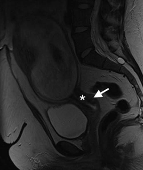
Fig. 20.1
Normal cervix and gravid uterus. Sagittal T2W image of the normal cervix in pregnancy. Note that signal intensity of the outer stroma (arrow) is greater than the inner stroma (asterisk)
Ovaries
The normal ovary in pregnancy has a low T2 signal intensity (T2SI) stroma and contains small follicles (Fig. 20.2). As pregnancy progresses the ovaries are displaced anteriorly and superiorly by the gravid uterus.
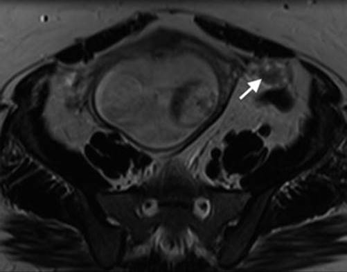
Fig. 20.2
Normal ovaries and gravid uterus. Axial T2W image of the pelvis showing a normal left ovary (arrow) displaced anteriorly and superiorly by the gravid uterus (18/40)
Myometrium
The myometrium returns intermediate signal intensity on T2W sequences, and the uterine wall is readily visible. Thin slices performed perpendicular to the myometrial-placental junction reduce partial voluming and allow distinction between the myometrium and placenta (a common pitfall with thicker slices).
20.4 Cervical Cancer in Pregnancy
Cervical cancer is the most commonly diagnosed gynaecologic malignancy during pregnancy. Incidence rates vary from 0.1 to 12 per 10,000 pregnancies [1].
Presentation of cervical cancer during pregnancy is the same as for non-pregnant patients, usually with painless post-coital bleeding. However, symptoms can be mistaken for complications of pregnancy, and this can lead to delay in diagnosis if clinical suspicion is low [34]. Cervical cancer does not adversely affect pregnancy, and pregnancy does not alter the course of the cancer.
MR is the single best imaging investigation that can accurately determine cervical tumour location, tumour size, depth of stromal invasion and extension into the lower uterine segment to assess local disease extent [48]. Accurate pretreatment evaluation of these (prognostic) factors is crucial in determining appropriate therapy.
Cervical cancer staging is based on the FIGO clinical and pathological staging system (Table 20.3) [41]. On T1W images, tumours are isointense to the normal cervix and are difficult to visualise. On T2W imaging, cervical cancer appears as a relatively hyperintense mass, easily distinguishable from low signal intensity cervical stroma (Fig. 20.3). Large tumours may be necrotic. MR features of cervical cancer in pregnant patients should be comparable to the non-pregnant patient [35].
Table 20.3
FIGO and TNM classification for cervical cancer [41]
|
FIGO stage |
TNM stage |
Extent of disease |
|
– |
Tis |
In situ |
|
I |
T1 |
Confined to uterus |
|
IA |
T1a |
Diagnosed only by microscopy |
|
IA1 |
T1a1 |
Depth ≤3 mm, horizontal spread ≤7 mm |
|
IA2 |
T1a2 |
Depth >3–5 mm, horizontal spread ≤7 mm |
|
IB |
T1b |
Clinically visible or microscopic lesion greater than T1a2 |
|
IB1 |
T1b1 |
≤4 cm |
|
IB2 |
T1b2 |
>4 cm |
|
II |
T2 |
Beyond uterus but not pelvic wall or lower third of vagina |
|
IIA |
T2a |
No parametrium |
|
IIA1 |
T2a1 |
≤4 cm |
|
IIA2 |
T2a2 |
>4 cm |
|
IIB |
T2b |
Parametrium |
|
III |
T3 |
Lower third vagina/pelvic wall/hydronephrosis |
|
IIIA |
T3a |
Lower third of vagina |
|
IIIB |
T3b |
Pelvic wall/hydronephrosis |
|
IVA |
T4 |
Mucosa of bladder/rectum; beyond true pelvis |
|
N1 |
Regional |
|
|
IVB |
M1 |
Distant metastasis |

Fig. 20.3
Cervix cancer in gravid uterus. (a) Sagittal T2W image of intermediate signal intensity barrel-shaped tumour of the cervix (arrow). (b) Axial oblique T2W image of the cervical tumour demonstrating left parametrial invasion (arrow). PL placenta, F foetus
However, it is important to note that the MR appearances of the normal cervix change as pregnancy progresses, and this can complicate interpretation when staging a cervical cancer. As described above the cross-sectional area of the cervix increases throughout pregnancy, the signal intensity of the external os is greater than the internal os and the signal intensity of the outer stroma is greater than the inner stroma [8, 23, 40]. Furthermore the MR signal intensity increases with gestational age and is maximal pre-partum. Therefore, in pregnancy the tumour may appear either isointense or hypointense against the physiological hyperintensity of the cervix. Nodal staging in the pelvis and para-aortic regions can be undertaken on MRI with moderate accuracy only. The presence of prominent pelvic veins due to the pregnancy may cause some difficulty in the interpretation of the pelvic lymph nodes and parametrial invasion.
Imaging for distant metastatic disease is planned depending on the stage of pregnancy and extent of cervical disease. In the future, whole body MRI may be used to investigate distant metastases (if the patient is able to tolerate the scan).
Traditionally, the treatment of cervical cancer in pregnancy was avoided, and the management strategy was termination of pregnancy if detected during the first two trimesters or delay of treatment until foetal maturity in the third trimester. Standard treatment was initiated post-partum. More recently, however, pregnancy preservation and treatment during pregnancy have become more common. Decision to treat and treatment schema clearly depend on disease stage, and the role of imaging in this setting is paramount to facilitate accurate staging and assessment of treatment response [21, 34].
Given the rarity of the disease and the complicated factors that must be taken into consideration on a case-by-case basis, standardisation of treatment is a challenge, and current guidelines are based on small case series and expert opinion. When a patient with cervical cancer presents, several issues must be considered by the multidisciplinary team – disease stage, nodal status, gestational age, obstetric complications and importantly the patient’s wishes regarding continuation versus termination of pregnancy [21].
20.4.1 Considerations Post-Fertility-preserving Surgery for Cervical Cancer
Historically patients with invasive cervical cancer stage IA up to IIA would be treated with a traditional Wertheim radical hysterectomy or radiation therapy. Higher-stage disease with parametrial invasion is treated with chemoradiation therapy. Although these radical therapies have good survival, they are associated with subsequent sterility. In recent years, surgical techniques have explored radical tumour resection without hysterectomy in young women with early-stage disease, thereby preserving fertility as well as the uterus and ovarian function. Radical trachelectomy with pelvic lymphadenectomy is a conservative but curative surgical procedure for early (stage 1B1 or lower) carcinoma of the cervix [47]. The surgical technique varies, but typically involves resection of the cervix (together with variable parametrial resection) and upper vaginal vault, and then anastomosis of the residual corpus uteri to the remainder of the vaginal vault (Fig. 20.4a). A cerclage suture is then placed around the corpus uteri at the anastomosis to maintain competency of the uterus in any subsequent pregnancies. Cerclage sutures may cause susceptibility artefact on MRI (Fig. 20.4b).
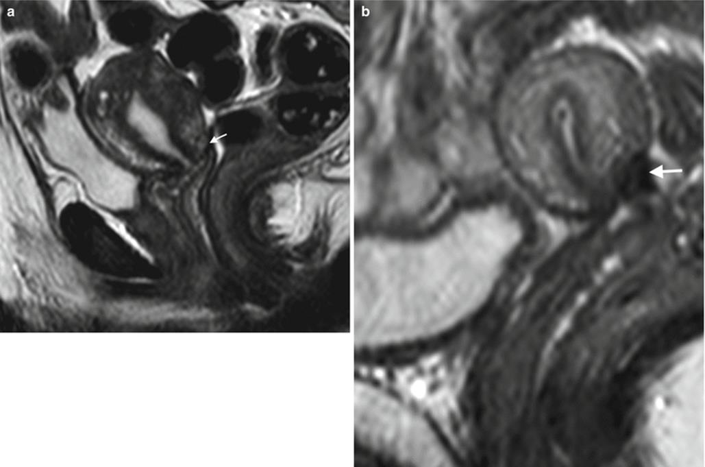
Fig. 20.4
Post-trachelectomy appearances. (a) Sagittal T2W image of a patient who underwent trachelectomy for stage 1B grade 1 adenocarcinoma of the cervix. Note the end-to-end anastomosis of the uterine body and vaginal vault (arrow). (b) Sagittal T2W image in a different patient who underwent trachelectomy demonstrates prominent susceptibility artefact from cerclage suture (arrow)
Several studies celebrate successful pregnancy outcomes for patients who have undergone trachelectomy [6, 45, 46, 47] but caution that there is an increased incidence of preterm premature rupture of the membranes, and therefore these pregnancies should be managed as high risk. Delivery should be by caesarian section.
Other complications may include an increased risk of miscarriage and bleeding. At our institution we managed a patient who presented at 12/40 with PV bleeding who had undergone trachelectomy for cervical cancer 6 years previously (Fig. 20.4). The bleeding resolved spontaneously, and there was no MR evidence of recurrence on follow-up imaging (Fig. 20.5). In this case, clinical examination in conjunction with MR was paramount.
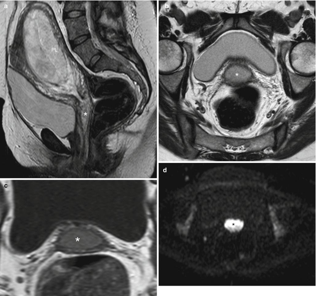

Fig. 20.5
Pregnancy post-trachelectomy with bleeding. (a) Following trachelectomy 6 years previously (Fig. 20.4a), this patient presented at 12/40 with vaginal bleeding. Sagittal T2W image demonstrates intermediate T2W signal material in the region of the cervix (asterisk) that is distending the vagina but not distorting its shape. (b) Axial oblique T2W image demonstrating intermediate T2W signal intensity ‘mass’ in the upper vagina (asterisk). (c) Axial T1W image where the ‘mass’ returns intermediate T1W signal (asterisk). (d, e) B 1200 image and corresponding ADC map demonstrate restricted diffusion within the ‘mass’ (asterisk). On clinical examination there was no visible recurrence, and these imaging appearances represent a blood clot. Repeat scan performed 6 weeks later (f) (T2W sagittal image) was normal, and the ‘mass’ was no longer visible. In this case clinical evaluation and imaging were vital and complementary to avoid pitfalls, exclude recurrence and identify complications. PL placenta
20.5 Adnexal Masses in Pregnancy
Adnexal masses are found in 1–2 % of pregnancies [44]. US is the first-line imaging modality and usually provides sufficient information for the treatment decision to be taken [7, 11]. Management is conservative or surgical depending on the imaging appearances of the mass, nature of presentation, gestational age, size of mass and patient preference. MR imaging may be helpful for characterisation of adnexal masses that remain indeterminate following US evaluation, for determining the origin of a pelvic mass and for surgical planning. In the acute setting, MR is particularly useful for evaluating the haemorrhagic content of adnexal masses, but clearly it should not delay surgical intervention. MR characteristics of adnexal masses that suggest benignity include high signal intensity on T1W sequences (indicating either fat, blood, or proteinaceous/mucinous content) with subsequent loss of signal intensity on fat-suppressed sequences (indicating fat), high signal intensity on T1W fat-suppressed images (indicative of blood) and low signal intensity on T2W images (indicative of fibrous tissue or haemosiderin) [32]. In particular, solid adnexal tissue that demonstrates low signal intensity on T2W sequences with low signal intensity on high b-value diffusion sequences has been shown to be highly suggestive of a benign non-invasive lesion [53]. In clinical practice, repeat imaging after pregnancy can be performed with gadolinium contrast enhancement to further support the diagnosis.
20.5.1 Cystic Adnexal Lesions
20.5.1.1 Simple and Haemorrhagic Cysts
Incidental US detection of a simple cyst in pregnancy is common. Size is the best indicator of whether the cyst requires monitoring or intervention as simple cysts that are smaller than 5 cm will resolve spontaneously in 90–100 % of cases [54]. Corpus luteal cysts enlarge during the first trimester, regress by the 12th week of gestation and disappear later on in the pregnancy. Haemorrhagic corpus luteal cysts have variable but characteristic US appearances due to the changing nature of the internal blood clot.
The decision to proceed to MR imaging in the context of a simple adnexal cyst is taken on a case-by-case basis. Larger cysts have an increased risk of torsion, rupture and obstetric complications, and in such cases MR is useful for accurate localisation and assessment of the position of the cyst and its relation to other structures. Simple ovarian cysts have thin featureless walls and return low signal intensity on T1W sequences and high signal intensity on T2W images. Corpus luteal cysts have thicker walls than simple cysts and have increased vascularity within the thickened wall seen as increased Doppler flow on US. They display more varied signal intensity on MR images because they are frequently haemorrhagic. MR is seldom required to aid diagnosis, but when used, haemorrhagic cysts return high signal intensity on T1W sequences and low signal intensity on heavily T2W sequences owing to T1 and T2 shortening. Haemorrhagic content is also demonstrated as hyperintense signal on T1 fat-saturated sequences [15, 37]. Adherent clot within a haemorrhagic corpus luteal cyst may mimic a solid nodule or papillary projection and raise concern regarding malignancy. In these cases the absence of a contrast-enhanced sequence makes this distinction challenging on MRI. Diffusion-weighted sequences can be helpful in this situation as, if the solid components return low SI on high b-value images, the likelihood of benignity is high. If adherent clot is suspected but not fully resolved on MRI and US, patients should be followed up with US to look for evolution and resolution of the haemorrhagic lesion.
20.5.1.2 Hyperstimulated Ovaries
Typically seen in patients who have undergone ovarian induction, the ovaries are enlarged with multiple cysts. These will resolve spontaneously in more than 90 % of patients [11]. Ovarian hyperstimulation syndrome (OHSS) appears as markedly enlarged ovaries with extravascular accumulation of fluid resulting in ascites, pleural effusions, intravascular volume depletion and dehydration. The enlarged ovaries are at risk of torsion and haemorrhage, but they usually regress later in pregnancy or after delivery. MR may be considered in the context of ovarian hyperstimulation syndrome to exclude ovarian torsion and differentiate from ovarian tumour. Hyperstimulated ovaries demonstrate a ‘spoke-wheel’ appearance with enlarged follicles peripherally located and central ovarian stroma [24]. The follicles return bilateral symmetrical low signal intensity on T1W sequences (high T1 signal intensity in some foci may be in keeping with haemorrhage). On T2W sequences, multiple cysts will return homogenous high signal intensity. Central ovarian stroma will return intermediate to low T2 signal (higher if oedematous). Ascites will be high signal on T2. Treatment is usually conservative.
20.5.1.3 Hyperreactio Luteinalis
Appearances of hyperreactio luteinalis are similar to that of hyperstimulated ovaries, but this rare condition is seen in patients who have not undergone ovulation induction. It is thought to result from hypersensitivity of the ovary to circulating human chorionic gonadotropin (hCG). Symptoms may be absent or can include maternal abdominal pain, abdominal distension, abnormal liver function tests, respiratory difficulties and hirsutism. MR can be used to distinguish hyperstimulated ovaries from ovarian neoplasm. Appearances of the ovaries are similar to OHSS but without ancillary extravasation of fluid, therefore no significant ascites or pleural effusion (Fig. 20.6).

Fig. 20.6
Hyperreactio luteinalis. (a, b) Ultrasound images of complex multilocular cystic masses in the right and left flanks. (c) Axial and (d) coronal T2W images demonstrated bilateral multilocular cystic masses (arrows). PL placenta
Similar appearances can be associated with a complete hydatidiform mole 14–30 % of the time (see later section).
20.5.2 Ovarian Torsion
Ovarian torsion is defined as partial or complete rotation of the ovarian vascular pedicle and causes obstruction to venous outflow and arterial inflow [9]. Torsion of an ovarian mass usually occurs in the mid to late first trimester when the gravid uterus is enlarging most rapidly [19] and occurs in 1 in 800 pregnancies [50]. Ovarian torsion can occur in an otherwise normal ovary, more often on the right-hand side [50]. MRI is indicated to establish a diagnosis if US is inconclusive and their clinical findings are not conclusive. MR appearances of ovarian torsion vary depending on the stage of the torsion. Early findings include oedematous enlargement of the ovary with peripheral displacement of follicles. Later findings are smooth thickening of the twisted ovarian mass and uterine deviation to the twisted side [10]. Massive ovarian oedema occurs when there is intermittent torsion of the ovary which interferes with venous and lymphatic drainage and causes ovarian enlargement [11]. MR appearances of the ovary demonstrate homogeneous low signal intensity on T1W images and high signal intensity on T2W images, and there may be peripheral displacement of the follicles. On T1W images there may be evidence of haemorrhage and necrosis. Associated findings include a thickened thrombosed pedicle which may demonstrate the ‘whirlpool’ sign, a blood-filled fallopian tube and a haemoperitoneum [30] (Fig. 20.7).

Fig. 20.7
Ovarian torsion. (a) Axial and coronal (b) T2W image demonstrating a right adnexal lesion (arrow) in keeping with an enlarged right ovary. (c) Axial T1W image demonstrates subtle high T1W signal within the enlarged ovary (asterisk), and appearances are suggestive of haemorrhagic products in this patient who presented with right-sided abdominal pain. (d) Axial T1FS image demonstrates high T1W signal within the enlarged right ovary (arrows). Appearances in keeping with haemorrhagic infarction. PL placenta
20.5.3 Luteoma of Pregnancy
A luteoma is a rare, tumour-like ovarian mass that occurs during pregnancy and is associated with elevated androgen levels. They are more common in the third and fourth decades and have increased prevalence in the African American population and in multiparous patients [12]. Luteomas can be asymptomatic or cause virilisation and should be suspected in a patient with a solid (or less commonly complex cystic) adnexal lesion and hirsutism in pregnancy. Luteomas are unilateral in 2/3 of cases and bilateral in 1/3 of cases. They have intermediate signal intensity on T1W sequences and low signal intensity on T2W sequences. Ancillary signs of malignancy are absent, and they classically regress post-partum; therefore, oophorectomy can be avoided [11].
20.5.4 Endometriomas
Although 30–50 % of women with endometriosis are infertile [17], an understanding of the behaviour of endometriosis in pregnancy is important. Because of its association with infertility, it is uncommon to find an unsuspected endometrioma at routine obstetric imaging. As in non-pregnant patients, MR has great specificity for the diagnosis of endometriomas, and the classical appearance is a high T1W signal intensity cystic lesion on fat-suppressed sequences. On T2W images the signal intensity of the endometrioma should be less than of simple fluid [55], and this is described as ‘T2 shading’ [49]. Ancillary findings including adhesions, endometriotic plaques and haematosalpinx may also be present.
Pregnancy can alter the appearance of endometriosis. The increased progesterone levels during pregnancy can stimulate hypertrophy of the endometrial stromal cells and formation of the vascular decidual lining of the uterus (decidualisation). Such hormonal stimulation may also affect ectopically located endometrial stromal cells within endometriomas and lead to enlargement and formation of vascular mural nodules leading to more complex appearances. Decidualised endometriosis has thus been described as a mimic of ovarian cancer on imaging [4] (Fig. 20.8). A diagnostic clue at MR imaging is that the T2 signal hyperintensity of the mural nodules is isointense to the thickened decidualised endometrium. Decidualised endometriosis can be managed conservatively and will usually resolve or regress after childbirth [49].

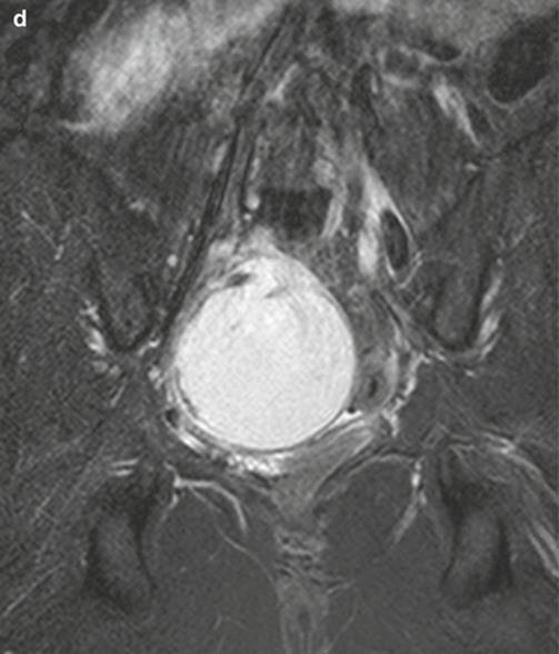
Fig. 20.8
Decidualised endometriotic cyst. (a) Ultrasound image demonstrating a central mass deep in the pelvis (measured). (b) Axial T2W image demonstrating a right adnexal mass (straight arrow) with internal high T2W signal and intermediate signal peripheral nodules and projections (curved arrows). (c) Axial T1W image demonstrates high T1W signal within the mass. (d) Coronal T1W with fat suppression returns intense high T1W signal within the mass. The mass was resected at caesarian section, and histology demonstrated decidualisation of an endometrioma. FL amniotic fluid, F foetus
20.5.5 Mature Cystic Teratomas of the Ovary
Mature cystic teratomas (dermoid cysts) have typical sonographic appearances but can cause a diagnostic conundrum on US if they are very large, and the lesion cannot be fully assessed, if there is minimal fat or if there are complex solid components [22]. In these situations, MR imaging is helpful to confirm the presence internal fat. On T1W MR sequences the fat-containing component of the lesion returns very high signal intensity (similar to retroperitoneal fat), and the calcification, bone, hair and fibrous tissues return low signal. The high signal of fat on T1 sequences is suppressed with T1W fat-suppressed images (Fig. 20.9). T1W sequences with ‘out-of-phase’ sequence improve diagnostic confidence in detecting the presence of minimal fat. MR can help determine the size of the mass if surgical removal is contemplated during pregnancy. Teratomas can be pedunculated and therefore more prone to torsion and rupture [11]. Torsion is suggested when the uterus is deviated to the affected side, engorged blood vessels are present on the twisted side, and there is a high signal intensity rim on T1W sequences [38]. Tumours can rupture leading to chemical peritonitis, a rare complication occurring in less than 1 % of cases [14, 38].
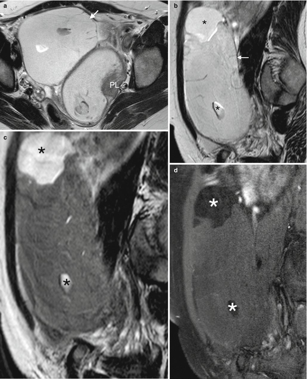
Fig. 20.9
Mature teratoma. (a) Axial T2W image demonstrates a right adnexal lesion (arrow) displacing the gravid uterus posteriorly. Right ovary (asterisk). (b) Sagittal T2W image demonstrates complex right adnexal mass (arrow), and note internal regions of higher T2W signal (asterisk). (c) Sagittal T1W image demonstrates low signal intensity of the mass in keeping with fluid. The internal regions of high T2W signal return high T1W signal (asterisk). (d) Sagittal T1W fat-saturated image shows suppressed signal in the internal regions of the mass in keeping with fat globules (asterisks). PL placenta
20.5.6 Hydrosalpinx
A hydrosalpinx has the imaging appearance of a cystic dilated tubular adnexal structure with incomplete septations that is separate from the ovary. It does not change over the course of pregnancy [11] but is associated with increased risk of tubal ectopic pregnancy. MR imaging can aid the non-invasive assessment of tubal dilatation and help distinguish dilated fallopian tubes from other cystic adnexal masses.
20.5.7 Ovarian Cancer
The risk of malignancy in an adnexal mass that is diagnosed during pregnancy is 2–3 % [20, 27]. Ovarian cancer is the second most common gynaecologic cancer complicating pregnancy after cervical cancer [36]. Patients are usually asymptomatic, and diagnosis is often based on incidental US findings (this commonly results in an earlier diagnosis and therefore better prognosis). Suspicious US appearance will prompt further investigation with tumour markers and MRI in indeterminate cases or for staging of disease. In the context of pregnancy, only serial measurements of Ca-125 showing an increase over time are helpful, since high levels of Ca-125 are expected during the first trimester [21, 29]. Staging is based on FIGO or TNM classification (Table 20.4) [33].
Table 20.4
FIGO and TNM classification for ovarian cancer 2014 [33]
|
FIGO stage |
TNM stage |
Extent of disease |
|
I |
T1 |
Limited to ovaries |
|
IA |
T1a |
One ovary, capsule intact. No tumour on surface, negative washings |
|
IB |
T1b |
Both ovaries, capsule intact. No tumour on surface, negative washings |
|
IC |
T1c |
Tumour limited to one or both ovaries |
|
IC1 |
Surgical spill |
|
|
IC2 |
Capsule rupture before surgery or tumour on ovarian surface |
|
|
IC3 |
Malignant cells in ascites or peritoneal washings |
|
|
II |
T2 |
Tumour involves one or both ovaries with pelvic extension or primary peritoneal cancer |
|
IIA |
T2a |
Uterus, tube(s) |
|
IIB |
T2b |
Other pelvic intraperitoneal tissues |
|
III |
T3 ± N1 |
Tumour involves one or both ovaries with spread to the peritoneum outside the pelvis and/or metastasis to the retroperitoneal lymph nodes |
|
IIIA |
T3a |
Positive retroperitoneal nodes and/or microscopic peritoneal metastasis |
|
IIIA1 |
Positive retroperitoneal lymph nodes only |
|
|
IIIA2 |
Microscopic, extrapelvic (above the brim) peritoneal involvement ± positive retroperitoneal lymph nodes |
|
|
IIIB |
T3b |
Macroscopic extrapelvic, peritoneal metastasis <2 cm. Includes extension to capsule of liver/spleen |
|
IIIC |
T3c ± N1 |
Macroscopic, extrapelvic peritoneal metastasis >2 cm in greatest dimension ± positive retroperitoneal lymph nodes, includes extension to capsule of liver/spleen |
|
IVA |
M1 |
Pleural effusion with positive cytology |
|
IVB |
Hepatic and/or splenic parenchymal metastasis, metastasis to extra-abdominal organs (including inguinal lymph nodes and lymph nodes outside the abdominal cavity) |
MR appearances of aggressive lesions that indicate malignant adnexal masses include the presence of both cystic and solid areas within a lesion, necrosis within a solid lesion, papillary projections from the wall or septum of a cystic lesion, an irregular septum or wall, multiple thickened (>3 mm) septations, a large size (>6 cm), bilateral lesions and ascites, peritoneal disease and lymphadenopathy [32]. Interpretation is limited due to artefact from foetal movement and the avoidance of using intravenous gadolinium. The DWI sequence is useful in this context: if a solid component demonstrates no signal return on high b-value sequences, then this is reassuring for benignity and follow-up imaging may be advocated. However, malignancy cannot be ruled out in cases where solid tissue in the adnexal mass has intermediate T2 signal intensity and high signal intensity on the high b-value image.
The management of women diagnosed with asymptomatic adnexal lesions that persist during pregnancy remains controversial, and ovarian malignancy diagnosed during pregnancy presents significant challenges as a result of the conflict between foetal well-being and optimal maternal therapy [36].
Ovarian metastases may present as ovarian masses with MR appearances characteristic of malignancy; we present two such cases from our institution (Figs. 20.10 and 20.11 [26]).
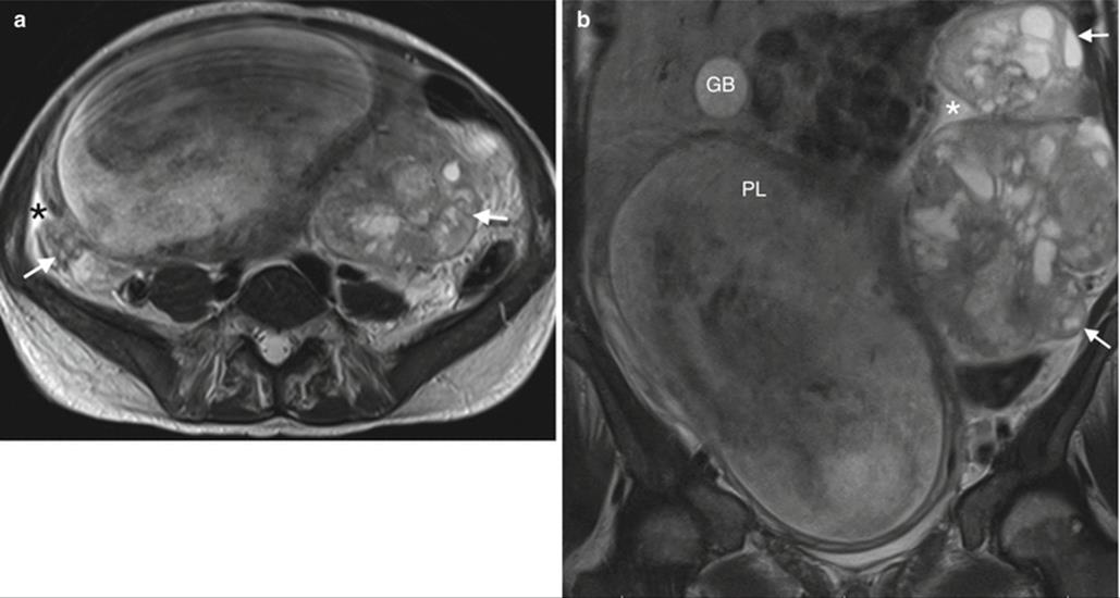
Fig. 20.10
Ovarian metastases. (a, b) Axial and coronal T2W images of bilateral complex adnexal masses with internal solid component (arrow). There is ascites (asterisk). Appearances are of an aggressive lesion. Post-partum histology was of a retroperitoneal leiomyosarcoma. GB gallbladder, PL placenta
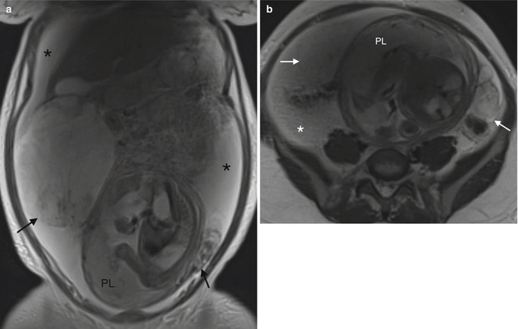
Fig. 20.11
Ovarian metastases. (a, b) Coronal and axial T2W image demonstrating a gravid uterus, complex bilateral adnexal masses (arrows) and ascites (asterisk). The patient was diagnosed incidentally following investigation for preeclampsia. The final diagnosis was of metastases from a primary cancer of unknown origin, most likely a gastric adenocarcinoma. PL placenta. For full case report see references [26]
20.6 Leiomyoma in Pregnancy
Leiomyomas, also known as fibroids or myomas, are common, benign smooth muscle tumours that contain varying amounts of connective tissue. They occur in 20–30 % of women of reproductive age [16]. The prevalence of uterine leiomyoma in pregnancy varies between 1.6 and 10.7 % depending upon the trimester of assessment and the size threshold [18, 28, 42, 51, 52], is higher in women of African origin and increases with age. Most leiomyomas are located within the wall of the uterus (intramural), but pedunculated subserosal and broad-ligament leiomyomas can mimic an ovarian mass.
Most women with leiomyomas do not have any related complications during pregnancy. Pain is the most common problem as leiomyomas may enlarge during pregnancy owing to increased oestrogen synthesis [43]. Evidence regarding obstetric complications in the presence of uterine leiomyomata varies. It is accepted that the presence of uterine leiomyomas is associated with an elevated risk of spontaneous abortion, and the loss rate is higher in patients with multiple fibroids [5, 13]. There is a questionable theoretical risk that subplacental lesions may represent an increased risk of placental abruption [39].
Ultrasonography is the preferred assessment tool for leiomyoma in pregnancy, and MR should be considered as a problem-solving adjunct. MR is the most accurate imaging modality for the detection and localisation of leiomyomas, particularly for those lesions arising in the posterior myometrium or deep in the pelvis beyond the field of view of the US probe. MR usually offers the confident differentiation from leiomyoma mimics, including adnexal masses.
MR appearances of non-degenerated uterine leiomyoma are of a well-circumscribed mass. Their internal appearance varies depending on their histological content. Those containing smooth muscle cells and collagen return homogenously decreased T2W signal intensity compared with the outer myometrium. If leiomyomas are more cellular, composed of compact smooth muscle cells, they can have relatively increased T2W signal intensity [16]. In the gravid uterus the size and location of each fibroid should be documented, as well as their relation to the placenta. This information aids surgical planning if the patient is due to undergo caesarian section (Fig. 20.12).

Fig. 20.12
Uterine fibroid. Coronal T2W image of a low uterine fibroid (arrow) in a gravid uterus. This patient underwent MRI for planning of caesarian section. PL placenta
One in five hundred pregnant women experiences acute abdominal pain with uterine tenderness due to leiomyoma-related complications [56]. As leiomyomas enlarge they may outgrow their blood supply and degenerate. Red degeneration is most common during pregnancy resulting from haemorrhagic infarction of a leiomyoma. Patients may present with pain and systemic symptoms such as fever and leucocytosis. There is point tenderness when the US probe is placed over the culprit lesion. US appearances of a degenerating leiomyoma are of a heterogenous hyperechoic lesion. Degenerated leiomyomas have variable appearances on T1 and T2W imaging. Leiomyomas that have undergone red degeneration return peripheral or diffuse high signal intensity on T1W images and variable signal intensity with or without low signal intensity on T2W imaging [25]. The high T1W signal intensity is due to the presence of methaemoglobin or the proteinaceous content of blood (Fig. 20.13). Histological correlation suggests that when the degenerated fibroid has a peripheral rim of high T1W signal intensity on MRI, the blood products are confined to the thrombosed vessels that surround the tumour [16]. Some lesions have a rim that returns high T2W signal, which corresponds to a zone of dilated lymphatic vessels, dilated veins and oedema due to vascular congestion [31]. Pain may also occur with torsion of a pedunculated leiomyoma due to infarction. Torsion is more likely in pregnancy due to the increasing size of the lesion due to oestrogen stimulation.

Fig. 20.13
Red degeneration in a fibroid. (a) This patient presented with acute abdominal pain in pregnancy. Sagittal T2W image of a low uterine fibroid (arrow) with internal high T2W signal intensity (asterisk). (b) Axial T1W image demonstrates high T1W signal within the fibroid (arrow) in keeping with blood products. PL placenta
20.7 Gestational Trophoblastic Neoplasia
Gestational trophoblastic neoplasia (GTN) is a spectrum of rare abnormalities where trophoblastic tissue, which is part of the blastocyst that normally invades the endometrium, proliferates in a more aggressive way than is normal. Proliferation can be localised and non-invasive (this is called hydatidiform mole) or invasive. In cases where invasion is localised to the uterus, these are termed invasive moles, but if metastases occur, the terminology is choriocarcinoma. Placental site trophoblastic tumour (PSTT) is the rarest form of GTN and represents neoplastic proliferation of trophoblasts that invade the myometrium at the placental site after pregnancy [2]. A full discussion of these entities is outside the scope of this chapter as there is usually no viable pregnancy; however, the imaging appearances of molar pregnancy will be briefly described as they may (rarely) coexist with a dizygotic twin pregnancy.
Molar pregnancy complicates about 0.1 % of pregnancies. It is relatively more common in older women. GTN should be suspected in patients with vaginal bleeding and rapid uterine enlargement who have markedly high serum hCG levels, and severe vomiting may occur [57]. US is the imaging modality of choice for diagnosis of GTN and should be obtained to exclude an intrauterine pregnancy before initiating chemotherapy. The classic description of the US appearance of molar pregnancies is ‘resembling a bunch of grapes’ (due to generalised swelling of chorionic villi) [2]. MR is indicated in difficult cases as a problem-solving tool, in cases of relapsed disease, suspected PSTT or in advanced disease. MR findings can be relatively non-specific, and it may be difficult to distinguish from an incomplete miscarriage or an ectopic pregnancy [3]. Primary molar tissue may be visualised as heterogeneously high signal intensity on T2W sequences and may distend the endometrium and enlarge the uterus with the ‘bunch of grapes’ appearance. A rim of normal hypointense myometrium may surround this. For a full description of the MR appearances throughout pregnancy and posttreatment, please refer to the reference article [2]. Figure 20.14 is a case of tubal molar pregnancy with associated endometrial hyperplasia secondary to the high b-HCG levels.

Fig. 20.14
Ectopic molar pregnancy. (a) Coronal T2W image of a complex left adnexal lesion (arrow) with a typical ‘bunch of grapes’ appearance and internal high T2W signal. Note the thickened endometrium (asterisk) as a result of hormonal stimulation. (b) Coronal T1W image demonstrating high T1W signal within the endometrial cavity (asterisk) and within the left adnexal lesion (arrow) in keeping with blood products
20.8 Conclusion
MR is a useful problem-solving tool in the evaluation of gynaecologic pathology in pregnant women. The MR examination should be specifically tailored to the clinical question, and the radiologist should be involved in scan sequence planning. MR can reduce diagnostic delay and improve patient care in a safe and cost-effective manner.
References
1.
Al-Halal H, Kezouh A, Abenhaim HA (2013) Incidence and obstetrical outcomes of cervical intraepithelial neoplasia and cervical cancer in pregnancy: a population-based study on 8.8 million births. Arch Gynecol Obstet 287(2):245–250. doi:10.1007/s00404-012-2475-3PubMed
2.
Allen SD, Lim AK, Seckl MJ, Blunt DM, Mitchell AW (2006) Radiology of gestational trophoblastic neoplasia. Clin Radiol 61(4):301–313. doi:10.1016/j.crad.2005.12.003PubMed
3.
Barton JW, McCarthy SM, Kohorn EI, Scoutt LM, Lange RC (1993) Pelvic MR imaging findings in gestational trophoblastic disease, incomplete abortion, and ectopic pregnancy: are they specific? Radiology 186(1):163–168. doi:10.1148/radiology.186.1.767797310.1148/radiology.186.1.7677973PubMed
4.
Bennett GL, Slywotzky CM, Cantera M, Hecht EM (2010) Unusual manifestations and complications of endometriosis—spectrum of imaging findings: pictorial review. Am J Roentgenol 194(6 Suppl):WS34–WS46. doi:10.2214/AJR.07.7142
5.
Benson CB, Chow JS, Chang-Lee W, Hill JA 3rd, Doubilet PM (2001) Outcome of pregnancies in women with uterine leiomyomas identified by sonography in the first trimester. J Clin Ultrasound 29(5):261–264. doi:10.1002/jcu.1031PubMed
6.
Bernardini M, Barrett J, Seaward G, Covens A (2003) Pregnancy outcomes in patients after radical trachelectomy. Am J Obstet Gynecol 189(5):1378–1382PubMed
7.
Bromley B, Benacerraf B (1997) Adnexal masses during pregnancy: accuracy of sonographic diagnosis and outcome. J Ultrasound Med 16(7):447–452PubMed
8.
Chan YL, Lam WW, Lau TK, Wong SP, Li CY, Metreweli C (1998) Cervical assessment by magnetic resonance imaging – its relationship to gestational age and interval to delivery. Br J Radiol 71(842):155–159. doi:10.1259/bjr.71.842.9579179PubMed
9.
Chang HC, Bhatt S, Dogra VS (2008) Pearls and pitfalls in diagnosis of ovarian torsion. Radiographics 28(5):1355–1368. doi:10.1148/rg.285075130PubMed
10.
Cheng KL, Tsao TF (2010) Ovarian torsion: appearance on MRI. Pediatr Radiol 40(Suppl 1):S104. doi:10.1007/s00247-010-1652-4PubMed
11.
Chiang G, Levine D (2004) Imaging of adnexal masses in pregnancy. J Ultrasound Med 23(6):805–819PubMed
12.
Choi JR, Levine D, Finberg H (2000) Luteoma of pregnancy: sonographic findings in two cases. J Ultrasound Med 19(12):877–881PubMed
13.
Ciavattini A, Clemente N, Delli Carpini G, Di Giuseppe J, Giannubilo SR, Tranquilli AL (2015) Number and size of uterine fibroids and obstetric outcomes. J Matern Fetal Neonatal Med 28(4):484–488. doi:10.3109/14767058.2014.921675PubMed
14.
Comerci JT Jr, Licciardi F, Bergh PA, Gregori C, Breen JL (1994) Mature cystic teratoma: a clinicopathologic evaluation of 517 cases and review of the literature. Obstet Gynecol 84(1):22–28PubMed
15.
Corwin MT, Gerscovich EO, Lamba R, Wilson M, McGahan JP (2014) Differentiation of ovarian endometriomas from hemorrhagic cysts at MR imaging: utility of the T2 dark spot sign. Radiology 271(1):126–132. doi:10.1148/radiol.13131394PubMed
16.
Deshmukh SP, Gonsalves CF, Guglielmo FF, Mitchell DG (2012) Role of MR imaging of uterine leiomyomas before and after embolization. Radiographics 32(6):E251–E281. doi:10.1148/rg.326125517PubMed
17.
Eskenazi B, Warner ML (1997) Epidemiology of endometriosis. Obstet Gynecol Clin North Am 24(2):235–258PubMed
18.
Exacoustos C, Rosati P (1993) US diagnosis of uterine myomas and complications in pregnancy. Obstet Gynecol 82(1):97–101PubMed
19.
Grendys EC Jr, Barnes WA (1995) Ovarian cancer in pregnancy. Surg Clin North Am 75(1):1–14PubMed
20.
Grigoriadis C, Eleftheriades M, Panoskaltsis T, Bacanu AM, Vitoratos N, Kondi-Pafiti A, Tsangkas A, Tympa A, Hassiakos D (2014) Ovarian cancer diagnosed during pregnancy: clinicopathological characteristics and management. G Chir 35(3–4):69–72PubMedCentralPubMed
21.
Han SN, Mhallem Gziri M, Van Calsteren K, Amant F (2013) Cervical cancer in pregnant women: treat, wait or interrupt? Assessment of current clinical guidelines, innovations and controversies. Ther Adv Med Oncol 5(4):211–219. doi:10.1177/1758834013494988PubMedCentralPubMed
22.
Hertzberg, B. S., & Kliewer, M. A. (1996). Sonography of benign cystic teratoma of the ovary: pitfalls in diagnosis. AJR Am J Roentgenol, 167(5):1127–1133. doi: 10.2214/ajr.167.5.8911163
23.
House M, O'Callaghan M, Bahrami S, Chelmow D, Kini J, Wu D, Patz S, Bhadelia RA (2005) Magnetic resonance imaging of the cervix during pregnancy: effect of gestational age and prior vaginal birth. Am J Obstet Gynecol 193(4):1554–1560. doi:10.1016/j.ajog.2005.03.042PubMed
24.
Jung BG, Kim H (2001) Severe spontaneous ovarian hyperstimulation syndrome with MR findings. J Comput Assist Tomogr 25(2):215–217PubMed
25.
Kawakami S, Togashi K, Konishi I, Kimura I, Fukuoka M, Mori T, Konishi J (1994) Red degeneration of uterine leiomyoma: MR appearance. J Comput Assist Tomogr 18(6):925–928PubMed
26.
Kim S-H, Abd Halim SR, Siddiqui N, Park W-HE (2014) Disseminated cancer in pregnancy: Krukenberg tumour. Case Rep Obstet Gynecol 2014:4. doi:10.1155/2014/216969
27.
Kondi-Pafiti A, Grigoriadis C, Iavazzo C, Papakonstantinou E, Liapis A, Hassiakos D (2012) Clinicopathological characteristics of adnexal lesions diagnosed during pregnancy or cesarean section. Clin Exp Obstet Gynecol 39(4):458–461PubMed
28.
Laughlin SK, Baird DD, Savitz DA, Herring AH, Hartmann KE (2009) Prevalence of uterine leiomyomas in the first trimester of pregnancy: an US-screening study. Obstet Gynecol 113(3):630–635. doi:10.1097/AOG.0b013e318197bbafPubMedCentralPubMed
29.
Marret H, Lhomme C, Lecuru F, Canis M, Leveque J, Golfier F, Morice P (2010) Guidelines for the management of ovarian cancer during pregnancy. Eur J Obstet Gynecol Reprod Biol 149(1):18–21. doi:10.1016/j.ejogrb.2009.12.001PubMed
30.
Masselli G, Derchi L, McHugo J, Rockall A, Vock P, Weston M, Spencer J (2013) Acute abdominal and pelvic pain in pregnancy: ESUR recommendations. Eur Radiol 23(12):3485–3500. doi:10.1007/s00330-013-2987-7PubMed
31.
Mittl RL Jr, Yeh IT, Kressel HY (1991) High-signal-intensity rim surrounding uterine leiomyomas on MR images: pathologic correlation. Radiology 180(1):81–83. doi:10.1148/radiology.180.1.205272810.1148/radiology.180.1.2052728PubMed
32.
Mohaghegh P, Rockall AG (2012) Imaging strategy for early ovarian cancer: characterization of adnexal masses with conventional and advanced imaging techniques. Radiographics 32(6):1751–1773. doi:10.1148/rg.326125520PubMed
33.
Mutch DG, Prat J (2014) 2014 FIGO staging for ovarian, fallopian tube and peritoneal cancer. Gynecol Oncol 133(3):401–404. doi:10.1016/j.ygyno.2014.04.013PubMed
34.
Nguyen C, Montz FJ, Bristow RE (2000) Management of stage I cervical cancer in pregnancy. Obstet Gynecol Surv 55(10):633–643PubMed
35.
Nicolet V, Carignan L, Bourdon F, Prosmanne O (2000) MR imaging of cervical carcinoma: a practical staging approach. Radiographics 20(6):1539–1549. doi:10.1148/radiographics.20.6.g00nv111539PubMed
36.
Oehler MK, Wain GV, Brand A (2003) Gynaecological malignancies in pregnancy: a review. Aust N Z J Obstet Gynaecol 43(6):414–420PubMed
37.
Outwater EK, Dunton CJ (1995) Imaging of the ovary and adnexa: clinical issues and applications of MR imaging. Radiology 194(1):1–18. doi:10.1148/radiology.194.1.7997533PubMed
38.
Outwater EK, Siegelman ES, Hunt JL (2001) Ovarian teratomas: tumor types and imaging characteristics. Radiographics 21(2):475–490. doi:10.1148/radiographics.21.2.g01mr09475PubMed
39.
Ouyang DW, Economy KE, Norwitz ER (2006) Obstetric complications of fibroids. Obstet Gynecol Clin North Am 33(1):153–169. doi:10.1016/j.ogc.2005.12.010PubMed
40.
Pates JA, Yost NP, Oliver Q, McIntire DD, Twickler DM (2007) Magnetic resonance signal characteristics of the cervix as pregnancy advances. Reprod Sci 14(5):440–444. doi:10.1177/1933719107306225PubMed
41.
Pecorelli S (2009) Revised FIGO staging for carcinoma of the vulva, cervix, and endometrium. Int J Gynaecol Obstet 105(2):103–104PubMed
42.
Qidwai GI, Caughey AB, Jacoby AF (2006) Obstetric outcomes in women with sonographically identified uterine leiomyomata. Obstet Gynecol 107(2 Pt 1):376–382. doi:10.1097/01.AOG.0000196806.25897.7cPubMed
43.
Rein MS, Barbieri RL, Friedman AJ (1995) Progesterone: a critical role in the pathogenesis of uterine myomas. Am J Obstet Gynecol 172(1 Pt 1):14–18PubMed
44.
Ribic-Pucelj M, Kobal B, Peternelj-Marinsek S (2007) Surgical treatment of adnexal masses in pregnancy: indications, surgical approach and pregnancy outcome. J Reprod Med 52(4):273–279PubMed
45.
Rodriguez M, Guimares O, Rose PG (2001) Radical abdominal trachelectomy and pelvic lymphadenectomy with uterine conservation and subsequent pregnancy in the treatment of early invasive cervical cancer. Am J Obstet Gynecol 185(2):370–374. doi:10.1067/mob.2001.115866PubMed
46.
Roy M, Plante M (1998) Pregnancies after radical vaginal trachelectomy for early-stage cervical cancer. Am J Obstet Gynecol 179(6 Pt 1):1491–1496PubMed
47.
Sahdev A, Jones J, Shepherd JH, Reznek RH (2005) MR imaging appearances of the female pelvis after trachelectomy. Radiographics 25(1):41–52. doi:10.1148/rg.251045047PubMed
48.
Sala E, Wakely S, Senior E, Lomas D (2007) MRI of malignant neoplasms of the uterine corpus and cervix. Am J Roentgenol 188(6):1577–1587. doi:10.2214/AJR.06.1196
49.
Siegelman ES, Oliver ER (2012) MR imaging of endometriosis: ten imaging pearls. Radiographics 32(6):1675–1691. doi:10.1148/rg.326125518PubMed
50.
Smorgick N, Pansky M, Feingold M, Herman A, Halperin R, Maymon R (2009) The clinical characteristics and sonographic findings of maternal ovarian torsion in pregnancy. Fertil Steril 92(6):1983–1987. doi:10.1016/j.fertnstert.2008.09.028PubMed
51.
Stout MJ, Odibo AO, Graseck AS, Macones GA, Crane JP, Cahill AG (2010) Leiomyomas at routine second-trimester US examination and adverse obstetric outcomes. Obstet Gynecol 116(5):1056–1063. doi:10.1097/AOG.0b013e3181f7496dPubMed
52.
Strobelt N, Ghidini A, Cavallone M, Pensabene I, Ceruti P, Vergani P (1994) Natural history of uterine leiomyomas in pregnancy. J Ultrasound Med 13(5):399–401PubMed
53.
Thomassin-Naggara I, Aubert E, Rockall A, Jalaguier-Coudray A, Rouzier R, Darai E, Bazot M (2013) Adnexal masses: development and preliminary validation of an MR imaging scoring system. Radiology 267(2):432–443. doi:10.1148/radiol.13121161PubMed
54.
Thornton JG, Wells M (1987) Ovarian cysts in pregnancy: does US make traditional management inappropriate? Obstet Gynecol 69(5):717–721PubMed
55.
Togashi K, Nishimura K, Kimura I, Tsuda Y, Yamashita K, Shibata T, Nakano Y, Konishi J, Konishi I, Mori T (1991) Endometrial cysts: diagnosis with MR imaging. Radiology 180(1):73–78. doi:10.1148/radiology.180.1.2052726PubMed
56.
Webb EM, Green GE, Scoutt LM (2004) Adnexal mass with pelvic pain. Radiol Clin North Am 42(2):329–348. doi:10.1016/j.rcl.2003.12.006PubMed
57.
Zhou Q, Lei XY, Xie Q, Cardoza JD (2005) Sonographic and Doppler imaging in the diagnosis and treatment of gestational trophoblastic disease: a 12-year experience. J Ultrsound Med 24(1):15–24