
Raphael E. Alford1, April A. Bailey2 and Diane M. Twickler2
(1)
Department of Radiology, University of Texas Southwestern Medical Center at Dallas, Dallas, TX, USA
(2)
Departments of Radiology and Obstetrics & Gynecology, University of Texas Southwestern Medical Center at Dallas, Dallas, TX, USA
Diane M. Twickler
Email: diane.twickler@utsouthwestern.edu
Keywords
Fetal central nervous systemFetal magnetic resonance imagingFetal CNS biometryFetal ventriculomegalyCallosal dysgenesisAgenesis of the corpus callosumLissencephalyPolymicrogyriaPosterior fossa malformationsNeural tube defectsDandy-WalkerHoloprosencephalyFetal cortical malformationsFetal intracranial ischemiaCongenital brain tumorsVein of Galen aneurysmal malformations
6.1 Introduction
Although ultrasound is the primary means of screening for fetal central nervous system (CNS) malformations, MRI has demonstrated added value in the evaluation of abnormalities not well assessed with sonography and in select circumstances for screening. In many fetal MRI practices, evaluation of the CNS is the most common indication for fetal MRI. MRI has demonstrated particular utility in evaluating the posterior fossa, midline structures, and cortex in the progressively ossifying fetal skull. The most common CNS indications are identified in Table 6.1 [98].
Table 6.1
Common indications for CNS imaging in the fetus
|
1. Congenital anomalies of the brain suspected or not adequately assessed by sonography including but are not limited to: |
|
(a) Ventriculomegaly |
|
(b) Agenesis of the corpus callosum |
|
(c) Holoprosencephaly |
|
(d) Posterior fossa anomalies |
|
(e) Cerebral cortical malformations |
|
In addition, MRI can be helpful in screening fetuses with a family risk for brain abnormalities such as tuberous sclerosis, corpus callosal dysgenesis, or lissencephaly |
|
2. Vascular abnormalities of the brain suspected or not adequately assessed by sonography including, but are not limited to: |
|
(a) Vascular malformations |
|
(b) Hydranencephaly |
|
(c) Infarctions |
|
(d) Monochorionic twin pregnancy complications |
Adapted from Reddy et al. [98]
6.2 Technique
Although there are no specific gestational age limits on fetal imaging, it is preferred the fetus be at least 18 weeks both due to the difficulty of imaging such small structures and because of more pronounced motion in younger gestations due to a higher ratio of amniotic fluid to fetus. It is our experience that there is a bimodal demand for fetal MRI, with the first peak before 24 weeks for management decisions and late in the third trimester in preparation for delivery − most often for non-CNS abnormalities. The timing of imaging must be determined on a case-by-case basis per clinical need [2].
All examinations are performed on a 1.5 tesla magnet utilizing a torso coil in most circumstances. Cardiac or body coils can be utilized depending on maternal and fetal size. The mother is placed feet first into the magnet either in prone or left decubitus positioning, per the patient’s comfort. We find the feet first position allows the mother’s head to be closer to the magnet opening, alleviating claustrophobia for some patients. First, 3-plane localizers are obtained relative to the maternal pelvis in the coronal, sagittal, and axial planes. The gravid uterus is then imaged in the maternal axial plane (7-mm slices, 0 gap) with a T2-weighted fast acquisition, typically a half-Fourier single-shot RARE (rapid acquisition with relaxation enhancement) technique, the mainstay of maternal pelvic and fetal imaging because of its high tissue resolution and rapid acquisition times. This ultrafast single-shot technique has an average acquisition time of less than 1 s, depending on imaging parameters. Other names for this sequence include single-shot fast spin echo (SSFSE) and half-Fourier acquisition single-shot turbo spin echo (HASTE), depending on vendor.
In the targeted examination of the CNS, 3-mm slice thickness, 0 gap T2-weighted half-Fourier RARE acquisitions are performed in the coronal, sagittal, and axial planes relative to the fetal brain. The thickness of the slice should be applied based on the size of the fetus, size of the area of interest, and considerations of the trade-off between the potential of improved conspicuity and signal-to-noise ratios. Additional sequences include axial T1-weighted breath hold images and diffusion-weighted images to evaluate for fat/subacute hemorrhage and restricted diffusion/ischemia, respectively. Sequence details can be found in Table 6.2 [23].
Table 6.2
Fetal brain imaging techniques
|
Acquisition |
Plane |
Slice thickness (mm) |
TR (ms) |
TE (ms) |
Flip angle |
FOV |
Matrix |
|
|
General |
T2 (SS-TSE) |
Axial to uterus |
7 |
1100 |
84 |
171 |
300 |
256 |
|
T1 (SS-SPE) |
Axial to uterus |
7 |
128 |
4.76 |
70 |
300 |
256 |
|
|
Fetal brain |
T2 (SS-TSE) |
Axial, sagittal, coronal |
3 |
1100 |
64 |
150 |
300 |
256 |
|
T1 (SS-SPE) breath hold |
Axial |
5 |
117 |
4.76 |
70 |
300 |
256 |
|
|
DWI |
Axial |
5 |
5100 |
104 |
150 |
300 |
192 |
This protocol is utilized on a Siemens Magnetom Avanto Syngo MRI 1.5 tesla MRI. All sequences but the localizer takes advantage of parallel imaging to shorten scan time. For a Philips protocol, the reader is referred to Brugger et al. [23]
In our practice a radiologist is present at the MRI control panel during all fetal imaging to ensure high-quality images and completeness of the exam. Given the dynamic nature of imaging a moving fetus, the radiologist ensures the images are of optimal orthogonal planes for accurate gestational age measurements and can adjust parameters such as slice thickness or field of view to optimize image quality. The radiologist should also evaluate for extracranial fetal or maternal anomalies that may require additional imaging.
6.3 Fetal CNS Biometry
Biometric measurements including biparietal diameter (BPD) and head circumference (HC) performed by MRI can be used to accurately estimate gestational age with sonographic nomograms [63, 99]. In the evaluation of normal intracranial structures, additional routine measurements include occipital frontal distance (OFD), ventricular atrial diameter, transverse cerebellar width, cisterna magna depth, corpus callosum length, cerebellar vermis height, and inferior vermian distance [62, 99].
The biparietal diameter is utilized to estimate gestational age and is defined on an axial image at the level of the thalami as the distance from the inner table of the skull on one side to the outer table of the skull on the contralateral side, as per ultrasound convention. The occipital frontal distance is also obtained at the level of the thalami and is defined as the midline anteroposterior distance, outer table to outer table (Fig. 6.1). The summation of biparietal diameter and occipitofrontal diameter (the midline anteroposterior distance from outer table to outer table by ultrasound convention) multiplied by π/2 can be used to generate an ellipsoid estimate of fetal head circumference; alternatively the circumference can be obtained directly in some software packages. We utilize the biparietal diameter and head circumference in their respective single variable polynomial equation, designed by Hadlock et al., to fit their respective nomograms for a simple estimate of gestational age [59]. Online calculators are also available. In contrast, obstetric ultrasonographers employ multiple parameters including abdominal circumference and femur length in providing a more reliable estimated gestational age. Significant deviations between an estimated gestational age generated solely by neurologic parameters and the “best” obstetric gestational age by history can indicate micro- or macrocephaly, or intrauterine growth restriction. An estimated gestational age is also crucial to evaluate cortical development (discussed below).

Fig. 6.1
Dashed line represents biparietal diameter (inner table to outer table), and the solid line represents occipitofrontal distance (outer table to outer table)
It is important to note that additional MRI-specific nomograms of the head have been proposed, which define separate cerebral biparietal, calvarial biparietal, and occiptofrontal diameter differently [52]. For the purpose of gestational age assignment, we prefer the more standard inclusion of the skull, as defined by sonographic nomograms. The brain itself can be measured when there is an abnormality and suspected parenchymal underdevelopment and compared to specific nomograms. Care should be taken to ensure the technique chosen matches that of the nomogram utilized.
In addition to the utility in generating an estimate of head circumference, occipitofrontal diameter can be used to calculate cephalic index, a ratio defined by biparietal diameter divided by occipitofrontal diameter, multiplied by 100. A normal cephalic index is between 70 and 85. Cephalic indices larger than this range (brachycephaly) and smaller than this range (dolichocephaly) may indicate the presence of craniosynostosis or syndromic disorder [55] (Fig. 6.2). In a small subset of patients with an abnormal cephalic index, an underlying intracranial abnormality may ultimately be identified.

Fig. 6.2
(a) Axial T2-weighted half Fourier images in a 21-week gestational age fetus with a biparietal diameter of 80 mm and an occipital frontal diameter of 87 mm, resulting in cephalic index of 92 %, consistent with brachycephaly. (b) 36-week gestational age fetus with a biparietal diameter of 88 mm, occipital frontal diameter of 132 mm, resulting in a cephalic index of 67 %, consistent with dolichocephaly
Ventricular measurement is typically obtained in the axial plane through the widest transverse segment of the ventricular atria at the level of the thalami, although some prefer measurement in the coronal plane [48, 108] (Fig. 6.3). Transverse cerebellar width, a measurement of greatest transverse cerebellar distance in axial or coronal plane, and vermian length, a measurement of the craniocaudal vermian height in the sagittal plane, aid in identifying underdevelopment of the cerebellum (Fig. 6.4). Assessment of the inferior vermian distance, the anteroposterior distance from the midbrain to the inferior most vermis, is a reproducible method of assessing for vermian elevation (or abnormally increased vermian angle) and should be <5 mm ( Twickler DM unpublished data). Cisterna magna measurements are obtained from the midline posterior aspect of the cerebellar vermis to the inner table of the occiput. Although gestational age dependent, reference values for normal cisterna magna fall between 4 and 10 mm [60]. The corpus callosum should be present by 20 weeks estimated gestational age (Fig. 6.4). Nomograms for multiple components of brain biometry by MRI, including the corpus callosum length and cerebellar vermis, have also been published (Fig. 6.5).
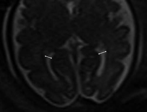
Fig. 6.3
Axial images at the level of the thalami measuring normal lateral ventricles, (Double sided arrow) at 34 weeks gestational age

Fig. 6.4
(a) Sagittal midline image demonstrating the vermian length (solid double arrow), inferior vermian distance (small arrow), and corpus callosum length in a 35-week fetus. (b) Axial image through the posterior fossa in a 34-week fetus demonstrating measurement of the transverse cerebellar distance (dashed arrow) and cisterna magna (solid arrow)
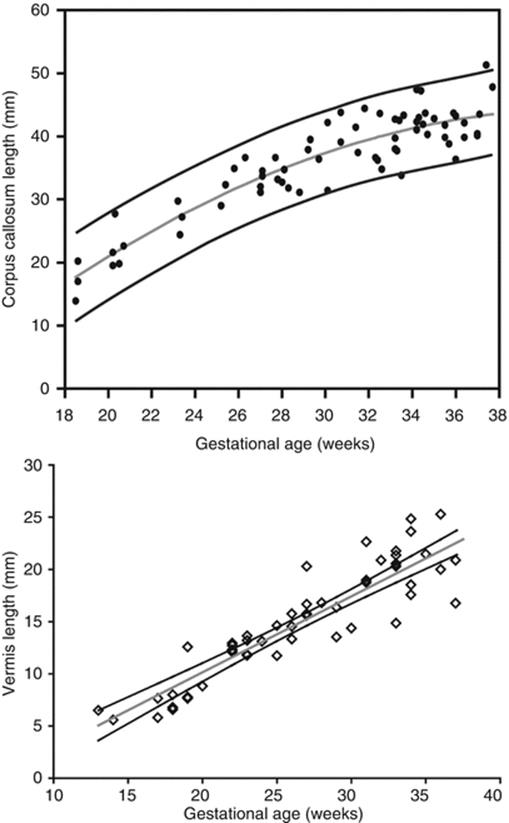
Fig. 6.5
(a) Nomogram depicting corpus callosum length by gestational age (Adapted from Harreld et al. [62]). (b) Nomogram depicting vermian length by gestational age (Twickler DM, unpublished data 2005)
6.4 Cortical Development
Cortical neurons migrate from the germinal zone, the inner most cortical layer, to form the cortical surface. Migration begins around 7 weeks and is mostly complete by 22 weeks [30]. During this span the normal germinal matrix can be seen as a T2 hypointense periventricular band, matching the cortical surface (Fig. 6.6). The developing white matter tissue between the germinal zone and cortical surface, or intermediate zones, demonstrates the opposite signal characteristics, T2 hyperintensity and T1 hypointensity. The germinal zone largely diminishes by 28 weeks [22].

Fig. 6.6
The appearance of the germinal matrix (identified by arrows) at 23 weeks gestational age
Cortical maturation can be evaluated by cerebral gyration and sulcation which tend to occur in a reliable pattern. The MR findings tend to lag behind the actual sulcal development by 1–2 weeks according to pathologic studies [79]. The cerebral hemispheres separate very early in gestation, a process that is typically complete by 12 weeks. The sylvian fissure and lateral sulcus are next to develop and are typically present by 20 weeks gestational age. The parietooccipital fissure is also usually present by 20 weeks. Beyond 20 weeks sulcation and gyration accelerate. The formation of the calcarine fissure is consistently visible around 24 weeks, the central sulcus usually by 26 weeks, precentral by 27 weeks and the postcentral by 28 weeks. Sulcation of the frontal and temporal gyri is usually well underway by 32 weeks and is required for normal development of the sylvian fissures. Some of the major sulcal milestones can be found in Fig. 6.7 and in our opinion are most easily assessed on sagittal and parasagittal images, although all planes can be useful. Twin pregnancies can result in delayed gyration of 2–3 weeks [29].


Fig. 6.7
(a) A 26-week fetus demonstrating parietooccipital sulcus (black arrow), calcarine sulcus (white arrow), central sulcus (white arrowhead), lateral sulcus, and sylvian fissure (long white arrow). (b) 28-week fetus demonstrating central sulcus (white arrow), cingulate sulcus (black arrow), precentral sulcus (long white arrow), and postcentral sulcus (white arrowhead). (c) A 32-week fetus and (d) a 34-week fetus demonstrating progressive interval gyration and sulcation. The black arrow in (c) identifies the developing frontal operculum
Myelination is the final stage of cerebral development, beginning during the second trimester and proceeding into the third decade of life [19]. Diffusion anisotropy has the potential to characterize pre-myelinating structures and can identify the corpus callosum and other white matter tracts [23]. At this point in time, more work needs be done to evaluate clinical utility in the developing fetus.
6.5 CNS Pathologies
6.5.1 Ventriculomegaly
Ventriculomegaly is one of the most common cerebral anomalies in the fetus and likely the most frequent indication for fetal MRI. The term ventriculomegaly describes the finding of lateral ventricular enlargement and is typically more accurate at the point of patient referral than the term hydrocephalus, a diagnostic term describing enlarged ventricles secondary to elevated ventricular pressures. A broad array of diagnoses can result in the imaging appearance of ventriculomegaly, and MRI aids in identifying the etiology and additional structural abnormalities, allowing for more precise patient counseling [107]. Ventriculomegaly is defined by enlargement of one or both ventricular atria to >10 mm (4 standard deviations above the mean) and is subdivided into categories of mild (10–12 mm), moderate (13–15 mm), and severe (>15 mm) [35, 45, 102, 104] (Fig. 6.8).
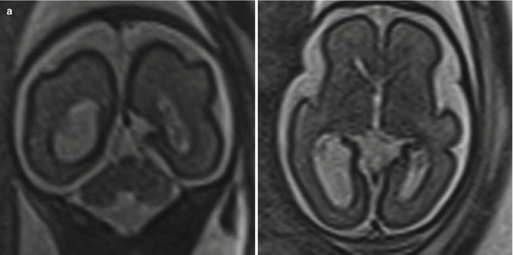

Fig. 6.8
(a) Mild isolated right ventriculomegaly of 12 mm in a 25-week fetus. (b) Moderate bilateral ventriculomegaly measuring 13 mm bilateral in a 29-week fetus with confirmed trisomy 21
The etiologies of fetal ventriculomegaly seen with imaging can be broadly classified into obstructive causes, neurologic malformations, and secondary to destructive processes. Effacement of subarachnoid spaces in the setting of ventriculomegaly is suggestive of obstructive hydrocephalus. Aqueductal stenosis is an obstructive cause of ventriculomegaly and can be recognized by distention of the lateral and third ventricles with a normal-appearing fourth ventricle and posterior fossa. Ventricular distention in aqueductal stenosis typically progresses with gestational age. There are multiple etiologies of aqueductal stenosis, including congenital, associated X-linked or autosomal recessive disorders, hemorrhage, and infection. X-linked aqueductal stenosis is the result of a mutation of Xq28, a gene responsible for a neural cell adhesion molecule [43]. X-linked aqueductal stenosis is nearly always associated with severe mental retardation and can be suggested by imaging in the setting of a male fetus with adducted thumbs [61] (Fig. 6.9). Other obstructive processes include Chiari 2 malformation, intraventricular hemorrhage, and mass effect from cystic or solid cerebral masses. Ventriculomegaly secondary to cerebrospinal fluid overproduction can be seen in the setting of choroid plexus papillomas, benign tumors which can have severe neurologic consequences secondary to their large size, and cerebrospinal fluid hypersecretion [25].
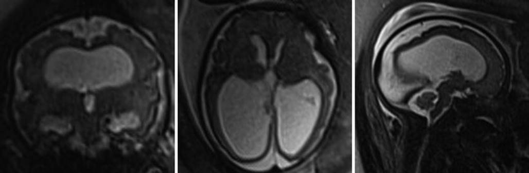
Fig. 6.9
Images of a 31-week male fetus with severe ventriculomegaly but normal-sized fourth ventricle in this patient with X-linked aqueductal stenosis
Additional cerebral anomalies can be found in 50–60 % of fetuses with ventriculomegaly and are more likely in proportion to severity of ventriculomegaly [45, 56, 75, 116]. The most common associated anomalies are Chiari 2 malformation, agenesis of the corpus callosum, Dandy-Walker malformation, holoprosencephaly, mass lesions, and destructive processes [31, 46]. Non-CNS abnormalities are present in one-third of cases [45]. Associated abnormalities are more common with more severe ventriculomegaly and are also associated with a worse prognosis [54, 117].
Isolated ventriculomegaly, cases of ventriculomegaly without additional abnormality, is the most common ultimate diagnosis [92]. Prognosis is dependent on the severity of ventriculomegaly. Normal neurologic outcomes are seen in 93–96 % of mild ventriculomegaly, 76 % of moderate ventriculomegaly, and 28 % of severe ventriculomegaly [46, 104, 116]. The incidence of perinatal and neonatal death also correlates with the degree of ventriculomegaly [45]. Ventricular enlargement throughout pregnancy is associated with increased incidence of developmental delay. Outcomes in asymmetric ventriculomegaly are also dependent on the severity of distention of the affected ventricle and can be equally good or poor when compared to symmetric ventriculomegaly [90, 116]. The risk of aneuploidy is low in the setting of isolated mild ventriculomegaly (1.8 %) [90], and thus far, the severity of ventriculomegaly has not been linked to increased risk of aneuploidy [45]. Intrauterine infection also appears to be an infrequent cause of ventriculomegaly (1.8 %) [90, 116]. Additional imaging findings related to CNS infection will be discussed later in this chapter.
A few authors have found male fetuses to have larger atria than female fetuses on average and a lower prevalence of developmental delay [93, 95, 116]. Other authors have found no significant differences between the sexes, suggesting this data be used with caution when counseling [58, 78].
It should also be noted that extreme ventricular dilatation can result in a thinned corpus callosum. Careful inspection for the presence or absence of the corpus callosum should be made in these cases as absence of the corpus callosum conveys its own associated anomalies.
Malformations of the developing brain can be divided into three broad categories: abnormal neurogenesis, abnormal neuron migration, and abnormal neuronal organization.
6.6 Abnormal Neuronal Migration
6.6.1 Callosal Dysgenesis and Agenesis
Fetal MR provides for the direct visualization of the entirety of the corpus callosum, the largest of the cerebral commissures. Normal development of the corpus callosum is a sensitive indicator for normal brain development. In studies comparing MRI to ultrasound for the evaluation of callosal abnormalities, MRI detects additional CNS abnormalities in up to 22.5 % of cases (most commonly posterior fossa malformations and cortical malformations) [105]. The corpus callosum begins forming around the 11th week of gestation and is typically completed by the 21–22th week [1, 97]. The corpus callosum is composed of five named regions: rostrum, genu, body, isthmus, and splenium. Malformation (hyperplasia, hypoplasia, or agenesis) of any region is termed callosal dysgenesis (Fig. 6.10); complete absence is termed agenesis of the corpus callosum.

Fig. 6.10
Callosal dysgenesis. Sagittal T2-weighted half Fourier RARE of a 31-week male fetus with thinned genu (black arrow) and body (white arrow) of the corpus callosum. The splenium and rostrum are not seen
The causes of callosal dysgenesis and agenesis are numerous and heterogeneous, as commissural formation relies on a complex series of steps that are each prone to insult/error. Complete agenesis is estimated to have a prevalence of 0.01–0.3 % [53, 114] and be found in 2–3 % of individuals with developmental disorders [70].
In complete agenesis both axial and coronal images will fail to depict crossing callosal fibers. The lateral ventricles will appear unusually parallel on axial images and often meet the criteria for moderate or severe ventriculomegaly. Unless ventriculomegaly is progressively worsening throughout pregnancy, concomitant ventricular enlargement does not portend a worse prognosis [51]. The cavum septi pellucidi, a space between thin walls of tissue that connect between the fornices and a normally formed corpus callosum, will not be seen. Coronal images demonstrate teardrop-shaped lateral ventricles (termed colpocephaly). Enlargement and rostral displacement of the third ventricle may appear to communicate to the median interhemispheric fissure. Midsagittal images will depict sulci radiating outward from the expected location of the corpus callosum (Fig. 6.11). Midline interhemispheric cysts are sometimes seen in the expected location of the cavum septi pellucidi and may or may not be in communication with the ventricular system (termed Types 1 and 2, respectively) [16]. If enlarged, these cysts can result in hydrocephalus or macrocephaly. Additional concomitant findings include periventricular nodular heterotopia, pericallosal lipomas, vermian hypoplasia, porencephaly, mega cisterna magna, nodular heterotopia, and abnormalities of sulcation and neuronal migration [76, 106].


Fig. 6.11
(a) T2 half Fourier RARE images of a 32-week fetus with an axial image demonstrating parallel lateral ventricles and colpocephaly, technically consistent with moderate bilateral ventriculomegaly. (b) Sagittal image through the midline depicting the radiating ray appearance. (c) Coronal T2 half Fourier RARE images obtained in a different 31 weeks estimated gestational age demonstrating lack of crossing callosal fibers and the viking helmet or longhorn appearance of the lateral ventricles
Callosal dysgenesis and agenesis are associated with numerous developmental malformations and syndromes, with the prognosis dependent on what additional findings are present. In a large California-based population study of fetal MR findings of callosal dysgenesis and agenesis, chromosomal anomalies were seen in 17.3 %, with 49 % having additional CNS abnormalities including cortical malformations, posterior fossa abnormalities, and intracranial cysts [53, 106]. When MRI, genetic, and serologic workup is negative for additional anomaly, callosal agenesis is described as isolated. Neurodevelopmental outcomes are normal in approximately 75 % of fetuses with isolated callosal agenesis, with the remaining 25 % at increased risk for moderate to severe learning disabilities [51, 105].
6.6.2 Lissencephaly Spectrum
Lissencephaly is a disorder of neuronal production or migration resulting in absence of gyral formation (agyria) or reduced gyration (pachygyria). Because the brain is agyric before 22 weeks, these malformations are often difficult to identify as significantly delayed before 30 weeks. Classic lissencephaly can be both inherited and sporadic, associated with isolated gene defects and Miller-Dieker syndrome – an autosomal dominant defect on chromosome 17p resulting in characteristic dysmorphic facies (wrinkled skin over the glabella, narrow forehead, prominent occiput, and small chin), severe mental retardation, and death in infancy or early childhood (OMIM #247200). In addition to a smooth gyral pattern, ventriculomegaly or colpocephaly and callosal dysgenesis agenesis are also seen in classic lissencephaly [32, 40]. The term variant lissencephaly describes a set of disorders morphologically less severe in appearance as compared to classic lissencephaly but with similarly poor neurologic outcomes [41]. The cobblestone complex (formerly termed Type 2 lissencephaly) refers to a disorganized “cobblestone” cortex and in contradistinction to classic lissencephalies is due to excessive neural migration secondary to defective glycosylation. These syndromes demonstrate a variable degree of pachygyria, gliotic white matter, and cerebellar hypoplasia. Associated diseases include the autosomal recessive Walker-Warburg syndrome, muscle-eye-brain disease, and Fukuyama congenital muscular dystrophy [71, 109].
6.7 Abnormal Postmigrational Organization
6.7.1 Polymicrogyria
Polymicrogyria is the result of abnormal post-migrational organization into numerous gyri and can result from infection (particularly CMV), maternal hypotension, and inherited disorders [10, 27]. Polymicrogyria appears as focal or diffuse abnormal infoldings of the T2 hypointense cortical surface and most commonly involves the frontal lobes and sylvian fissures. Polymicrogyria is also more commonly bilateral (Fig. 6.12). Polymicrogyria is associated with developmental delay, epilepsy, and focal neurologic defects depending on the involved structures [7, 64].

Fig. 6.12
(a) Axial T2 half Fourier RARE images obtained at 35 weeks estimated gestational age depicting bilateral polymicrogyria (arrowheads). (b) Normal axial T2 half Fourier RARE image for comparison
6.7.2 Schizencephaly
Schizencephaly is the result of abnormal neuronal migration and results in parenchymal clefts lined by dysplastic cortex that communicate with the lateral ventricles and can be unilateral or bilateral. Mutations altering the expression of the EMX2 homeobox gene in proliferating neuroblasts have been associated with schizencephaly [24]. Infectious, traumatic, and toxic etiologies are also suggested [89]. In Type 1, or closed-lip schizencephaly, the walls of the cleft are nearly apposed to each other with the parenchymal cleft communicating with the pia-ependymal seam, a structure continuous with the lateral ventricle. In Type 2, or open-lip schizencephaly, large cleft extends from the cortical surface to the lateral ventricle (Fig. 6.13). The identification of gray matter lining the clefts differentiates schizencephaly from large areas of parenchymal destruction seen in porencephaly. Additional findings sometimes seen include absent cavum septum pellucidum and ventriculomegaly. Polymicrogyria and optic hypoplasia are also associated but are usually identified postnatally. The degree of impairment depends on the size, bilaterality of the lesions, and the involved structures, with large bilateral defects having a worse prognosis. Clinical sequelae can include developmental delay, paralysis, and seizures. Seizure disorder but otherwise normal neurodevelopmental outcome has been seen in patients with small closed-lip defects [11]. As many as 47 % of patients with the appearance of an open-lip defect will have a closed-lip defect postnatally [87].

Fig. 6.13
Axial (a) and coronal (b) T2-weighted half Fourier RARE images demonstrating bilateral open-lip schizencephaly in a 37-week gestational age fetus
6.7.3 Subependymal Lesions
Gray matter heterotopia describes the abnormal localization of gray matter as a result of failure of neuronal migration, resulting in periventricular (subependymal), subcortical, and leptomeningeal heterotopia. Periventricular heterotopias occur by sporadic or multiple inherited genetic defects [36]. At imaging nodules of tissue isointense to the cortex outline the ventricles, projecting into the ventricular lumen, and may be focal or diffuse (seen in the autosomal recessive form Xq28 gene defect) (Fig. 6.14) [84]. Care must be taken to not confuse an involuting germinal matrix, which occurs between 26 and 28 weeks gestation, and will not protrude into the lateral ventricles. Subependymal hamartomas of tuberous sclerosis are a differential consideration and appear hyperintense on T1-weighted images [17]. There may be mild to moderate ventriculomegaly and additional findings of callosal agenesis and rarely encephaloceles. Subcortical and leptomeningeal heterotopias also occur, but have rarely been documented antenatally due to the limited spatial resolution of MRI.
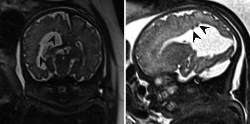
Fig. 6.14
Axial and coronal T2-weighted half Fourier RARE images demonstrating subependymal heterotopia (arrowheads) in a 34-week fetus with right posterior open-lip schizencephaly
6.7.4 Abnormal Neurogenesis
Microcephaly is a descriptive term rather than a diagnosis and may be due to a primary disorder of neurogenesis or secondary to systemic insult or injury. Microcephaly is defined by most as a head circumference less than 3 standard deviations below the mean for gestational age (Fig. 6.15). In the primary forms of microcephaly (microcephaly vera), additional findings often seen include a simplified gyral pattern (microlissencephaly), callosal dysgenesis, and gray matter heterotopia [73]. Secondary microcephaly can be the result of ischemic events, phenylketonuria, fetal alcohol syndrome, infections (CMV, toxoplasmosis), and additional toxins.
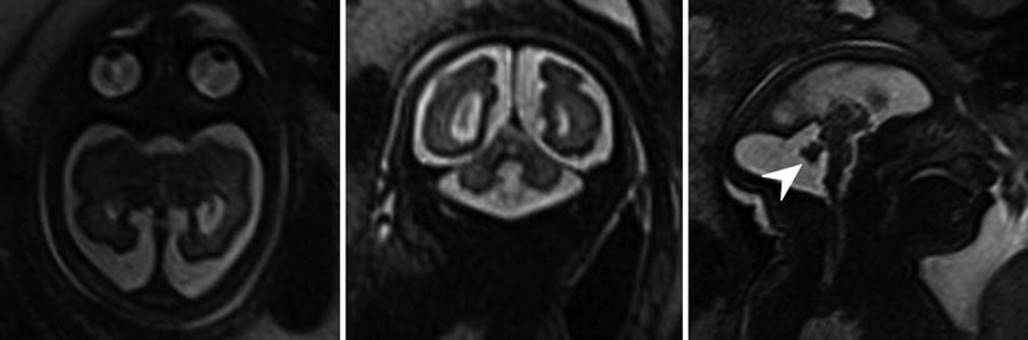
Fig. 6.15
Axial, sagittal, and coronal T2-weighted half Fourier RARE images microcephaly with microlissencephaly in a 35-week gestational age fetus. Anterior sloping of the forehead and vermian hypoplasia (arrowhead) are also seen in this case
Megalencephaly describes increased brain volume and to our knowledge has not been diagnosed antenatally. The few etiologies of megalencephaly described have been collectively categorized as megalencephaly capillary malformation-polymicrogyria syndromes until more is discovered about their pathogenesis [9]. Hemimegalencephaly is hamartomatous overgrowth of a cerebral hemisphere and is distinct from the etiologies of megalencephaly [38]. Hemimegalencephaly is sporadic and also associated with a number of syndromes including Klippel-Trenaunay-Weber, epidermal nevus, and Proteus syndrome. Children typically demonstrate severe mental retardation, seizures, and hemiplegia [37].
6.8 Posterior Fossa Malformations
6.8.1 Fluid Collections of the Posterior Fossa
A common indication for fetal MRI is cystic expansion of the posterior fossa, an ultrasonographic finding that elicits an evaluable differential diagnosis list with different clinical implications and commonly includes Dandy-Walker malformation, hypoplastic cerebellar vermis, Blake pouch cyst, mega cisterna magna, and arachnoid cysts. Depending on the extent of the cystic expansion and associated mass effect, distinguishing between these abnormalities can be challenging and may not be possible if cyst walls are too small for visualization.
A Dandy-Walker malformation is classically described as a triad of partial or complete vermian agenesis, an enlarged posterior fossa with elevation of the tentorium and confluence of sinuses (e.g., torcula), and a large cystic appearing in the fourth ventricle which extends dorsally (Fig. 6.16). Mass effect from the cyst often results in compression of the brainstem against the clivus. Dandy-Walker malformations are associated with de novo deletions of 3q and have not demonstrated Mendelian inheritance patterns [57, 86]. The term Dandy-Walker variant, historically utilized to describe vermian hypoplasia without enlargement of the posterior fossa, has fallen out of favor as more recent embryologic data suggests each of the findings in Dandy-Walker malformation can have different degrees of severity but are attributable to the same genetic aberrations [88]. The term Dandy-Walker complex is preferred for when a similar constellation of findings are present, but to a lesser severity than the findings of true Dandy-Walker malformation. Commonly associated cerebral abnormalities include ventriculomegaly, callosal dysgenesis, occipital encephaloceles, and gray matter heterotopias. Dandy-Walker malformation in the presence of additional developmental defects is often associated with chromosomal abnormalities (up to 46 %) [34, 45]. Neurodevelopmental outcome in children with isolated Dandy-Walker malformation is variable and has most commonly been associated with mental retardation, hypotonia, and epilepsy. In milder expressions of Dandy-Walker complex, outcomes may be normal in 50–80 % of patients , with normal vermian lobulations [47, 77].

Fig. 6.16
Axial and sagittal T2-weighted half Fourier RARE images of a 26-week gestational age fetus with severe vermian hypoplasia (white arrowhead), an elevated torcula (black arrow), and cystic dilatation of the posterior fossa, consistent with Dandy-Walker malformation
A Blake pouch cyst is an ependymal-lined outpouching of the dorsal and inferior membrane of the fourth ventricle (e.g., inferior medullary velum). This results in upward rotation of a normally developed vermis in the setting of a normal appearance of the tentorium and torcula. Blake pouch cysts are included in the Dandy-Walker continuum by some authors, and when very large, it can be difficult to evaluate the vermian integrity. Mass effect from large Blake pouch cysts is possible but extremely rare, and in isolation, these lesions neither have neurodevelopmental consequences nor require treatment [51].
In addition to Blake pouch cyst, mega cisterna magna and arachnoid cysts of the posterior fossa also have a good prognosis, allowing for appropriate patient counseling following their diagnosis. Mega cisterna magna is abundant retrocerebellar fluid within focally enlarged subarachnoid space, defined as a distance between the inner table of the occiput and the cerebellum as >10 mm after 18 weeks gestational age (Fig. 6.17). Mega cisterna magna is in communication with the ventricular system and can be considered a normal variant [20, 120]. Congenital arachnoid cysts are collections of CSF that arise within duplications of the arachnoid membrane (Fig. 6.18). Arachnoid cysts can produce mass effect on the adjacent cerebellum and vermis and can obstruct CSF flow leading to hydrocephalus. Retrocerebellar arachnoid cysts are considered by some to be part of the Dandy-Walker continuum, making a thorough evaluation for additional cerebral anomaly important when an arachnoid cyst is suspected. When very large, arachnoid cysts are managed with shunt placement in infancy but generally have a good prognosis [3]. Approximately 1/3 of both Blake pouch cysts and mega cisterna magna will resolve in utero [47], and the majority will not require further treatment [42].

Fig. 6.17
Axial and coronal T2-weighted half Fourier RARE images of a 34-week gestational age fetus with mega cisterna magna (double headed line) measuring 19 mm

Fig. 6.18
Axial and coronal T2-weighted half Fourier RARE images of a 37-week gestational age fetus with an arachnoid cyst (arrowheads) producing mass effect on the developing cerebellum
The vermis may be aplastic or hypoplastic as identified by comparison with cerebellar gestational biometry. The fourth ventricle is not enlarged in the setting of vermian hypoplasia. The primary fissure, the fissure separating the anterior lobe from the posterior lobe, should be visible by 28 weeks and the secondary fissures of the cerebellum by 32 weeks [3]. Absence of fissures is suggestive of vermian hypoplasia. When hypoplastic the vermis often appears rotated and the inferior vermian distance is increased, but the posterior fossa is not enlarged and the torcula is in a normal location (Fig. 6.19). The cerebellar hemispheres are usually spared, and the transcerebellar width can be normal [3]. Vermian agenesis may be isolated or syndromic. It is currently controversial as to whether a subset of these patients have a mild form of Dandy-Walker complex. Up to one-third of patients diagnosed with suspected isolated vermian hypoplasia by prenatal MRI had a normal vermian length postnatally, suggesting the need for follow-up MRIs in these cases [80]. Rhombencephalosynapsis, vermian aplasia with midline fusion of the cerebellar hemispheres, is a rare malformation that can be identified by a keyhole-shaped fourth ventricle and contiguous transverse cerebellar folia on a coronal view (Fig. 6.20). Rhombencephalosynapsis has been associated with midline supratentorial abnormalities such as holoprosencephaly and septal agenesis. Cognitive impairment is mild or absent, but ataxia and other movement disorders are often present [96]. Joubert’s syndrome and related disorders are in a category of autosomal and X-linked recessive syndromes that also include vermian hypoplasia. In children these syndromes demonstrate the “molar tooth sign” – a mid- and hindbrain malformation that resembles a molar tooth on axial images. Postnatally these patients suffer from ataxia, developmental delay, and oculomotor and respiratory abnormalities [39].

Fig. 6.19
Axial and sagittal T2-weighted half Fourier RARE images of a 21-week gestational age fetus with vermian hypoplasia

Fig. 6.20
Axial, sagittal, and coronal T2-weighted half Fourier RARE images of a 37-week gestational age fetus with rhombencephalosynapsis. The vermis is absent and the cerebellar hemispheres are fused. Sagittal image depicts absence of the primary fissure. White arrowhead demonstrates the “keyhole” appearance of the fourth ventricle. Multiple black arrowhead demonstrates continuous transverse cerebellar folia on the coronal view
A multitude of rare disorders can result in cerebellar hypoplasia, defined by diminished transverse cerebellar width and vermian length. Cerebellar hypoplasia is associated with infection (CMV), aneuploidy, and PHACE syndrome (Posterior fossa abnormalities, Hemangioma, Arterial lesions, Cardiac abnormalities, Eye abnormalities). When there is concomitant brainstem hypoplasia (a diminished appearance of the pons), pontocerebellar hypoplasia is a consideration [50].
6.8.2 Neural Tube Defects
Neural tube closure begins at the embryologic correlate of the cervical spine and proceeds both rostrally and caudally. Neural tube defects affect 0.5–2 per 1000 pregnancies worldwide [85], the most common of which are Chiari 2 malformations.
Chiari 2 malformation describes a failure of rostral neural tube closure resulting in a lumbosacral (but sometimes higher) myelomeningocele and caudal herniation of the cerebellum through the foramen magnum. This obstructs cerebrospinal fluid flow and hydrocephalus. Callosal dysgenesis and ventral displacement of the tentorium cerebelli are commonly seen. Chiari 2 malformations occur in nearly all myelomeningoceles (Fig. 6.21). Associated malformations include callosal dysgenesis, polymicrogyria, and heterotopias [83]. The severity of hindbrain herniation by MRI is significantly associated with childhood seizure activity, high-risk bladder dysfunction, and lack of independent ambulation [28]. Prenatal myelomeningocele repair has been reported to improve motor outcomes but is associated with maternal and fetal risks [4].
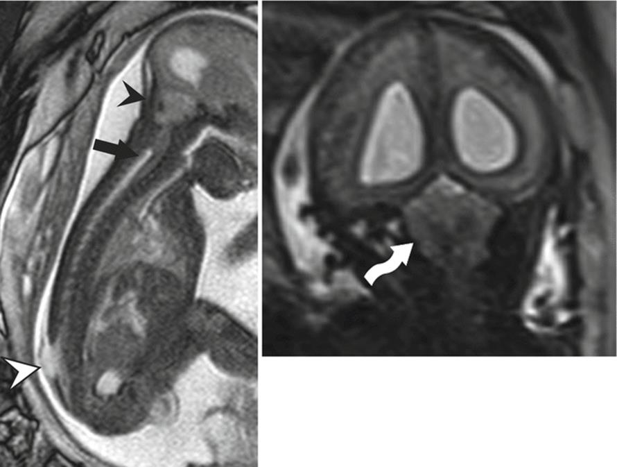
Fig. 6.21
Sagittal true FISP image through the brain and spine in 27-week gestational age fetus depicts a small posterior fossa with effaced subarachnoid spaces (black arrowhead) and herniation of the cerebellar tonsils to approximately the C5–C6 level (black arrow). A lumbar meningocele is present (white arrowhead). Coronal half Fourier RARE image depicts effaced subarachnoid spaces (wavy arrow) and moderate bilateral ventriculomegaly
Chiari 3 malformation describes a caudal neural tube defect with a high cervical/occipital meningoencephalocele, containing parts of the cerebellum and sometimes the occipital lobes (Fig. 6.22). Commonly associated abnormalities include a small posterior fossa with hypoplastic cerebellar structures, ventriculomegaly, and callosal dysgenesis [26]. With surgery, involving the closure of the meningoencephalocele and often shunting, the condition is survivable but the functional prognosis is poor.

Fig. 6.22
Sagittal half Fourier image through the brain and spine in an 18-week gestational age fetus depicting an occipital meningoencephalocele (arrowhead), consistent with a Chiari 3 malformation
Chiari 1 malformation describes the herniation of the cerebellar tonsils through the foramen magnum and a small posterior fossa (some investigators additionally require tonsillar deformity) and is not a neural tube defect. The etiology is controversial but thought to be secondary to underdevelopment of the posterior fossa and alterations in CSF flow. Chiari 1 malformations often demonstrate hydrosyringomyelia of the central canal of the spinal cord (e.g., syrinx). Chiari 1 malformations are rarely diagnosed prenatally.
Of the remaining spectrum of malformations following failure of neural tube closure, the most common is anencephaly. The prevalence of anencephaly in the United States is approximately 2.6 per 10,000 [91]. In anencephaly the rostral neural tube fails to close resulting in failure of calvarial formation. The forebrain is subsequently exposed to amniotic fluid and fails to form or is destroyed. These patients will have elevated serum alpha fetoprotein, and often the diagnosis will be made by ultrasound. When, however, there is diagnostic uncertainty regarding this lethal diagnosis, MRI can be obtained for confirmation.
Cephaloceles occur following failure of mesodermal development and result in a calvarial defect, usually occurring over the midline occiput and containing herniated cranial contents. When herniated brain is present, they are termed encephaloceles. Cephaloceles occur in isolation and are present in some syndromes such as the autosomal recessive Meckel-Gruber, a syndrome with a primary triad of occipital encephalocele, bilateral polycystic kidneys, and hepatic fibrosis [81].
Iniencephaly is an infrequently encountered differential consideration for anencephaly. Iniencephaly demonstrates a variable deficit of the occipital bones resulting in an enlarged foramen magnum, fixed hyperextension of the cervical spine with cervical fusion (termed “stargazer” appearance), and spina bifida or rachischisis of the cervical spine. Associated neurologic defects include microcephaly and encephaloceles. Associated extra-CNS malformations include cleft lip and palate, diaphragmatic hernia, omphalocele, club foot, and cardiovascular malformations [101]. Anencephaly with fixed cervical hyperextension is a differential possibility, but does not have cervical fusion as can be seen in iniencephaly. Klippel-Feil, an inherited condition in which there are two or more fused consecutive cervical vertebrae, is another differential consideration.
6.8.3 Holoprosencephaly
The holoprosencephalies arise from lack of appropriate separation of the prosencephalon into the left and right cerebral hemispheres, resulting in a spectrum of fusion anomalies of the cerebral hemispheres and ventricles across the midline. The prevalence of holoprosencephaly has been estimated at approximately 1:1300 gestations in the United States [72]. Classically there are three types of holoprosencephaly, alobar, semilobar, and lobar. Alobar holoprosencephaly is the result of complete failure of cleavage of the cerebral hemispheres, resulting in union of the cerebral hemispheres and a single monoventricle. The falx and interhemispheric fissure are completely absent and the thalami are commonly fused (Fig. 6.23). Distinguishing between semilobar and lobar holoprosencephaly prenatally can be a challenge as the malformation demonstrates a continuum of findings. In semilobar holoprosencephaly, there is persistent fusion of the anterior cortical structures but partial separation of the posterior cerebral hemispheres and posterior ventricles (Fig. 6.24). The falx is partially present posteriorly. A dorsal cyst may also be present. In lobar holoprosencephaly, the falx can be seen both posteriorly and anteriorly. The frontal lobes are dysmorphic, whereas the temporal lobes are fully formed. The appearance of a fused fornix traveling through the third ventricle has been associated with lobar holoprosencephaly [112] (Fig. 6.25). The septum pellucidum fails to form in all forms of holoprosencephaly. Associated facial dysmorphism is most commonly seen in alobar holoprosencephaly, the most severe of which is cyclopia, but includes hypotelorism and cleft lip/palate. Facial dysmorphism is usually mild or absent in semilobar and lobar holoprosencephaly [94]. Aneuploidies (mainly trisomies 18 and 13) are associated with holoprosencephaly [72]. Additionally mutations in at least 12 genetic loci have been implicated with both recessive and dominant inheritance and variable expressivity [113]. Nongenetic associations of holoprosencephaly include maternal diabetes and ethyl alcohol and retinoic acid exposure [94].
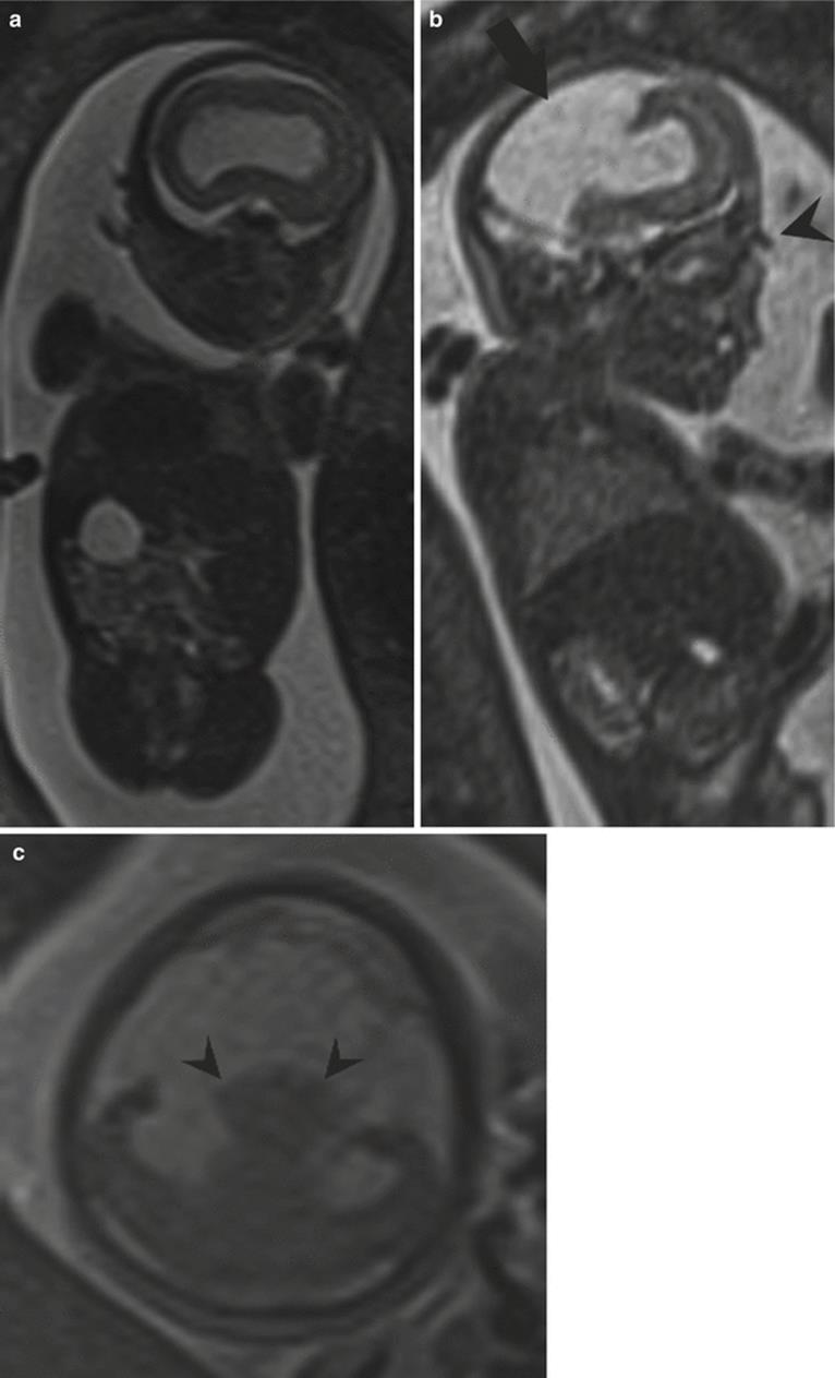
Fig. 6.23
(a) T2-weighted half Fourier RARE images of a 22-week gestational age fetus with alobar holoprosencephaly demonstrating a monoventricle. (b) Sagittal image shows a large dorsal cyst filling the calvarium (black arrow). As is suggested on this slightly parasagittal image, no normal nose was seen. Black arrowhead identifies a small proboscis. (c) Axial image near the level of the skull base demonstrating fused thalami (black arrowheads). This fetus demonstrated no orbital development (not shown)
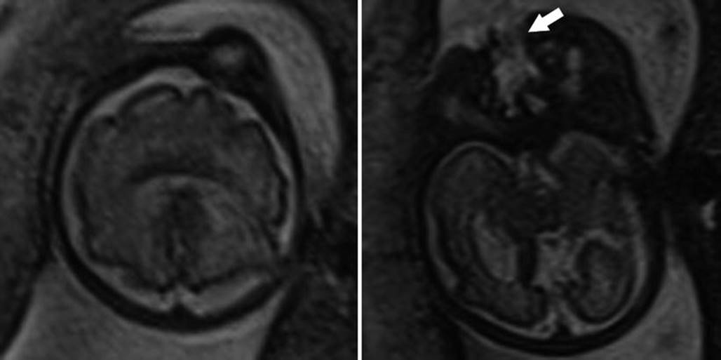
Fig. 6.24
Two axial T2-weighted half Fourier RARE images of a 20-week gestational age fetus with semilobar holoprosencephaly demonstrating contiguous frontal lobes with a monoventricle but division of the hemispheres posteriorly. A midline facial cleft was present (white arrow)
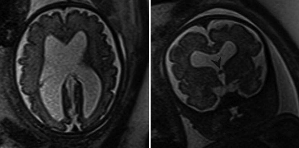
Fig. 6.25
Axial and coronal T2-weighted half Fourier RARE images of a 30-week gestational age fetus with lobar holoprosencephaly demonstrating apparent division of the cerebral hemispheres with large communication of the lateral ventricles with the third ventricle. A fused fornix appears to travel within the third ventricle (black arrowhead)
Middle interhemispheric variant of holoprosencephaly (also known as syntelencephaly) demonstrates fused posterior frontal and parietal lobes, with separation of the anterior frontal lobes, occipital lobes, and basal forebrain (Fig. 6.26). The genu and splenium of the corpus callosum typically form, whereas the body of the corpus callosum is usually absent [13]. A large dorsal cyst may also be present.

Fig. 6.26
(a) Axial T2-weighted half Fourier RARE image of a 34-week gestational age fetus with middle interhemispheric variant of holoprosencephaly. The falx is present both anteriorly and posteriorly, and the deep gray nuclei are separated. (b) Axial image near the apex demonstrates non-separation of the posterior frontal lobes (white arrowhead). (c) Coronal image depicting both the posterior frontal lobe continuity (black arrowhead) and ample communication between the lateral and third ventricles
6.9 Destructive Processes
6.9.1 Ischemia and Hemorrhage
The sequela of ischemic injury is dependent upon stage in pregnancy. Hydranencephaly, the appearance of cystic cavities replacing the bilateral cerebral hemispheres in the setting of a normal falx, is believed to be the result of bilateral internal carotid artery occlusion early in the second trimester. Smaller ischemic events during the second trimester can also interfere with neuronal migration resulting in abnormalities such as polymicrogyria and schizencephaly [15, 89]. An insult occurring early to mid-second trimester can also result in a porencephalic cyst, a smooth margined cavity within the parenchyma that does not undergo a reparative process due to early gestational age. The periventricular germinal zone is most active between 13 and 26 weeks gestation. Ischemic events occurring during this time result in periventricular leukomalacia, a form of hemorrhagic necrosis. By 26 gestational weeks, the brain has developed reparative processes and infarcts may demonstrate smaller cystic components and septations. Before 34–36 weeks, fragile immature penetrating arteries from the cortical surface supply the periventricular white matter, resulting in increased vulnerability to hypoxic and traumatic events [6, 111]. Around 34–36 weeks, deep cerebrovascular supply begins to develop and take over perfusion of the deep cerebral structures, and the territory supplied by cortical vasculature begins to recede to the cortical surface, resulting in increased sensitivity of the subcortical white matter during this period [49].
There are maternal, placental, and fetal etiologies of CNS ischemic injury. Maternal causes commonly include preeclampsia, hypoxia, hypotension, drug use, and abdominal trauma. Fetal causes include infection, thrombocytopenia, and anasarca [49, 100]. In the setting of monochorionic multiple gestation, the in utero demise of a fetal twin places the surviving twin at risk for ischemic events, suspected to be secondary to hypoperfusion or embolic events [67, 69].
The imaging appearance of ischemia can be subtle, especially when diffuse, due to the inherently high signal on T2-weighted images in normal parenchyma. Ischemic injury is suggested by focal or asymmetric T2 hyperintensity and by loss of gray white differentiation if the brain has matured sufficiently. Laminar necrosis and periventricular leukomalacia can appear hyperintense on T1-weighted images [21, 44, 49]. With time post injury, there is ex vacuo dilatation of the ventricle on the injured side. A GRE T2* sequence can be added to evaluate for hemosiderin deposition seen in chronic hemorrhage. The prognosis depends on both the etiology of hemorrhage/infarction and the involved structures.
Intracranial fetal hemorrhage is most often related to ischemia, infections, vascular malformations, and clotting disorders. In the developing brain, reperfusion following infarction very commonly results in capillary rupture and hemorrhage. Intraventricular hemorrhage has a prevalence of 0.04–2.0 %, while intraparenchymal hemorrhage has a prevalence of 0.04–0.4 % [65, 114].
6.9.2 Mass Lesions
Congenital brain tumors, those that are observed prenatally or before the second month of life, are extremely rare. Teratomas are reported to account for half of all congenital tumors. Other tumors listed in decreasing frequency include astrocytomas, lipomas, choroid plexus papillomas, and primitive neuroectodermal tumors [110]. The most common presenting finding regardless of tumor type is macrocephaly. Hydrocephalus, polyhydramnios, and hydrops are also commonly seen. Unlike the pediatric population, intracranial fetal tumors are most often supratentorial. It can be difficult to distinguish the intracranial tumors prenatally. Teratomas often have a complex cystic and solid appearance and sometimes contain fat or calcification (Fig. 6.27). A T2* sequence can be helpful in identifying associated hemorrhage, a finding most commonly seen in astrocytomas [118]. Choroid plexus papillomas are lobular intraventricular masses and can overproduce CSF resulting in hydrocephalus (Fig. 6.28). Midline intracranial lipomas are benign tumors associated with agenesis of the corpus callosum and will depict signal characteristics of fat (hyperintense signal on T1 and T2). Excepting choroid plexus papillomas and lipomas, the prognosis of an intracranial tumor is poor, with tumors identified earlier in gestation having even worse outcomes [118].

Fig. 6.27
Axial and sagittal half Fourier RARE images through the brain of a 28-week gestational age fetus with giant intracranial teratoma

Fig. 6.28
Axial and sagittal half Fourier RARE images in a 31-week gestational age fetus demonstrating heterogeneous cystic and solid choroid plexus papilloma (arrowheads) resulting in severe hydrocephalus secondary to overproduction of cerebrospinal fluid
6.9.3 Vein of Galen Aneurysmal Malformations
Vein of Galen aneurysmal malformations (VGAMs) describes an arteriovenous fistula between either anterior or posterior cerebral arterial circulation and an aberrantly persistent prosencephalic vein, occurring in the expected location of the vein of Galen. Aneurysmal dilatation of the vein of Galen is usually found. On MRI this will appear as an oval-shaped structure posterior to the third ventricle, hypointense on T1 and T2 images secondary to flow voids (Fig. 6.29). Clinical sequelae depend on the extent of arteriovenous shunting and can result in high output heart failure and hydrops. Hydrocephalus is also frequently seen and is attributed to mass effect on the aqueduct of Sylvius. Infarction and subsequent porencephaly is another associated finding. When the malformation is large or there is extensive shunting, the prognosis is poor [66].

Fig. 6.29
Axial and sagittal T2-weighted half Fourier RARE images in 39-week gestational age fetus with a vein of Galen malformation (arrowheads). Hypointensity within the dilatated structure is indicative of a flow void
6.9.4 Infection
Toxoplasma gondii, Rubella, Cytomegalovirus (CMV), and Herpes virus, collectively summarized by the acronym TORCH, are historically the most common infections associated with congenital malformation and can have devastating neurologic sequelae. The “O” in TORCH represents an “other” category, including syphilis, parvovirus B19, and more recently lymphocytic choriomeningitis (LCM virus). The most common congenital infection today is cytomegalovirus (CMV), accounting for over half of all prenatal infections with an estimated annual incidence of 2.9–10 per 1000 live births (90 % of which are asymptomatic) [5]. Toxoplasma gondii infection is the second most frequently encountered infection, with rates of 0.1–1 per 1000 live births. In countries where immunization against Rubella is widely available, congenital rubella has nearly been eliminated, but does still remain a problem in developing nations [5, 82]. Congenital syphilis does present with certain findings on antenatal imaging, but none have been observed in the central nervous system. The remaining congenital infections are less frequently encountered.
The CNS findings of prenatal infection overlap, and differentiation between infection and syndromic malformation is difficult by imaging alone. The sequela and severity of the neurologic injury vary depending on the point of infection during gestation, with earlier infections resulting in more severe injury. Early second trimester CMV infection is speculated to disrupt neuronal migration and result in cortical malformations such as agyria and lissencephaly, cerebellar hypoplasia, marked ventriculomegaly, and microcephaly. CMV infection after 26 weeks is associated with polymicrogyria, focal temporal lobe and periventricular T2 hyperintensities, or symmetric and less pronounced ventriculomegaly [12]. Septations within the ventricles can be suggestive of ventriculitis but are also sometimes seen following intraventricular hemorrhage. The periventricular calcifications well associated with congenital CMV are poorly visualized with MR as compared to ultrasound, but MR has been shown to be more sensitive to cortical gyration abnormalities [33]. Less commonly, cerebellar hemorrhage has been associated with CMV infection. Toxoplasmosis lacks the gyration abnormalities seen in CMV per neonatal literature [14]. Microcephaly, intracranial calcifications, and ventriculomegaly have also been seen in the setting of toxoplasmosis, congenital rubella, herpes simplex, and LCM virus [5, 14, 119]. MRI findings may also be normal in congenital infection [8].
6.10 Future Directions
Thus far, the majority of clinical fetal neuroimaging is accomplished through morphologic analysis, yet many physiologic imaging techniques are beginning to show promise. In pregnant sheep, blood oxygen level-dependent MRI has been utilized to evaluate cerebral oxygenation [103, 115]. Spectroscopy has been utilized to evaluate brain maturity [18]. Functional MRI has been used to delineate developing fiber tracks such as the corpus callosum to whole brain maps [68, 74]. These applications are promising but require increased validation before entering the clinical realm.
References
1.
Achiron R, Achiron A (2001) Development of the human fetal corpus callosum: a high-resolution, cross-sectional sonographic study. Ultrasound Obstet Gynecol 18(4):343–347. doi:10.1046/j.0960-7692.2001.00512.xPubMed
2.
ACR–SPR Practice Parameter for the Safe and Optimal Performance of Fetal Magnetic Resonance Imaging (MRI). Amended 2014 (Resolution 39)
3.
Adamsbaum C, Moutard ML, Andre C, Merzoug V, Ferey S, Quere MP, Lewin F, Fallet-Bianco C (2005) MRI of the fetal posterior fossa. Pediatr Radiol 35(2):124–140. doi:10.1007/s00247-004-1316-3PubMed
4.
Adzick NS, Thom EA, Spong CY, Brock JW 3rd, Burrows PK, Johnson MP, Howell LJ, Farrell JA, Dabrowiak ME, Sutton LN, Gupta N, Tulipan NB, D’Alton ME, Farmer DL, Investigators M (2011) A randomized trial of prenatal versus postnatal repair of myelomeningocele. N Engl J Med 364(11):993–1004. doi:10.1056/NEJMoa1014379PubMedCentralPubMed
5.
Bale JF Jr (2009) Fetal infections and brain development. Clin Perinatol 36(3):639–653. doi:10.1016/j.clp.2009.06.005PubMed
6.
Ballabh P (2010) Intraventricular hemorrhage in premature infants: mechanism of disease. Pediatr Res 67(1):1–8. doi:10.1203/PDR.0b013e3181c1b176PubMedCentralPubMed
7.
Barkovich AJ (2010) Current concepts of polymicrogyria. Neuroradiology 52(6):479–487. doi:10.1007/s00234-009-0644-2PubMedCentralPubMed
8.
Barkovich AJ, Girard N (2003) Fetal brain infections. Childs Nerv Syst 19(7–8):501–507. doi:10.1007/s00381-003-0763-8PubMed
9.
Barkovich AJ, Guerrini R, Kuzniecky RI, Jackson GD, Dobyns WB (2012) A developmental and genetic classification for malformations of cortical development: update 2012. Brain. doi:10.1093/brain/aws019
10.
Barkovich AJ, Hevner R, Guerrini R (1999) Syndromes of bilateral symmetrical polymicrogyria. AJNR Am J Neuroradiol 20(10):1814–1821PubMed
11.
Barkovich AJ, Kjos BO (1992) Schizencephaly: correlation of clinical findings with MR characteristics. AJNR Am J Neuroradiol 13(1):85–94PubMed
12.
Barkovich AJ, Lindan CE (1994) Congenital cytomegalovirus infection of the brain: imaging analysis and embryologic considerations. AJNR Am J Neuroradiol 15(4):703–715PubMed
13.
Barkovich AJ, Quint DJ (1993) Middle interhemispheric fusion: an unusual variant of holoprosencephaly. AJNR Am J Neuroradiol 14(2):431–440PubMed
14.
Barkovich AJ, Raybaud C (2012) Pediatric neuroimaging. Wolters Kluwer/Lippincott Williams & Wilkins, Philadelphia [etc.]
15.
Barkovich AJ, Rowley H, Bollen A (1995) Correlation of prenatal events with the development of polymicrogyria. AJNR Am J Neuroradiol 16(4 Suppl):822–827PubMed
16.
Barkovich AJ, Simon EM, Walsh CA (2001) Callosal agenesis with cyst: a better understanding and new classification. Neurology 56(2):220–227. doi:10.1212/wnl.56.2.220PubMed
17.
Baron Y, Barkovich AJ (1999) MR imaging of tuberous sclerosis in neonates and young infants. AJNR Am J Neuroradiol 20(5):907–916PubMed
18.
Bartha AI, Yap KR, Miller SP, Jeremy RJ, Nishimoto M, Vigneron DB, Barkovich AJ, Ferriero DM (2007) The normal neonatal brain: MR imaging, diffusion tensor imaging, and 3D MR spectroscopy in healthy term neonates. AJNR Am J Neuroradiol 28(6):1015–1021. doi:10.3174/ajnr.A0521PubMed
19.
Benes FM, Turtle M, Khan Y, Farol P (1994) Myelination of a key relay zone in the hippocampal formation occurs in the human brain during childhood, adolescence, and adulthood. Arch Gen Psychiatry 51(6):477–484PubMed
20.
Bolduc ME, Limperopoulos C (2009) Neurodevelopmental outcomes in children with cerebellar malformations: a systematic review. Dev Med Child Neurol 51(4):256–267. doi:10.1111/j.1469-8749.2008.03224.xPubMed
21.
Brisse H, Fallet C, Sebag G, Nessmann C, Blot P, Hassan M (1997) Supratentorial parenchyma in the developing fetal brain: in vitro MR study with histologic comparison. AJNR Am J Neuroradiol 18(8):1491–1497PubMed
22.
Brisse H, Fallet C, Sebag G, Nessmann C, Blot P, Hassan M (1997) Supratentorial parenchyma in the developing fetal brain: in vitro MR study with histologic comparison. Am J Neuroradiol 18(8):1491–1497PubMed
23.
Brugger PC, Stuhr F, Lindner C, Prayer D (2006) Methods of fetal MR: beyond T2-weighted imaging. Eur J Radiol 57(2):172–181. doi:10.1016/j.ejrad.2005.11.017PubMed
24.
Brunelli S, Faiella A, Capra V, Nigro V, Simeone A, Cama A, Boncinelli E (1996) Germline mutations in the homeobox gene EMX2 in patients with severe schizencephaly. Nat Genet 12(1):94–96PubMed
25.
Buxton N, Punt J (1997) Choroid plexus papilloma producing symptoms by secretion of cerebrospinal fluid. Pediatr Neurosurg 27(2):108–111PubMed
26.
Caldarelli M, Rea G, Cincu R, Di Rocco C (2002) Chiari type III malformation. Childs Nerv Syst 18(5):207–210. doi:10.1007/s00381-002-0579-yPubMed
27.
Chang BS, Piao X, Bodell A, Basel-Vanagaite L, Straussberg R, Dobyns WB, Qasrawi B, Winter RM, Innes AM, Voit T, Grant PE, Barkovich AJ, Walsh CA (2003) Bilateral frontoparietal polymicrogyria: clinical and radiological features in 10 families with linkage to chromosome 16. Ann Neurol 53(5):596–606. doi:10.1002/ana.10520PubMed
28.
Chao TT, Dashe JS, Adams RC, Keefover-Hicks A, McIntire DD, Twickler DM (2010) Central nervous system findings on fetal magnetic resonance imaging and outcomes in children with spina bifida. Obstet Gynecol 116(2 Pt 1):323–329. doi:10.1097/AOG.0b013e3181e666e8PubMed
29.
Chi JG, Dooling EC, Gilles FH (1977) Gyral development of the human brain. Ann Neurol 1(1):86–93. doi:10.1002/ana.410010109PubMed
30.
Chong BW, Babcook CJ, Salamat MS, Nemzek W, Kroeker D, Ellis WG (1996) A magnetic resonance template for normal neuronal migration in the fetus. Neurosurgery 39(1):110–116PubMed
31.
D’Addario V, Pinto V, di Cagno L, Pintucci A (2007) Sonographic diagnosis of fetal cerebral ventriculomegaly: an update. J Matern Fetal Neonatal Med 20(1):7–14. doi:10.1080/14767050601036188PubMed
32.
Dobyns WB, Reiner O, Carrozzo R, Ledbetter DH (1993) Lissencephaly: a human brain malformation associated with deletion of the lis1 gene located at chromosome 17p13. JAMA 270(23):2838–2842. doi:10.1001/jama.1993.03510230076039PubMed
33.
Doneda C, Parazzini C, Righini A, Rustico M, Tassis B, Fabbri E, Arrigoni F, Consonni D, Triulzi F (2010) Early cerebral lesions in cytomegalovirus infection: prenatal MR imaging. Radiology 255(2):613–621. doi:10.1148/radiol.10090749PubMed
34.
Ecker JL, Shipp TD, Bromley B, Benacerraf B (2000) The sonographic diagnosis of Dandy–Walker and Dandy–Walker variant: associated findings and outcomes. Prenat Diagn 20(4):328–332. doi:10.1002/(SICI)1097-0223(200004)20:4<328::AID-PD806>3.0.CO;2-OPubMed
35.
Falip C, Blanc N, Maes E, Zaccaria I, Oury JF, Sebag G, Garel C (2007) Postnatal clinical and imaging follow-up of infants with prenatal isolated mild ventriculomegaly: a series of 101 cases. Pediatr Radiol 37(10):981–989. doi:10.1007/s00247-007-0582-2PubMed
36.
Ferland RJ, Batiz LF, Neal J, Lian G, Bundock E, Lu J, Hsiao YC, Diamond R, Mei D, Banham AH, Brown PJ, Vanderburg CR, Joseph J, Hecht JL, Folkerth R, Guerrini R, Walsh CA, Rodriguez EM, Sheen VL (2009) Disruption of neural progenitors along the ventricular and subventricular zones in periventricular heterotopia. Hum Mol Genet 18(3):497–516. doi:10.1093/hmg/ddn377PubMedCentralPubMed
37.
Flores-Sarnat L, Sarnat HB (2002) Hemimegalencephaly: part 1. Genetic, clinical, and imaging aspects. J Child Neurol 17(5):373PubMed
38.
Flores-Sarnat L, Sarnat HB, Dávila-Gutiérrez G, Álvarez A (2003) Hemimegalencephaly: part 2. Neuropathology suggests a disorder of cellular lineage. J Child Neurol 18(11):776–785. doi:10.1177/08830738030180111101PubMed
39.
Fluss J, Blaser S, Chitayat D, Akoury H, Glanc P, Skidmore M, Raybaud C (2006) Molar tooth sign in fetal brain magnetic resonance imaging leading to the prenatal diagnosis of Joubert syndrome and related disorders. J Child Neurol 21(4):320–324. doi:10.1177/08830738060210041001PubMed
40.
Fong KW, Ghai S, Toi A, Blaser S, Winsor EJ, Chitayat D (2004) Prenatal ultrasound findings of lissencephaly associated with Miller-Dieker syndrome and comparison with pre- and postnatal magnetic resonance imaging. Ultrasound Obstet Gynecol 24(7):716–723. doi:10.1002/uog.1777PubMed
41.
Forman MS, Squier W, Dobyns WB, Golden JA (2005) Genotypically defined lissencephalies show distinct pathologies. J Neuropathol Exp Neurol 64(10):847–857PubMed
42.
Forzano F, Mansour S, Ierullo A, Homfray T, Thilaganathan B (2007) Posterior fossa malformation in fetuses: a report of 56 further cases and a review of the literature. Prenat Diagn 27(6):495–501. doi:10.1002/pd.1722PubMed
43.
Fransen E, Vits L, Camp GV, Willems PJ (1996) The clinical spectrum of mutations in L1, a neuronal cell adhesion molecule. Am J Med Genet 64(1):73–77. doi:10.1002/(SICI)1096-8628(19960712)64:1<73::AID-AJMG11>3.0.CO;2-PPubMed
44.
Fusch C, Ozdoba C, Kuhn P, Durig P, Remonda L, Muller C, Kaiser G, Schroth G, Moessinger AC (1997) Perinatal ultrasonography and magnetic resonance imaging findings in congenital hydrocephalus associated with fetal intraventricular hemorrhage. Am J Obstet Gynecol 177(3):512–518PubMed
45.
Gaglioti P, Danelon D, Bontempo S, Mombro M, Cardaropoli S, Todros T (2005) Fetal cerebral ventriculomegaly: outcome in 176 cases. Ultrasound Obstet Gynecol 25(4):372–377. doi:10.1002/uog.1857PubMed
46.
Gaglioti P, Oberto M, Todros T (2009) The significance of fetal ventriculomegaly: etiology, short- and long-term outcomes. Prenat Diagn 29(4):381–388. doi:10.1002/pd.2195PubMed
47.
Gandolfi Colleoni G, Contro E, Carletti A, Ghi T, Campobasso G, Rembouskos G, Volpe G, Pilu G, Volpe P (2012) Prenatal diagnosis and outcome of fetal posterior fossa fluid collections. Ultrasound Obstet Gynecol 39(6):625–631. doi:10.1002/uog.11071PubMed
48.
Garel C, Alberti C (2006) Coronal measurement of the fetal lateral ventricles: comparison between ultrasonography and magnetic resonance imaging. Ultrasound Obstet Gynecol 27(1):23–27. doi:10.1002/uog.2666PubMed
49.
Garel C, Delezoide AL, Elmaleh-Berges M, Menez F, Fallet-Bianco C, Vuillard E, Luton D, Oury JF, Sebag G (2004) Contribution of fetal MR imaging in the evaluation of cerebral ischemic lesions. AJNR Am J Neuroradiol 25(9):1563–1568PubMed
50.
Garel C, Fallet-Bianco C, Guibaud L (2011) The fetal cerebellum: development and common malformations. J Child Neurol 26(12):1483–1492. doi:10.1177/0883073811420148PubMed
51.
Garel C, Moutard ML (2014) Main congenital cerebral anomalies: how prenatal imaging AIDS counseling. Fetal Diagn Ther 35(4):229–239. doi:10.1159/000358519PubMed
52.
Garel CR, Garel CR (2004) MR imaging of the fetal brain : normal development and cerebral pathologies. Springer, Berlin/New York
53.
Glass HC, Shaw GM, Ma C, Sherr EH (2008) Agenesis of the corpus callosum in California 1983–2003: a population-based study. Am J Med Genet A 146A(19):2495–2500. doi:10.1002/ajmg.a.32418PubMedCentralPubMed
54.
Graham E, Duhl A, Ural S, Allen M, Blakemore K, Witter F (2001) The degree of antenatal ventriculomegaly is related to pediatric neurological morbidity. J Matern Fetal Neonatal Med 10(4):258–263. doi:10.1080/jmf.10.4.258.263
55.
Gray DL, Songster GS, Parvin CA, Crane JP (1989) Cephalic index: a gestational age-dependent biometric parameter. Obstet Gynecol 74(4):600–603PubMed
56.
Griffiths PD, Reeves MJ, Morris JE, Mason G, Russell SA, Paley MN, Whitby EH (2010) A prospective study of fetuses with isolated ventriculomegaly investigated by antenatal sonography and in utero MR imaging. AJNR Am J Neuroradiol 31(1):106–111. doi:10.3174/ajnr.A1767PubMed
57.
Grinberg I, Northrup H, Ardinger H, Prasad C, Dobyns WB, Millen KJ (2004) Heterozygous deletion of the linked genes ZIC1 and ZIC4 is involved in Dandy-Walker malformation. Nat Genet 36(10):1053–1055. doi:http://www.nature.com/ng/journal/v36/n10/suppinfo/ng1420_S1.htmlPubMed
58.
Haddad S, Peleg D, Matilsky M, Ben-Ami M (2001) Cerebral lateral ventricular atrial diameter of male and female fetuses at 20–24 weeks’ gestation. Ultrasound Obstet Gynecol 18(2):155–156. doi:10.1046/j.1469-0705.2001.00418.xPubMed
59.
Hadlock FP, Deter RL, Harrist RB, Park SK (1984) Estimating fetal age: computer-assisted analysis of multiple fetal growth parameters. Radiology 152(2):497–501. doi:10.1148/radiology.152.2.6739822PubMed
60.
Haimovici JA, Doubilet PM, Benson CB, Frates MC (1997) Clinical significance of isolated enlargement of the cisterna magna (>10 mm) on prenatal sonography. J Ultrasound Med 16(11):731–734PubMed
61.
Halliday J, Chow CW, Wallace D, Danks DM (1986) X linked hydrocephalus: a survey of a 20 year period in Victoria, Australia. J Med Genet 23(1):23–31PubMedCentralPubMed
62.
Harreld JH, Bhore R, Chason DP, Twickler DM (2011) Corpus callosum length by gestational age as evaluated by fetal MR imaging. Am J Neuroradiol 32(3):490–494. doi:10.3174/ajnr.A2310PubMed
63.
Hatab MR, Zaretsky MV, Alexander JM, Twickler DM (2008) Comparison of fetal biometric values with sonographic and 3D reconstruction MRI in term gestations. AJR Am J Roentgenol 191(2):340–345. doi:10.2214/AJR.07.2623PubMed
64.
Hayashi N, Tsutsumi Y, Barkovich AJ (2002) Polymicrogyria without porencephaly/schizencephaly. MRI analysis of the spectrum and the prevalence of macroscopic findings in the clinical population. Neuroradiology 44(8):647–655. doi:10.1007/s00234-002-0793-zPubMed
65.
Heibel M, Heber R, Bechinger D, Kornhuber HH (1993) Early diagnosis of perinatal cerebral lesions in apparently normal full-term newborns by ultrasound of the brain. Neuroradiology 35(2):85–91PubMed
66.
Heuer G, Gabel B, Beslow L, Stiefel M, Schwartz E, Storm P, Ichord R, Hurst R (2010) Diagnosis and treatment of vein of Galen aneurysmal malformations. Childs Nerv Syst 26(7):879–887. doi:10.1007/s00381-009-1063-8PubMed
67.
Hu LS, Caire J, Twickler DM (2006) MR findings of complicated multifetal gestations. Pediatr Radiol 36(1):76–81. doi:10.1007/s00247-005-0021-1PubMed
68.
Jakab A, Schwartz E, Kasprian G, Gruber GM, Prayer D, Schopf V, Langs G (2014) Fetal functional imaging portrays heterogeneous development of emerging human brain networks. Front Hum Neurosci 8:852. doi:10.3389/fnhum.2014.00852PubMedCentralPubMed
69.
Jelin AC, Norton ME, Bartha AI, Fick AL, Glenn OA (2008) Intracranial magnetic resonance imaging findings in the surviving fetus after spontaneous monochorionic cotwin demise. Am J Obstet Gynecol 199(4):398.e391–398.e395. doi:http://dx.doi.org/10.1016/j.ajog.2008.06.062
70.
Jeret JS, Serur D, Wisniewski K, Fisch C (1986) Frequency of agenesis of the corpus-callosum in the developmentally disabled population as determined by computerized-tomography. Pediatr Neurosci 12(2):101–103
71.
Jissendi-Tchofo P, Kara S, Barkovich AJ (2009) Midbrain-hindbrain involvement in lissencephalies. Neurology 72(5):410–418. doi:10.1212/01.wnl.0000333256.74903.94PubMedCentralPubMed
72.
Kagan KO, Staboulidou I, Syngelaki A, Cruz J, Nicolaides KH (2010) The 11-13-week scan: diagnosis and outcome of holoprosencephaly, exomphalos and megacystis. Ultrasound Obstet Gynecol 36(1):10–14. doi:10.1002/uog.7646PubMed
73.
Kaindl AM, Passemard S, Kumar P, Kraemer N, Issa L, Zwirner A, Gerard B, Verloes A, Mani S, Gressens P (2010) Many roads lead to primary autosomal recessive microcephaly. Prog Neurobiol 90(3):363–383. doi:http://dx.doi.org/10.1016/j.pneurobio.2009.11.002PubMed
74.
Kasprian G, Brugger PC, Weber M, Krssak M, Krampl E, Herold C, Prayer D (2008) In utero tractography of fetal white matter development. Neuroimage 43(2):213–224. doi:10.1016/j.neuroimage.2008.07.026PubMed
75.
Kelly EN, Allen VM, Seaward G, Windrim R, Ryan G (2001) Mild ventriculomegaly in the fetus, natural history, associated findings and outcome of isolated mild ventriculomegaly: a literature review. Prenat Diagn 21(8):697–700PubMed
76.
Kim TH, Joh JH, Kim MY, Kim YM, Han KS (2002) Fetal pericallosal lipoma: US and MR findings. Korean J Radiol 3(2):140–143. doi:10.3348/kjr.2002.3.2.140PubMedCentralPubMed
77.
Klein O, Pierre-Kahn A, Boddaert N, Parisot D, Brunelle F (2003) Dandy-Walker malformation: prenatal diagnosis and prognosis. Childs Nerv Syst 19(7–8):484–489. doi:10.1007/s00381-003-0782-5PubMed
78.
Kramer RL, Yaron Y, Johnson MP, Evans MI, Treadwell MC, Wolfe HM (1997) Differences in measurements of the atria of the lateral ventricle: does gender matter? Fetal Diagn Ther 12(5):304–305PubMed
79.
Levine D, Barnes P (1999) Cortical maturation in normal and abnormal fetuses as assessed with prenatal MR imaging. Radiology 210(3):751–758. doi:10.1148/radiology.210.3.r99mr47751PubMed
80.
Limperopoulos C, Robertson RL, Estroff JA, Barnewolt C, Levine D, Bassan H, du Plessis AJ (2006) Diagnosis of inferior vermian hypoplasia by fetal magnetic resonance imaging: potential pitfalls and neurodevelopmental outcome. Am J Obstet Gynecol 194(4):1070–1076. doi:10.1016/j.ajog.2005.10.191PubMedCentralPubMed
81.
Logan C, Abdel-Hamed Z, Johnson C (2011) Molecular genetics and pathogenic mechanisms for the severe ciliopathies: insights into neurodevelopment and pathogenesis of neural tube defects. Mol Neurobiol 43(1):12–26. doi:10.1007/s12035-010-8154-0PubMed
82.
McLean HQ, Fiebelkorn AP, Temte JL, Wallace GS, Centers for Disease C, Prevention (2013) Prevention of measles, rubella, congenital rubella syndrome, and mumps, 2013: summary recommendations of the Advisory Committee on Immunization Practices (ACIP). Morb Mortal Wkly Rep Recomm Rep 62(RR-04):1–34
83.
Miller E, Widjaja E, Blaser S, Dennis M, Raybaud C (2008) The old and the new: supratentorial MR findings in Chiari II malformation. Childs Nerv Syst 24(5):563–575. doi:10.1007/s00381-007-0528-xPubMed
84.
Mitchell LA, Simon EM, Filly RA, Barkovich AJ (2000) Antenatal diagnosis of subependymal heterotopia. Am J Neuroradiol 21(2):296–300PubMed
85.
Mitchell LE (2005) Epidemiology of neural tube defects. Am J Med Genet C: Semin Med Genet 135C(1):88–94. doi:10.1002/ajmg.c.30057
86.
Murray JC, Johnson JA, Bird TD (1985) Dandy-Walker malformation: etiologic heterogeneity and empiric recurrence risks. Clin Genet 28(4):272–283PubMed
87.
Nabavizadeh SA, Zarnow D, Bilaniuk LT, Schwartz ES, Zimmerman RA, Vossough A (2014) Correlation of prenatal and postnatal MRI findings in schizencephaly. Am J Neuroradiol 35(7):1418–1424. doi:10.3174/ajnr.A3872PubMed
88.
Nelson MD Jr, Maher K, Gilles FH (2004) A different approach to cysts of the posterior fossa. Pediatr Radiol 34(9):720–732. doi:10.1007/s00247-004-1253-1PubMed
89.
Oh KY, Kennedy AM, Frias AE Jr, Byrne JL (2005) Fetal schizencephaly: pre- and postnatal imaging with a review of the clinical manifestations. Radiographics 25(3):647–657. doi:10.1148/rg.253045103PubMed
90.
Ouahba J, Luton D, Vuillard E, Garel C, Gressens P, Blanc N, Elmaleh M, Evrard P, Oury JF (2006) Prenatal isolated mild ventriculomegaly: outcome in 167 cases. BJOG 113(9):1072–1079. doi:10.1111/j.1471-0528.2006.01050.xPubMed
91.
Parker SE, Mai CT, Canfield MA, Rickard R, Wang Y, Meyer RE, Anderson P, Mason CA, Collins JS, Kirby RS, Correa A, National Birth Defects Prevention N (2010) Updated National Birth Prevalence estimates for selected birth defects in the United States, 2004–2006. Birth Defects Res A Clin Mol Teratol 88(12):1008–1016PubMed
92.
Patel MD, Filly AL, Hersh DR, Goldstein RB (1994) Isolated mild fetal cerebral ventriculomegaly: clinical course and outcome. Radiology 192(3):759–764. doi:10.1148/radiology.192.3.7520183PubMed
93.
Patel MD, Goldstein RB, Tung S, Filly RA (1995) Fetal cerebral ventricular atrium: difference in size according to sex. Radiology 194(3):713–715. doi:10.1148/radiology.194.3.7862967PubMed
94.
Petryk A, Graf D, Marcucio R (2015) Holoprosencephaly: signaling interactions between the brain and the face, the environment and the genes, and the phenotypic variability in animal models and humans. Wiley Interdiscip Rev Devel Biol 4(1):17–32. doi:10.1002/wdev.161
95.
Pilu G, Falco P, Gabrielli S, Perolo A, Sandri F, Bovicelli L (1999) The clinical significance of fetal isolated cerebral borderline ventriculomegaly: report of 31 cases and review of the literature. Ultrasound Obstet Gynecol 14(5):320–326. doi:10.1046/j.1469-0705.1999.14050320.xPubMed
96.
Poretti A, Alber FD, Bürki S, Toelle SP, Boltshauser E (2009) Cognitive outcome in children with rhombencephalosynapsis. Eur J Paediatr Neurol 13(1):28–33. doi:http://dx.doi.org/10.1016/j.ejpn.2008.02.005PubMed
97.
Rakic P, Yakovlev PI (1968) Development of the corpus callosum and cavum septi in man. J Comp Neurol 132(1):45–72. doi:10.1002/cne.901320103PubMed
98.
Reddy UM, Abuhamad AZ, Levine D, Saade GR, Fetal Imaging Workshop Invited P (2014) Fetal imaging: executive summary of a joint Eunice Kennedy Shriver National Institute of Child Health and Human Development, Society for Maternal-Fetal Medicine, American Institute of Ultrasound in Medicine, American College of Obstetricians and Gynecologists, American College of Radiology, Society for Pediatric Radiology, and Society of Radiologists in Ultrasound Fetal Imaging Workshop. J Ultrasound Med 33(5):745–757. doi:10.7863/ultra.33.5.745PubMed
99.
Reichel TF, Ramus RM, Caire JT, Hynan LS, Magee KP, Twickler DM (2003) Fetal central nervous system biometry on MR imaging. AJR Am J Roentgenol 180(4):1155–1158. doi:10.2214/ajr.180.4.1801155PubMed
100.
Rorke LB, Zimmerman RA (1992) Prematurity, postmaturity, and destructive lesions in utero. Am J Neuroradiol 13(2):517–536PubMed
101.
Sahid S, Sepulveda W, Dezerega V, Gutierrez J, Rodriguez L, Corral E (2000) Iniencephaly: prenatal diagnosis and management. Prenat Diagn 20(3):202–205PubMed
102.
Salomon LJ, Ouahba J, Delezoide AL, Vuillard E, Oury JF, Sebag G, Garel C (2006) Third-trimester fetal MRI in isolated 10- to 12-mm ventriculomegaly: is it worth it? BJOG 113(8):942–947. doi:10.1111/j.1471-0528.2006.01003.xPubMed
103.
Schoennagel BP, Yamamura J, Fischer R, Tavares de Sousa M, Weyhmiller M, Birkelbach M, Kooijman H, Adam G, Wedegaertner U (2015) BOLD MRI in the brain of fetal sheep at 3T during experimental hypoxia. J Magn Reson Imaging 41(1):110–116. doi:10.1002/jmri.24555PubMed
104.
Signorelli M, Tiberti A, Valseriati D, Molin E, Cerri V, Groli C, Bianchi UA (2004) Width of the fetal lateral ventricular atrium between 10 and 12 mm: a simple variation of the norm? Ultrasound Obstet Gynecol 23(1):14–18. doi:10.1002/uog.941PubMed
105.
Sotiriadis A, Makrydimas G (2012) Neurodevelopment after prenatal diagnosis of isolated agenesis of the corpus callosum: an integrative review. Am J Obstet Gynecol 206(4):337.e331–335. doi:10.1016/j.ajog.2011.12.024
106.
Tang PH, Bartha AI, Norton ME, Barkovich AJ, Sherr EH, Glenn OA (2009) Agenesis of the corpus callosum: an MR imaging analysis of associated abnormalities in the fetus. Am J Neuroradiol 30(2):257–263. doi:10.3174/ajnr.A1331PubMed
107.
Twickler DM, Magee KP, Caire J, Zaretsky M, Fleckenstein JL, Ramus RM (2003) Second-opinion magnetic resonance imaging for suspected fetal central nervous system abnormalities. Am J Obstet Gynecol 188(2):492–496. doi:10.1067/mob.2003.100PubMed
108.
Twickler DM, Reichel T, McIntire DD, Magee KP, Ramus RM (2002) Fetal central nervous system ventricle and cisterna magna measurements by magnetic resonance imaging. Am J Obstet Gynecol 187(4):927–931PubMed
109.
van der Knaap MS, Smit LM, Barth PG, Catsman-Berrevoets CE, Brouwer OF, Begeer JH, de Coo IF, Valk J (1997) Magnetic resonance imaging in classification of congenital muscular dystrophies with brain abnormalities. Ann Neurol 42(1):50–59. doi:10.1002/ana.410420110PubMed
110.
Vazquez E, Castellote A, Mayolas N, Carreras E, Peiro J, Enríquez G (2009) Congenital tumours involving the head, neck and central nervous system. Pediatr Radiol 39(11):1158–1172. doi:10.1007/s00247-009-1369-4PubMed
111.
Volpe JJ (2001) Neurobiology of periventricular leukomalacia in the premature infant. Pediatr Res 50(5):553–562PubMed
112.
Volpe P, Campobasso G, De Robertis V, Rembouskos G (2009) Disorders of prosencephalic development. Prenat Diagn 29(4):340–354. doi:10.1002/pd.2208PubMed
113.
Wallis D, Muenke M (2000) Mutations in holoprosencephaly. Hum Mutat 16(2):99–108. doi:10.1002/1098-1004(200008)16:2<99::AID-HUMU2>3.0.CO;2[-‐]0PubMed
114.
Wang LW, Huang CC, Yeh TF (2004) Major brain lesions detected on sonographic screening of apparently normal term neonates. Neuroradiology 46(5):368–373. doi:10.1007/s00234-003-1160-4PubMed
115.
Wedegartner U, Kooijman H, Yamamura J, Frisch M, Weber C, Buchert R, Huff A, Hecher K, Adam G (2010) In vivo MRI measurement of fetal blood oxygen saturation in cardiac ventricles of fetal sheep: a feasibility study. Magn Reson Med 64(1):32–41. doi:10.1002/mrm.22344PubMed
116.
Weichert J, Hartge D, Krapp M, Germer U, Gembruch U, Axt-Fliedner R (2010) Prevalence, characteristics and perinatal outcome of fetal ventriculomegaly in 29,000 pregnancies followed at a single institution. Fetal Diagn Ther 27(3):142–148. doi:10.1159/000304735PubMed
117.
Weichert J, Hartge D, Krapp M, Germer U, Gembruch U, Axt-Fliedner R (2010) Prevalence, characteristics and perinatal outcome of fetal ventriculomegaly in 29,000 pregnancies followed at a single institution. Fetal Diagn Ther 27(3):142–148PubMed
118.
Woodward PJ, Sohaey R, Kennedy A, Koeller KK (2005) From the archives of the AFIP: a comprehensive review of fetal tumors with pathologic correlation. Radiographics 25(1):215–242. doi:10.1148/rg.251045156PubMed
119.
Wright R, Johnson D, Neumann M, Ksiazek TG, Rollin P, Keech RV, Bonthius DJ, Hitchon P, Grose CF, Bell WE, Bale JF Jr (1997) Congenital lymphocytic choriomeningitis virus syndrome: a disease that mimics congenital toxoplasmosis or Cytomegalovirus infection. Pediatrics 100(1), E9PubMed
120.
Yildiz H, Yazici Z, Hakyemez B, Erdogan C, Parlak M (2006) Evaluation of CSF flow patterns of posterior fossa cystic malformations using CSF flow MR imaging. Neuroradiology 48(9):595–605. doi:10.1007/s00234-006-0098-8PubMed