
Neil U. Lall1, Mariana L. Meyers2 and David M. Mirsky3
(1)
Department of Radiology, University of Colorado School of Medicine, Aurora, CO, USA
(2)
Department of Radiology, Children’s Hospital Colorado, University of Colorado School of Medicine, Aurora, CO, USA
(3)
Department of Radiology, Children’s Hospital Colorado, University of Colorado School of Medicine, 13123 East 16th Avenue, B125, Aurora, CO 80045, USA
David M. Mirsky
Email: David.Mirsky@childrenscolorado.org
Keywords
MacrocephalyMicrocephalyCraniosynostosisHypotelorismHypertelorismProptosisMicrophthalmiaColobomaCleft lip and palateMicrognathiaBranchial cystThymic cystRanulaThyroglossal duct cystDermoidEpidermoidGoiterLymphatic malformationHemangiomaTeratomaEpignathusEpulisNeuroblastomaRhabdomyosarcoma
7.1 Indications for MRI of Fetal Skull, Face, and Neck
MRI of the fetal skull, face, and neck is used in the evaluation of numerous entities, many of which cannot be completely assessed with sonography. These anomalies range from dysmorphisms (such as macro-/microcephaly, hypo-/hypertelorism, or cleft lip/palate) to lymphovascular malformations to neoplasms (such as hemangiomas, teratomas, and neuroblastomas). MRI can be particularly helpful in assessing cases with potential airway obstruction to improve delineation of anatomy, an issue of increased importance in planning potential fetal or perinatal surgical intervention and in planning time and type of delivery [1, 2]. Additionally, as central nervous system abnormalities are commonly associated with pathology of skull, face, and neck, MRI also allows for detailed evaluation of intracranial contents for concurrent pathology or extension of extracranial pathology [3].
7.2 Abnormalities of Calvarial Size or Shape
7.2.1 Macrocephaly
Macrocephaly, defined as head circumference greater than two standard deviations above the mean, is most commonly related to benign enlargement of the brain, megalencephaly; however, as macrocephaly can also be a manifestation of underlying pathology, fetal MRI is important in assessing for related abnormalities. Hydrocephalus, hemorrhage, tumor, vein of Galen aneurysm, or other cause of intracranial mass effect may all result in macrocephaly (Fig. 7.1a, b). Generalized disorders of overgrowth represent another category of etiologies of macrocephaly, including Sotos syndrome (cerebral gigantism), Beckwith–Wiedemann syndrome, Weaver syndrome, and achondroplasia [3]. Additionally, macrocephaly has been seen in association with multiple neurocutaneous disorders, most consistently with multiple hemangiomatosis syndrome, CLOVES syndrome (congenital lipomatous overgrowth, vascular malformations, epidermal nevi, and skeletal/scoliosis/spinal anomalies), and Bannayan–Riley–Ruvalcaba syndrome (Fig. 7.2a, b). Though less common, macrocephaly may occur unilaterally due to either hamartomatous enlargement of a single cerebral hemisphere or due to a unilateral cause of intracranial mass effect such as tumor or porencephaly [4].
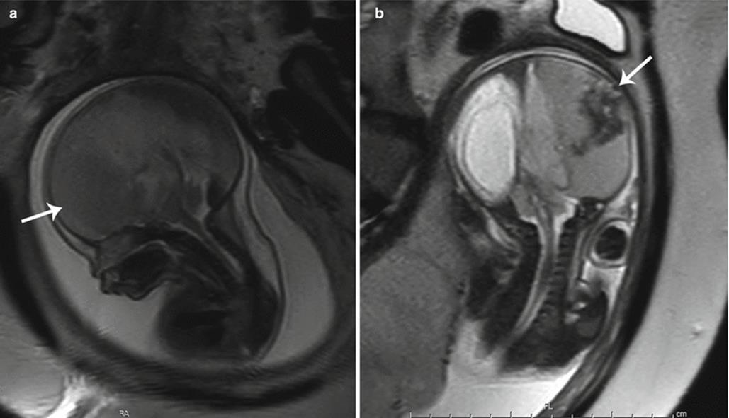
Fig. 7.1
Macrocephaly. (a, b) Sagittal and coronal T2 single-shot haste (SSH) MR images demonstrate an enlarged head secondary to a large left-sided extra-axial hematoma (white arrows)

Fig. 7.2
CLOVES syndrome. (a, b) Axial and coronal T2 SSH MR images of the fetal head demonstrate hemihypertrophy of the right cerebral hemisphere (white arrows). A large venolymphatic malformation (black arrow) of the right axilla was also noted in this patient with CLOVES syndrome
7.2.2 Microcephaly
Microcephaly is a manifestation of an underlying small brain which may be secondary to arrest of growth or due to volume loss from an insult such as infection, ischemia, or hemorrhage (Fig. 7.3a, b). Causes of growth arrest are numerous relating to drugs, maternal disease, and genetic/syndromic disorders among other causes.
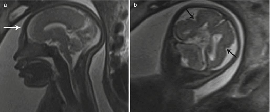
Fig. 7.3
Microcephaly. (a) Sagittal T2 SSH MR image of the fetal brain demonstrates a small cranium (white arrow). (b) Coronal T2 SSH MR of this patient’s brain revealed bilateral perisylvian polymicrogyria (black arrows)
7.2.3 Abnormal Skull Shape
Abnormal calvarial configuration has a number of causes, many of which are related to genetic abnormalities and may have fairly characteristic appearances [3]. A “lemon” shape of the skull has been identified in association with neural tube defects due to loss of subarachnoid spaces with collapse of calvarial bones around the brain (Fig. 7.4). A more triangular “strawberry” configuration is described in association with trisomy 18. Following fetal demise, collapse of the brain may result in overlapping calvarial bones termed Spalding sign. Cases of severe oligohydramnios or anhydramnios may also result in abnormal calvarial morphology, with configuration of the distortion varying depending on the resultant distribution of external pressure.

Fig. 7.4
Lemon-shaped head. Axial T2 SSH MR image illustrates flattening of the bilateral frontal bones (white arrows) in the setting of Chiari II malformation
Craniosynostoses are a specific collection of skull abnormality defined by premature fusion of 1 or more cranial sutures, which prevents calvarial growth in planes perpendicular to the abnormally fused suture(s). A characteristic morphologic abnormality of the skull and face corresponds to each specific fusion location. For example, the most common form of craniosynostosis involves premature fusion of the sagittal suture resulting in generalized elongation of the calvarium with narrowing of the skull width, termed scaphocephaly or dolichocephaly [5]. Coronal craniosynostosis may result in plagiocephaly or brachycephaly, if bilateral, while metopic craniosynostosis results in trigonocephaly (Fig. 7.5). Craniosynostoses may exist independently or as part of a syndrome. Syndromic craniosynostoses include Apert, Crouzon, Pfeiffer, Saethre–Chotzen, Jackson–Weiss, and Antley–Bixler. Apert syndrome is characterized by coronal craniosynostosis with possible involvement of other sutures, midface hypoplasia, and syndactyly of the hands and feet (mitten hands) [6, 7].
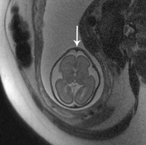
Fig. 7.5
Trigonocephaly. Axial T2 SSH MR image demonstrates a triangular appearance of the forehead secondary to underlying metopic synostosis (white arrow) (Courtesy of Dr. Tamara Feygin, CHOP)
7.3 Scalp Masses
There are numerous causes of fetal scalp masses, the most common being hemangiomas, lymphatic malformations, and congenital inclusion cysts such as dermoids/epidermoids. These masses are not limited strictly to the scalp and are discussed in detail below. Cephalocele is another potential cause of a fetal scalp mass and is an entity in which MRI can be of particular benefit as it may both confirm a calvarial defect (which sonography may falsely detect due to scanning angle) and also define the intracranial anatomy. Evaluation of intracranial anatomy is especially helpful in confirmation of diagnosis of a cephalocele with identification of associated venous sinus anomalies (Fig. 7.6a, b) [8].

Fig. 7.6
Occipital encephalocele. (a) Sagittal T2 SSH MR image shows a midline occipital calvarial defect with herniation of brain and meninges (white arrow). (b) Postnatal sagittal T2-weighted MR image confirms the prenatal findings (white arrow)
7.4 Abnormal Orbits/Globes
7.4.1 Hypotelorism
Hypotelorism is characterized by eyes which are abnormally close together. This can be quantified by measurement of interocular diameter (IOD: medial-to-medial margin of the vitreous) and binocular diameter (BOD: lateral-to-lateral margin of the vitreous) below the 5th percentile for gestational age (Fig. 7.7a, b) [9]. Caution should be used in comparing only to standardized growth charts specifically for MRI measurements, as ultrasound measurements rely upon the bony margins rather than vitreous margins, resulting in larger BOD and smaller IOD when calculated with ultrasound compared to MRI [9, 10]. An alternative simplified approach to recognizing hypotelorism is to compare the IOD to a single orbital width, both of which should be roughly equal.
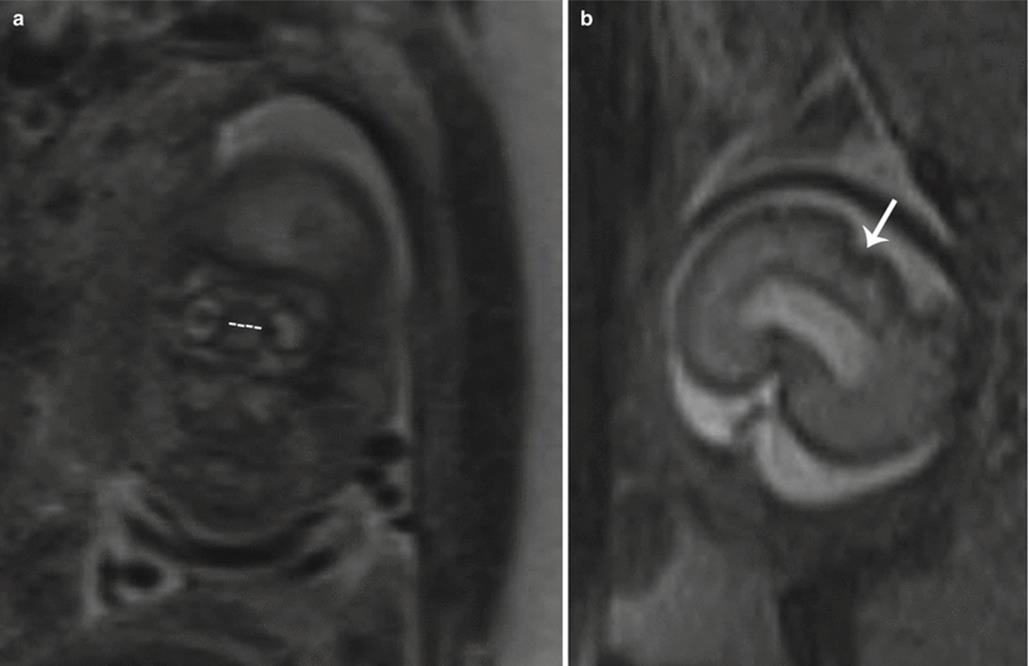
Fig. 7.7
Hypotelorism. (a) Coronal T2 SSH MR image demonstrates a shortened interocular distance (dotted line) in a fetus with trisomy 13. (b) Axial T2 SSH MR image of this patient’s brain revealed lack of normal forebrain cleavage and a monoventricle indicative of holoprosencephaly (white arrow)
As over half of these patients have an underlying aneuploidy, hypotelorism identified on fetal MRI should prompt evaluation for intracranial anomalies. Often associated are midline malformations of the brain such as holoprosencephaly. Secondary hypotelorism may be caused by abnormal calvarial development, such as metopic synostosis or microcephaly [11].
7.4.2 Hypertelorism
Hypertelorism, contrastingly, is characterized by eyes which are abnormally wide set. This is quantified by IOD and BOD above the 95th percentile for gestational age [9]. Similarly, hypertelorism can be inferred when the IOD is larger than the orbital width.
Primary hypertelorism is rarely present in isolation and often is accompanied by an underlying chromosomal derangement or syndrome. Secondary hypertelorism may result due to craniosynostoses, anterior encephalocele (Fig. 7.8a, b), or midline facial masses [11]. Maternal intake of antiepileptic drugs has also been suggested as a cause of hypertelorism.

Fig. 7.8
Hypertelorism. (a) Axial T2 SSH MR image shows a frontonasal encephalocele (white arrow) with consequent widening of the interocular distance. (b) Postnatal axial T2-weighted MR image confirms the findings (white arrow)
7.4.3 Proptosis
Proptosis is defined by anterior displacement of the globes. This appearance may be caused by shallow orbits in association with a craniosynostosis. Rarely, proptosis may be the presenting feature of an underlying orbital mass or orbital encephalocele [3].
7.4.4 Microphthalmia/Anophthalmia
Microphthalmia is characterized by a small globe below the fifth percentile in size for gestational age. Severe microphthalmia may be mistaken for anophthalmia, a far less common entity with complete absence of the globe and the neuroectodermal tissue of the orbit. True anophthalmia may occur from failure of formation of the optic vesicle and requires preservation of non-neuroectodermally derived tissue, such as the eyelids, conjunctiva, lacrimal apparatus, and extraocular muscles; a small cystic structure may be present within the orbit that does not represent a small globe. Fetal MRI is useful in determining the degree of microphthalmia and delineating the internal structure of the globe and orbital contents in addition to assessing for associated brain findings (Fig. 7.9) [12, 13].
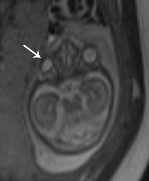
Fig. 7.9
Microphthalmia. Axial T2 SSH MR image demonstrates a small right eye (white arrow) and associated hypertelorism in this patient with the genetic diagnosis of Fasier syndrome
Microphthalmia/anophthalmia is a feature of many hereditary conditions and is frequently seen with aneuploidy, such as trisomy 13 (Patau syndrome). They are also a feature of various syndromes such as Aicardi syndrome, CHARGE syndrome (coloboma, heart defects, choanal atresia, retarded growth and development, genital anomalies, and ear anomalies), Walker–Warburg syndrome, and oculocerebrocutaneous syndrome. Often the syndrome is unknown and a diagnosis of multiple congenital anomalies (MCA) is applied. Microphthalmia/anophthalmia may also be acquired with etiologies including congenital infection, fetal alcohol syndrome, and congenital cataracts.
7.4.5 Optic Disc Coloboma
Optic disc coloboma is characterized by a focal defect of the posterior globe at the site of optic nerve head insertion resulting in outpouching of the vitreous fluid. Coloboma may be sporadic or inherited, with the former typically unilateral and the latter typically bilateral. Coloboma may be seen in association with numerous syndromes including CHARGE syndrome, Walker–Warburg syndrome, Aicardi syndrome, Goldenhar syndrome, and Noonan syndrome [14]. Imaging findings consist of a focal outpouching of high-signal vitreous on T2-weighted imaging at the optic nerve insertion site (Fig. 7.10a, b). A retrobulbar “cyst” that communicates with globe may be present. Optic disc coloboma may be associated with microphthalmia and, at times, retinal detachment [15].
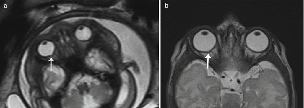
Fig. 7.10
Coloboma. (a) Axial T2 SSH MR image suggested a small outpouching along the posterior aspect of the right eye (white arrow) in this fetus that was otherwise normal. (b) Postnatal axial T2-weighted MR image confirms the suspicion for coloboma (white arrow)
7.4.6 Persistent Hyperplastic Primary Vitreous
Persistent hyperplastic primary vitreous (PHPV), or persistent fetal vasculature, results from a failure of gestational involution of the embryonic hyaloid vascular system within Cloquet’s canal. A triangular retrolental soft tissue mass remains, which, in the majority of cases, extends posteriorly to the optic disc. It is usually isolated and unilateral. Bilateral lesions may be associated with trisomy 13 or syndromes such as Norrie and Walker–Warburg [16]. Postnatally, children with PHPV present with leukocoria and may go on to develop glaucoma, cataract, intraocular hemorrhage, or retinal detachment. As PHPV may be often confused with retinoblastoma, CT imaging may be employed to detect intralesional calcifications which are rare in PHPV.
7.4.7 Neoplastic and Nonneoplastic Masses of the Orbits
Primary tumors of the fetal orbit are rare, with reports of retinoblastoma, teratoma, and sarcoma. More commonly, the orbit is secondarily affected by extension of an intracranial or cervical neoplasm. Depending upon the size and directional growth of the tumor, the globe may be completely obscured and the orbit may be destroyed. MRI is helpful in determining intracranial extension.
While retinoblastoma represents the most common intraocular malignancy of childhood, prenatal detection of congenital retinoblastoma is limited. The orbit is a rare location for primary teratoma development. Orbital teratomas are characteristically massive tumors which appear as complex cystic and solid masses on imaging. Despite being benign tumors, teratomas are locally aggressive and result in severe facial deformity. General characteristics of teratomas are discussed in further detail below under “Cervical Teratomas” and “Epignathus.”
At the most benign end of the spectrum, a dacryocystocele, or lacrimal duct cyst, may in rare instances resemble a cystic orbital mass. In the axial plane, it appears as a round cyst resembling an “extra eye.” In the coronal and sagittal planes, it can be tubular in configuration when there is nasal extension. Though typically unilateral, dacryocystoceles may be bilateral. Bilateral involvement with nasal extension has been reported to cause nasal obstruction soon after birth. Pathologically, it results from nasolacrimal duct obstruction with distention of the duct and sac. As an isolated finding, these are almost always sporadic. Most resolve without surgery, either in utero or within the 1st year of life.
7.5 Cleft Lip and Palate
Cleft lip is the most common fetal facial anomaly, occurring in 1 in 700 live births. While the two may occur separately, cleft lip is accompanied by cleft palate in 50–85 % of affected patients [4, 17]. Orofacial clefting primarily results as a consequence of failed fusion of the facial mesenchyme during embryogenesis. When complete, the cleft extends through the upper lip to involve the hard palate. Clefts may be unilateral (left more common than right), bilateral, or midline, with respectively increasing rates of associated aneuploidy (Fig. 7.11a, b). Complex facial clefts, not conforming to the pattern of developmental clefts, may occur with amniotic band syndrome (Fig. 7.12a, b).
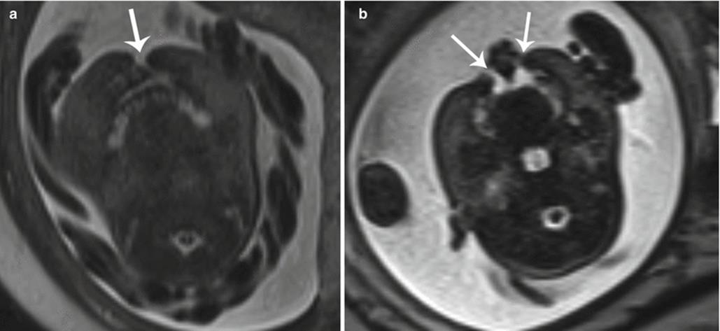
Fig. 7.11
Cleft lip/palate. (a) Axial T2 SSH MR image demonstrates a unilateral cleft lip (white arrow). (b) In a different fetus, bilateral cleft lip/palate (white arrows) with an intervening triangular segment was observed on this axial T2 SSH MR image
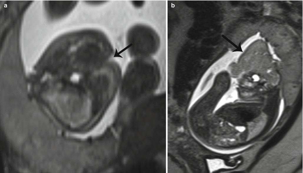
Fig. 7.12
Complex cleft lip/palate. (a) Axial T2 SSH MR image shows an atypical slash defect of the lip and palate (black arrow) in a patient with amniotic band syndrome. (b) This patient also exhibited exencephaly with presence of dysmorphic brain material (black arrow)
Fetal MRI is very good at defining an amniotic fluid-filled cleft [18]. The multiplanar capability of MRI also makes it particularly useful in visualizing the posterior soft palate and in determining overall extent of involvement for surgical planning [19, 20]. However, unilateral cleft lip without cleft palate, as well as closely apposed cleft orofacial cleft, can nonetheless be difficult to visualize on fetal MRI. Bilateral orofacial cleft has a characteristic premaxillary protrusion on profile view resulting from elevation of the median nasal prominence; in the axial plane, a midline triangular tissue lies between the two clefts.
More than 400 syndromes are associated with orofacial clefting; the most common associated chromosomal abnormality is trisomy 13 and trisomy 18 [4, 17]. Various infections and teratogens have also been implicated as the underlying etiology of this deformity. Midline orofacial clefts are often associated with midface hypoplasia. Additionally, midline defects have an increased association with holoprosencephaly and as such should prompt careful scrutiny of the intracranial contents [3].
7.6 Abnormal Mandible
7.6.1 Micrognathia
Micrognathia, or mandibular hypoplasia, is related to a defect in the first and second branchial arches. It is typically coupled with retrognathia, abnormal posterior positioning of the mandible with receding chin. This deformity can be subjectively observed but is further enhanced with calculation of an inferior facial angle <50°, by calculating the intersection of a line orthogonal to the forehead and another drawn from the tip of the mentum to the anterior border of the upper lip [21]. It may develop in isolation or most often is present in conjunction with a variety of conditions including Goldenhar syndrome (hemifacial microsomia), Pierre Robin sequence, and Treacher Collins syndrome [22]. There is a high association with chromosomal abnormalities involving up to 66 % of fetuses with micrognathia, commonly trisomy 13 and trisomy 18. Given the common association with other abnormalities, identification of micrognathia on fetal MRI may raise concern to closely evaluate for an underlying syndrome or genetic abnormality, which can be of importance in genetic counseling for the parents (Fig. 7.13a, b).
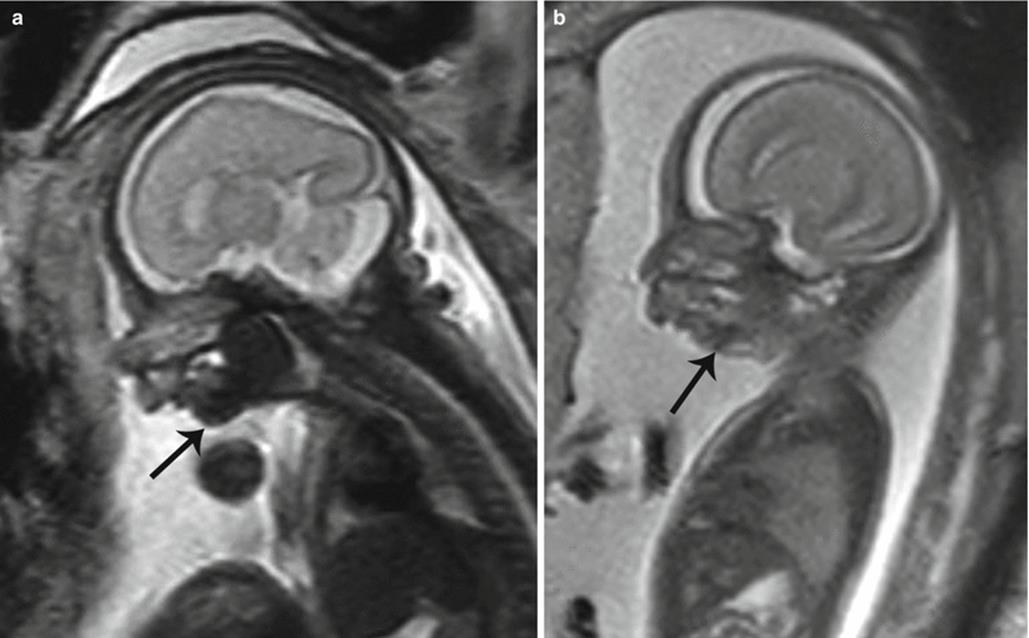
Fig. 7.13
Micrognathia. Sagittal T2 SSH MR images demonstrate (a) mild and (b) severe mandibular hypoplasia (black arrows) in two different fetuses
Caution should be taken to avoid overdiagnosis in early imaging; the mandible may often normally appear small in younger gestational age fetuses, as significant mandibular growth occurs during the third trimester. Additionally, asymmetric angulation of acquisition can give a false appearance of a small mandible [3].
Micrognathia yields a decreased size of the oral cavity with resultant posterior and superior displacement of the tongue and incomplete palatal fusion. Affected children frequently have impaired swallowing; as such polyhydramnios can be the initial presentation. As they are at risk for airway compromise, many of these patients require scheduled deliveries using the ex utero intrapartum treatment (EXIT) procedure.
7.6.2 Agnathia
Agnathia is an exceedingly rare and typically fatal malformation which is also related to a defect in the first branchial cleft. It is commonly associated with microstomia (small mouth), absent tongue, and midfacial location of the ears (Fig. 7.14a, b). A spectrum of concurrent abnormalities can occur including holoprosencephaly, genitourinary anomalies, cardiovascular anomalies, skeletal malformations, and situs inversus [23].

Fig. 7.14
Agnathia. (a) Sagittal T2 SSH MR image in a fetus with complete absence of the mandible (white arrow). Only the primordial teeth of the maxilla are seen. (b) Postmortem photograph of the same fetus demonstrates agnathia, microtia, a broadened nose, low-set abnormal ears, and slanted palpebral fissures
7.7 Lesions of the Oral Cavity
While masses of the fetal oral cavity are rare, they are of concern due to the high risk of airway obstruction and impairment of swallowing. This can result in polyhydramnios and/or respiratory distress at delivery [24]. As such, many of these patients may require delivery via the EXIT procedure with a pediatric head and neck surgeon present at time of delivery. Such lesions include congenital epithelial inclusion cysts (such as dermoid and epidermoid cysts), enteric duplication cysts, ranulas, teratomas (epignathus), epulis, and venolymphatic malformations, all of which are discussed in further detail below.
7.8 Congenital Lesions of the Neck
Congenital lesions of the neck detected in utero are overall very rare. These are varied in etiology and can be cystic (such as branchial anomalies, thymic cysts, meningocele, esophageal atresia, and thyroglossal duct cyst), solid (such as ectopic thymus, hemangioma, and sarcoma), or mixed cystic/solid (such as neuroblastoma and teratoma, both of which may also be completely solid) in composition.
7.8.1 Branchial Anomalies
Defects in the embryogenesis of the branchial apparatus may manifest as sinuses, fistulas, or cysts, the latter of which may be seen in conjunction with the former two. Prenatal diagnosis of branchial apparatus anomalies is very rare with few cases described in the literature [25–28].
Branchial cleft cysts are hypointense on T1-weighted sequences and hyperintense on T2-weighted sequences. If initially detected on US, they may appear as round/ovoid anechoic or hypoechoic thin-walled cysts often with movement of internal echoes. They may be located in a number of different anatomic locations, depending upon the branchial cleft of origin, and may rarely lead to airway obstruction. The most common lesion is a second branchial cyst, in the anterolateral neck (more frequently on the left), anterior to the sternocleidomastoid, posterior to the submandibular gland, and lateral to the carotid sheath [25]. First branchial cysts are located in the preauricular region or about the angle of the mandible. Third and fourth branchial cysts are exceedingly rare. Branchial cleft cysts are usually isolated lesions; however, they have been reported with branchio-oto-renal syndrome [29].
7.8.2 Cervical Thymic Cyst
Thymic cysts are remnants of the thymopharyngeal tracts which may be encountered in the neck, thoracic inlet, or mediastinum [30]. The thymic buds migrate inferiorly to form the thymopharyngeal ducts, which extend along the carotid sheath from the angle of the mandible to the superior mediastinum with attachment to the pericardium. In addition to thymic cysts, rests of ectopic thymic tissue can be found along the normal path of descent [31].
Thymic cysts are very rare and infrequently diagnosed in utero (Fig. 7.15a, b). They are commonly multilocular, but may be unilocular with size ranging from 1.4 to 8 cm. Internal signal is typically hypointense on T1-weighted images and hyperintense on T2 but may be heterogeneous if complicated by hemorrhage [32]. They are more common on the left and are located along the carotid space, splaying the carotid artery and jugular vein [31]. Up to 50 % have a mediastinal connection. Differential diagnoses include lymphatic malformation, thyroid cyst, thyroglossal duct cyst, and branchial cleft cyst.

Fig. 7.15
Cervical thymic cyst. (a, b) Coronal and axial SSH MR images of a twin pregnancy reveal a bilobed cystic mass extending from the anterior mediastinum into the left neck along the course of the thymopharyngeal duct in twin A (white arrows) (Courtesy of Dr. Christopher Cassady, TCH)
7.8.3 Ranula
A ranula is a mucocele or retention cyst of a sublingual gland or a minor salivary gland duct located at the floor of the mouth which may extend into the submandibular/cervical region. They are classified according to location into either simple (intraoral) or plunging (oral/cervical) types. They have well-delineated borders and demonstrate internal low-T1- and high-T2-weighted signal. Congenital ranulas may elevate the floor of the mouth, displacing the tongue and rarely obstructing the airway [33, 34]. Differential diagnosis includes lymphatic malformation, thyroglossal duct cyst, and dermoid because of midline/paramidline positioning.
Incidence of congenital ranula is estimated at 0.7 %, with prenatal diagnosis being very rare [33]. Congenital ranulas are theorized to arise secondary to salivary gland ductal atresia or ostial adhesion [33].
7.8.4 Thyroglossal Duct Cyst
While thyroglossal duct cysts represent the most common midline cervical anomaly, constituting 70 % of all congenital neck masses, prenatal diagnosis is rare [35, 36]. The exact incidence is unknown; however, 7 % of the population has been shown to have a thyroglossal duct remnant [36].
Prior to its involution, the embryologic thyroglossal duct extends from the foramen cecum of the tongue, along the anterior surface of the hyoid to the pyramidal lobe of the thyroid. Persistence of the duct results in cyst or sinus formation at any point of descent; infrequently, ectopic thyroid tissue can be found in the path of descent. The majority of duct remnants are midline or parasagittal, adjacent to the hyoid bone [37]. Rarely, these cysts are found within the tongue or the floor of the mouth, with the potential for airway obstruction [35]. The lesion can be occasionally associated with Cowden syndrome.
The classic appearance of a thyroglossal duct cyst is a thin-walled unilocular lesion with high internal signal on T1- and T2-weighted sequences; however, internal signal may vary depending on protein content [38, 39]. Some may even appear pseudosolid mimicking ectopic tissue [38]. Demonstration of a normal thyroid gland is recommended to exclude the diagnosis of ectopic tissue.
7.8.5 Dermoid/Epidermoid
Although they represent 7 % of head and neck lesions and 25 % of midline cervical anomalies, prenatal diagnosis of dermoid and epidermoid is rare [40]. Both develop from inclusion of ectodermal tissue during the fusion of the branchial arches; dermoid cysts contain both ectoderm and mesoderm, while epidermoids consist of only the ectoderm [41].
Dermoids and epidermoids present as well-circumscribed, thin-walled T1 hyper- to isointense, and T2 hyperintense unilocular lesions. They may occasionally have fat–fluid or fluid–fluid levels. Diffusion-weighted imaging can confirm diagnosis with demonstration of restricted diffusion [39]. While the majority of these cysts are found around the orbit or adjacent to the nose, approximately 11 % will present in the midline floor of the mouth in the submandibular space and may cause airway obstruction (Fig. 7.16a, b) [40].
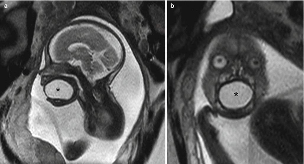
Fig. 7.16
Oral inclusion cyst. (a, b) Sagittal and coronal SSH MR images of a fetus with a large cystic lesion occupying the oral cavity (asterisks)
7.9 Inflammatory Lesions
7.9.1 Thyroid Goiter
Congenital thyroid goiter is a very rare condition that presents with diffuse enlargement of the thyroid gland associated with either thyroid dysfunction or, rarely, with normal (euthyroid) function [42].
While the normal thyroid gland is hyperintense on T1-weighted images relative to muscle and isointense on T2-weighted images, with fetal goiter, there is symmetric thyroid enlargement with increased T1 signal due to the intrinsic iodine content [43]. T2 signal greater than that of muscle suggests thyroid dysfunction; however, intrinsic thyroid T1 hyperintensity is unchanged despite thyroid functional state [44]. Enlargement is best quantified using ultrasound for which there are established nomograms to compare gestational age and biparietal diameter to thyroid gland size [45–47].
MRI is useful in the evaluation of degree of neck hyperextension as well as extent of tracheal and/or esophageal compression by the goiter which may result in airway compromise and polyhydramnios (Fig. 7.17). Neck hyperextension may necessitate cesarean section owing to risk of dystocia.
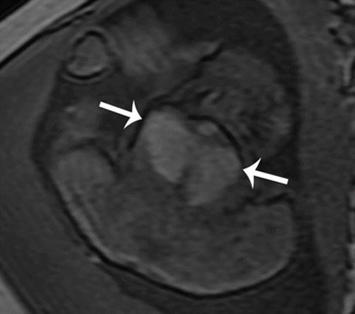
Fig. 7.17
Thyroid goiter. Axial T1-weighted MR image demonstrates diffuse enlargement of the thyroid gland, characteristically T1 hyperintense in signal (white arrows)
7.10 Vascular Malformations
Vascular malformations are classified according to hemodynamics into high-flow and low-flow malformations and are further classified according to the type of vessel present (arteries, veins, or lymphatics). Low-flow vascular malformations include pure or combined malformations of capillary, lymphatic, and venous channels. High-flow vascular malformations include both arteriovenous malformations and fistulas.
7.10.1 Lymphatic Malformations
Lymphatic malformations, often referred to as lymphangiomas or in the neck as cystic hygromas, are the most common fetal posterior cystic neck mass. They may evolve secondary to incomplete or inadequate venolymphatic connections with stasis of lymphatic fluid leading to dilated lymphatic channels [48]. This forms a subcutaneous accumulation of lymphatic fluid and forming a multiseptated collection containing dysplastic but mature lymphatic channels [49]. Though typically located within the posterior subcutaneous tissues, lymphatic malformations frequently wrap around laterally and may involve only one side of the neck, resulting in postural abnormality. They can be massive, transspatial masses insinuating between vessels and other normal structures [31]. They are classified into microcystic, macrocystic, and combined lesions.
MRI demonstrates a multilocular cystic transspatial mass (Fig. 7.18a, b). The cystic component is typically isointense to muscle on T1 and hyperintense on T2-weighted images, unless complicated by protein or hemorrhage, in which case there may also be fluid–fluid levels. Seventy-five percent of lymphatic malformations occur in the neck, with the majority originating in the posterior triangle or oral cavity and more commonly on the left side [50]. Lymphatic malformations often coexist with venous malformations, termed venolymphatic malformations, with the venous portion appearing more serpentine and having imaging characteristics as described below in “Venous Malformations.” As the deep component of the malformation is often poorly delineated by ultrasound, on MRI close attention should be paid to the deep extent and airway involvement to guide perinatal management, as 3–10 % of infants with LM have respiratory compromise or mediastinal extension which may require EXIT procedure [50, 51]. Additionally, cardiac and skeletal malformations may be present in up to 34 % of cases, and lymphatic malformations may be frequently associated with fetal hydrops [52].

Fig. 7.18
Lymphatic malformation. (a) Coronal T2 SSH MR image of the fetal head and neck reveals a large, complex cystic lesion in a beard-like distribution (black arrow). (b) Axial T2 SSH MR image, in the same patient, illustrates the deep neck extension by the lesion, displacing the airway rightward (white arrow)
Nuchal lymphatic malformations diagnosed during the first trimester are highly associated with chromosomal abnormalities (51 %), most commonly trisomy 21, and carry up to a 25 % risk of death. Other associations include Turner syndrome, trisomies 13 and 18, and deletions such as 13q and 18p [52]. Non-nuchal lymphatic malformations are usually isolated but may be associated with syndromes such as Gorham–Stout disease, Klippel–Trenaunay syndrome, and generalized lymphatic anomaly syndrome [1]. Only 17 % of fetuses with a lymphatic malformation will result in a healthy newborn; however, absence of aneuploidy or of other associated anomalies in the second trimester has been resulted with approximately 95 % chance of normal pediatric outcome [52].
7.10.2 Venous Malformations
Venous malformations are slow-flow vascular anomalies which contain dysplastic but mature venous channels. They may be associated with a lymphatic malformation (termed venolymphatic malformation). Venous malformations occur in approximately 1 % of all people, of which approximately 40 % occur in the head and neck [1]. They may occur at any time during angiogenesis and are always present by birth; however, most are small and often go unrecognized on prenatal imaging.
While typically isointense to muscle on T1-weighted and hyperintense on T2-weighted images, they may appear heterogeneous if thrombus or hemorrhage is present and may have associated fluid–fluid levels. T2* GRE images can be useful to demonstrate blooming artifact from phleboliths or hemorrhage. Similar to lymphatic malformations, they are often infiltrative and cross fascial planes.
Venous malformations can be associated with blue rubber bleb nevus syndrome, Maffucci syndrome, and Klippel–Trenaunay, and Parkes Weber syndrome [1].
7.10.3 Arteriovenous Fistula or Malformation
An arteriovenous malformation (AVM) is a tangle of abnormal thin-walled vessels connecting dilated high-flow feeding arteries to a draining vein (which is often enlarged due to increased pressures and high flow). They are believed to arise from failure of regression of arteriovenous channels in the primitive retiform plexus [53]. AVMs are most common in the head and neck, yet the true incidence is unknown. Arteriovenous fistulas (AVFs) lack a tangle of dysplastic vessels, instead having a direct connection between the feeding artery and the enlarged draining vein, and are more often acquired rather than congenital.
A high degree of arteriovenous shunting may result in cardiac failure and hydrops. These lesions are treated with transarterial embolization or surgery. Syndromic associations include Cowden syndrome, Bannayan–Riley–Ruvalcaba syndrome, Parkes Weber syndrome, and hereditary hemorrhagic telangiectasia (HHT) [1].
7.11 Neoplasms
7.11.1 Congenital Hemangioma
Hemangiomas arise from abnormal cellular proliferation of vascular endothelial cells and are the most common vascular tumor in infancy. They affect approximately 10 % of infants with a high female predilection, and greater than 50 % of hemangiomas involve the head and neck region [54]. Congenital hemangiomas (CHs) are rare tumors which grow in utero and typically stabilize in size in the third trimester, in contradistinction to infantile hemangiomas, which typically appear postnatally around 2 weeks of age [55]. In utero growth may be rapid or gradual. Two subtypes of CH exist, the rapidly involuting CH (RICH), which usually involutes by 8–14 months of postnatal life, and the noninvoluting CH (NICH), which may persist into late childhood, often requiring surgical treatment [56, 57].
Congenital hemangiomas may be visualized as early as 12 weeks’ gestation and typically involve the posterolateral neck. They appear as well-defined heterogeneous solid masses with intermediate T1 signal and hyperintense T2 signal intensity (Fig. 7.19a, b) [58]. The high T2 signal intensity likely results from slow vascular flow through small vascular spaces of the hemangioma. Heterogeneous flow voids may be seen in high-flow vessels within the mass and may make appearance similar to that of a teratoma [58]. Imaging features may also be difficult to distinguish from congenital fibrosarcoma, which also appears as a well-defined heterogeneous mass with flow voids [58, 59]. Unlike infantile hemangiomas, CHs may contain vascular aneurysms, intravascular thrombi, and arteriovenous shunting [60]. RICHs and NICHs cannot be differentiated based on imaging features, location, or size.
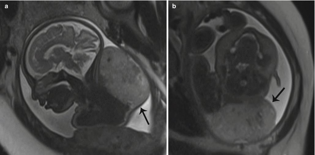
Fig. 7.19
Congenital hemangioma. (a, b) Sagittal and axial T2 SSH MR images demonstrate a large, mildly heterogeneous mass (black arrows) arising from the soft tissues of the posterior neck extending to the upper back
As brain cortical malformations have been shown to be associated with arterial anomalies, careful scrutiny of the brain should also be sought [58, 61]. Hemangiomas can also be seen as part of the PHACES syndrome (posterior fossa malformations, hemangiomas, arterial anomalies, cardiac defects, eye abnormalities, and sternal cleft).
7.11.2 Cervical Teratoma
Teratomas are germ cell tumors composed of tissues derived from all 3 embryonic layers, ectoderm, mesoderm, and endoderm.
The neck is the second most common location for teratomas, which present as anterior midline neck masses which can infiltrate the surrounding structures. They are usually large tumors and can range in diameter from 4 to 12 cm. Fetal MRI is excellent in determining the anatomic extent of these masses. With a complex composition often containing both multiloculated cystic and solid components in addition to calcification and hemorrhage, teratomas typically demonstrate mixed signal intensity (Fig. 7.20a, b). The calcifications are virtually pathognomonic of teratomas, and they can be recognized as areas of susceptibility on EPI [31].
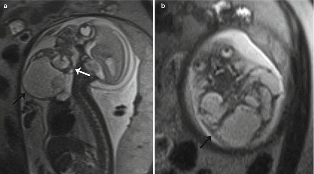
Fig. 7.20
Cervical teratoma. (a, b) Sagittal and coronal T2 SSH MR images of a fetus with a large, complex cystic and solid anterior neck mass (black arrows) causing compression of the airway and esophagus (white arrow) as well as hyperextension of the neck. There was hyperexpansion of the lungs and polyhydramnios (not shown) secondary to airway/esophageal compression
While imaging characteristics could be confused for LMs and branchial cleft cysts, these lesions predominantly occur in the posterolateral neck, whereas cervical teratomas occur anteriorly. Additionally, LMs are much more pliable than teratomas, resulting in less mass effect and compression of the neck vasculature and hypopharynx. As discussed above, teratoma may also appear similar to congenital hemangioma or goiter if more solid in appearance, though goiter is less likely to cause as severe a degree of mass effect upon the airway and esophagus.
7.11.3 Epignathus
Epignathus is a rare teratoma of the oropharyngeal region, arising from the soft or hard palate, and is believed to derive from pluripotential cells in Rathke pouch that grow in a disorganized manner [62]. Epignathus has similar imaging characteristics as those described for cervical teratomas. They usually present during the second trimester as unidirectional or bidirectional masses, involving only the oral cavity and protruding forward but may also extend intracranially. They may vary in size from a few centimeters to as large as the fetal head or body, potentially requiring EXIT procedure to secure the airway.
7.11.4 Epulis
Congenital epulis is a rare benign tumor, occurring almost exclusively in females, which arises from the gingival mucosa of the alveolar ridge of the maxilla and less commonly the mandible [71]. It presents as a well-defined pedunculated mass protruding through the fetal mouth with hypointense signal T2-weighted images (Fig. 7.21a, c). If the lesion becomes large, it may lead to impair fetal swallowing and polyhydramnios and/or airway obstruction [63]. While typically solitary, few cases of synchronous epulides have been described [63, 64]. Surgical excision is curative with no reported recurrence in the literature [65]. Small lesions have been reported to regress spontaneously [63].
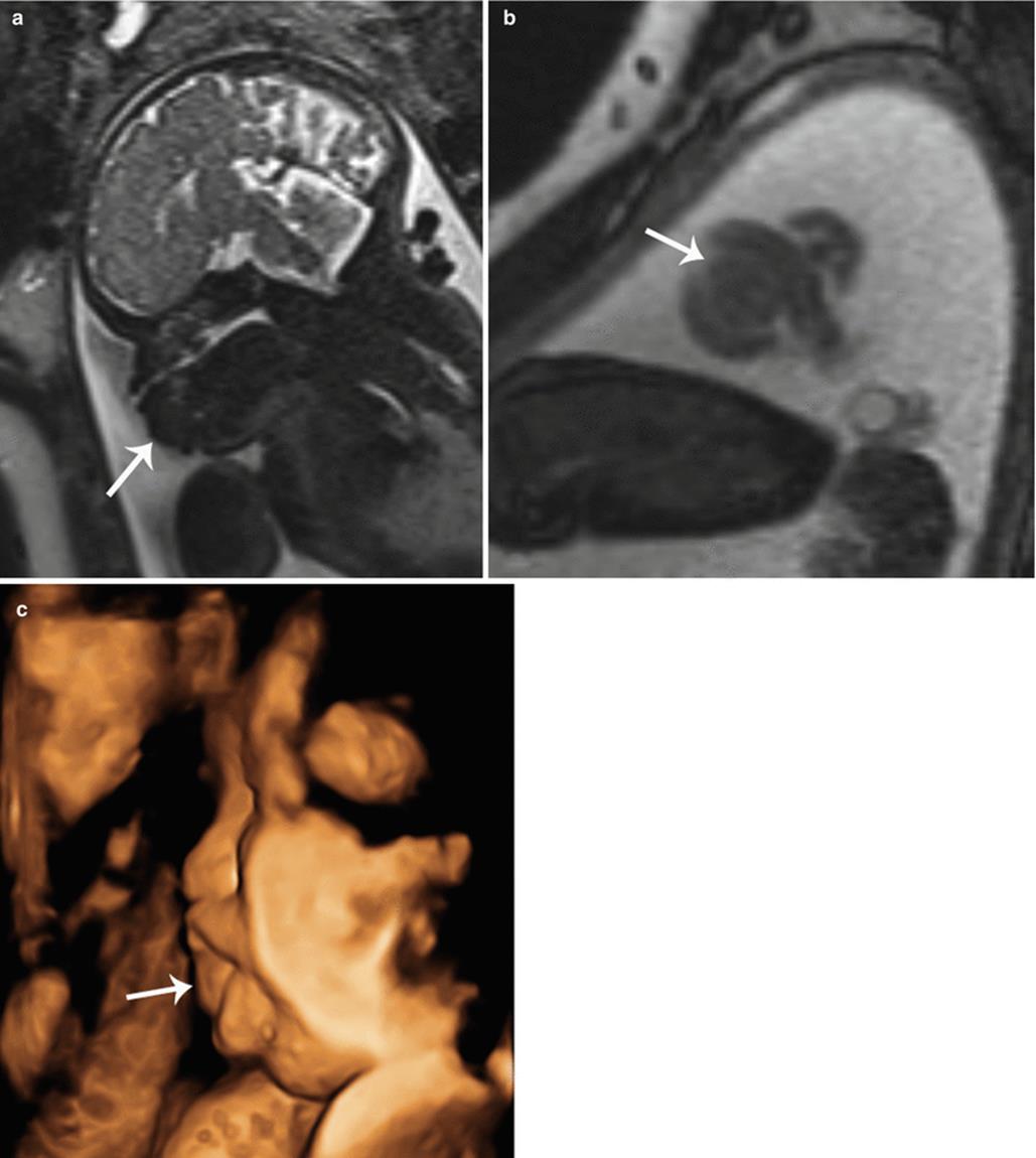
Fig. 7.21
Epulis. (a, b) Sagittal SSH and coronal BTFE MR images reveal a rounded lesion arising from the oral cavity (white arrows). (c) 3D volume rendered US image of the facial profile, in the same fetus, illustrates the lesion protruding through the mouth (white arrow)
The etiology of epulis is unknown and controversial. The terms epulis and congenital gingival granular cell tumor have been interchangeably used in the literature; however, they have differing histology and epidemiology; epulides are gingival tumors of infancy, while granular cell tumors occur in adulthood [63, 66].
7.11.5 Neuroblastoma
Congenital cervical neuroblastoma is a rare solid or mixed solid/cystic mass of the lateral neck. It accounts for only 4.4 % of congenital neuroblastomas, which are most commonly located in the adrenal gland [67].
7.11.6 Rhabdomyosarcoma
Congenital rhabdomyosarcoma of the head and neck is an extremely rare solid tumor with little published information regarding prenatal diagnosis [71]. As with other masses of the head and neck, they may result in airway obstruction, which may be assessed on fetal MRI [68]. Prenatal metastases to the sacral region have been reported [69]. As they are often associated with chromosomal translocations, amniocentesis for fetal karyotype should be performed. Associations have been reported with Robert syndrome, Beckwith–Wiedemann syndrome, and neurofibromatosis [70].
References
1.
Kathary N, Bulas DI, Newman KD, Schonberg RL (2001) MRI imaging of fetal neck masses with airway compromise: utility in delivery planning. Pediatr Radiol 31:727–731. doi:10.1007/s002470100527CrossRefPubMed
2.
Stevens GH, Schoot BC, Smets MJW, Kremer B, Manni JJ, Gavilanes AWD, Wilmink JT, van Heurn LWE, Hasaart THM (2002) The ex utero intrapartum treatment (EXIT) procedure in fetal neck masses: a case report and review of the literature. Eur J Obstet Gynecol Reprod Biol 100:246–250. doi:10.1016/S0301-2115(01)00467-5CrossRefPubMed
3.
Mirsky DM, Shekdar KV, Bilaniuk LT (2012) Fetal MRI: head and neck. Magn Reson Imaging Clin N Am 20:605–618. doi:10.1016/j.mric.2012.06.002CrossRefPubMed
4.
Bianchi D, Crombleholme T, D’Alton M, Malone F (2010) Fetology: diagnosis and management of the fetal patient: diagnosis and management of the fetal patient. McGraw Hill Professional, New York, NY, USA 10010
5.
Benson ML, Oliverio PJ, Yue NC, Zinreich SJ (1996) Primary craniosynostosis: imaging features. Am J Roentgenol 166:697–703. doi:10.2214/ajr.166.3.8623653CrossRef
6.
Hansen WF, Rijhsinghani A, Grant S, Yankowitz J (2004) Prenatal diagnosis of Apert syndrome. Fetal Diagn Ther 19:127–130. doi:10.1159/000075135CrossRefPubMed
7.
Delahaye S, Bernard JP, Rénier D, Ville Y (2003) Prenatal ultrasound diagnosis of fetal craniosynostosis. Ultrasound Obstet Gynecol 21:347–353. doi:10.1002/uog.91CrossRefPubMed
8.
Lau TK, Leung TN, Leung TY, Pang MW, Tam WH (2001) Fetal scalp cysts: challenge in diagnosis and counseling. JUM 20:175–177
9.
Paquette LB, Jackson HA, Tavaré CJ, Miller DA, Panigrahy A (2009) In utero eye development documented by fetal MR imaging. AJNR Am J Neuroradiol 30:1787–1791. doi:10.3174/ajnr.A1664CrossRefPubMed
10.
Robinson AJ, Blaser S, Toi A, Chitayat D, Pantazi S, Keating S, Viero S, Ryan G (2008) MRI of the fetal eyes: morphologic and biometric assessment for abnormal development with ultrasonographic and clinicopathologic correlation. Pediatr Radiol 38:971–981. doi:10.1007/s00247-008-0929-3CrossRefPubMed
11.
Trout T, Budorick NE, Pretorius DH, McGahan JP (1994) Significance of orbital measurements in the fetus. JUM 13:937–943
12.
Levine D, Barnes PD, Madsen JR, Li W, Edelman RR (1997) Fetal central nervous system anomalies: MR imaging augments sonographic diagnosis. Radiology 204:635–642. doi:10.1148/radiology.204.3.9280237CrossRefPubMed
13.
Poutamo J, Vanninen R, Partanen K, Ryynänen M, Kirkinen P (1999) Magnetic resonance imaging supplements ultrasonographic imaging of the posterior fossa, pharynx and neck in malformed fetuses. Ultrasound Obstet Gynecol 13:327–334. doi:10.1046/j.1469-0705.1999.13050327.xCrossRefPubMed
14.
Dutton GN (2004) Congenital disorders of the optic nerve: excavations and hypoplasia. Eye 18:1038–1048. doi:10.1038/sj.eye.6701545CrossRefPubMed
15.
Righini A, Avagliano L, Doneda C, Pinelli L, Parazzini C, Rustico M, Triulzi F, Bulfamante G (2008) Prenatal magnetic resonance imaging of optic nerve head coloboma. Prenat Diagn 28:242–246. doi:10.1002/pd.1955CrossRefPubMed
16.
Katorza E, Rosner M, Zalel Y, Gilboa Y, Achiron R (2008) Prenatal ultrasonographic diagnosis of persistent hyperplastic primary vitreous. Ultrasound Obstet Gynecol 32:226–228. doi:10.1002/uog.5385CrossRefPubMed
17.
Merritt L (2005) Part 1. Understanding the embryology and genetics of cleft lip and palate. Adv Neonatal Care 5:64–71CrossRefPubMed
18.
Smith AS, Estroff JA, Barnewolt CE, Mulliken JB, Levine D (2004) Prenatal diagnosis of cleft lip and cleft palate using MRI. Am J Roentgenol 183:229–235. doi:10.2214/ajr.183.1.1830229CrossRef
19.
Ghi T, Tani G, Savelli L, Colleoni GG, Pilu G, Bovicelli L (2003) Prenatal imaging of facial clefts by magnetic resonance imaging with emphasis on the posterior palate. Prenat Diagn 23:970–975. doi:10.1002/pd.737CrossRefPubMed
20.
Arangio P, Manganaro L, Pacifici A, Basile E, Cascone P (2013) Importance of fetal MRI in evaluation of craniofacial deformities. J Craniofac Surg 24:773–776. doi:10.1097/SCS.0b013e318286988cCrossRefPubMed
21.
Nemec U, Nemec SF, Brugger PC, Weber M, Bartsch B, Bettelheim D, Gruber M, Prayer D (2014) Normal mandibular growth and diagnosis of micrognathia at prenatal MRI. Prenat Diagn. 2015;35(2):108–116. doi:10.1002/pd.4496
22.
Bromley B, Benacerraf BR (1994) Fetal micrognathia: associated anomalies and outcome. J Ultrasound Med 13:529–533PubMed
23.
Hisaba WJ, Milani HJF, Araujo Júnior E, Passos JP, Barreto EQS, Carvalho NS, Helfer TM, Pares DBS, Nardozza LMM, Moron AF (2014) Agnathia-otocephaly: prenatal diagnosis by two- and three-dimensional ultrasound and magnetic resonance imaging. Case report. Med Ultrasound 16:377–379
24.
Edwards RM, Chapman T, Horn DL, Paladin AM, Iyer RS (2013) Imaging of pediatric floor of mouth lesions. Pediatr Radiol 43:523–535. doi:10.1007/s00247-013-2620-6CrossRefPubMed
25.
Lind RC, Hulscher JBF, van der Wal JE, Dikkers FG, de Langen ZJ (2010) A very rare case of a giant third branchial pouch remnant discovered in utero. Eur J Pediatr Surg 20:349–351. doi:10.1055/s-0029-1246194CrossRefPubMed
26.
Robichaud J, Papsin BC, Forte V (2000) Third branchial cleft anomaly detected in utero. J Otolaryngol 29:185–187PubMed
27.
Suchet IB (1995) Ultrasonography of the fetal neck in the second and third trimesters. Part 3. Anomalies of the anterior and anterolateral nuchal region. Can Assoc Radiol J 46:426–433PubMed
28.
Tsai P-Y, Chang C-H, Chang F-M (2003) Prenatal imaging of the fetal branchial cleft cyst by three-dimensional ultrasound. Prenat Diagn 23:605–606. doi:10.1002/pd.639CrossRefPubMed
29.
Smith RJ, Pagon RA, Adam MP, Ardinger HH (1993) Branchiootorenal spectrum disorders. GeneReviews Seattle (WA): University of Washington, Seattle; 1993–2015
30.
Nguyen Q, deTar M, Wells W, Crockett D (1996) Cervical thymic cyst: case reports and review of the literature. Laryngoscope 106:247–252. doi:10.1097/00005537-199603000-00001CrossRefPubMed
31.
Cigliano B, Baltogiannis N, Marco MD, Faviou E, Antoniou D, Luca UD, Soutis M, Settimi A (2007) Cervical thymic cysts. Pediatr Surg Int 23:1219–1225. doi:10.1007/s00383-006-1822-5CrossRefPubMed
32.
De Miguel CE, Casanova A, Urbano J, Delgado Carrasco J (1997) Congenital thymic cyst: prenatal sonographic and postnatal magnetic resonance findings. J Ultrasound Med 16:365–367CrossRef
33.
Gul A, Gungorduk K, Yildirim G, Gedikbasi A, Ceylan Y (2008) Prenatal diagnosis and management of a ranula. J Obstet Gynaecol Res 34:262–265. doi:10.1111/j.1447-0756.2008.00767.xCrossRefPubMed
34.
Chan DFY, Lee CH, Fung TY, Chan DLW, Abdullah V, Ng PC (2006) Ex utero intrapartum treatment (EXIT) for congenital giant ranula. Acta Paediatr 95:1303–1305. doi:10.1080/08035250600580545CrossRefPubMed
35.
Lindstrom DR, Conley SF, Arvedson JC, Beecher RB, Carr MH (2003) Anterior lingual thyroglossal cyst: antenatal diagnosis, management, and long-term outcome. Int J Pediatr Otorhinolaryngol 67:1031–1034. doi:10.1016/S0165-5876(03)00195-2CrossRefPubMed
36.
Keizer AL, Deurloo KL, van Vugt JMG, Haak MC (2011) A prenatal diagnosis of a thyroglossal duct cyst in the fetal anterior neck. Prenat Diagn 31:1311–1312. doi:10.1002/pd.2872CrossRefPubMed
37.
Hsieh Y-Y, Hsueh S, Hsueh C, Lin J-N, Luo C-C, Lai J-Y, Huang C-S (2003) Pathological analysis of congenital cervical cysts in children: 20 years of experience at Chang Gung Memorial Hospital. Chang Gung Med J 26:107–113PubMed
38.
Kutuya N, Kurosaki Y (2008) Sonographic assessment of thyroglossal duct cysts in children. JUM 27:1211–1219
39.
Mossa-Basha M, Yousem DM (2013) Congenital cystic lesions of the neck. Appl Radiol 42:8–22
40.
Masters F, Given CA II (2008) Cystic neck masses: a pictorial review of unusual presentations and complicating features. Appl Radiol 37:26
41.
Lev S, Lev MH (2000) Imaging of cystic lesions. Radiol Clin North Am 38:1013–1027CrossRefPubMed
42.
Jain V, Sharma R, Verma S, Agarwal R (2010) Fetal euthyroid goiter. Indian J Pediatr 76:1259–1260. doi:10.1007/s12098-009-0331-7CrossRef
43.
Karabulut N, Martin DR, Yang M, Boyd BK (2002) MR imaging findings in fetal goiter caused by maternal graves disease. J Comput Assist Tomogr 26:538–540CrossRefPubMed
44.
Kondoh M, Miyazaki O, Imanishi Y, Hayakawa M, Aikyou M, Doi H (2004) Neonatal goiter with congenital thyroid dysfunction in two infants diagnosed by MRI. Pediatr Radiol 34:570–573. doi:10.1007/s00247-004-1145-4CrossRefPubMed
45.
Ho SSY, Metreweli C (1998) Normal fetal thyroid volume. Ultrasound Obstet Gynecol 11:118–122. doi:10.1046/j.1469-0705.1998.11020118.xCrossRefPubMed
46.
Ranzini AC, Ananth CV, Smulian JC, Kung M, Limbachia A, Vintzileos AM (2001) Ultrasonography of the fetal thyroid: nomograms based on biparietal diameter and gestational age. JUM 20:613–617
47.
Gietka-Czernel M, Dębska M, Kretowicz P, Dębski R, Zgliczyński W (2012) Fetal thyroid in two-dimensional ultrasonography: nomograms according to gestational age and biparietal diameter. Eur J Obstet Gynecol Reprod Biol 162:131–138. doi:10.1016/j.ejogrb.2012.02.013CrossRefPubMed
48.
Chervenak FA, Isaacson G, Blakemore KJ, Breg WR, Hobbins JC, Berkowitz RL, Tortora M, Mayden K, Mahoney MJ (1983) Fetal cystic hygroma: cause and natural history. N Engl J Med 309:822–825. doi:10.1056/NEJM198310063091403CrossRefPubMed
49.
Koyuncu FM, Tamay AG, Bugday S (2010) Intrauterine diagnosis and management of fetal goiter: a case report. J Clin Ultrasound 38:503–505. doi:10.1002/jcu.20717CrossRefPubMed
50.
Fordham LA, Chung CJ, Donnelly LF (2000) Imaging of congenital vascular and lymphatic anomalies of the head and neck. Neuroimaging Clin N Am 10:117–136, viiiPubMed
51.
Oosthuizen JC, Burns P, Russell JD (2010) Lymphatic malformations: a proposed management algorithm. Int J Pediatr Otorhinolaryngol 74:398–403. doi:10.1016/j.ijporl.2010.01.013CrossRefPubMed
52.
Malone FD, Ball RH, Nyberg DA, Comstock CH, Saade GR, Berkowitz RL, Gross SJ, Dugoff L, Craigo SD, Timor-Tritsch IE, Carr SR, Wolfe HM, Dukes K, Canick JA, Bianchi DW, D’Alton ME (2005) First-trimester septated cystic hygroma: prevalence, natural history, and pediatric outcome. Obstet Gynecol 106:288–294. doi:10.1097/01.AOG.0000173318.54978.1fCrossRefPubMed
53.
Kohout MP, Hansen M, Pribaz JJ, Mulliken JB (1998) Arteriovenous malformations of the head and neck: natural history and management. Plast Reconstr Surg 102:643–654CrossRefPubMed
54.
Tseng J-J, Chou M-M, Chen W-H (2007) Prenatal 3- and 4-dimensional ultrasonographic findings of giant fetal nuchal hemangioma. J Chin Med Assoc 70:460–463. doi:10.1016/S1726-4901(08)70040-6CrossRefPubMed
55.
Marler JJ, Fishman SJ, Upton J, Burrows PE, Paltiel HJ, Jennings RW, Mulliken JB (2002) Prenatal diagnosis of vascular anomalies. J Pediatr Surg 37:318–326. doi:10.1053/jpsu.2002.30831CrossRefPubMed
56.
Boon LM, Enjolras O, Mulliken JB (1996) Congenital hemangioma: evidence of accelerated involution. J Pediatr 128:329–335. doi:10.1016/S0022-3476(96)70276-7CrossRefPubMed
57.
Enjolras O, Mulliken JB, Boon LM, Wassef M, Kozakewich HPW, Burrows PE (2001) Noninvoluting congenital hemangioma: a rare cutaneous vascular anomaly. Plast Reconstr Surg 107:1647–1654. doi:10.1097/00006534-200106000-00002CrossRefPubMed
58.
Robson CD, Barnewolt CE (2004) MR imaging of fetal head and neck anomalies. Neuroimaging Clin N Am 14:273–291, viii. doi:10.1016/j.nic.2004.03.005
59.
Boon LM, Fishman SJ, Lund DP, Mulliken JB (1995) Congenital fibrosarcoma masquerading as congenital hemangioma: report of two cases. J Pediatr Surg 30:1378–1381CrossRefPubMed
60.
Alamo L, Beck-Popovic M, Gudinchet F, Meuli R (2011) Congenital tumors: imaging when life just begins. Insights Imaging 2:297–308. doi:10.1007/s13244-011-0073-8PubMedCentralCrossRefPubMed
61.
O’Connor SC, Rooks VJ, Smith AB (2012) Magnetic resonance imaging of the fetal central nervous system, head, neck, and chest. Semin Ultrasound CT MR 33:86–101. doi:10.1053/j.sult.2011.10.005CrossRefPubMed
62.
Sumiyoshi S, Machida J, Yamamoto T, Fukano H, Shimozato K, Fujimoto Y, Kaetsu A (2010) Massive immature teratoma in a neonate. Int J Oral Maxillofac Surg 39:1020–1023. doi:10.1016/j.ijom.2010.04.008CrossRefPubMed
63.
Koch BL, Myer C, Egelhoff JC (1997) Congenital epulis. AJNR Am J Neuroradiol 18:739–741PubMed
64.
Roy S, Sinsky A, Williams B, Desilets V, Patenaude YG (2003) Congenital epulis: prenatal imaging with MRI and ultrasound. Pediatr Radiol 33:800–803. doi:10.1007/s00247-003-1024-4CrossRefPubMed
65.
Jiang L, Hu B, Guo Q (2011) Prenatal sonographic diagnosis of congenital epulis. J Clin Ultrasound 39:217–220. doi:10.1002/jcu.20765CrossRefPubMed
66.
López de Lacalle JM, Aguirre I, Irizabal JC, Nogues A (2001) Congenital epulis: prenatal diagnosis by ultrasound. Pediatr Radiol 31:453–454CrossRefPubMed
67.
Isaacs H (2007) Fetal and neonatal neuroblastoma: retrospective review of 271 cases. Fetal Pediatr Pathol 26:177–184. doi:10.1080/15513810701696890CrossRefPubMed
68.
Skelton VA, Goodwin A (1999) Perinatal management of a neonate with airway obstruction caused by rhabdomyosarcoma of the tongue. Br J Anaesth 83:951–955. doi:10.1093/bja/83.6.951CrossRefPubMed
69.
Yoshino K, Takeuchi M, Nakayama M, Suehara N (2005) Congenital cervical rhabdomyosarcoma arising in one fetus of a twin pregnancy. Fetal Diagn Ther 20:291–295. doi:10.1159/000085088CrossRefPubMed
70.
Önderoğlu LS, Yücel A, Yüce K (1999) Prenatal sonographic features of embryonal rhabdomyosarcoma. Ultrasound Obstet Gynecol 13:210–212. doi:10.1046/j.1469-0705.1999.13030210.xCrossRefPubMed
71.
Meyers ML, Dannull KA (2015) The Neck, in: Kline-Fath B, Bahado-Singh R, Bulas D (Eds.), Fundamental and Advanced Fetal Imaging: Ultrasound and MRI. Wolters Kluwer Health. Philadelphia, PA 19103