
Daniel Albo
Presentation
An 83-year-old patient with known history of diabetes mellitus, hypertension, chronic renal failure, and congestive heart failure presents with a 2-week history of crampy abdominal pain following meals with thin-caliber bowel movements but no nausea and vomiting. His vital signs were normal. On physical exam, his abdomen was nontender, somewhat distended, with a palpable mass in the left upper quadrant of his abdomen. His rectal exam revealed guaiac-positive brown stool. His laboratory exams were significant for a hemoglobin of 10, a white cell blood count of 9, a creatinine of 2.9, and an albumin of 2.9.
Differential Diagnosis
An elderly patient presenting with symptomatic anemia, a high-grade partial bowel obstruction, and a palpable left upper-quadrant mass is highly suggestive of a distal colon cancer. However, the differential diagnosis is relatively broad and includes inflammatory conditions such as diverticulitis (less likely in this patient due to the lack of a fever and of left-sided abdominal tenderness on exam and with a normal white cell blood count), ischemic colitis (although he has risk factors for vascular disease, he did not present with bloody diarrhea, and he had a palpable left upper-quadrant mass), an enlarged spleen (such as seen in hematologic malignancies), and other neoplastic processes involving the left upper quadrant (gastric, pancreatic tail, adrenal or kidney cancer as well as retroperitoneal sarcomas).
Workup
The patient underwent a computed tomography (CT) scan of the abdomen and the pelvis (with oral contrast only due to his elevated creatinine), which revealed a large, obstructing mass in the splenic flexure of the colon (Figure 1), with a moderately distended cecum (<10 cm) and small bowel, with no evidence of liver metastases or carcinomatosis. A colonoscopy revealed a nearly completely obstructing mass at the level of the splenic flexure of the colon (Figure 2). The mass could not be traversed and they were unable to evaluate the proximal colon. A biopsy was obtained that showed a moderately to poorly differentiated adenocarcinoma. A carcino embrionic antigen (CEA) level was obtained and it was 34.6. This workup confirmed the diagnosis of an obstructing splenic flexure colon cancer.
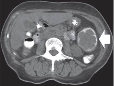
FIGURE 1 • CT scan of the abdomen showing a large, obstructing splenic flexure colon cancer (arrow). This CT scan was obtained with oral contrast only due to the patient’s impaired kidney function.
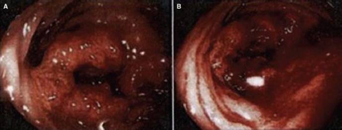
FIGURE 2 • Colonoscopy. A: Splenic flexure of the colon showing a large, fungating circumferential mass that is obstructing the lumen of colon. B: Descending colon, showing a normal colonic lumen distal to the splenic flexure mass and some evidence of bleeding from the tumor.
Discussion
Carcinoma of the transverse colon (of which splenic flexure colon cancer is a subset) accounts for 10% of all colorectal cancers. Diagnosis is often delayed and complicated forms (perforation, fistulization, obstruction) occur in 30% to 50% of cases. The progression of symptoms is often insidious and tumors may be quite large by the time of diagnosis. These tumors can also extend or fistulize into adjacent organs. Distal transverse cancers may be small annular lesions, which are prone to obstruction.
From a surgeon’s perspective, it is important to consider two types of complete colonic obstructions, those with a competent or with an incompetent ileocecal valve. In patients with an incompetent ileocecal valve, the pressure that builds up proximal to the splenic flexure tumor partially decompresses into the small bowel, allowing for preoperative stabilization of the patient (by nasogastric tube decompression, rehydration, and correction of underlying electrolyte disorders) and performance of a full workup, including a colonoscopy. Patients that present with a complete large bowel obstruction with a competent ileocecal valve, on the other hand, represent a true surgical emergency. In these patients, the pressure that builds up proximal to the splenic flexure tumor cannot decompress into the small bowel due to the competency of the ileocecal valve. In this situation, the proximal colon will progressively get more dilated and the tension on the proximal colonic wall will increase. According to Laplace’s law, the increase in tension on the wall of the cavity will be directly proportional to the fourth potency of radius of the cavity. Therefore, the part of the proximal colon with the largest radius, the cecum, will suffer the greatest increase in colonic wall tension. This increase in wall tension will lead to collapse of the intramural capillaries and ischemia, eventually producing a catastrophic cecal perforation. Patients with a complete colonic obstruction that present with right lower-quadrant abdominal pain should be operated on emergently, since this likely represent a sign of an impending cecal rupture. Colonoscopy in these patients is formally contraindicated, since it could lead to a critical delay before surgery, and the increase in intraluminal pressure due to insufflation during the colonoscopy could lead to a full-blown cecal perforation.
Workup
Clinical presentation permitting, the preoperative staging workup should include a CT scan of the abdomen and the pelvis with oral and intravenous contrast, a chest x-ray or a CT scan of the chest, a CEA blood level, and a full colonoscopy. In patients with bulky splenic flexure tumors in which it is not possible to evaluate the proximal colon by colonoscopy, a barium enema is indicated to rule out synchronous colon cancer in the proximal colon, although it frequently is either not feasible or reliable. Although the use of on-table colonoscopy through the appendiceal orifice has been described, this is often not practical. The incidence of a synchronous proximal colon cancer, though, is only about 5%, and only a subset of these synchronous tumors will not be detected by either preoperative CT scan or intraoperative palpation of the colon. Therefore, although evaluation of the full colon preoperatively by colonoscopy is preferred, in patients in whom evaluation of the proximal colon is just not possible, it is acceptable to perform surgery for the splenic flexure tumor and perform a short-interval colonoscopy postoperatively (6 months after surgery) to fully evaluate the reminder of the colon.
Surgical Approach
Colonic stenting is the best option either for palliation or as a bridge to surgery in patients with a complete bowel obstruction due to a splenic flexure colon cancer with a competent ileocecal valve. This approach reduces morbidity and mortality rate and it eliminates the need for temporary colostomy. Nevertheless, surgical management remains relevant as colonic stenting has a small rate of failure, and it is not always available. There are various surgical options for dealing with patients with obstructing splenic flexure colon cancer. One-stage primary resection and anastomosis is the preferred choice for low-risk patients. It is paramount to not only resect the bowel segment involving the splenic flexure of the colon with negative margins of resection, but to also include the entire lymphovascular pedicles associated with the splenic flexure of the colon (a minimum of 12 lymph nodes should be obtained). These include the left colic vessels, the left branches of the middle colic vessels, and the inferior mesenteric vein. Subtotal colectomy is useful in cases of proximal bowel damage (i.e., cecal perforation) or synchronous tumors. A two-stage procedure, with a resection and colostomy as a first step and colostomy takedown as a second step, should be reserved for high-risk patients. A three-stage procedure, with the creation of simple colostomy as a first stage, has no role other than for use in very ill patients who are not fit for any other procedure. The multistage approach is marred by a higher cumulative rate of perioperative morbidity and mortality and frequent failure to complete the planned sequence of operations and a resulting high permanent stoma rate (up to 40% of patients).
One of the most significant changes in colon surgery over the last decade has been the increased application of minimally invasive surgery (MIS) techniques. MIS offers several key short-term patient advantages to open surgery (Table 1), including less perioperative pain, smaller incisions (Figure 3B), shorter hospital stays, faster recovery, and lower wound morbidity (including wound infections and incisional hernias). In elderly patients, and in patients with significant comorbidities, it may also reduce the incidence of postoperative pulmonary and cardiac complications. In addition, we believe that laparoscopic surgery offers a superior visualization in the left upper quadrant of the abdomen and an easier, more controlled mobilization of the splenic flexure versus open surgery (Figure 3A). Due to these advantages, for surgeons with the requisite skill and experience, we feel strongly that MIS should be considered the preferred approach to patients with colon cancer, including those with splenic flexure tumors.
TABLE 1. Advantages of Minimally Invasive Surgery for Colon Cancer
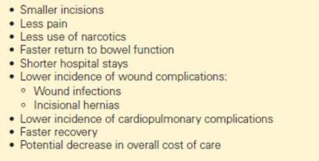
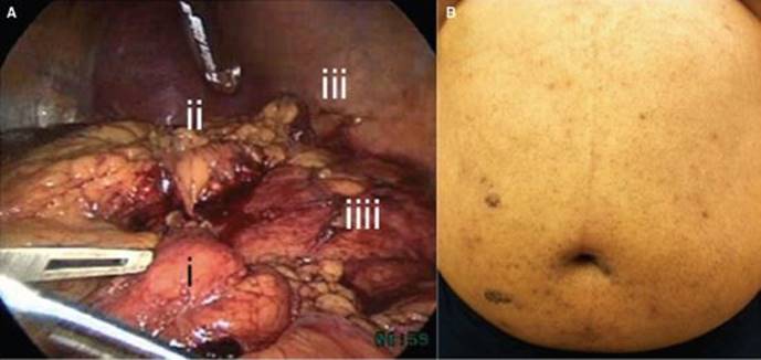
FIGURE 3 • HALS for colon cancer of the splenic flexure. A: Full mobilization of the splenic flexure showing the splenic flexure of the colon (i) fully mobilized to the midline, the tail of the pancreas (ii), the spleen (iii), and Gerota’s fascia (iv). B: 10 days postoperatively, the extraction site (placed as small transverse suprapubic incision hiding in a skin crease) and the 5-mm umbilical port site are no longer visible. The 5-mm and the 12-mm right-sided port sites are noticeable only due to the Dermabond that we use to seal the incisions; after the Dermabond falls off, these ports will be barely noticeable.
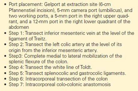
Patients with large splenic flexure tumors can be particularly challenging even for advanced laparoscopic surgeons. The position of these tumors deep in the left upper quadrant and the close proximity to the spleen, the pancreas, and the stomach can make these cases extremely complex. As a result, surgeons tend to shy away from laparoscopic surgery in the management of these tumors. An alternative to conventional laparoscopic surgery is hand-assisted laparoscopic surgery (HALS). In this technique, the surgeon has the option of introducing a hand in the abdomen during surgery through a special port (called a Gelport) that can be inserted in the expected extraction site. This provides the surgeon with tactile feedback and allows for greater versatility during surgery. While preserving all the known short-term advantages of conventional laparoscopic surgery versus open surgery, HALS offers several key advantages over conventional laparoscopic surgery (Table 2), including higher percentage of utilization of MIS, lower conversion to open rates, shorter operating times, and lower estimated blood loss rates. Key operative principles of MIS for the surgical management of splenic flexure colon cancer are summarized in Table 3.
TABLE 2. Advantages of HALS Over Conventional Laparoscopy in the Management of Splenic Flexure Tumors
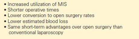
TABLE 3. Key Technical Steps During HALS for Splenic Flexure Colon Cancer (HALS left hemicolectomy)
Adjuvant Therapy
Adjuvant chemotherapy is reserved for patients with stage III (positive lymph nodes) and stage IV (distant metastases) disease. Node-negative patients that may be considered for adjuvant chemotherapy include patient with high-risk characteristics, including lymphovascular invasion, perineural invasion, and lack of microsatellite instability. For patients with borderline lesions, genetic testing (such as Oncotype evaluation) is rapidly emerging as a potentially useful tool in determining which node-negative patients may benefit from additional therapy. Current adjuvant therapy protocols often times include oxalloplatin and 5-fluorouracil (FOLFOX) chemotherapy. The use of biological cancer therapy (such as Avastin) is also emerging as a potential treatment option in addition to more standard chemotherapy regimens.
Surveillance
Eighty five percent of colon cancer recurrences occur within 3 years of surgery, with the majority of the remaining recurrences occurring between years 3 and 5. The 2005 American Society of Clinical Oncology surveillance recommendations for colon cancer include an annual CT scan of the chest and abdomen for 3 years, a colonoscopy at 3 years, a history and physical examination and risk assessment every 3 to 6 months for the first 3 years and then every 6 months during years 4 and 5, and a CEA level checked every 3 months for at least 3 years after surgery.
Case Conclusion
The patient underwent a hand-assisted laparoscopic left hemicolectomy with a primary stapled midtransverse colon-sigmoid anastomosis. During surgery, we found a very large mass in the splenic flexure of the colon, with no direct extension into contiguous organs, and no evidence of synchronous colonic lesions in the right colon, liver metastases, or carcinomatosis. The estimated blood loss was <50 mL, and the operative time was 120 minutes. The patient recovered uneventfully and was discharged home on postoperative day 4, tolerating a regular diet, ambulating, with minimal incisional pain, and having bowel movements. The final pathology revealed an ulcerated, moderately differentiated adenocarcinoma involving the entire lumen of the colon that extended through the colonic wall and into the pericolonic adipose tissue with negative proximal, distal, and radial margins. There were 26 lymph nodes with no evidence of malignancy. There was no evidence of lymphovascular or perineural invasion. The tumor was positive for MLH-1, MSH-2, MSH-6, and PMS-2 by immunoperoxidase staining (microsatellite instability).
Due to the patient’s advanced age, adequate lymphadenectomy negative for cancer, and lack of lymphovascular and perineural invasion, and the presence of microsatellite instability in the primary tumor, the medical oncologist decided that chemotherapy was not indicated in this patient.
TAKE HOME POINTS
· Diagnosis of transverse colon cancer is often delayed and complicated forms (perforation, fistulization, obstruction) occur in 30% to 50% of cases.
· Patients that present with a complete large bowel obstruction with a competent ileocecal valve represent a true surgical emergency.
· Clinical presentation permitting, the preoperative staging workup should include a CT scan of the abdomen and the pelvis with oral and intravenous contrast, a chest x-ray or a CT scan of the chest, a CEA blood level, and a full colonoscopy.
· Colonic stenting is the best option either for palliation or as a bridge to surgery in high-risk patients with a complete bowel obstruction due to a splenic flexure colon cancer with a competent ileocecal valve.
· In paitents undergoing surgery, it is paramount to not only resect the bowel segment involving the splenic flexure of the colon with negative margins of resection, but to also include the entire lymphovascular pedicles associated with the splenic flexure of the colon (a minimum of 12 lymph nodes should be obtained). This should include include high ligation of the left colic vessels, the left branches of the middle colic vessels, and the inferior mesenteric vein.
· Subtotal colectomy is useful in cases of proximal bowel damage (i.e., cecal perforation) or synchronous tumors.
SUGGESTED READINGS
Comparison of self-expanding metal stents and urgent surgery for left-sided malignant colonic obstruction in elderly patients. Dig Dis Sci. 2011;56:2706–10.
Guo MG, Feng Y, Zheng Q, Di JZ, Wang Y, Fan YB, Huang XY.
NCCN Clinical Practice Guidelines in Oncology: colon cancer. National Comprehensive Cancer Network. J Natl Compr Canc Netw. 2009 Sep;7(8):778–831.
Stipa F, Pigazzi A, Bascone B, Cimitan A, Villotti G, Burza A, Vitale A. Management of obstructive colorectal cancer with endoscopic stenting followed by single-stage surgery: open or laparoscopic resection? Surg Endosc. 2008;22:1477–81.