
Natasha S. Becker
Samir S. Awad
Presentation
The patient is a 78-year-old gentleman with a past history of hypertension, peripheral vascular disease, and diabetes. Two weeks ago, he underwent carotid endarterectomy and was recently treated as an outpatient with ciprofloxacin for a UTI. He presents back to the hospital with abdominal pain and profuse watery diarrhea. He describes this as 10 bowel movements a day and states that the stool is foul smelling. He denies nausea/vomiting, fever, melena, or hematochezia. He denies any unusual food intake or recent travel, and he drinks only city tap water. No one else in his household is sick. Upon exam, he is afebrile, mildly tachycardic, but normotensive. He appears dehydrated with tacky mucous membranes and dry skin. His abdomen is soft, mildly distended, and diffusely tender without peritoneal signs. He has no abdominal scars.
Differential Diagnosis
Acute onset of profuse diarrhea is most commonly from an infectious cause. Infectious causes include viral infections such as norovirus, rotavirus, and adenovirus. Bacterial infections usually cause more severe diarrhea and in adults are usually due to Vibrio, Escherichia coli, Salmonella, Campylobacter or Shigella. However, in patients with recent antibiotic use, Clostridium difficile infection should be considered. Parasitic infections are also possible and can be due to Giardia lamblia, Cryptosporidium, and Entamoeba histolytica. Noninfectious causes should be considered if no pathogen is found and if diarrhea persists for longer than 10 to 14 days. Noninfectious causes can include osmotic antibiotic-associated diarrhea, carcinoid syndrome, collagenous colitis, and pancreatic insufficiency.
Workup and Treatment
The patient in this scenario was admitted to the internal medicine service and is resuscitated and kept NPO. Initial lab work shows WBC count of 14, Cr of 1.5, and CO2 of 15. Stool is sent for culture and ova and parasites. Enzyme immunoassay is sent for C. difficile toxin, and empiric PO metronidazole is started due to high clinical suspicion for C. difficile colitis. This test comes back positive on hospital day 2. On hospital day 3, the patient’s diarrhea resolves, but he begins to complain of increasing abdominal pain and fever to 101.7. He becomes more tachycardic and hypotensive. His abdomen also becomes more distended. He is transferred to the ICU for more intensive monitoring. Plain films are obtained that show a distended ascending and transverse colon (7 cm) (Figure 1). PO vancomycin and IV metronidazole are started, but after 24 hours, the patient remains unstable and is requiring norepinephrine support despite adequate fluid status. WBC count rises to 23 K, and CT of the abdomen is obtained, which shows pancolonic thickening with dilation to 7 cm (Figure 2). A surgical consultation is called, and the patient is prepared for surgery.
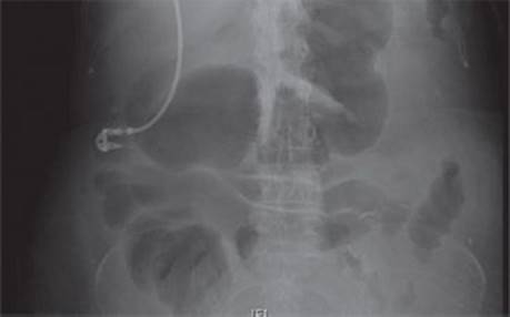
FIGURE 1 • Flat KUB showing distended colon.

FIGURE 2 • CT scan showing thickened colon.
C. difficile colitis most commonly presents as watery diarrhea with abdominal tenderness in a patient who has recently been on antibiotics. It is a gram-positive spore forming anaerobe bacteria that is most likely part of the normal colonic flora. Broad-spectrum antibiotics may disrupt the normal anaerobic flora and allow C. difficile to flourish. Certain strains of C. difficile produce toxins. Toxins A and B have been identified and are believed to cause glycosylation of proteins that normally maintain cell membrane integrity. Cell membrane breakdown then leads to secretory diarrhea. Recently, there have been more virulent strains of C. difficile identified that are believed to produce more toxins. These include the BI/NAP1/027 strain that causes more severe disease and higher mortality. There have also been reports of C. difficile colitis occurring in patients with no history of antibiotic use.
Diagnosis is based on clinical suspicion and history. It can be confirmed with stool toxin enzyme immunoassay that detects toxins A and B. Sensitivity of this test ranges from 63% to 94%, with a specificity of 75% to 100%. There are also enzyme immunoassays against glutamate dehydrogenase that is a common C. difficile antigen. Other diagnostic tests include cell-culture cytotoxic assays and C. difficile culture. More recently, the use of polymerase chain reaction (PCR) for amplification of C. difficile DNA in stool was approved by the FDA and can be performed in a few hours. PCR appears to be more sensitive than enzyme immunoassays and also have high specificity. However, many individuals are asymptomatically colonized with C. difficile, so careful patient selection for this type of testing is very important. Endoscopy can also be used to diagnose C. difficile colitis; however, because of concern for bowel perforation, it is usually reserved for cases in which the diagnosis is in question.
Once a patient is diagnosed with C. difficile, or if there is high clinical suspicion, any broad-spectrum antibiotics should be stopped (if possible) and treatment started. Most cases can be successfully treated with metronidazole, although there are recent reports of treatment failures in more severe cases. PO vancomycin is also effective although more expensive, and there is concern about the development of vancomycin-resistant enterococcus. In patients with severe illness requiring ICU admission, IV metronidazole and PO vancomycin combination therapy should be started. In 95% of patients, this will be adequate treatment; however in 3% to 5%, fulminant colitis can occur.
Fulminant C. difficile colitis is defined as severe systemic toxic effects from infection resulting in toxic megacolon, need for ICU admission, need for colectomy, or death. Mortality rate is reported as 35% to 60% in most series. Surgical intervention may be curative in cases of fulminant colitis and is recommended if patients with signs of severe systemic toxicity fail to improve after 24 to 48 hours of maximal therapy. Other specific indications for surgery include organ failure, vasopressors requirement, worsening CT scan findings, and peritonitis. Some experts advocate earlier surgical intervention as mortality after colectomy for C. difficile is increased in patients in multisystem organ failure, those requiring vasopressors, and those requiring ventilatory support. As the disease process involves the entire colon, the recommended surgery is total abdominal colectomy with end ileostomy.
Surgical Approach
The patient in our scenario is now intubated and requiring pressor support. As there is no improvement even with full intensive care unit support, consent is obtained from his family for exploratory laparotomy, total abdominal colectomy, and end ileostomy (Table 1).
TABLE 1. Key Technical Steps and Potential Pitfalls for Total Abdominal Colectomy

Patients with fulminant C. difficile colitis requiring surgery are often extremely ill and in multisystem organ failure. Total abdominal colectomy removes most of the diseased organ and toxin-producing bacteria and often results in dramatic improvement. Although the residual rectum may contain disease, most patients are too sick to allow for extensive pelvic dissection. Reanastomosis in this setting would be too risky given the patient’s already tenuous state and likelihood of leak; therefore, end ileostomy is almost always preferred.
If possible, the patient is marked for ileostomy prior to being brought to the operating room. The patient is placed into modified lithotomy position to allow for proctoscopy. This may be performed prior to incision in order to evaluate the rectal mucosa. Exploratory laparotomy is conducted under general anesthesia through a midline incision. Care should be taken upon entry into the abdomen to avoid the dilated colon. Once in the abdomen, the colon should be inspected for any areas of perforation and the remainder of the abdomen should be quickly inspected for any other pathology. In some cases, only a portion of the colon will appear grossly diseased at laparotomy. However, given the high likelihood of pancolonic involvement and the poor condition of the patients, they will likely not tolerate a second laparotomy. Therefore, total abdominal colectomy should still be performed. The colon is then mobilized from its peritoneal attachments. The distal ileum is transected and the mesentery is divided with ligature of the ileocolic, right colic, middle colic, left colic, and sigmoid vessels. The distal colon is then divided at the pelvic brim with care taken to resect the whole sigmoid colon if the sigmoidal vessels have been ligated. The rectal stump may be oversewn. In rare cases, the rectum may be the source of bleeding or such severe disease that staples or sutures will cut through the bowel. In this case, further resection of the rectum or a total proctocolectomy should be considered. If gross spillage has occurred, the abdomen should be irrigated. The ileostomy is then brought through the abdominal wall, and after the fascia of the midline is closed, this is matured.
Special Intraoperative Considerations
If the colon is so dilated as to interfere with safe mobilization, consideration should be given to decompression either through the colon or through the ileum. If the patient is extremely unstable, a damage control operation may be performed in which the abdomen is left open after colon resection and the patient returns to the ICU for resuscitation. The patient is brought back to the operating room after 24 to 48 hours for closure and maturation of the ileostomy. In cases of colonic perforation where there is a large amount of contamination, the abdomen should be well irrigated after colon removal and the skin may be left open with wet to dry dressing changes.
Postoperative Management
Mortality rate after abdominal colectomy for C. difficile is very high (35% to 50%) and patients require intensive supportive care in the perioperative period. Respiratory, cardiac, and renal support/replacement may be required. Postoperatively the patient is kept in the ICU until their hemodynamic stabilize. Antibiotic treatment against C. difficile is continued and the patient is kept NPO until bowel function returns. Recurrent C. difficile infection can occur in the residual rectum or in the small bowel, and this should be kept in mind if the patient relapses or continues to have symptoms of sepsis.
Case Conclusion
The patient is taken to surgery and dilated colon without signs of perforation or ischemia is found. Total abdominal colectomy with end ileostomy is performed. Postoperatively, his condition stabilizes and he is extubated on POD 2. His pressor requirements decrease and they are discontinued by postoperative day 2 also. He is transferred to the step down unit. He experiences a prolonged ileus and requires TPN support. On POD 13, his ileostomy begins to function and he is started on a diet. He tolerates this well and is discharged home on POD 16 (Figure 3).

FIGURE 3 • Dilated colon seen at surgery.
TAKE HOME POINTS
· C. difficile colitis should be considered in patents with profuse diarrhea, especially if they have received recent antibiotic therapy.
· Diagnosis can be confirmed through stool enzyme immunoassay or PCR testing.
· Most cases can be treated medically, but early consideration should be given to surgical intervention in patients with fulminant disease.
· Patients undergoing surgery for C. difficile colitis are very ill and usually in multiple organ failure. Perioperative mortality is very high.
· Given the pancolonic involvement and the poor health status of the patient, the procedure of choice is total abdominal colectomy with end ileostomy.
· The colon may grossly appear normal at colectomy but should still be removed in its entirety.
SUGGESTED READINGS
Ananthakrishnan AN. Clostridium difficile infection: epidemiology, risk factors, and management. Nat Rev Gastroenterol Hepatol. 2011;8:17–26.
Chan S, Ng K, Lyon D, et al. Acute bacterial gastroenteritis: a study of adult patients with positive stool cultures treated in the emergency department. Emerg Med J. 2003;20:335–338.
Couturier MR, Lee B, Zelyas N, et al. Shiga-toxigenic Escherichia coli detection in stool samples screened for viral gastroenteritis in Alberta, Canada. J Clin Microbiol. 2011;49(2):574–578.
Gash K, Brown E, Pullyblank A. Emergency subtotal colectomy for fulminant Clostridium difficile colitis–is a surgical solution considered for all patients? Ann R Coll Surg Engl. 2010;92:56–60.
Hall JF, Berger D. Outcome of colectomy for Clostridium difficile colitis: a plea for early surgical management. Am J Surg. 2008;196(3):384–388.
Jaber M, Olafsson S, Fung W, et al. Clinical review of the magagement of fulminant Clostridium difficile infection. Am J Gastroenterol. 2008;103:3195–3203.
Kufelnicka A, Kirn T. Effective utilizaion of evolving methods for the laboratory diagnosis of Clostridium difficile infection. Clin Infect Dis. 2011;52(12):1451–1457.
Kuntz JL, Chrischiles EA, Pendergast JF, et al. Incidence of and risk factors for communnity-associated Clostridium difficile infection: a nested case-control study. BMC Infect Dis. 2011;11:194.
Perera AD, Akbari RP, Cowher MS, et al. Colectomy for fulminant C. difficile colitis: predictors of mortality. Am Surg. 2010;76:418–421.
Rothenberger DA, Bullard KM. Surgery for toxic megacolon. In: Fischer JE, ed. Mastery of Surgery. Philadelphia, PA: Lippincott, Williams and Wilkins, 2007:1465–1474.
Sailhamer EA, Carson K, Chang Y, et al. Fulminant Clostridium difficile colitis. Arch Surg. 2011;144(5):433–439.
Sayedy L, Kothari D, Richards R. Toxic megacolon associated Clostridium difficile colitis. World J Gastrointest Endosc. 2010;2(8):293–297.