
Paul A. Gardner and Carl H. Snyderman
INTRODUCTION
Guiot first proposed using the endoscope for transsphenoidal procedures in 1957, and Apuzzo reported this approach in 1977. However, it was not until 1984 that Griffith and Veerappen used the pure endonasal technique to resect a pituitary adenoma of the sella. Jho, a neurosurgeon, began to define this purely endoscopic technique in 1997 by working with the otolaryngologists Carrau and Snyderman. However, it was not until 2001 that Jho reported the first endoscopic, endonasal resection of an intracranial meningioma compressing the optic nerve. Other neurosurgeons, such as DeDivittis and Cappabianca, have adopted and refined these techniques, widening their acceptance. Kelly, in 2004, reported on three patients in whom the endoscopic endonasal approach was used to resect suprasellar meningiomas with good short-term results, in this case using an endoscopic-“assisted” approach.
There remains controversy about the use of the endoscopic technique for suprasellar meningiomas because of concern for vascular involvement or adherence. However, all of these approaches, starting with the transsphenoidal approach to the sella, are based on the simple anatomic relationship of the tumor and its region of origin with the sinus(es) used for access. A natural extension of this concept is using the ethmoid sinuses as a corridor to access the entire anterior cranial fossa.
Yuen, also in 1997, described an endoscopic-assisted craniofacial resection applying the principles and instruments developed for inflammatory endoscopic sinus surgery to surgically address a malignant neoplasm. Stammberger described a completely endoscopic resection of an esthesioneuroblastoma (ENB) in 1999. Since then, multiple case series have documented good results following endoscopic resection of ENBs and other neoplasms of the skull base. These surgeons and others began working regularly as surgical teams of otolaryngologists and neurosurgeons to advance the field and refine these approaches. Not only were tumors of sinus origin such as ENBs being addressed, but tumors with even more significant intracranial extension and origin involving the anterior cranial base, such as meningiomas, were increasingly being shown to be safely accessible.
Adoption of the transcribriform approach has lagged behind that of extended transsphenoidal approaches, possibly due to the lack of adequate transnasal access to the area using a microscope, due to the difficulties with reconstruction, and partly due to the need for a surgical team composed of an otolaryngologist and neurosurgeon. The addition of an endoscope and its application by otolaryngologists and neurosurgeons working together to cross areas of anatomic understanding has allowed some surgical teams to resect most tumors involving the anterior cranial base.
HISTORY
Patients with tumors of the anterior cranial base can present with varying signs and symptoms, depending on the origin and extension of the tumor. Meningiomas can have subtle presentations with gradual loss of cognitive function and inhibition and are often mistaken as dementia. Personality changes and memory difficulties are often the most identifiable symptoms, even though they often go unrecognized by the patient. As a result, it is important to try to discuss these symptoms with family members or friends who may have noticed subtle changes that can only be identified retrospectively. Patients may become increasingly short tempered, impatient, or disinhibited. Alternatively, abulia may be the overriding presentation and often goes completely unnoticed.
Vision changes (blurred or double vision or loss of vision), headache, loss of taste or smell, and epistaxis are more objective symptoms that can help guide diagnosis and treatment. The period of onset and rapidity of progression also provide important clues and suggest malignancy, though indolent tumors such as meningiomas can reach a critical mass where they rapidly create more noticeable symptoms.
PHYSICAL EXAMINATION
A full neurologic examination is critical to look for other signs of involvement. Many more aggressive sinonasal tumors can have occult cranial nerve involvement that is not readily apparent radiographically. Very large tumors or those with significant associated edema can cause papilledema and associated vision loss. This is classically described in Foster Kennedy syndrome, where direct compression causes optic atrophy on one side with a central scotoma, while mass effect and edema result in papilledema on the other. It is also associated with anosmia and cognitive deficits. Full neuro-ophthalmologic evaluation is important to assess for papilledema as early cerebrospinal fluid (CSF) diversion or tumor treatment can prevent long-term loss of vision. In addition, until the increased intracranial pressure is resolved, the risk of postoperative complications associated with failure of reconstruction (such as CSF leak) is increased. Careful examination can also detect signs of orbital involvement such as proptosis.
Sinonasal tumors are more likely to cause anosmia than are meningiomas, but any tumor that involves the cribriform plate can affect smell and taste. These can be tested in the office with coffee or nonnoxious items. The degree of olfactory loss can be quantified with a “scratch and sniff” olfactory test (Sensonics, Inc., Haddon Heights, NJ). Nasal endoscopy for sinonasal tumors can add valuable information about involvement of nasal structures or the presence of associated infection that can affect the route of approach or timing of surgery.
INDICATIONS
The indications for surgical treatment depend upon multiple factors, including the diagnosis and biologic behavior of the neoplasm, stage of disease, patient comorbidities, patient preference, and prior treatment. Often, biopsy of sinonasal tumors can be done preoperatively by an otolaryngologist under endoscopic guidance in the office. Imaging is recommended prior to biopsy, though, to ensure that overly vascular tumors are not biopsied in a setting in which they cannot be controlled or appropriately managed. With the aid of a skilled skull base radiologist, the differential diagnosis can usually be narrowed enough to guide management in combination with intraoperative pathologic examination/confirmation. Generally, low-grade malignant tumors and high-grade neoplasms that are early stage are initially treated with surgical excision followed by adjuvant radiotherapy if indicated. Unresectable malignant tumors, high-grade tumors, or those with distant metastases are treated initially with radiation therapy ± chemotherapy. Alternatively, aggressive extradural debulking of the tumor can be performed at the time of the biopsy to relieve symptoms and decrease symptoms from nasal obstruction, orbital compression, or neural involvement or to provide decreased tumor burden prior to radiation and/or chemotherapy (though this is of unproven benefit).
The majority of midline tumors of the anterior cranial base can be addressed endonasally depending upon their extension and neurovascular involvement/relationships. Complete removal with negative margins is the goal for the treatment of malignant sinonasal tumors. This can generally be accomplished equally well endonasally as with open or combined approaches. No approach provides true “en bloc” removal, and the confirmation of histologically negative margins is critical. There are no long-term data available addressing purely endonasal resection of ENBs, but there is growing experience suggesting equivalence for tumor control. It is important to have a surgical team capable of combining an open approach for tumor extension beyond the reach of an endonasal approach, such as superiorly behind the frontal sinus or laterally past the midorbit. The approach should not limit the ability to achieve clear surgical margins.
Tumors such as olfactory groove meningiomas are generally best treated with resection if they are symptomatic from mass effect on either the frontal lobes or optic nerves. In addition, if there is radiographic evidence of frontal lobe or optic compression, resection is indicated as the primary treatment, even if the patient is minimally symptomatic, unless their comorbidities dictate otherwise. If tumors appear benign and are small and incidental, radiosurgery could be considered as the initial treatment, though a period of observation is usually warranted for these tumors in reliable patients given their typically indolent growth curve.
Complete resection of olfactory meningiomas can often be achieved via an endonasal approach, depending upon lateral extension and vascular involvement. If there is tumor extension beyond the meridian of the midorbit, consideration should be given to an open approach to ensure complete removal. In older patients with significant comorbidities, it is often reasonable to achieve subtotal or near-total resection. In these cases, symptom relief by removing mass effect rather than cure becomes the primary goal.
CONTRAINDICATIONS
Other than an active sinus infection, there are few absolute contraindications to an endoscopic endonasal approach. The infection can usually be treated with antibiotics with minor delay of surgery. Sometimes, however, surgical treatment of sinus disease is required for adequate drainage. At other times, infection is associated with tumor involvement and obstruction of natural drainage pathways. In these cases, partial tumor resection to allow drainage may be necessary with judgment required to determine delay of intradural tumor resection, depending on the urgency of patient symptomatology, tumor growth, or need for other treatment.
Preservation of olfaction is rarely possible with tumors that extensively involve the anterior cranial base. Indeed, resection of ENBs with skull base involvement includes both olfactory tracts and bulbs, thereby destroying all olfaction. Small, early-stage tumors can sometimes be removed endonasally with preservation of olfaction on the contralateral side, but oncologic margins should never be compromised to preserve olfaction. Small, unilateral planum or olfactory meningiomas likely carry greater risk of olfactory compromise when approached endonasally. This risk should be clarified with patients as part of the discussion of resection options and compared with the potential complications associated with frontal lobe retraction or manipulation associated with open, transcranial approaches.
Dense fibrous tumor tissue and vascularity are NOT contraindications to an endonasal resection. In fact, most of the same instruments and devices available for tumor debulking and coagulation that are available with open approaches are also available with extended shafts, pistol grips, or thin tips for endonasal use. The wide exposures provided with expanded approaches allow the use of these instruments in the same fashion as an open approach.
Encasement of the anterior cerebral vasculature is a relative contraindication and source of controversy. Certainly, if a tumor has contact with or encasement of arteries, resection becomes more difficult and requires a team that is more advanced in their learning curve. Microdissection techniques can be applied endonasally in the same way they are from an open approach. Arterial repair with suture is not an option via an endonasal approach, but, given the caliber of the vessels involved, it is unlikely that salvage of an injured vessel with suture is likely regardless of approach. Microclips and bipolar partial coagulation remain good strategies to employ in this situation. Nevertheless, this group of tumors should be approached in the way that the surgical team feels most comfortable.
The true advantage of the endonasal approach is the displacement of neurovascular structures to the outside of the tumor, minimizing or completely avoiding all manipulation of these structures. The only limitations are tumor extension beyond these, such as lateral to the bifurcation of the ICA. Most tumors of the anterior skull base do not extend past the optic chiasm but rather elevate it. Therefore, the only limitations are, anterosuperiorly, into the upper frontal sinuses and associated dura or, laterally, past the meridian of the orbit. By removing the lamina papyracea, the periorbita can be safely retracted laterally to allow access to tumor or involved dura extending over the orbit. The limitation becomes the vertical plane of the optic nerve (the midorbit). Most importantly, oncologic principles, especially that of negative margins, must not be compromised by the approach. This may require combining an endonasal approach with an open approach, especially if the tumor or resection margin extends beyond the midorbit or into the frontal sinus or associated dura.
PREOPERATIVE PLANNING
Tumors of the anterior skull base should be thoroughly evaluated with both MRI and CT angiography (CTA). MRI is helpful to determine the degree of involvement of the skull base and sinuses as well as demonstrating associated frontal lobe edema. CTA is often more valuable, though, as it demonstrates osseous changes in the anterior skull base, involvement and erosion of the skull base and orbit (lamina papyracea), and vascular relationships with the tumor (Fig. 12.1). These studies can also be used intraoperatively with image guidance.

FIGURE 12.1 Sagittal CT (computed tomography) angiogram showing the relationship of the anterior cerebral arteries (arrow) with a large olfactory groove meningioma. Notice also the hyperostosis (dashed arrow) of the planum.
Preoperative embolization is not necessary for most tumors of the anterior skull base when the tumor is approached endonasally. The main blood supply is usually the anterior and/or posterior ethmoid arteries. These are easily accessed during an endoscopic endonasal approach, providing good control and early devascularization.
If the patient has significant papilledema, especially when associated with ventriculomegaly, ventriculoperitoneal shunt placement should be considered to allow for maximum decompression and preservation of vision while awaiting resolution of cerebral edema and also to assist with healing of the resultant skull base defect following tumor removal.
SURGICAL TECHNIQUE (VIDEO 12.1)
Positioning is critical for an endoscopic approach to the cribriform area. The patient's head must be adequately extended to allow access to the anterior-most aspect of the anterior cranial base without the operator's hands and instruments colliding with the patient's chest. This will also help the flap or reconstruction graft to stay in place with gravity as an aid during placement.
The major advantage of an endonasal approach to the anterior skull base is based on the fact that the tumor originates from, involves, or is in contact with the skull base directly adjacent to the ethmoid sinuses. Therefore, the approach is via a tailored ethmoidectomy. This can range from sphenoidotomy and posterior ethmoidectomy for access to the planum sphenoidale to full anterior and posterior ethmoidectomies with frontal sinusotomy. Lateral extension can be accessed by adding maxillary antrostomies and removal of the laminae papyraceae. When in doubt, wider exposure and access is always valuable as it improves the visualization, orientation, and working room. If a tumor extends close to the posterior table of the frontal sinus, a frontal sinusotomy (Lothrop or Draf 3) should be performed. This significantly improves orientation by providing a clear anterior border/landmark and may prevent postoperative obstruction of the frontal sinus.
The technique for reconstruction must be planned prior to the surgery. With aggressive malignancies such as many cancers of the sinonasal tract, a nasal septal flap is not a good option due to possible tumor involvement. However, in select ENBs, if there is a radiographic, gross endoscopic, and histopathologically negative margin on a contralateral septal flap, it can be used. If the flap is inadequate for complete coverage, it can be supplemented by a nonvascularized autograft such as fascia lata or allograft-like cadaveric dermis (Alloderm) covering the entire defect with the vascularized flap overlying (superficial to) a portion of this graft. The vascularized flap seems to greatly improve healing of the nonvascularized portion. Otherwise, an extracranial pericranial flap should be planned and used without hesitation (Fig. 12.2). This flap can be placed “extracranially” without a craniotomy by passing it through an opening at the level of the nasion below the plane of the skull base (in contrast to a traditional pericranial flap that is technically intracranial).


FIGURE 12.2 Intraoperative photographs showing a vascularized pericranial flap (PF) after harvest (A) and after tunneling through a nasion osteotomy (B). Arrow points to edge of osteotomy.
Once tailored sphenoethmoidectomies have been performed, the overlying skull base can be removed. The first step in this is to locate, control, and ligate the ethmoid arteries (Fig. 12.3). This also serves to devascularize the tumor as these vessels typically supply the vast majority of any cribriform tumor. The posterior ethmoid artery can be found approximately one-half centimeter anterior to the optic nerve, and it runs slightly posteriorly as it passes from lateral to medial. The anterior ethmoid artery (AEA) (which runs obliquely anteriorly as it passes from lateral to medial) can also be located by removing adjacent lamina papyracea and ligating the artery at its exiting foramen. Care should be taken to completely coagulate the stump of the ethmoid arteries to avoid postoperative retrobulbar hematoma. Once the arteries are controlled, osteotomies can be performed bilaterally with a high-speed drill and coarse diamond burr just medial to the orbit (Fig. 12.4). These are connected posteriorly across the planum sphenoidale or tuberculum sellae as needed. Finally, anterior osteotomies are made just behind the posterior wall of the frontal sinus, extending to the crista galli. Once complete, these will allow bilateral anterior cranial plates, separated by the cribriform plate, to be dissected free from the overlying dura and removed. The planum sphenoidale and tuberculum sella may be removed separately.

FIGURE 12.3 Intraoperative endoscopic view showing bipolar coagulation of a ligated right anterior ethmoidal artery (AEA).

FIGURE 12.4 Intraoperative endoscopic view following complete ethmoidectomy showing the anterior cranial base from below. The osteotomies that have been drilled are highlighted with dotted lines.
Removal of bone laterally can be extended with a 45-degree Kerrison rongeur to the midorbit if the lamina papyracea has been removed. This will allow complete exposure of all involved dura in most cases, thereby obviating the need for an open approach. The dura and bulky tumor can now be coagulated with pistol-grip bipolar electrocautery to maximize hemostasis and tumor devascularization. Finally, the crista galli can be dissected free and then removed with either a drill and dissectors or Kerrison rongeur, depending upon the height. Ideally, the dura will be dissected free before the crista galli is detached, as this dissection becomes much more difficult once it is free floating.
Dura is opened just medial to the lamina papyracea (lateral to the olfactory nerve in an anterior craniofacial resection and in the middle of one side of an olfactory groove meningioma). For meningiomas, this should be done bilaterally up to the falx for maximal debulking and then coagulated and cut. Care should be taken not to expose the anterior frontal lobes until late in the operation, as they can sag downward obscuring the view. During an anterior craniofacial resection (e.g., ENB), the dura is opened from lateral to medial up to the falx at the level of the crista galli before the incision is extended posterior to the planum. The falx will then need to be coagulated and transected, angling posteriorly in order to avoid following it up the posterior wall of the frontal sinus.
Extracapsular dissection of the tumor is now performed. With ENBs and other sinonasal malignancies, the olfactory nerves and tracts should be resected with the dura, carefully dissected free from the overlying frontal lobe vasculature. They are transected posteriorly at the level of the planum along with the dura (Fig. 12.5).


FIGURE 12.5 Intraoperative endoscopic view showing the dural defect following resection of (A) olfactory neuroblastoma and (B) olfactory groove meningioma. Note the preservation of the fronto-orbital branch (arrow) of the anterior cerebral artery.
Surgical margins of the dura are then examined histologically to ensure complete, microscopic resection. When excising olfactory groove meningiomas, the tumor is carefully rolled inward, using a Cottonoid patty, dissector, or gentle suction to develop a plane between the tumor and the overlying, compressed, gliotic cortex. Laterally, an angled endoscope improves visualization and can help avoid unnecessary venous bleeding. As the dissection proceeds posteriorly, the anterior cerebral arteries (ACAs) become a concern, and proximal control should be gained by dissecting the planum/tuberculum portion of the tumor until these arteries are identified. Further extracapsular dissection can then take place with care and knowledge of their location. If necessary, a small amount of tumor capsule can be left on the anterior communicating complex to avoid injury to the small but critical perforators such as the artery of Huebner.
Reconstruction begins with the placement of an inlay Duragen graft, often draped slightly over the remaining falx. The patient's head must be in adequate extension so that gravity will hold each layer in place. The entire defect is then covered with a vascularized flap or a free graft; the flap must be in contact with either bone or soft tissue edge circumferentially to heal. Care should be taken to ensure that the pedicle of the flap is in contact with the lateral nasal wall or sinus along its entire course to prevent contraction. Often, turning the flap so that the pedicle runs up the ipsilateral lateral nasal wall provides the best option, with the flap lying obliquely. If an extracranial pericranial flap is used (see Video 12.2), it should be tucked between the orbits and overlying dura for separation. Surgicel and tissue glue are placed along the edges followed by Gelfoam and usually Merocel tampons. A Foley balloon often does not fit well in this rectangular defect.
POSTOPERATIVE MANAGEMENT
Nasal packing (either Foley balloon or Merocel tampons, depending on the size or shape of the defect) is removed approximately 7 days postoperatively following intradural surgery. One to two days of intravenous broad-spectrum antibiotics is recommended, but patients should be maintained on appropriate antibiotics (generally oral cephalosporins or equivalent antibiotic to cover nasal flora) as long as the packing is in place to avoid toxic shock or other associated infection. Once the packing is removed, endoscopy is performed in the outpatient setting to look for an overt CSF leak or defect. The flap should be pulsatile, and, generally, little debridement is performed at this time.
Repeat endoscopy is then performed 3 weeks postoperatively, at the same time that the Silastic nasal splints are removed. More vigorous debridement can be performed at this point to remove any remaining foreign substances such as residual tissue glue or Gelfoam. Crusts are generally allowed to heal without disturbance.
Thromboembolic complications are common, and patients should be routinely monitored for venous thrombosis in an attempt to avoid pulmonary embolus. Subcutaneous heparin can be started safely as early as 24 hours postoperatively in high-risk patients (immobilized or known hypercoagulable states).
COMPLICATIONS
Intraoperative arterial injury is perhaps the most dreaded endoscopic endonasal complication. This is most likely to occur when tumors of the anterior cranial base involve or encase the anterior cerebral arteries. Avoidance of injury is best achieved by strict maintenance of standard microsurgical dissection technique. Significant debulking and sequential extracapsular dissection are performed prior to dissecting the tumor from the arteries.
CSF leak is the best known complication of skull base resection, especially endonasal surgery. Vascularized reconstruction is key to rapid healing and maintaining a low rate of postoperative CSF leak. In many cases, a large nasal septal flap can be raised, but when this is not available, an extracranial pericranial flap provides a good alternative. If a CSF leak or pneumocephalus occurs (typically after packing is removed), it should be re-explored immediately to prevent further complication. With an obvious leak or increase in pneumocephalus, CSF diversion alone is not advisable as it is unlikely to resolve the fistula and may exacerbate the problem by entraining air. Removal of remaining absorbable packing such as tissue glues and Gelfoam will allow thorough inspection to locate the site of the leak. Often, a simple adjustment of the position of the flap is all that is needed, but augmentation with an adipose tissue graft or suture/clip may be needed if there is a pinhole defect in the flap itself. If the flap is inadequate due to retraction or septal perforation, Alloderm or other allograft material can be placed to cover the entire defect before replacing the vascularized flap over it.
Close collaboration between the otolaryngologist and the neurosurgeon remains critical in the postoperative period to remain vigilant for complications. Infectious complications such as sinusitis should be treated promptly to avoid meningitis. Epistaxis is a rare but known complication. Intracranial arterial dissection during tumor removal should be kept in mind as a possible source for postoperative epistaxis. Other hemorrhagic complications include retrobulbar hematoma from avulsion of the ethmoid artery or inadequate coagulation/ligation. Treatment of this should prompt an ophthalmology consultation and may require a canthal release to relieve pressure.
The usual postoperative complications can and do occur following endonasal skull base surgery as happens with traditional approaches. Patients can be safely anticoagulated as early as the first postoperative day in the event of thromboembolism. Obviously, this should be done with care without boluses and with slightly lower levels of anticoagulation. Seizures seem to occur at a lower rate than with traditional approaches, though this has never been thoroughly studied. Anticonvulsant therapy can often be avoided altogether in patients undergoing endoscopic anterior craniofacial resection for meningioma or sinonasal malignancy without significant cerebral edema.
RESULTS
We recently performed a preliminary review of 35 patients who underwent endoscopic endonasal resection of ENB at the University of Pittsburgh Medical Center (UPMC) over a period of 9 years. The Kadish classification, reconstruction, postoperative complications, adjuvant treatment, and outcomes were analyzed.
The mean age at the time of the surgery was 48 years (16–79), and 60% were male. The majority of patients were Kadish C (63%); 34% were Kadish B, 3% Kadish A, and 0% Kadish D. Two patients required a combined open and endoscopic approach to achieve negative dural margins. The majority of reconstructions were done with a septal flap (57%) and/or pericranial flap (17%). In 14%, no reconstruction was done as dural resection was not needed. Lumbar drainage was used in only five patients (14%). Postoperative complications occurred in 21% of the patients including diplopia (6%), CSF leak/meningitis (3%), epistaxis (3%), intracranial hematoma (3%), orbital emphysema (3%), and cosmetic deformity from a pericranial flap (3%). Postoperatively, 63% received radiation therapy, and 14% received chemotherapy. Negative margins were achieved in 94% of patients. Local/regional recurrence occurred in one patient (3%) and regional recurrence in one patient (3%). The mean follow-up period was 35 months (1–110), and 100% are currently disease-free after surgical salvage. This represents a preliminary review with short follow-up that will need to be peer-reviewed.
Devaiah and Andreoli performed a meta-analysis of available literature to compare EES with open surgery and found that there were greater published survival rates with endoscopic surgery in 361 patients (p= 0.0019).
EES was similarly evaluated for the removal of 44 olfactory groove meningiomas between November 2002 and February 2012 at the UPMC. Gross total resection (GTR) was achieved in 24 (54.5%) patients (Fig. 12.6), and 7 more (15.9%) were near total. The remaining 13 patients (29.5%) had a subtotal resection. The majority of these were intentional subtotal removals in elderly patients, but one was due to vascular injury and one due to poor visualization from venous bleeding very early in our experience. Tumors were removed in 2 stages in 16 (36.4%) patients. The GTR rates increased over time, from 48% to 69%, demonstrating a learning curve with this approach.


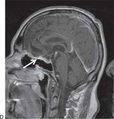
FIGURE 12.6 Preoperative coronal postcontrast, T1-weighted MRI (A) and sagittal CT angiogram (B) showing a medium-sized olfactory groove meningioma. Postoperative coronal (C) and sagittal (D), postcontrast, T1-weighted MRI showing complete resection of the olfactory groove meningioma. Note the enhancing nasoseptal flap (arrow).
The most common complication was a CSF leak, occurring in 17 patients (39%), but this has decreased significantly in recent years with the use of the vascularized nasal septal flap. Three patients subsequently developed meningitis and one a frontal abscess from superinfection of a Teflon granuloma. All were treated without further neurologic sequelae. There were two major complications: one intraoperative anterior cerebral injury, which suffered a rupture of a delayed pseudoaneurysm requiring craniotomy and endovascular treatment, and the second an octogenarian patient with a large planum/olfactory meningiomas who developed apparent vasospasm and multiple, distant vascular territory infarcts.
PEARLS
· Perform a full frontal sinusotomy (Draf 3 or Lothrop) for any tumor with extension close to the frontal sinus as this provides a reliable anterior anatomic landmark.
· Coagulation and ligation of the ethmoid arteries provides early devascularization of any tumor of the anterior cranial base.
· Vascularized reconstruction, such as a nasal septal flap, extracranial pericranial flap, or a combination, should be used whenever possible.
· Removal of the lamina papyracea provides additional lateral access out to the coronal level of the midorbit by retraction of the periorbita.
· The removal of large, benign tumors can often be staged to decrease single anesthesia time, blood loss, and surgeon fatigue.
PITFALLS
· Tumors with intracranial vascular involvement should be approached with caution since intracranial dissection requires a surgical team with significant experience with endonasal dissection.
· Endoscopic anterior craniofacial resection should not limit the margins of resection and should be used in combination with open approaches whenever necessary for oncologic purposes.
INSTRUMENTS TO HAVE AVAILABLE
· A full set of sinus instruments
· High-speed drill
· Zero- and 45-degree endoscopes
· A microdebrider
· Fine and angled tip, pistol-grip bipolars (Storz) are critical for extra- and intradural hemostasis.
· Extendable tip neurodissectors (KLS Martin)
· Fine, pistol-grip microscissors (Storz)
· CUSA (Integra) and Sonopet (Stryker) ultrasonic aspirators, both with extended tips
SUGGESTED READING
Casiano RR, Numa WA, Falquez AM. Endoscopic resection of esthesioneuroblastoma. Am J Rhinol 2001;15:271.
Cook SW, Smith Z, Kelly DF. Endonasal transsphenoidal removal of tuberculum sellae meningiomas: technical note. Neurosurgery 2004;55:239–246.
Kassam A, Snyderman CH, Mintz A, et al. Expanded endonasal approach: the rostrocaudal axis. Part I. Crista galli to the sella turcica. Neurosurg Focus 2005;19(1):E3.
Castelnuovo PG, Delu G, Sberze F, et al. Esthesioneuroblastoma: endonasal endoscopic treatment. Skull Base 2006;16:25–30.
Gardner PA, Kassam AB, Thomas A, et al. Endoscopic endonasal resection of anterior cranial base meningiomas. Neurosurgery 2008;63(1):36–52; discussion 52–54.
Snyderman C, Carrau R, Kassam A, et al. Endoscopic skull base surgery: principles of endonasal oncological surgery. J Surg Oncol 2008;97:658–664.