
Richard J. Harvey and Charles Teo
INTRODUCTION
While endoscopic endonasal access to the sphenoid sinus and skull base has dramatically changed the operative approach and patient recovery for many skull base pathologies, there are still many (and perhaps the majority) of neoplastic lesions involving the ventral skull base that require more than the endonasal approach alone. The development of the endoscope for cranial base surgery has made transfacial approaches nearly obsolete, but the concept of open craniotomy access remains viable. While transfacial approaches are still used for tumors that involve superficial structures such as skin, premaxillary adipose tissue, lacrimal sac, and anterior orbit, in our institution they are rarely applied outside of these situations.
The innovative use of traditional and minimal access craniotomy approaches has expanded the breadth of skull base pathologies that are now addressed with combined endoscopic-assisted craniofacial resections. There is no “attempt” to perform craniofacial resections “endoscopically or not”; the surgery is merely predetermined by the pathology, anatomical regions that need to be approached to resect the tumor, and reconstructive considerations. While we consider the endoscope helpful for osteotomy placement from above, with the avoidance of transfacial incisions that can have deleterious side effects apart from cosmesis (Fig. 19.1), the endoscope allows better access to the medial orbital wall to floor transition and still offers, in our opinion, better visualization of pathology in and around the sphenoid sinus. None of these advantages, however, dictate the application of endoscopic-assisted craniofacial surgery. We combine the endoscopic and open craniotomy approaches based on sensible anatomical guides that offer a practical approach to endoscopic-assisted craniofacial surgery, whether the combination is for access, reconstruction, or vascular control.

FIGURE 19.1 An example of the potential complications from transfacial incision. An extended lateral rhinotomy approach was used, and the medial canthal region broke down after radiotherapy. Such complications are rare with endoscopic-assisted craniofacial resections.
The majority of the tumors of the cranial base managed with endoscopic-assisted resections are malignant tumors or high-grade meningiomas, and as with other malignancy, the presentation may be related to local, regional, or distant disease. Defining the histology of the tumor and its stage is the key goal of investigations. Finally, treatment is generally multimodal with a combination of surgery and radiotherapy as the mainstay for most lesions. The proximity of critical structures, specifically the orbit, brain, and cranial nerves, dictate the morbidity from curative interventions.
HISTORY
The most important element in accurate diagnosis of sinonasal malignancy is clinical suspicion. The insidious onset of unilateral symptoms, the lack of previous inflammatory sinus disease or rhinitis, and the relative age of the patient (>50 years for tumors compared to <50 years for inflammatory disease) should be key features that prompt exclusion of neoplasia as a cause for a patient's symptoms. Although the presentation can be with regional symptoms (mass in the neck, orbital changes, diplopia, epiphora or cranial nerve dysfunction) and/or distant metastasis, this is relatively uncommon for most tumors, and the more common presenting symptoms (nasal obstruction, bleeding, discharge, and hyposmia) are local. These symptoms share common presenting complaints of patients with inflammatory sinonasal disease, which again highlights the importance of initial clinical suspicion. Unilateral eustachian tube dysfunction can also occur. Gross macroscopic changes to the mucosa of the hard palate or the skin are uncommon in developed countries. True involvement of the cavernous sinus by malignancy is generally a contraindication to surgery and curative treatment. Involvement of the proximal trigeminal nerves or the other upper cranial nerves should be evaluated. Numbness of the palate and midface dysesthesia are the signs of involvement in the pterygopalatine fossa and V2 involvement in the orbital floor or the roof of the maxillary sinus. Along with asking about orbital symptoms (visual acuity changes, diplopia, and displacement of globe), the inclusion of epiphora is important as this is an anterior limit often missed and needs to be addressed via a transfacial approach.
General health, nutrition, smoking status, bleeding risks, and prior nasal/sinus and cranial procedures should also be noted.
PHYSICAL EXAMINATION
The evaluation of a patient with skull base neoplasia should include the limits of the tumor. Superiorly, the involvement of the dura and brain parenchyma is a radiologic assessment in most patients. Inferiorly, the palate should be evaluated to see whether a total maxillectomy is necessary. Anteriorly, the lacrimal sac and premaxillary tissue should be evaluated bimanually via the gingival buccal sulcus. Laterally, above the orbital floor, signs of restricted ocular movements, conjunctival congestion, and orbital displacement are signs of involvement of a space-occupying tumor. The presence of trismus is an important sign that the masticator space/pterygoid muscles are involved.
Endoscopic examination reveals a mass within the nasal cavity (Fig. 19.2). Preoperative endoscopic examination can be useful to inspect for uninvolved areas of the paranasal sinuses. Many lesions appear extensive on imaging, but they are predominantly exophytic lesions with only a small area of invasion. If the lesion is unilateral, then assessment of the contralateral septum and sphenoethmoidal recess can greatly assist surgical planning. Look for evidence of prior surgery on the septum, sinus, or turbinates. The local reconstructive options available should be noted on examination. Examination of the neck for cervical lymph node metastases is important as these are not always accurately assessed on imaging and may provide an easy route for tissue diagnosis.


FIGURE 19.2 The classic endoscopic appearances of a SCC (A) and an olfactory neuroblastoma (B).
INDICATIONS
Skull base pathologies requiring endoscopic-assisted surgery are neoplasms: primarily malignant tumors of the paranasal sinuses (Fig. 19.3A). Some intracranial lesions such as atypical/aggressive meningioma are also addressed (Fig. 19.3B). The general principle of surgical access is important here. If the surgical approach crosses the axis of critical neural or vascular structures, then an alternate route should be considered.


FIGURE 19.3 Typical aggressive SCC ex-inverting papilloma with extension into the lateral aspect of the frontal sinus (A) and an aggressive atypical meningioma with involvement of the roof of the orbit (B). Both required endoscopic-assisted resections.
Classic absolute indications to combined endoscopic and open cranial resection:
1.Involvement of the dura beyond the midorbital roof and intraorbital extension
2.Involvement of the anterior table or lateral recess of the frontal sinus
3.Significant involvement of the posterior table of the frontal sinus, where preservation of the sinus is not practical and cranialization should be performed
4.Tumor superior or lateral to the optic nerve canal
5.Tumor lateral to the carotid artery or involvement around bifurcations
6.Extension into facial or orbital soft tissues (usually transfacial approaches apply)
Relative indications:
1.Gross involvement of brain parenchyma
2.Lateral extension of tumor to the lateral wall of the maxillary sinus and infratemporal fossa
CONTRAINDICATIONS
Tumors that are situated entirely medial to the cavernous internal carotid artery (ICA) are most accessible using an endonasal approach and do not need an open approach. Where bacterial colonization is associated with the pathology, preoperative antibiotic therapy should be given. Severe local mucosal involvement from concomitant inflammatory conditions should warrant consideration of an alternate route.
PREOPERATIVE PLANNING
Radiologic Evaluation
Imaging should always be focused on what is trying to be achieved, namely tumor staging. Accurate information on local tissue involvement is critical for “T” staging. Most patients will undergo both computed tomography (CT) and magnetic resonance imaging (MRI) for several reasons. The T2 MRI will highlight edematous mucus and retained secretions compared to the tumor as defined on T1 post–contrast imaging (Fig. 19.4A). The involvement of the periorbita is determined by bone loss on CT and enhancement of adipose tissue on MRI. Thirdly, involvement of the dura (and brain parenchyma) is defined by bone loss on CT and dural enhancement on MRI (Fig. 19.4). Perineural involvement of cranial nerves is usually defined using fine-slice adipose tissue–saturated T1 post–contrast MRI. Finally, the relationship of the tumor to the intracranial course of the ICA and its branches is defined by either CT or MR angiography. Formal angiography of the ICA should be considered when there is tumor involvement or ectasia. The merits of preoperative balloon occlusion testing are very limited and should be applied when a sacrifice of the carotid artery and bypass is being considered and not just because of the risk of carotid bleeding. Where possible, include a scan series (CT or MRI) for future image-guided surgery (IGS) in the initial assessment.

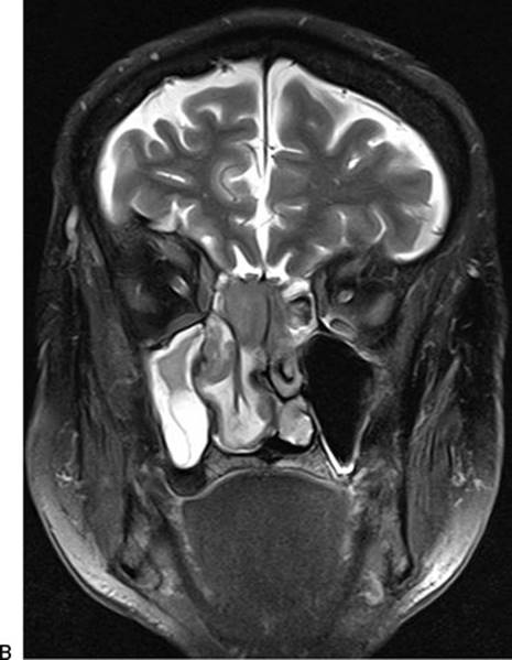
FIGURE 19.4 The T1 (A) and T2 post–contrast (B) MRI characteristics of an invasive right sphenoethmoid SCC. The differentiation of tumor, mucus, and mucosal edema can be seen.
Regional and distant metastases may be defined with a positron emission tomography/CT assessment. This technique is a combination of full-body CT and assessment of focal radioactive glucose uptake (18FDG) by cells. This is the most efficient form of staging and can provide standard uptake value information for subsequent follow-up. Imaging of the neck, chest, and abdomen as well as blood tests for calcium and alkaline phosphatase would also suffice. Specific investigations for bone, brain, or other metastasis are usually driven by clinical suspicion rather than being routine.
Histopathologic Diagnosis
Obtaining histopathologic confirmation of malignancy and its subtype is critical prior to therapy. This is usually done prior to treatment decision making, and the use of “frozen” or intraoperative specimens is ill advised and not recommended for a definitive diagnosis. This is important as some tumors such as lymphomas are radiosensitive and do not require surgery, whereas a diagnosis of melanoma would prompt an aggressive search for metastasis prior to local treatment.
The WHO classification is listed in Table 19.1 with the most common subtypes included. The epithelial versus nonepithelial distinction is easy and reflects the frequency of tumors. Epithelial tumors are the most common with squamous cell carcinoma (SCC), adenocarcinoma, and adenocystic carcinoma most commonly reported. The nonepithelial tumors are lymphoma (hematologic), olfactory neuroblastoma (neuroectodermal), chondrosarcoma (bone/cartilage), and mucosal melanoma (neuroectodermal).
Table 19.1 The WHO Classification System for Sinonasal Malignancies
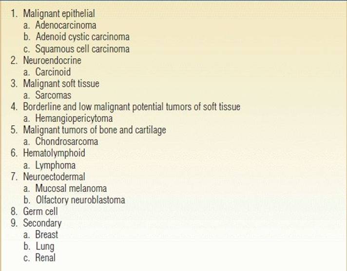
The Main Subtypes are Described Here with Common Pathologies.
Endoscopic biopsy can be performed in either a clinic or an operating room. The ability to manage a biopsy site that bleeds is critical. When lesions have already been imaged and the pathology is in the proximal nasal cavity anterior to the middle turbinate, we routinely biopsy these in the clinic as we have bipolar forceps and compression dressings available. This greatly assists in organizing further investigations and treatment decision making.
Patients with distant metastases rarely undergo surgical therapy. The focus of treatment in these patients is symptom control (palliation), and short courses of radiotherapy are often given. With the exception of lymphomas, chemotherapy/radiotherapy or combinations are not curative but given as adjuncts to surgical resection with curative intent. Radiotherapy is used to control local and regional disease. There are complex lymphatic channels in the paranasal sinuses and skull base that prevent full excision of the lymphatic compartments during surgery. Additionally, close surgical resection margins occur next to the orbits, carotid arteries, and cranial nerves that benefit from additional local therapy. Some centers use chemoradiotherapy for sensitive tumors such as olfactory neuroblastoma prior to surgery, as this is generally quick to initiate and can reduce tumor size, making surgery less technically demanding (less bulk and bleeding). It is a misjudgment to believe that with this approach, a lesser region of the skull base can be removed as the resection must still follow the originally involved anatomical sites.
SURGICAL TECHNIQUE
Image Guidance
The accessibility and ease of use with image guidance systems make them almost mandatory for modern craniofacial units. Although no surgery should be delayed because the IGS system is not functioning, an available IGS unit should always be used. We prefer to have patients in a Mayfield pin fixation when working in the sphenoclival area or when extensive drilling is required. The image guidance reference point is fixed to the clamp. For simpler tumors of the anterior cranial fossa with frontal sinus and orbital roof involvement, we use a bone-fixed reference point (Fig. 19.5). IGS is useful to define the limits of the frontal sinus and extent of the tumor, identify the ICA near tumor, and define the location of any cranial window to introduce a pericranial flap.

FIGURE 19.5 An option to Mayfield pin fixation is the direct-to-bone fixation. The registration of the image guidance system then has to occur prior to the remainder of the coronal incision being raised.
Preparation
a.0.5 × 3 inch cotton pledgets soaked in 1:2,000 adrenaline + 1% ropivacaine
i.10 pledgets soaked by 10 mL of solution.
ii.As many as possible placed in each nasal cavity immediately after intubation.
iii.Remaining pledgets join the setup for use intraoperatively during the extradural approach.
b.1% ropivacaine and 1:100,000 adrenaline mixture
i.2 × 10 mL solutions made by 9 mL of 1% ropivacaine and 1 mL of 1:10,000 adrenaline.
ii.Inject any external scalp or eyebrow incision lines with one syringe.
iii.The other is used endonasally via 22-gauge spinal needle on 10-mL syringe.
1.The distal 6 mm of the needle is bent at 45 degrees with bevel up to facilitate injection. The shaft is bayoneted to improve hand position.
iv.Start posterior and inferior first to avoid bleeding onto the surgical field.
v.Injection: sphenoid rostrum, choana inferiorly, septum, nasal floor, middle turbinate root.
c.Perform the endonasal injections immediately when the endoscope is set up. Replace cotton pledgets under endoscopic control after injecting and complete set up of the surgical field (i.e., microdebriders, bipolars forceps, check that devices are working, check required instruments—as much time as possible).
d.Wait for the systemic effects of injection to resolve before surgery starts if the endoscopic component is first. Usually this occurs when the heart rate is below 70 bpm rather than a measure of mean arterial pressure.
The endonasal component of the resection proceeds by defining uninvolved sinuses to allow for visualization of the medial aspect of the orbits, nasofrontal recesses, and sphenoid sinuses. Bone landmarks (optic canals, carotid canals) are identified. The margins of resection are defined. When the resection is anterior to the anterior ethmoidal artery, visualization of the posterior wall of the frontal sinus via a Draf III frontal sinusotomy is critical, and anatomical landmarks are used to guide the surgeon instead of the variable anatomy of the frontal recess (Fig. 19.6).


FIGURE 19.6 A large atypical meningioma (A) demonstrates the importance of a Draf III frontal sinusotomy when exposing the posterior table (B) and thus orientating the surgeon to the location of the skull base in patients with a large tumor and a history of prior surgery.
We routinely visualize the orbital floor as a key landmark to define the sphenoid/posterior ventral skull base. Once this occurs, the orbital axis from the medial wall to the optic canal can be localized. Ligation of the internal maxillary artery is usually performed on the predominant side of the tumor to help control bleeding and gain access to the pterygopalatine and infratemporal fossa (Fig. 19.7). A Draf III frontal sinusotomy is performed to identify the posterior table and anterior ventral skull base. The ethmoidal arteries are controlled.
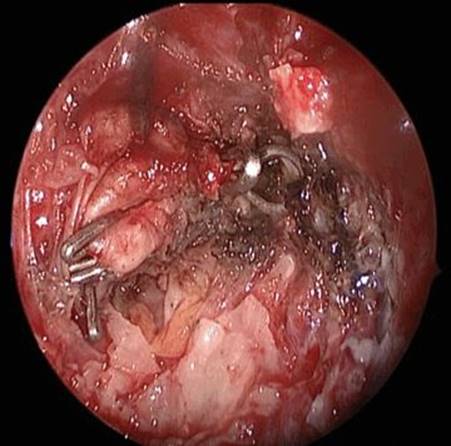
FIGURE 19.7 Transmaxillary ligation of the right internal maxillary artery occurs early in the dissection to control bleeding. Such ligation removes the utility of local mucosal flaps on that side that are often involved in tumor.
After consideration as to whether a nasoseptal flap is appropriate, a full posterior septectomy is used when dealing with tumors in the anterior cranial fossa. The sphenoid ostium is widened superiorly so that the level of the planum/roof can be visualized on the uninvolved side or it is identified by debulking and using the orbital floor as a guide. A large backbiting forceps is used to engage the posterior edge of the septum as close to the floor of the nose as possible. This nasal floor release of the septum comes anteriorly to the anterior wall of the frontal sinus and in line with where the anterior limit of the Draf III frontal sinusotomy will be. The 2-mm Kerrison rongeur is then engaged at the anterior limit of this backbiting forceps-made incision, and a vertical channel is made in the septum to the level of the roof of the sphenoid (now in view). The microdebrider can be used to remove some mucosa of the septum to view the contralateral turbinates/nasal cavity (although this may be primarily tumor). An inferior vertical channel of sphenoid bone is removed on either side of the midline. A large straight Mayo scissor, double action or thru-cutting, is used to define a superior incision in the remaining septum and continued into the intersinus septum of the sphenoid sinus. The roof of the sphenoid sinus, which is visible, is the guide. A large grasping forceps is used to remove the septum and sphenoid rostrum. The remaining anterior wall of the sphenoid is removed or tumor debulked laterally and superiorly to expose the roof and lateral opticocarotid recess. This creates a true posterior septectomy with a vertical anterior limit down to the floor of the nose and is ideal for instrument access.
The options for the cranial access are determined by what is required to manage the pathology.
1. No local reconstruction option available
When no local mucosal flap is available, the coronal approach can be used primarily to provide the pericranial flap based on one supraorbital vessel pedicle or the temporoparietal fascia based on the superficial temporal artery. These fascial flaps can be introduced to the anterior skull base through a simple window in the nasion region for the pericranial flap (Fig. 19.8) or passed via an infratemporal fossa tunnel to the posterior maxilla for a temporoparietal fascial flap. No formal craniotomy is required, and it avoids the potential for necrosis of bone flaps in patients undergoing radiotherapy, additional dural or venous injury, and subdural collections.
2. Tumor with limited orbital roof, superolateral optic canal involvement, and ICA control
The supraorbital approach is ideal when the tumor is located laterally and superiorly over the optic canal, orbital roof, and ICA. Here, the intradural approach offers limited brain retraction, the ability of both surgeons to work concomitantly, great cosmesis, and the ability to manage pathology lateral to the ICA that ensures good vascular control (Fig. 19.9).
The head is placed in the Mayfield pin fixation with the head turned 30 degrees to the contralateral side and the head extended to bring the malar prominence uppermost. The side of the approach is determined by the lateral projection of the tumor. The skin incision is placed in the superior border of the eyebrow, lateral to the supraorbital nerve (Fig. 19.9). A pericranial flap is reflected inferiorly and the keyhole exposed after skin and muscle dissection, which are retracted superiorly using fish hook retractors. A single burr hole is made and a free supraorbital bone flap, approximately 20 mm in height or approximately the width of an open bipolar shaft, is created. The inner table of the supraorbital rim is then drilled down along with the bony protuberances of the floor of the anterior cranial fossa to gain additional exposure. The dura is opened in a C-shaped manner and reflected inferiorly. Under microscopic vision, the dura is identified over the orbit and the arachnoid of the carotid cistern is identified and opened to allow release of cerebrospinal fluid (CSF). No fixed brain retraction is used. Standard microsurgical techniques of tumor removal are used and the optic canals opened when required for complete removal of the tumor and decompression of the optic nerves. After removal of the tumor, the dura is closed watertight, and the bone flap replaced using two Craniofix clamps (Aesculap Inc., Center Valley, PA). The bone flap is approximated flush to the upper border of the craniotomy and the resulting inferior margin bone defect filled with Surgicel (Ethicon Inc., Johnson and Johnson, Piscataway, NJ), to reduce depressions in the forehead. The pericranial flap is carefully reapproximated, and the galea and skin are closed without tension. A subcuticular nylon running suture provides the best cosmetic outcome for skin closure.
3. Excision required of the orbital roof and anterior wall of the frontal sinus or need for cranialization of the frontal sinus
A coronal incision is performed to expose the orbital rims. A “widow's peak” in the incision allows for easy reorientation on closure. A “stepped” or “broken” incision can be useful to avoid an obvious incision when the hair is wet on the sides (Fig. 19.10). The incision is made posteriorly across the scalp in men when male pattern baldness is predicted. In women, a hairline trichophytic incision and closure can sometimes be used.


FIGURE 19.8 Pericranial flap being introduced through a craniotomy window when only pericranial fascia is required from the open exposure. A. Small bone window at the level of nasion and below the level of cranial base. B. Pericranial flap inserted through the bone window.


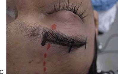
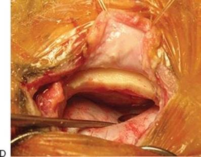


FIGURE 19.9 An example of a meningioma lateral to the carotid bifurcation (A). The A1 take off from the bifurcation is only seen in the distance with a 30-degree endoscope and does not represent safe access (B). The supraorbital incision (C) and approach (D) can allow both surgeons to work simultaneously (E). Visualization and control of the internal carotid artery via the craniotomy (F). Total removal (G) and an imperceptible incision (H) on the patient's right.

FIGURE 19.10 A “widow's peak” and a staggered incision during the coronal incision can assist with closure and cosmesis.
The traditional craniotomy can be modified (often via IGS) to encompass the need of the surgical access. When simple cranialization needs to occur, then a limited craniotomy (Fig. 19.11) can be used. This must be extended laterally to manage extension to the roof of the orbit. The bone flap must be discarded in cases of tumor involvement of the anterior wall of the sinus. Split calvarial grafts work well to avoid a cosmetic defect. Only in cases of extensive removal of the wall of the orbit is it reconstructed with titanium mesh. Contrary to traditional teaching, large segments of the medial wall and roof can be discarded without reconstruction with preservation of orbital stability.
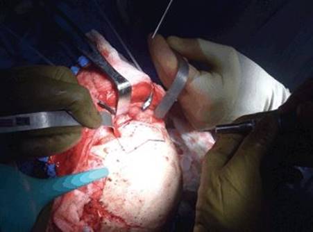
FIGURE 19.11 An example of exposure with release of the supraorbital vessels from their notch. The craniotomy was prepared so that the frontal sinus could be cranialized in a patient with a sinonasal undifferentiated carcinoma.
Closure
Overall, reconstruction follows the principles of dural reconstruction and preventing CSF leaks, separating the intracranial cavity and the aerodigestive tract, covering nerves and blood vessels of the neck and skull base with well-vascularized tissue, supporting the orbit, separating the nasal cavity from the oral cavity, and restoring facial contour and function.
The dural defect is directly closed from above where possible. Vascularized flaps are preferred over free grafts. When a local fascial flap is used, it is an underlay flap on top of the skull base defect. Duragen, fascia, or other collagen substitute is used as a second layer to close the defect on the cranial side superior to the flap. When a local mucosal graft is used, the Duragen is placed intracranial (but not extradural), and the mucosal graft is an onlay flap. Tissue glue is used only on the nasal cavity side. Silastic sheets 0.5 mm (Medtronic, Jacksonville, FL) are used to cover the septum and any donor mucosal sites. It is secured with a 2/0 Prolene or 4/0 silk suture anteriorly through and through the anterior septum. Tie the knot on the left as this makes dividing the right loop in the clinic simple and easy for removal. Dissolvable packing (Gelfoam, Spongistan, Nasopore) is placed against the reconstruction. A Foley 16-gauge urinary catheter is used to compress the Gelfoam pack against the flap. The balloon is used to hold the Gelfoam in place. There is no uniform direct pressure applied or required on the flap by the balloon.
POSTOPERATIVE MANAGEMENT
Postoperative lumbar drainage is considered in all patients but rarely used unless the patient is at high risk for a postoperative leak. Those at high risk are obese patients or those with known elevated intracranial pressure, postradiotherapy patients, and those with a resection cavity that is in free communication with a high-flow CSF cistern.
When used, the balloon is removed during the hospital stay. Most of the patients who have had a transdural approach are hospitalized for 5 to 7 days. Blowing the nose is avoided for 3 weeks when the dura or periorbita has been reconstructed. Silastic 0.5-mm sheets are used to cover any mucosal flap donor site for 3 weeks. The balloon is removed on days 3 to 4, and patients are observed for a further 48 hours. This may be conservative for some centers, but we feel that if a CSF leak has not occurred in the first 72 hours, then the chance of breakdown of the reconstruction after this point is uncommon. The risk of meningitis is also significantly reduced beyond 5 days. Patients are seen in clinic at 3 weeks post-op, and the silastics are removed. There is no need to see patients earlier with this suggested routine.
Nasal saline irrigation starts on day 7. There is no role for simple nasal sprays except for moisturizing crusts in the first 7 days. All postoperative recoveries must include high-volume positive pressure saline irrigations used twice a day. These are isotonic, and we do not promote special mixtures such as Ringer lactate, or hypertonic or hypotonic solutions as there is little clinical evidence that they offer any additional benefit over isotonic saline. Bactroban 2% ointment (the water-soluble propylene glycol version) is used topically in the nasal vestibule twice/day to minimize crusting and reduce Staphylococcus aureus colonization. Antibiotic coverage is given for 10 days postsurgery or as long as dressings remain in the paranasal sinus. Our philosophy of long-term antibiotic use is not to prevent perioperative complications (such as meningitis) but to reduce the bacterial colonization that occurs in heavily operated and nonfunctioning sinuses.
COMPLICATIONS
The most common immediate complications are nasal bleeding, CSF leak (+/− pneumocephalus), and meningitis. Epistaxis is usually from a transected septal branch that has not been cauterized with bipolar diathermy during widening of the sphenoid inferolaterally; it usually occurs on the contralateral side to the septal flap. CSF leak is almost always a technical error or shift in reconstructive layers when they occur early (<48 hours) and a mucosal flap has been used. Early reexploration and revision are recommended. Lumbar drainage can be used when there appears to be good early healing but an event, such as the patient straining or blowing their nose, occurs and a small amount of CSF leak is evident. Cautious use of lumbar drainage may provide an avenue for closure without reexploration. Long-term follow-up of tumor recurrence and secondary effects such as postradiotherapy hypopituitarism is ongoing. It may take up to 12 months before mucosal crusting fully resolves if there has been an extensive resection and radiotherapy. Ongoing nasal saline irrigations provide great assistance with this, and the goal is to provide a long-term functional cavity that is easy to evaluate (Fig. 19.12).

FIGURE 19.12 An endoscopic view of the sinonasal cavity 1 year postexcision and radiotherapy of a T4 SCC of the septum. An inferior turbinate flap was used in the reconstruction. Leaving the cavity in this state allows for easy irrigation by the patient and surveillance by the physician.
RESULTS
Comparable results have been noted for completely endoscopic, endoscopic-assisted, and standard craniofacial approaches for properly selected tumors. The selection of a surgical approach depends on multiple factors including the experience and comfort of the surgical teams.
PEARLS
· Always take great care in raising the pericranial flap. If the pedicle is compromised, a temporoparietal flap based on the superficial temporal may be harvested to replace it. Undermine the posterior incision to increase its length. One can never have too much.
· If the entire sphenoid sinus is full of pathology, then use a transethmoid approach on the least affected side and use the orbital floor to locate the sphenoid sinus at a safe level below the skull base.
· Create your surgical access so that the procedure proceeds with a 0-degree endoscope and straight instruments. Avoid angled endoscopes; although the view may be good, the dexterity, manipulation, and access with instruments are often limited.
· Cover all demucosalized nasal regions with an occlusive dressing (usually silastic sheets 0.5 mm) to prevent crusting and allow for quick granulation to occur in the first 21 days. Reepithelialization occurs quickly over granulation tissue but slowly over dry crusts.
PITFALLS
· In patients receiving induction chemotherapy prior to surgery, it is a misjudgment to believe that a lesser region of the skull base can be removed as the resection must still follow the originally involved anatomical sites.
· Visualization of the posterior wall of the frontal sinus via a Draf III frontal sinusotomy is critical, and anatomical landmarks are used to guide the surgeon instead of the variable anatomy of the frontal recess.
· Postoperative epistaxis is usually from a transected septal branch of the sphenopalatine artery that has not been cauterized with bipolar diathermy during widening of the sphenoid inferolaterally.
INSTRUMENTS TO HAVE AVAILABLE
Useful instruments for endoscopic-assisted craniofacial resections.
· Endoscopic drill: 5-mm 15-degree coarse diamond suction–irrigated ASB burr and protected high-speed irrigation burrs.
· Coblation is an excellent transnasal endoscopic tissue removal device and bipolar instrument. The Procise EZ Wand is malleable, low profile, and most suited to this work.
· Doppler ultrasound probe.
SUGGESTED READING
Harvey RJ, Gallagher RM, Sacks R. Extended endoscopic techniques for sinonasal resections. Otolaryngol Clin North Am 2010;43(3):613–638.
Lund VJ, Stammberger H, Nicolai P, et al. European position paper on endoscopic management of tumours of the nose, paranasal sinuses and skull base. Rhinol Suppl 2010(22):1–143.
Patel MR, Stadler ME, Snyderman CH, et al. How to choose? Endoscopic skull base reconstructive options and limitations. Skull Base 2010;20(6):397–404.
Harvey RJ, Winder M, Parmar P, et al. Endoscopic skull base surgery for sinonasal malignancy. Otolaryngol Clin North Am 2011;44(5):1081–1140.
Rawal RB, Gore MR, Harvey RJ, et al. Evidence-based practice: endoscopic skull base resection for malignancy. Otolaryngol Clin North Am 2012;45(5):1127–1142.