
Franco DeMonte
INTRODUCTION
Neoplasms of the nasal cavity and paranasal sinuses account for only 0.2% to 0.8% of all malignancies and 2% to 3% of all head and neck cancers. The incidence is between 0.3 and 1 case per 100,000 population. Tumors in this location are rare in childhood with the incidence beginning to increase in the fourth decade. The median age of diagnosis is 62 years in men and 72 years in women. There is a slight male predominance.
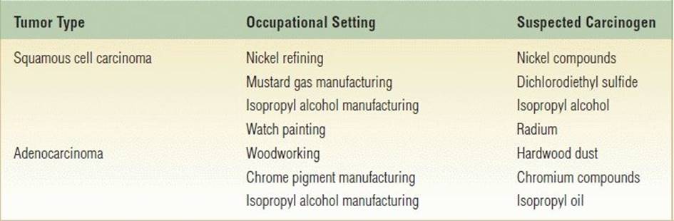
The majority of tumors of the nasal cavity and paranasal sinuses arise from the mucous membranes lining these air spaces. The most common pathology in North American and Asian society is squamous cell carcinoma with olfactory neuroblastoma, adenocarcinoma, adenoid cystic carcinoma, and sinonasal undifferentiated carcinoma being other pathologies common in larger surgical series (Table 18.1). It is often difficult to determine the exact site of origin of these tumors given that more than 90% of them may have invaded at least one sinus wall and the disease may extend well beyond the original sinus. The maxillary sinus is the most common primary site of origin in 55% of cases. Ten percent of these tumors arise in the ethmoid sinus and only 1% in the sphenoid and frontal sinuses. Thirty-five percent of tumors originate from within the nasal cavity. Epidemiologic studies have identified a variety of environmental hazards that have been associated with the development of sinonasal tumors. A significant association between sinonasal tumors and cigarette smoking has been identified in a large case control analysis of white American males. Various occupational hazards have also been linked to the development of sinonasal tumors. The tumor type, occupational setting, and suspected carcinogens are outlined in Table 18.2.
Table 18.1 Histologic Distribution of 209 Patients with Paranasal Sinus Tumors Treated by Craniofacial Resection at M. D. Anderson Cancer Center, 1992–2008
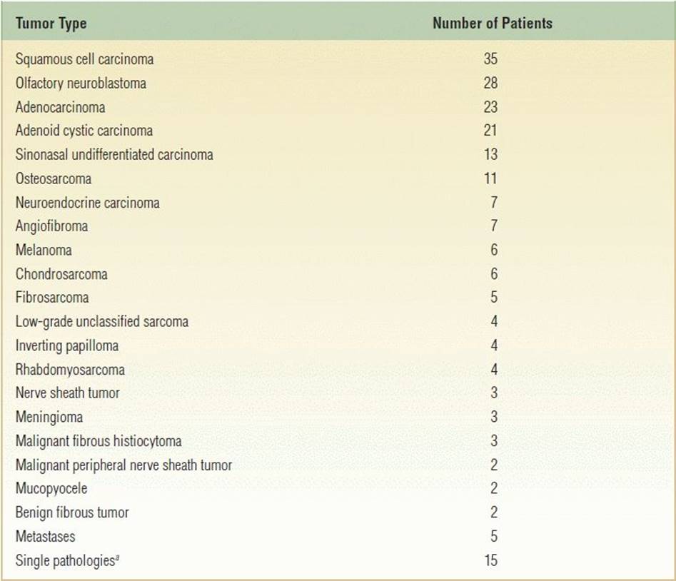
aIncludes mucoepidermoid carcinoma, mesenchymal chondrosarcoma, teratocarcinosarcoma, angiosarcoma, osteoma, leiomyosarcoma, fibrovascular polyp, ameloblastic carcinoma, PNET/Ewing sarcoma, high-grade unclassified sarcoma, malignant solitary fibrous tumor, germ cell tumor, myoepithelial carcinoma, liposarcoma, hemangioma.
Table 18.2 Occupational Hazards Associated with Paranasal Sinus Tumors
The transcranial approach for anterior craniofacial resection is a technique used to manage these sinonasal malignancies when the neoplasm extends to or through the base of the skull to involve the parameningeal space, the meninges themselves, or the intradural and intracerebral structures. The technique provides excellent visualization, the ability to manage the orbits safely and completely, and the opportunity for robust dural and cranial base repair.
HISTORY
The most common initial symptoms of malignant sinonasal disease are nasal airway obstruction that is frequently unilateral, chronic nasal discharge, and epistaxis. Unfortunately, these early symptoms of malignant disease are identical to those of benign nasal and paranasal sinus disease. Studies have shown a combined physician and patient delay ranging from 3 to 14 months due to this nonspecific symptom complex. The liberal use of imaging studies and early biopsy has lessened the diagnostic delay and increased the percentage of tumors being diagnosed at an earlier stage.
Tumors of the superior aspect of the nasal cavity and ethmoid sinus may be associated with anosmia and headache. Carcinoma of the frontal sinus may present as an acute frontal sinusitis with pain, swelling, and evidence of bone erosion. Nasal obstruction, epistaxis, and nasal discharge may be absent when the tumor is located in the sphenoid sinus, and patients typically suffer from headache, diplopia, and cranial neuropathy.
PHYSICAL EXAMINATION
A thorough examination of the head and neck, including endoscopic evaluation of the sinonasal and nasopharyngeal regions, is an important part of the evaluation of patients suspected of having a malignancy of the sinonasal tract. Middle ear effusion may indicate tumor involvement of the nasopharynx, eustachian tube, pterygoid plates, or tensor veli palatini muscle. The cranial nerves must be evaluated. A full ophthalmologic examination should be completed. Eye movements should be carefully assessed to identify any restriction of movement consistent with involvement of the orbital tissues or ocular motor nerves. Specific attention should be paid to examination of the trigeminal nerve. The not infrequent occurrence of perineural extension of malignancy to the branches of the trigeminal nerve may be identified by areas of hypesthesia associated at times with paresthesia. Biopsy of any suspicious lesions should be performed.
INDICATIONS
Parameters to consider when planning management for a patient with a malignancy of the paranasal sinuses include tumor pathology and biologic aggressiveness, extent of disease, the availability and potential success rates of adjuvant therapies, and the potential for functional impairment and esthetic deformity with extirpative surgery. Currently, most patients are treated with surgery and radiation therapy as a combined treatment modality, but other adjuvant therapies such as chemotherapy and radiosurgery may be indicated (Table 18.3). Optimal patient outcome is only achieved through multidisciplinary assessment and management.
Table 18.3 Management Paradigms and Applicable Malignancies
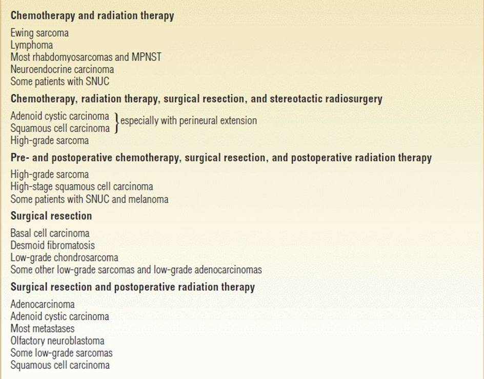
MPNST, malignant peripheral nerve sheath tumor; SNUC, sinonasal undifferentiated carcinoma.
CONTRAINDICATIONS
Patients with comorbidities, such as severe cardiovascular disease; markedly debilitated or demented patients; patients with coagulopathy; or those with end-stage renal or pulmonary disease will probably not benefit by excision of these tumors. Although patients with metastatic disease are usually not candidates for craniofacial resection, those with oligometastatic disease of low- to intermediate-grade tumors are occasionally selected for craniofacial resection for palliation of specific symptoms such as optic neuropathy and pain. I do not recommend craniofacial resection for patients with involvement of the intracranial internal carotid artery by high-grade malignancy.
PREOPERATIVE PLANNING
Computed tomography (CT) and magnetic resonance imaging (MRI) are complementary studies and the imaging methods of choice for assessing sinonasal malignancies. CT imaging is particularly useful for assessing bone changes, especially erosion. Direct coronal CT is best for evaluating the integrity of the anterior skull base including the roof of the orbit, cribriform plate, and planum sphenoidale (Fig. 18.1). The extent of tumor is best assessed with MRI, which can differentiate tumor from inflamed mucosa, blood, or inspissated mucus in most cases (Fig. 18.2). Obliteration of adipose tissue planes in the pterygopalatine fossa, infratemporal fossa, and nasopharynx usually indicates tumor involvement of these regions (Fig. 18.3). Dural thickening and enhancement are usually an indication of tumor invasion (Fig. 18.4). Similarly, thickening and enhancement of cranial nerves indicate perineural spread of the malignancy (Fig. 18.5). The identification by MRI of signal voids within the tumor or proximity of the neoplasm to the internal carotid artery may be an indication for preoperative angiography to assess tumor vascularity and plan surgical treatment. PET–CT along with MRI may help to identify lymph node involvement or distant metastasis. This, however, occurs in less than 10% of patients at initial evaluation.
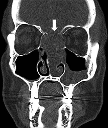
FIGURE 18.1 Coronal CT with bone window algorithm. The arrow identifies erosion of the cribriform plate in this patient with sinonasal neuroendocrine carcinoma. (All figures are property of the Department of Neurosurgery, The University of Texas M.D. Anderson and are used with permission.)
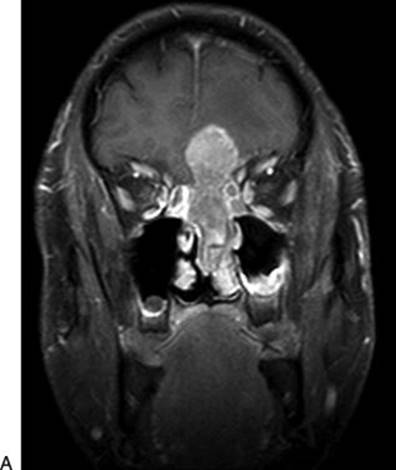
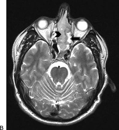
FIGURE 18.2 A. Coronal postcontrast T1-weighted MRI reveals an enhancing tumor of the sinonasal cavity with transcranial extension into the left subfrontal region. (Same patient as in Fig. 18.1) (All figures are property of the Department of Neurosurgery, The University of Texas M.D. Anderson and are used with permission.) B. Axial T2-weighted MRI reveals a tumor of the sinonasal cavity of intermediate signal intensity (arrowhead) and adjacent high T2 signal intensity sinus secretions (arrow) (Same patient as in Fig. 18.1) (All figures are property of the Department of Neurosurgery, The University of Texas M.D. Anderson and are used with permission.)
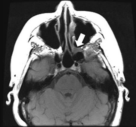
FIGURE 18.3 Axial noncontrast T1-weighted MRI of patient with sinonasal adenoid cystic carcinoma. Note the expansion of the left pterygomaxillary fossa (arrow) with loss of fat hyperintensity. (All figures are property of the Department of Neurosurgery, The University of Texas M.D. Anderson and are used with permission.)
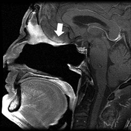
FIGURE 18.4 Sagittal postcontrast T1-weighted MRI identifies dural thickening and nodular enhancement consistent with recurrent sinonasal adenocarcinoma (arrow). (All figures are property of the Department of Neurosurgery, The University of Texas M.D. Anderson and are used with permission.)
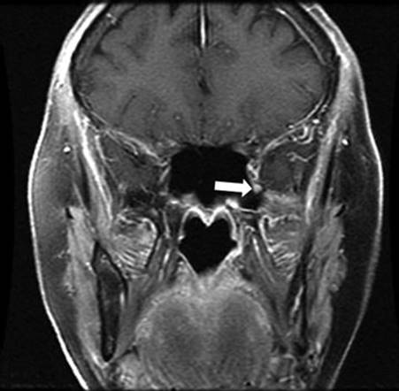
FIGURE 18.5 Coronal postcontrast T1-weighted MRI. The arrow highlights the enlarged and contrast-enhancing left maxillary nerve in this patient with adenoid cystic carcinoma of the maxillary sinus. Pathologic confirmation of perineural extension of malignancy was made at the time of surgery. (All figures are property of the Department of Neurosurgery, The University of Texas M.D. Anderson and are used with permission.)
Optimal management plans can only be constructed with accurate pathologic diagnosis of the neoplasm. Endoscopy permits access to most tumors of the sinonasal cavities. Occasionally, deep-seated lesions can be sampled by CT-guided needle biopsy. The importance of evaluation of the biopsy specimen by an experienced pathologist cannot be overemphasized.
SURGICAL TECHNIQUE
The goal of surgical management is to achieve a microscopically complete tumor resection with acceptable morbidity. Recent evidence suggests that this may not necessarily need to be done in an en bloc fashion. Certainly neurologic function should not be risked in an attempt to achieve an en bloc resection. In the presence of tumor extension to the cavernous sinus, internal carotid artery, and optic chiasm and in patients with distant metastases, surgery is usually not indicated except in highly select circumstances.
The transcranial approach for anterior craniofacial resection has as its goals the basal exposure of the medial anterior cranial fossa. It can be used as a stand-alone approach in tumors that do not extend lateral to the medial third of the maxillary sinuses, but it is commonly used in concert with nasal endoscopic techniques or occasionally with open transfacial approaches. The size of the craniotomy used depends on individual anatomic variability (especially of the frontal sinuses), tumor size, and extent. The use of intraoperative frameless stereotactic navigation allows for the optimization of the size of the craniotomy and its placement. The contours of the frontal sinus can easily be identified if a direct transfrontal sinus approach is chosen.
The patient is placed supine under general endotracheal anesthesia. The endotracheal tube is placed such that it will not interfere with any endoscopic or transfacial approaches. A tracheostomy is sometimes required. If facial incisions are used, then tarsorrhaphy sutures are placed on the side of the lateral rhinotomy or bilaterally. The head is elevated to allow improved venous drainage and gravitational relaxation of the cerebrum. Lumbar spinal drains are not used. A bicoronal incision extends from just in front of the tragus bilaterally posterior to the patient's hairline. The incision is deepened down through skin and subcutaneous tissue until the galea aponeurotica is identified. The galea is opened sharply to expose the subgaleal space. Once the incision has been completed, skin hooks or rakes are used to elevate the bifrontal scalp flap. Sharp dissection with the #15 blade is used to dissect the underside of the galea away from the loose connective tissue layer maximizing the thickness of the combined periosteal and loose connective tissue layer. This separation should not extend below approximately 1 to 1.5 cm above the orbital rims in order to protect the distal branches of the facial nerve supplying the frontalis muscles. The scalp flap is deflected anteriorly over a roll of lap sponges to prevent acute folding of the scalp. The scalp posterior to the incision is then undermined in the same layer to allow for the elevation of a large vascularized pericranial flap. The pericranial flap is then separated from the superior temporal lines bilaterally and posteriorly from beneath the scalp flap and with subperiosteal dissection is reflected forward (Fig. 18.6). Incisions are then made through both superficial and deep layers of the temporalis fascia beginning at the superior temporal line and traveling posteriorly toward the root of zygoma for approximately 5 cm. This incision should begin approximately 1.5 to 2 cm above the “keyhole” region and travel parallel to the zygomatic arch. Subfascial dissection allows exposure of the anteriormost aspect of the temporal fossa. The anterior aspect of the temporalis muscle can be dissected from the temporal fossa with periosteal elevators to expose the “keyhole” region bilaterally (Fig. 18.7). Entry holes are placed in both “keyhole” regions to expose the dura of the frontal lobe and the floor of the anterior cranial fossa. One or two entry holes are placed over the midline exposing the posterior most frontal dura lateral to the superior sagittal sinus bilaterally. The dura is then dissected from the underside of the calvarium, and a power osteotome is used to divide the calvarium from the central burr hole down to each entry hole in the “keyhole” regions. The power osteotome is used to create osteotomies superior to the orbital rims bilaterally. These osteotomies are carried to approximately the inner third of the orbits bilaterally. Osteotomies are then placed inferiorly from the medial aspect of these osteotomies to the region of the frontal nasal suture. The bone is then divided at the level of the frontal nasal suture. Usually, this allows for a controlled fracture of the posterior wall of the frontal sinus. If this is not easily accomplished, the use of a fine power osteotome through the previously constructed osteotomies with scoring of the posterior wall of the frontal sinus allows its controlled fracture. The bifrontal bone flap is then elevated. In patients in which the dura is tightly adherent to the calvarium, it may be best to remove the anterior wall of the frontal sinus as a separate osteotomy and then remove the posterior wall of the frontal sinus under direct vision. Cottonoids are used to protect the dural surface, and the dura is dissected from the posterior wall of the frontal sinuses bilaterally to the level of the foramen cecum. Careful epidural hemostasis is achieved. The frontal sinuses are then completely demucosalized and cranialized. With this very basal approach, the dura of the floor of the anterior cranial fossa is dissected and elevated from the floor. This is greatly facilitated by elevation of the patient's head, by the use of hyperventilation to an arterial PCO2 of 28 mm Hg, and, if necessary, by the bilateral opening of the frontal dura to allow egress of cerebrospinal fluid (CSF). The dura is elevated to the level of the crista galli and over both orbits. The crista galli is removed with a high-speed drill under magnified vision. The dural sleeves extending through the cribriform plate are cut, and elevation of the dura continues posteriorly to expose the frontal–sphenoid suture. The posterior ethmoidal arteries are identified at the posterolateral aspects of the cribriform plate in line with the frontal–sphenoid suture. After coagulation and division of the posterior ethmoidal arteries, further elevation of the dura is possible. If the dura is not involved by neoplasm, it is then repaired primarily. If a larger defect is present, then a dural replacement graft is inserted and microscopically sutured to achieve a watertight closure. Much more commonly, however, the dura is involved by neoplasm, and in these circumstances, an intradural exposure is necessary. In this situation, bilateral frontal dural incisions are made, and the anteriormost superior sagittal sinus is ligated. Any tumor extending into the brain is removed in a piecemeal fashion. Involved cerebrum is resected using standard neuromicrosurgical techniques. Incisions are then made in the dura of the floor of the anterior cranial fossa circumferentially around the involved dura. The involved dura is allowed to remain fixed to the underlying tumor while the free edges are elevated off the floor. At this point, a dural replacement graft is inserted and microscopically sutured. With a high-speed drill, osteotomies are made in the floor of the anterior cranial fossa. If the tumor involves the entire ethmoid sinus complex, the osteotomies are placed through the medial aspect of the roof of the orbit to enter the orbits bilaterally lateral to the lamina papyracea (Fig. 18.8). The posterior osteotomy should be at the level of the frontal–sphenoid suture. This suture typically overlies the posterior end of the posterior ethmoid air cells although it may overlie the anterior aspect of a large sphenoidal sinus. The posterior ethmoid arteries are identified, coagulated, and divided. Of note is that the exit of the optic nerve from the optic canal can be found approximately 5 to 7 mm posteromedial to the posterior ethmoid neurovascular bundle. The anterior ethmoid arteries are also identified intraorbitally, coagulated, and divided. From above, the periorbita is dissected from the lamina papyracea bilaterally, and a small curved osteotome is placed medial to the orbital tissues (Fig. 18.8). Osteotomies are made at the inferior aspect of the lamina papyracea just where the bone turns to form the floor of the orbit. This osteotomy is placed bilaterally. An osteotomy is then carried out across the lamina papyracea posteriorly with half of the osteotome placed in the orbit and half in the posterior ethmoid sinus. The posterior bony nasal septum is then divided with this curved osteotome. If the posterior osteotomy is through the planum sphenoidale, it enters the sphenoid sinus and needs to be continued through the anterior wall of the sphenoid to the floor of the sinus. Bringing the osteotomy through the floor of the anterior sphenoid sinus separates the face and rostrum of the sphenoid from the cranial base. The anterior osteotomy is performed across the base of the frontal sinuses and enters the nasal cavity (Fig. 18.8). The anterior aspect of the nasal septum is exposed and divided with heavy scissors. This allows removal of the specimen transcranially. Residual air cells are removed, and any involved or questionable periorbital tissue is resected. Small periorbital defects are well tolerated, but large defects should be repaired with temporalis fascia. I do not use nasal packing but prefer to use bipolar cautery and the judicial use of topical hemostatic agents to achieve complete hemostasis. Small drill holes are then placed through the residual bone of the planum sphenoidale (Fig. 18.9). The pericranial flap is then tailored to the appropriate length and sutured to the drill holes placed in the planum and in the medial orbital roofs if available (Fig. 18.10). The bone flap is then placed in its anatomical position and rigidly fixated taking great care to avoid compression of the pericranial flap between the edges of the bone. The pericranium should be able to move freely between the edges of the bone. The bilateral incisions of the temporalis fascia are closed as is the scalp. The scalp is typically closed in two layers with absorbable suture in the galea and either staples or a nonabsorbable suture in the skin. Drains are not typically placed.
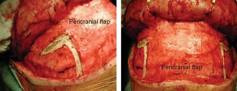
FIGURE 18.6 Intraoperative photographs of the incisions necessary to elevate the vascularized pericranial flap from the calvarium. Bilateral incisions through both the superficial and deep layers of the temporalis fascia free the flap from the superolateral orbits. The bilateral incisions at the superior temporal lines are connected posteriorly beneath the scalp posterior to the incision line. (All figures are property of the Department of Neurosurgery, The University of Texas M.D. Anderson and are used with permission.)
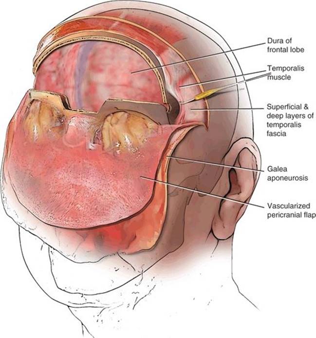
FIGURE 18.7 Subfrontal approach for transcranial resection of a sinonasal malignancy. The vascularized pericranial flap has been elevated and reflected anteriorly, and the anterior temporalis muscle has been deflected posteriorly. A bifrontal craniotomy has been performed with inferior extension to just above the frontonasal suture. The next step would be to cranialize and demucosalize the frontal sinus and gain the subfrontal exposure by dissection of the dura off of the floor of the anterior cranial fossa.
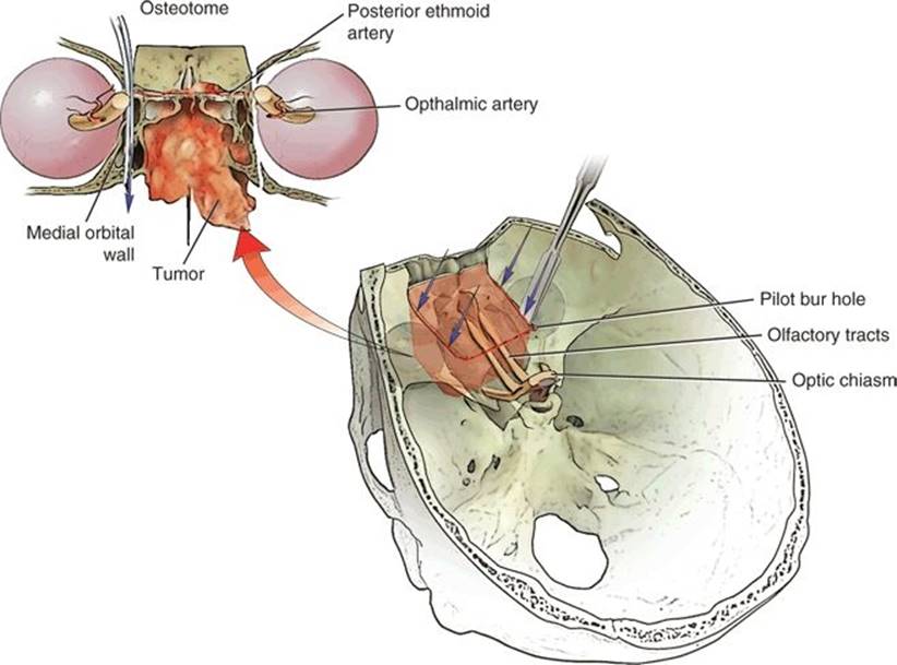
FIGURE 18.8 The placement of osteotomies (arrows and dashed line) needed in order to free the central skull base and sinonasal cavities from the craniofacial skeleton. A high-speed drill is used to make osteotomies into both orbits. The posterior osteotomy is made at the level of the frontosphenoid suture. The osteotome divides the posterior and anterior lamina papyracea and the nasal septum. A curved malleable retractor protects the orbital tissues, while osteotomies are made in the medial floor of the orbit.
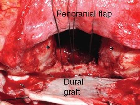
FIGURE 18.9 Intraoperative photo following closure of the dural defect with allograft material. Drill holes have been placed in the planum sphenoidale, and the pericranial flap is being secured to these holes with sutures. (All figures are property of the Department of Neurosurgery, The University of Texas M.D. Anderson and are used with permission.)
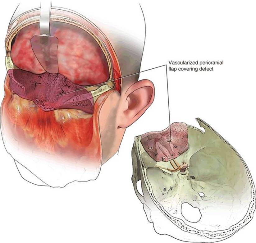
FIGURE 18.10 The reconstruction of the anterior cranial base with the vascularized pericranial flap. The flap is fixed by sutures to holes that have been drilled in the planum sphenoidale and medial aspect of the orbit.
POSTOPERATIVE MANAGEMENT
Following extubation in the operating room, the patient is transferred to the intensive care unit for observation overnight. Hourly neurologic examinations are performed to assess for any changes in neurologic status. Assessment of the amount and nature of any nasal drainage is also important. It should be expressly communicated to all caregivers that nothing should be inserted into the nose. Also, if the orbits were entered during surgery, postoperative evaluations of visual acuity and pupillary function are necessary. Careful electrolyte balance is maintained since hyponatremia is not uncommon. Antibiotic coverage, begun preoperatively, is continued for 24 hours. Postoperative MRI is performed, preferably within 24 to 48 hours following surgery in order to avoid misinterpretation due to surgically induced enhancement of tissues.
COMPLICATIONS
The most concerning potential postoperative complication following craniofacial resection is that of tension pneumocephalus. There seems to be a particular risk for this complication following resection of the cribriform plate when compared with larger degrees of resection of the anterior cranial base. The relatively smaller degree of skull base resection needed for removal of the cribriform plate may contribute to a ball-valve mechanism that predisposes the patient to the accumulation of air under tension. This complication has become almost nonexistent since I stopped using lumbar spinal drainage. If it should occur, treatment consists of percutaneous aspiration of air and, in some cases, intubation. Much more commonly seen as a consequence of lumbar spinal drainage of CSF has been the syndrome of intracranial hypotension. In its mildest form, it consists of severe and occasionally incapacitating postural headache. Management is fluid hydration and slow mobilization. A high-volume (30 mL) epidural blood patch may be required if a lumbar drain has been used. The incidence of complications in the literature is difficult to evaluate because of a lack of uniformity in reporting. Most series report complications in 25% to 40% of patients undergoing craniofacial resection with mortality rates ranging from 0% to 7%. The most commonly identified complications include wound infections, meningitis, CSF leakage, delayed return of neurologic function, and pneumocephalus. Most of these series have spans of many years. In my series, dating back to 1992, in which aggressive antibiotic coverage is used routinely, no cases of osteomyelitis have been identified, and the incidence of meningitis is less than 1%. Similarly, consistent attention toward watertight closure of the dura, the use of vascularized grafts, and the liberal use of free tissue transfer have resulted in a 1% incidence of CSF leakage in my patients undergoing anterior craniofacial resection.
RESULTS
Recent large series of craniofacial resection have reported overall 5-year survival rates of 47% to 70%. The International Collaborative Study noted an overall survival of 48.3% at 5 years, with a 5-year disease-specific survival rate of 53.3% and a 5-year recurrence-free survival rate of 45.8%. Outcome is histology specific. At my institution, the 5-year overall survival rate for mucosal melanoma was found to be 38.7%, while that for olfactory neuroblastoma was 89%. Pathologically positive margins, involvement of the orbit, and invasion of the brain have also been shown to be predictors of local recurrence and a shorter survival time. However, well-selected patients with transdural extension of malignancy can have outcomes similar to those without intradural tumoral spread. In this group of patients, the most important factors affecting overall and progression-free survival were the ability to achieve microscopically negative margins and the lack of direct invasion of the brain. Although advanced age has often been identified as a negative prognostic factor, Hentschel et al. found no difference in oncologic outcome in a population of patients aged 70 years or greater when compared to a younger cohort. The cohort of elderly patients did, however, have a significantly greater incidence of cardiovascular comorbidities and systemic complications. Involvement of the intracranial internal carotid artery by high-grade malignancy remains the major contraindication to surgical excision of these tumors. Surgical resection did not provide a survival advantage in this subgroup of patients with malignancy of the anterolateral skull base.
PEARLS
· The first step in management is obtaining an accurate pathologic diagnosis.
· CT and MRI are complementary studies, both of which contribute to the ability to best plan the surgical removal of a sinonasal malignancy.
· The use of lumbar subarachnoid drains for withdrawal of CSF is unnecessary and is associated with a high rate of complications.
· Sharp dissection immediately below the galea aponeurotica maximizes the thickness of the loose connective tissue layer and thus the pericranial flap.
· The optic canal lies 5 to 7 mm posterior and medial to the posterior ethmoid foramen located at the lateral end of the frontosphenoid suture.
PITFALLS
· Failure to completely cranialize and demucosalize the frontal sinus can result in formation of a mucocele.
· Replacement of the frontal craniotomy without leaving adequate space for the pericranial flap can result in necrosis of the flap and an elevated risk of CSF leakage and infection.
INSTRUMENTS TO HAVE AVAILABLE
Standard neuromicrosurgical set, dural replacement graft, and frameless stereotactic navigation for some patients.
SUGGESTED READING
DeMonte F. Evolving role of skull base surgery for patients with low and high grade malignancies. J Neurooncol 2004;69: 191–198.
Feiz-Erfan I, Suki D, Hanna E, et al. Prognostic significance of transdural invasion of cranial base malignancies in patients undergoing craniofacial resection. Neurosurgery 2008;61:1178–1185.
Hanna E, DeMonte F, Ibrahim S, et al. Endoscopic resection of sinonasal cancers with and without craniotomy. Arch Otolaryngol Head Neck Surg 2009;135:1219–1224.
Hentschel SJ, Vora Y, Suki D, et al. Malignant tumors of the anterolateral skull base. Neurosurgery 2010;66(1):102–112; discussion 112.
Hanasono MM, Silva A, Skoracki RJ, et al. Skull base reconstruction: an updated approach. Plast Reconstr Surg 2011;128(3): 675–686.