
Lawrence J. Marentette
INTRODUCTION
A midface degloving approach was first described in 1927. Casson et al. in 1974 described the current technique that is presented here. Conley and Price as well as many other authors have described this versatile approach. It is extremely useful in exposing the nasal cavity and the maxillary, ethmoid, and sphenoid sinuses. When combined with the coronal approach, access can be gained to the entire craniofacial skeleton. This is useful not only in tumor surgery but also for extensive midface reconstructions. Although it can also be applied in cases of panfacial fractures, a sublabial approach is usually sufficient for this. The operative technique is actually a combination of closed rhinoplasty dissection along with a sublabial approach.
HISTORY
The typical patient who is a candidate for midfacial degloving is one who presents with a complaint of nasal obstruction or epistaxis as a result of neoplasm of the nasal cavity, ethmoid, or maxillary sinus. The presentation is often quite insidious. Involvement of the second division of the trigeminal nerve is often a sign of rather advanced disease. Proptosis, diplopia, and visual loss are signs of invasion of the orbit.
PHYSICAL EXAMINATION
Intranasal examination may reveal a lesion of the nasal septum or of the wall of the nasal cavity involving the turbinates up to the superior aspect of the middle turbinate. It may also extend through the medial wall of the maxillary sinus into the sinus itself. Involvement at or above the middle meatus suggests involvement of the ethmoid sinuses.
INDICATIONS
The approach is typically indicated for patients with a neoplasm of the nasal cavity that does not reach the anterior cranial base. This may involve the wall of the nose, the turbinates, ethmoid sinuses, and the nasal septum. The nasal cavity is also quite readily accessible with this approach, and complete visualization extending to the nasopharynx can be achieved. If the mass involves the anterior cranial base, a midface degloving can be combined with an open technique such as a subcranial approach.
CONTRAINDICATIONS
This approach by itself is contraindicated in patients who present with a neoplasm that traverses the cribriform plate or roof of the ethmoid sinus. It may be used in conjunction with a subcranial approach with removal of the frontal bar, which would then provide complete exposure to the entire nasal cavity as well as the anterior cranial fossa.
PREOPERATIVE PLANNING
Maxillofacial CT scan with contrast including axial, coronal, and sagittal views is essential to determine the extent of tumor involvement. Any areas of bone destruction should be readily apparent. A magnetic resonance imaging (MRI) of the maxillofacial skeleton and cranial cavity is also important as visualization of soft tissue by MRI is far superior to that of a CT scan. This study allows viewing of the cranial nerves, and any enhancement of these areas would suggest perineural spread of malignant tumor.
SURGICAL TECHNIQUE
A sublabial incision is created extending from first molar to first molar along the maxillary alveolus approximately 6 to 7 mm from the gingival mucosal junction (Fig. 21.1). This allows for an adequate cuff of tissue during the closure. Retractors are used to evert the lips and the soft tissues of the cheek, and the first incision is made with electrocautery at right angles to the mucosa only. The second incision is made through this incision at right angles to the underlying bone going through submucosa and the periosteum. Using a periosteal elevator, the soft tissues are elevated over the canine fossa and medial and lateral buttresses up to the inferior orbital rim and onto the body of the zygoma taking care to preserve the infraorbital nerve bilaterally. Attention is then turned to the nose. A complete transfixation incision is made through the membranous columella extending from the tip of the nasal septum along its caudal border down to the anterior nasal spine (Fig. 21.2). This incision is then carried along the floor of the nose down onto the bone of the piriform aperture and then continuing in an superior direction incising between the upper and lower lateral cartilages. At this point, it is helpful to use a double hook inserted at the alar rim with the finger used to evert the lower level cartilage, thus making the junction of the lower and upper level cartilages much easier to identify. This incision continues superiorly and medially between this inner cartilaginous region until it joins the nasal septal incision connecting with the membranous septum. Small curved iris scissors are then used to elevate the skin from the nasal dorsum taking care to leave the attachment of the upper lateral cartilages to the nasal bones intact (Fig. 21.3). This is done bilaterally at this point; attention is then redirected to the intraoral incision. Using wide retractors everting the lips, the entire midfacial complex is then elevated superiorly using a periosteal elevator to release any remaining soft tissue attachments (Fig. 21.4). With complete exposure of the piriform aperture, osteotomies of the medial buttress with its removal will allow resection of a tumor of the nasal wall. The inferior osteotomy through the medial buttress is made with a saw blade more than 5 mm superior to the canine root tip. A vertical osteotomy is made through the lateral buttress. A transverse osteotomy connects the lateral buttress with the piriform aperture just inferior to the infraorbital nerve. Using a curved osteotome placed in the nasal cavity behind the medial buttress, the anterior maxilla is removed exposing the maxillary sinus and nasal wall. Reconstruction is then accomplished either by replacing the medial buttress or with an outer table calvarial graft. The skin is then redraped over the nasal dorsum. Absorbable sutures are used to close all intranasal incisions. Attention is then turned to the intraoral incision where prior to the main closure, one or two absorbable sutures are placed transversely in the midline so as to recreate the frenulum of the upper lip. This avoids the tethering effect of the upper lip to the underlying maxilla. Using a 3-0 absorbable suture starting from the midline and working laterally on each side, the suture is used to close the incision going through mucosa and submucosa in a horizontal running fashion. Once this is accomplished on each side, 4-0 absorbable sutures are used to close the mucosal edges in a simple running fashion catching only the mucosal edges. At the end of the closure, the incision is everted with this suture technique. This avoids any long-term scar contracture that can result in gingival recession and exposure of tooth roots.

FIGURE 21.1 A sublabial incision is made between the first molars.

FIGURE 21.2 Circumvestibular incisions are made allowing the lower lateral cartilages to be reflected with the nasal skin.
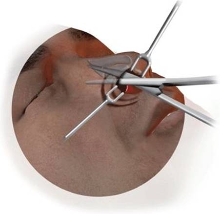
FIGURE 21.3 Scissors are used to elevate the nasal skin in a fashion similar to a closed rhinoplasty.

FIGURE 21.4 The cheek and nasal skin is elevated exposing the anterior maxilla and piriform aperture. Note the preservation of the inferior orbital nerves.
POSTOPERATIVE MANAGEMENT
Postoperative management is applying an ointment to the incisions and also saline irrigations not only to keep the incisions clean but also to clean the cavities in cases of tumor resection. Nasal saline spray is also useful to maintain moisture in the nasal cavity itself. After 1 to 2 weeks, nasal irrigation with saline spray is also quite useful to prevent crusting of the nasal cavity.
COMPLICATIONS
Almost all of these patients have decreased sensation in the distribution of the infraorbital nerve; however, with the nerves being preserved, resolution may take up to 18 months. On rare occasions there may be midfacial weakness from excessive retraction of the soft tissues and injury of the buccal branches of the facial nerve. Vestibular stenosis may occur; however, with meticulous mucosal closure, this should not occur. If vestibular stenosis does occur, it is most commonly seen in patients who have had postoperative radiotherapy.
RESULTS
The midface degloving approach affords excellent exposure to the anterior maxilla and nasal cavity. It can be combined with a Le Fort I osteotomy, which results in extending the field into the nasopharynx. The postoperative cosmetic result is superior to the lateral rhinotomy and Weber-Ferguson approaches.
PEARLS
· Elevation of the soft tissues of the midface may be done quickly with the surgeon standing at the patient's head and using two elevators scooping toward the surgeon. This does require having an assistant holding retractors and another assistant maintaining the suction.
· Each end of a Penrose drain can be placed through each nostril and out then through the upper lip to use as a retractor.
· Preoperative and postoperative steroids are given to reduce the amount of postoperative edema.
PITFALLS
· The main pitfall of this technique is vestibular stenosis, which in some cases is unavoidable when patients are receiving postoperative radiotherapy.
· Excessive retraction of the midfacial soft tissues may result in temporary anesthesia in the distribution of the second division of the trigeminal nerve followed by paresthesia as sensation returns.
· Excessive retraction may also result rarely in weakness of the buccal branches of the facial nerve with a temporary loss of movement of the midfacial soft tissues that may take anywhere from 1 to 6 months to recover.
INSTRUMENTS TO HAVE AVAILABLE
· Standard head and neck tray
· Retractors
· Rhinoplasty tray
SUGGESTED READING
Portmann G, Retrouvey H. Le cancer du nez. Paris, France: Gaston Doin et cie, 1927.
Casson PR, Bonnano PC, Converse JM. The midfacial degloving procedure. Plast Reconstr Surg 1974;53:102–113.
Conley J, Price JC. Sublabial approach to the nasal and nasopharyngeal cavities. Am J Surg 1979;138:615–618.
Price JC. Facial degloving. In: Rhinology. New York, NY: John Wiley & Sons Inc., 1987:1098–1123, Chapter 37.