
Guy J. Petruzzelli
INTRODUCTION
The term lateral rhinotomy does not refer to a specific surgical procedure; rather it describes an incision placed on the face between the medial canthus and the anterior midline of the nose within the nasal–facial groove. The lateral rhinotomy incision can be used either in combination with the bifrontal craniotomy to manage the inferior aspect of tumors of the paranasal sinuses and medial orbit or as a stand-alone approach to craniofacial resection for tumors of the paranasal sinuses with limited intracranial extension anteriorly. In this section, the discussion is limited to a description of the technique as it applies to a combined approach with a separate craniotomy.
The lateral rhinotomy approach has been the traditional method of access and exposure of tumor of the superior ethmoid sinus and lateral nasal wall and was used in the early series of combined craniofacial resections. Detractors of this approach have cited the facial incision as a significant cause for using alternative methods. I find that with meticulous suturing technique and attention to detail, the incision can be minimized yielding an excellent aesthetic result. The decision on how best to manage the inferior and medial components of a tumor requiring a craniofacial resection is based on several factors including (1) anatomic site of origin of the tumor, (2) degree of involvement of the medial orbital structures, and (3) inferior tumor extension and involvement of the lateral nasal wall.
HISTORY
Patients often present with a long-standing history of nasal obstruction and rhinorrhea, frequently refractory to over-the-counter systemic or topic nasal decongestants. Patients with tumors of the ethmoid sinus, lateral nasal wall, and medial wall of the orbit requiring craniofacial resection with lateral rhinotomy will present with a history of unilateral nasal symptoms of obstruction, epistaxis, and/or purulent rhinorrhea. Masses extending laterally into the medial orbit or inferiorly into the inferior meatus may obstruct the lacrimal apparatus resulting in unilateral epiphora. Generalized symptoms of headache, (retro-)orbital pain, and facial pain can be due to blockage of the sinuses with inspissated secretions; however, consideration should also be given to extension of tumor to the periorbita or dura of the anterior cranial fossa.
PHYSICAL EXAMINATION
Patients with sinonasal malignancies involving the anterior cranial base should have a comprehensive head and neck examination including nasal endoscopy and flexible fiberoptic nasopharyngolaryngoscopy. Visual fields, extraocular movements, and visual acuity should be documented, and any patient with any abnormal findings should have a comprehensive ophthalmologic examination. A mass in the medial canthus, broadening of the nasal dorsum, or distortion of the normal anatomy of the nasomaxillary crease can be observed (Fig. 22.1). Orbital extension can result in diplopia, restriction of gaze, proptosis, or changes in visual acuity. Skin involvement indicates an aggressive malignancy (Fig. 22.2). All patients should undergo biopsy prior to treatment. Tumors of this region are characteristically friable and tend to hemorrhage when manipulated; therefore, for safety purposes, consideration of biopsy in the operating room is warranted (Fig. 22.3). A comprehensive physical examination including risk stratification for patients with cardiopulmonary disease is necessary in all patients being considered for craniofacial resection.
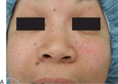
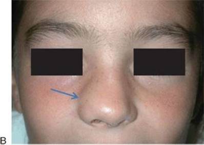
FIGURE 22.1 Patients with a tumor of the anterior cranial base with significant intranasal component presenting with a mass in the left medial canthal region (A) and an external nasal deformity with blunting of the right nasal–facial crease (arrow) (B).

FIGURE 22.2 Advanced sinonasal carcinoma with skin involvement.

FIGURE 22.3 Malignant tumor of the anterior cranial base with hemorrhage prolapsing out of the nasal cavity.
INDICATIONS
The role and timing of surgery in patients with malignant neoplasms of the anterior cranial base depends upon the biology of the primary neoplasm and the patient's willingness to accept craniofacial resection and its possible complications. Surgery remains the standard of care in the management of patients with minor salivary gland tumors of the anterior cranial base and lacrimal region, some neuroendocrine carcinomas, and adenocarcinomas. Sinonasal undifferentiated carcinoma, poorly differentiated squamous cell carcinomas, and other very high-grade neoplasms may be treated with multidrug neoadjuvant chemotherapy followed by definitive chemoradiation. The management of olfactory neuroblastoma (ONB) remains somewhat controversial in that some groups favor primary nonsurgical therapy with surgical salvage while others believe ONB is most amenable to treatment with definitive surgery.
CONTRAINDICATIONS
Contraindications for craniofacial resection via lateral rhinotomy include aggressive histology with poor prognosis, extensive involvement of the facial skin (limiting ability to obtain negative margins), radiographic evidence of extensive perineural invasion, poor performance status, and extensive involvement of the brain parenchyma and clinical or radiographic evidence of metastatic disease. A relative contraindication is lateral rhinotomy and partial maxillectomy in an only-seeing eye.
PREOPERATIVE PLANNING
Computed tomography (CT) and magnetic resonance imaging (MRI) should be viewed as complementary imaging modalities, and both are helpful in the preoperative evaluation of a patient undergoing craniofacial resection with lateral rhinotomy. The superior bony detail rendered by the CT provides useful information regarding the amount of bone destruction of the ethmoid labyrinth and nasal wall particularly on the contralateral side of the tumor. The MRI is able to more clearly identify transcranial extension of tumor and to delineate tumor from retained secretions in the sinuses (Fig. 22.4). The role of combined positron emission tomography–computed tomography (PET–CT) scanning in the preoperative evaluation of advanced skull base tumors is limited to the detection of cervical and retropharyngeal adenopathy and distant metastatic disease. I obtain PET–CT scan on patients with high-grade malignances and any patient presenting with cervical lymphadenopathy.

FIGURE 22.4 Postcontrast MRI of ONB with penetration of the cribriform plate and fovea ethmoidalis and involvement of the dura of the anterior cranial fossa.
SURGICAL TECHNIQUE
When combined with bifrontal craniotomy for anterior craniofacial resection, the lateral rhinotomy is usually performed after the intracranial portion of the procedure, assessment of intracranial and intradural margins, and the reconstruction of the dura of the anterior cranial fossa.
The skin incision is marked prior to infiltration with local anesthetic (lidocaine 1:100,000 with epinephrine) to avoid distortion of the aesthetic units of the nose that are integral to the design of the incision. Ophthalmic ointment is placed in both eyes, and a suture tarsorrhaphy can be placed on the contralateral side to protect the cornea. I place a plastic corneal protector on the ipsilateral side to obviate the need for a tarsorrhaphy, which could distort the skin and make monitoring the eye more difficult. The incision begins under the ipsilateral eyebrow just medial to the supraorbital notch palpated in the supraorbital rim. The incision is beveled parallel to the hair follicles of the upper eyebrow and camouflaged along the inferior border of the eyebrow. The incision is extended inferiorly into the medial canthus where it is broken with the soft “W” or “Z” that will prevent webbing of the medial canthus. The apices of the broken incisions should be broad enough to prevent necrosis of the distal tip of the skin triangle, and monopolar cautery should be avoided in this portion of the incision. Failure to break the incision in the medial canthus or necrosis of the distal portions of the incision will lead to unsightly webbing of the medial canthus. The incision is then extended inferiorly respecting the aesthetic units of the nose in a vertical line along the nasal–maxillary crease, around the alar crease and into the philtrum. Traditionally, the lateral rhinotomy incision terminated into the nasal vestibule; however, this places the integrity of the lateral attachment of the lower lateral nasal cartilage at risk resulting in nasal valve collapse. Based on the extent of resection necessary, a separate ipsilateral gingiva–buccal incision can be added to increase exposure to the maxillary antrum. Alternatively, the incision can be extended inferiorly to include an upper lip split if a radical maxillectomy is required. I have found that the subciliary extension (Weber-Fergusson incision) often results in an unfavorable functional and aesthetic result and is rarely needed even for radical maxillectomy. Upper and lower eyelid transconjunctival incisions can be added for orbital exenteration. Should bilateral exposure of the superior ethmoid vault be necessary, the incision can be extended across the midline with a 15- to 20-mm horizontal extension across the nasal dorsum (Fig. 22.5) at the level of the nasion.

FIGURE 22.5 Incision placement for lateral rhinotomy. Incision can be extended across the midline with a 15- to 20-mm horizontal extension across the nasal dorsum (dashed line).
Following the skin and ipsilateral gingiva–buccal sulcus incisions, subperiosteal dissection proceeds with exposure of the anterior face of the maxilla medial and lateral to the infraorbital neurovascular bundle, the bone of the nasal dorsum, the medial inferior orbital rim, and the pyriform aperture. The medial attachment of the medial canthal tendon to the anterior lacrimal crest is marked to facilitate the correct reapproximation of the tendon, and a medial canthotomy is performed tagging the tendon with a 4-0 clear nylon suture. The lamina papyracea and medial orbit are inspected, and, if needed, the medial periorbita can be resected and assessed with frozen sections. If there is no involvement of the periorbita, sharp subperiosteal dissection proceeds posteriorly into the orbit along the frontoethmoidal suture with identification of the anterior and posterior ethmoidal arteries. If not managed during the intracranial portion of the procedure, the vessels should be bipolar coagulated and transected. A small amount of the anterior lacrimal crest is removed with a Kerrison rongeur to facilitate the exposure and subsequent delivery of the lacrimal sac. The lacrimal sac is transected at the junction of the lacrimal sac and nasolacrimal duct and opened widely. The dacryocystorhinostomy is completed by suturing the open sac to the periorbita anteriorly and posteriorly to maintain patency. If this procedure is being performed for a malignant tumor, then I place Crawford Silastic lacrimal stents into the superior and inferior lacrimal ducts delivered into the nose to maintain patency of the lacrimal system. If bilateral ethmoid and anterior cranial base resection is being performed, a subperiosteal plane must be developed over the nasal dorsum to the contralateral medial canthus, paying particular attention to keep the opposite medial canthal tendon intact. Finally, a portion of the anterior wall of the maxilla is removed and the maxillary antrum is inspected. Frozen sections of the antrum can be evaluated as necessary.
Complete mobilization of the soft tissue envelope provides the necessary access to the facial skeleton for the required osteotomies. Osteotomies are made with a high-speed reciprocating saw and antibiotic irrigation. Beginning inferiorly, a horizontal osteotomy is made from the maxillary antrum across the floor of the nose in the inferior meatus below the inferior turbinate. The second osteotomy is vertically oriented and placed 5 to 8 mm medial to the inferior orbital foramen and extends superiorly through the inferior orbital rim and the residual anterior maxillary wall into the orbit. Smooth malleable retractors and 1 × 3 cm neurosurgical pledgets soaked in lidocaine 1%, 1:100,000 with epinephrine are used to protect the orbital contents. Placement of the nasal osteotomies depends on whether the procedure is unilateral or bilateral. In unilateral cases, an osteotomy is placed in the nasofrontal suture; for bilateral procedures, the osteotomy is extended across the midline at the nasal dorsum with the saw and a contralateral osteotomy of the frontonasal sutures is performed with an osteotome as if one were performing a rhinoplasty. For a classical medial maxillectomy for an extracranial tumor of the nasal cavity, the superior osteotomy is made with the saw 5 mm inferior to the frontoethmoidal suture. When the medial maxillectomy is performed in conjunction with a transcranial approach, the superior osteotomy is more commonly made from the intracranial aspect through the fovea ethmoidalis and the roof of the orbit. Placing the osteotomies in this manner allows for the en bloc resection of the cribriform plate, fovea ethmoidalis, medial wall of the orbit, and lateral wall of the nose. Following the osteotomies, the remaining soft tissue attachments are divided with curved Mayo or right-angled scissors and the specimen is delivered anteriorly. Since the bony anterior cranial base is being removed as a part of the specimen, care must be taken to not disturb the anterior fossa dural reconstruction or compress the frontal lobes during the mobilization of the specimen. Visualizing the defect both intra- and extracranially will avoid such an injury. Brisk bleeding accompanies the transection of the branches of the internal maxillary artery; this is initially controlled with tamponade and then with bipolar electrocautery or titanium hemoclips. The anterior face of the sphenoid sinus and the nasofrontal recess are opened widely to ensure ventilation of the sinuses. The mucosa of the maxillary antrum and sphenoid sinus are assessed histologically for final surgical margins. Following establishment of definitive hemostasis, the wound is closed (Fig. 22.6).

FIGURE 22.6 En bloc resection of lateral nasal wall and anterior cranial base. (#, inferior turbinate; *, crista galli.)
Meticulous attention to detail at the time of closure prevents aesthetic and functional complications as discussed below (Fig. 22.7). The previously tagged medial canthal tendon should be approximated to the residual nasal bone for proper alignment of the orbit. The dacryocystorhinostomy should be reinspected and the lacrimal stents secured. The wound is closed in three layers beginning with approximation of the periosteum with buried polyglactin sutures. The subcutaneous tissue is closed with buried absorbable suture as well. The skin is closed with fine (6-0) monofilament nylon suture, a fast-absorbing chromic suture, or subcuticular closure; the latter two require reinforcement with Steri-Strips. The gingiva–buccal incision is closed in two layers with polyglactin suture for the periosteum and chromic suture for the mucosa. If the lip has been split, a multilayer closure beginning with meticulous realignment of the vermilion is performed; magnification with loupes is helpful to ensure a good cosmetic result. If bilateral nasal osteotomies are required, a firm external nasal splint is applied to stabilize the nasal bone fragments, and after assessing vision in the recovery room, the ipsilateral eye is patched and a light pressure dressing is applied to reduce facial and eyelid edema. The denuded surfaces of the maxillary and sphenoid sinuses can be lightly lined with Gelfoam soaked in thrombin to reduce oozing. If there is persistent bleeding from bone or mucosal edges, the defect can be packed with antibiotic-impregnated nasal tampons or gauze.

FIGURE 22.7 Favorable functional and aesthetic result following lateral rhinotomy.
POSTOPERATIVE MANAGEMENT
The additional of the lateral rhinotomy adds little to the routine postoperative care of a patient undergoing a craniofacial resection. Patients are extubated at the completion of the procedure to allow for a neurologic examination and assessment of vision. The eye on the ipsilateral side is patched, and a light pressure dressing is applied to the face overnight to reduce edema, and the head can be elevated 15 to 30 degrees. Normal saline nasal irrigations can begin on the 3rd postoperative day, and the packing can begin to be removed on the 5th postoperative day. The skin incision should be cleaned three times a day with hydrogen peroxide; topical antibiotics are not needed as they may cause local irritation. Appropriate ophthalmic lubrication should be used, and if a gingiva–buccal sulcus incision was needed, mouth rinses with 0.12% chlorhexidine gluconate mouthwash are prescribed.
COMPLICATIONS
Complications from craniofacial resection have been described previously and include meningitis, intraparenchymal hemorrhage, cerebral venous or arterial insufficiency, bone loss, cranial deformity, and systemic complications such as pneumonia, sepsis, and respiratory failure. Particular complications from the lateral rhinotomy separate from those of the craniofacial resection can be divided into hemorrhage, ophthalmologic, and aesthetic. Cerebrospinal fluid fistula has been described as a complication of the medial maxillectomy performed via lateral rhinotomy when the superior horizontal saw cut or osteotomy is made at or above the frontoethmoidal suture. In cases in which the lateral rhinotomy is performed in conjunction with craniofacial resection, the anterior fossa floor and superior medial orbit are resected and dura should be reconstructed in a watertight fashion. Vascular or hemorrhagic complications result from failure to control the internal maxillary artery or its branches. Large-caliber vessels should be ligated with titanium clips immediately upon removal of the specimen before they retract into bony canals and become more difficult to manage.
Loss of vision can occur if any of the osteotomies are carried too far posteriorly and result in damage to the optic nerve at the orbital apex. Excessive traction on the specimen during delivery can result in enough indirect tension of the globe to cause a traction injury to the optic nerve. Failure to reattach or accurately reposition the medical canthal tendon can result in enophthalmos, dystopia, and diplopia. A wide dacryocystorhinostomy and lacrimal stenting is necessary to prevent stenosis of the lacrimal apparatus, particularly during postoperative external beam radiation. Delayed epiphora may require dilation of the puncta and lacimal ducts or, rarely, revision (conjunctivo-) dacryocystorhinostomy.
When meticulous closure techniques are applied, the healed lateral rhinotomy incision is barely perceptible. The skin incisions must be placed in such a way as to respect the aesthetic units of the nose. Minimal use of the monopolar cautery and gentle manipulation of the tissue edge are needed to prevent skin necrosis and unsightly scarring. Fine sutures should be used for the subcuticular and skin closures (Fig. 22.8).


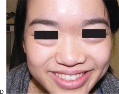
FIGURE 22.8 Illustrative case of resection of a fibrosarcoma of the nose and anterior cranial base solely via lateral rhinotomy: (A) preoperative appearance, (B) exposure at the time of resection, (C) en bloc lateral nasal wall and cribriform specimen and (D) postoperative appearance at 9 years free of disease.
RESULTS
The craniofacial approach to tumors of the anterior skull base was originally described by Ketchum in 1963. The lateral rhinotomy component of this approach provides excellent exposure of the sinonasal complex. Using this exposure, a medial maxillectomy procedure is carried out. The transcranial approach to the skull base may be done prior to or following transfacial surgery and the entire specimen removed from below.
With meticulous attention to surgical detail, complications such as penetration of the periorbita or dura can be avoided. A dacryocystorhinostomy will prevent epiphora, and fixation of the medial canthal tendon to the bone will prevent telecanthus.
PEARLS
· Mark skin incision prior to infiltration with lidocaine–epinephrine.
· Carefully mark attachment of medial canthal tendon and reattach with nonabsorbable suture.
· Control internal maxillary artery bleeding immediately.
· Use broad gingiva–buccal incision to facilitate lateral exposure.
· No need to sacrifice inferior orbital nerve.
· Anchor the base of the lower lateral cartilage to the lateral periosteum of the face to prevent nasal valve collapse and a misshapen ala.
PITFALLS
· A skin graft placed in the intranasal defect will produce malodorous crusting.
· Inaccurate reattachment of the medical canthal ligament will result in telecanthus.
· Excessive retraction on the globe may result in blindness.
· Failure to do a dacryocystorhinostomy will result in epiphora.
INSTRUMENTS TO HAVE AVAILABLE
· High-speed reciprocating saw
· Sharp osteotome
· Malleable retractors
· Lacrimal dilators, probes
· Silastic lacrimal stents
· Topical hemostatic materials (Gelfoam, thrombin)
· Curved Mayo or right-angled scissors
SUGGESTED READING
Schramm VL, Myers EN. Lateral rhinotomy. Laryngoscope 1978;88:1–4.
Schramm VL, Myers EN, Maroon JC. Anterior skull base surgery for benign and malignant disease. Laryngoscope 1979;89: 1077–1091.
Shah JP, Sundaresan N, Galicich J, et al. Craniofacial resection for tumors involving the base of the skull. Am J Surg 1987;154: 352–357.
Weisman R. Lateral rhinotomy and medial maxillectomy. Otolaryngol Clin North Am 1995;28:1145–1156.
Lund V. Surgical management of midfacial tumors: transfacial degloving, midface degloving, or endoscopic approach? Curr Opin Otolaryngol Head Neck Surg 2001;9:95–99.