
Kurt Laedrach
INTRODUCTION
Anterior craniofacial resection is defined as an open surgical procedure for the treatment of malignant tumors arising from the nasal cavity and paranasal sinuses, involving or transgressing the anterior skull base and the orbits. The procedure combines a frontal craniotomy with additional transfacial approaches, such as a lateral rhinotomy. In 1963, Ketcham published the first series and concluded that this exposure allowed appropriate staging of the extent of the tumor along with successful en bloc resection. Craniofacial resection has become the method of choice and gold standard for approaching malignant tumors of the anterior skull base over the years; however, later reports continued to document a considerable complication and mortality rate with this procedure. Numerous papers by others described different technical variations and modifications. Cantu summarized them in four categories: (1) classical transcranial/transfacial, (2) transcranial only, (3) transfacial only, and (4) subcranial, avoiding facial incisions.
In 1972, Derome was the first to describe an anterior extradural transbasal craniotomy-only approach allowing neurosurgeons to remove midline tumors of the anterior cranial fossa transcranially, yet avoiding transfacial procedures. He further noted that more centrally located structures, such as the clivus, potentially could be reached by this procedure. As a major disadvantage, the olfactory filaments were sacrificed, resulting in permanent anosmia. Further modifications of the original transbasal approach consisted of variations of the frontal craniotomy, the extent of orbital and nasal osteotomies, as well as the detachment of the medial canthal ligaments.
The subcranial extended approach was introduced by Raveh in 1978 mainly for the treatment of traumatic anterior disruptions of the skull base. Later adaptations included the correction of congenital and acquired craniofacial anomalies and finally, in 1980, the removal of benign and malignant neoplasms of the skull base. The differences from most earlier reported transfrontal techniques do not primarily lie in the location of the craniotomy and extension of the naso-orbital osteotomy lines but much more in the surgical exposure and direction of the approach.
The major advantages are the broad anterior and inferior exposure of the skull base plane including the roof of the ethmoid and sphenoid up to the clivus, laterally across the roof of both orbits toward the temporal bones, as well as caudally along the nasal and maxillary sinus region to the floor of the palate. This wide exposure enables optimal demarcation of the borders of the tumor and facilitates intra- and extradural radical removal of the tumor. Dural detachment is possible without major retraction of the frontal lobes, while preserving vital structures such as optic nerves, the optic chiasm, and carotid arteries.
The extended anterior subcranial approach may be combined with additional well-known procedures such as the midfacial degloving, Le Fort I down-fracture, or orbitozygomatic approaches, for even more extensive tumor invasion into the inferior maxilla, the orbit, or the middle cranial fossa and lower clivus. Nevertheless, disfiguring facial incisions are still avoided or invisibly hidden intraorally or transconjunctivally.
The efficiency of the Raveh technique has been confirmed by many others. In particular, publications by Fliss et al. provided numerous useful modifications, results and survival data including quality of life studies.
A uniformly accepted terminology regarding open surgical approaches to the anterior skull base does not exist in the literature, and the basic differences and similarities of the various modifications are often confusing. This lack of a common understanding inhibits communication and results in many passionate discussions.
HISTORY
Patients with sinonasal tumors often present with nonspecific symptoms that can be difficult to differentiate from inflammatory conditions. Common presenting symptoms of sinonasal tumors include nasal obstruction, rhinorrhea, epistaxis, loss of olfaction, epiphora, and headache. Extension laterally to the orbit can displace the orbit and result in diplopia or proptosis. Intracranial extension is often silent but may be associated with subtle changes in personality and mood, impairment of higher cognitive abilities, and memory loss. Loss of smell and taste may be the only symptom of a frontal lobe tumor such as an olfactory groove meningioma.
PHYSICAL EXAMINATION
A complete examination of the head and neck is performed. Nasal endoscopy is important to assess the extent of intranasal tumor and to obtain tissue for diagnosis. The orbit is examined for evidence of proptosis and decreased extraocular motility. Diplopia may result from displacement of the orbital tissues or from paralysis of extraocular muscles. Widening of the nasal dorsum may result from remodeling of bone from a slowly growing benign tumor or soft tissue invasion by a malignant tumor. Erosion of the anterior table of the frontal sinus may result in frontal swelling or a palpable defect in the bone. A complete neurologic assessment is performed with testing of visual acuity, olfactory function (if appropriate), and sensory and motor function (cranial nerves 3, 4, 5, 6, and 7). Involvement of multiple cranial nerves is a poor prognostic sign and suggests involvement of the skull base.
INDICATIONS
Indications for the extended subcranial approach include craniofacial trauma requiring repair of cranial, orbital, and midface fractures; correction of congenital and acquired craniofacial anomalies; and the removal of benign and malignant neoplasms of the base of the skull. The extended subcranial approach is an alternative to the standard craniofacial resection for most benign and malignant tumors. It can also be applied to the treatment of inflammatory disease of the frontal sinus when obliteration or cranialization is necessary.
CONTRAINDICATIONS
There are few contraindications to the extended subcranial approach. If tumor pathology extends to the inferior maxilla, the orbit or the middle cranial fossa, and lower clivus, the extended anterior subcranial approach may be combined with additional well-known procedures such as the midfacial degloving, Le Fort I down-fracture, or orbitozygomatic approaches. Additionally, many tumors with involvement of the skull base can now be effectively treated with endoscopic endonasal techniques.
PREOPERATIVE PLANNING
Preoperative assessment of pathology often requires both CT and MRI for evaluation of bone and soft tissues. These are obtained using a navigation protocol for intraoperative image guidance. If the tumor is accessible for endoscopic biopsy, this is preferred. Biopsies are not performed if there is concern about the vascularity of the tumor or there is a risk of a cerebrospinal fluid (CSF) leak. A complete ophthalmologic consultation is obtained in patients with visual symptoms. This may include visual field testing.
SURGICAL TECHNIQUE
The Subcranial Extended Anterior Approach—Raveh Technique
The patient is placed under general oral endotracheal anesthesia in the supine position. For stabilization and fixation, the head is fixed in a skull clamp (MAYFIELD Modified Skull Clamp System), prepped and draped with the upper face exposed. The BrainLAB bone anchored reference array is then fixed to the scalp through a small incision with a self-tapping screw behind the hairline in view of the camera. The three support pins prevent rotation during surgery. The surface anatomy is scanned with the wireless Z-touch laser device to calculate the registration algorithm for intraoperative navigation. The standard bicoronal flap is then elevated with subperiosteal dissection up to the orbital rims, taking care to preserve the pericranium for possible use during reconstruction (Fig. 23.1). The supraorbital nerves are released from their canals using a Kerrison rongeur. The flap is dissected down to the frontozygomatic suture lines bilaterally and to the rhinion and piriform apertures in the midline. The periorbita is dissected from the superior, medial, and lateral walls of the orbit back to the apex on either side, and the anterior ethmoid arteries are clipped and cut. Raising the scalp and face flap induces traction on the orbital contents to a certain degree which causes no harm. In contrast, a procedure such as lateral retraction of the globes that puts pressure on the orbital contents should be performed carefully and only intermittently. The outline of the frontal sinus is identified with light from an endoscope and the intended bone flap is traced. The line of the nasofrontal segment may be deliberately extended cranially and laterally depending on the tumor extension (Fig. 23.2). The thin blade of a powerful high speed oscillating saw (TPS, Stryker SA, Montreux, Switzerland) is then used under water irrigation to perform the osteotomies across the frontal bone, down to and along the orbital roofs and medial orbital wall into the nasomaxillary groove just anterior to the lacrimal crest (Fig. 23.3). The lacrimal duct is taken out of its bony canal. A vertical chisel osteotomy performed from the side just anterior to the crista galli allows detachment of the frontonasal segment and exposure of the anterior skull base. The nasofrontal segment is now elevated under direct vision. While careful attention is paid to the removal of all mucosa to expose the nasal and sphenoethmoidal extracranial tumor part, using its lateral and caudal borders as guides, broad access is now provided to mobilize the caudal part of the tumor (Fig. 23.4). Cranial burr holes are then performed with diamond drills near the junction of the posterior wall of the frontal sinus and the roof of the ethmoid and dissectors inserted along the roofs of the orbits to protect the dura. Using the tumor borders as guides, more bone is removed, and the dura is divided circumferentially around the olfactory groove and the involved dura, facilitating exposure of the planum sphenoidale. The medial wall of the optic nerve canal is also unroofed by this access, and the optic chiasm and nerves can be exposed bilaterally. The last step consists of the microscopic intracranial neurosurgical dissection and finally en bloc removal of the tumor. The exposed nasosphenoethmoidal resection cavity as well as the clival aspect are cleaned of remaining mucosa, and frozen section specimens are taken from critical locations.
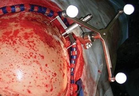
FIGURE 23.1 The BrainLAB® bone anchored reference array for intraoperative navigation is fixed to the scalp with a self-tapping screw. The bicoronal flap is elevated.

FIGURE 23.2 The outline of the nasofrontal segment depends upon the anatomy of the frontal sinus, the size of the tumor, and the location with regard to the intracranial extension. Type A osteotomy leaves the posterior wall of the frontal sinus intact to be removed as a second step, whereas type B osteotomy includes the posterior wall in a one-step procedure. (Adapted from an illustration courtesy of Professor Dan M. Fliss.)

FIGURE 23.3 Osteotomies are performed across the frontal bone, down to and along the roofs of the orbit, medial wall of the orbit into the nasomaxillary groove just anterior to the lacrimal crest. The lacrimal duct is removed from its canal. (Adapted from an illustration courtesy of Professor Dan M. Fliss.)

FIGURE 23.4 Following separation just anterior to the crista galli using a chisel, the frontonasal segment is detached providing direct vision to the nasal and sphenoethmoidal extracranial part of the tumor. Further removal of the posterior wall of the frontal sinus allows exposure of the planum sphenoidale and intracranial neurosurgical dissection. (Adapted from an illustration courtesy of Professor Dan M. Fliss.)
Resection of a tumor mass creates a large dead-space defect with communication between the sterile intracranial space and the sinonasal cavity. Satisfaction regarding functional and cosmetic outcome always depends as much on the reconstruction as it does on the tumor resection. Dural defects resulting from intradural tumor involvement must be closed to restore a water- and airtight barrier between the intracranial contents and any communication with the sinonasal cavity or nasopharynx to avoid a CSF leak, pneumocephalus, or meningitis. Free fascia is the standard, harvested from fascia lata, pericranium, or the superficial temporalis fascia. The first layer is adapted and sutured to the edges of the resected dura. Further overlapping sheaths are meticulously applied and sealed with fibrin glue intracranially under the bone level between the lateral resection borders of the planum sphenoidale, orbital roofs, and frontal cranial vault. An additional collagen sponge coated with fibrinogen and thrombin coagulation factors (TachoSil, Nycomed, Denmark) is glued over the superficial fascia layer toward the sinonasal cavity, which reaches from the exposed frontal lobes up to the clivus (Fig. 23.5A–D).

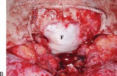


FIGURE 23.5 A–D. Dural repair. A. Defect resulting from intradural tumor resection, showing posterior wall of the sphenoid sinus (S) and planum sphenoidale (PS). B. First layer of fascia lata (F) is adapted and sutured to the edges of the resected dura. C. Further overlapping layers of fascia lata (F) are applied over the bony borders of the resection. D. An additional collagen sponge (T) coated with fibrinogen and thrombin coagulation factors (TachoSil®, Nycomed, Denmark) is glued over the superficial layer of fascia toward the sinonasal cavity, which reaches from the exposed frontal lobes up to the clivus. (Adapted from an illustration courtesy of Professor Dan M. Fliss.)
Significant osseous defects are likely to produce functional disturbances or aesthetic contour deformities such as enophthalmos or diplopia, if left unrepaired. The anatomical restoration of the missing medial wall and floor of the orbit to their original internal shape is therefore crucial to achieve a normal position of the globe. For this purpose, a high molecular resorbable L-lactide-co-glycolide preshaped triangle form orbital plate (Synthes Polymax) is heated in a hot water bath to a malleable state, then contoured to the desired shape before becoming rigid in air temperature again. This self-reinforced resorbable material shows high initial strength and mechanical stability similar to titanium and is consequently suitable for reconstructive procedures in non–load-bearing areas, even if later radiation is planned. A stable bridge of soft tissue should remain after complete degradation within 12 to 24 months.
Finally, I apply a vaseline-coated gauze along the skull base planes to provide additional support against brain pulsation, being removed transnasally after 8 to 10 days. The frontal sinus must be cranialized by removing the posterior table and removing all of the mucosa from the undersurface of the nasofrontal segment. The bone flap is then repositioned precisely to reproduce the original anatomical position and fixed with three-dimensional titanium microplates. Telecanthus should be prevented by bilateral fixation of the canthal ligaments. This is accomplished by passing a nonabsorbable suture through the medial canthal ligament and then guiding it under the nasofrontal segment to the contralateral anterior wall of the frontal sinus. Bilateral tightening of the suture results in medial, downward, and inward traction, thus achieving correct positioning of the canthal ligaments. The last step of the procedure consists in the application of suction drains and closure of the bicoronal incision in layers. In the absence of manifest signs of early postoperative CSF leakage, no lumbar drainage is instituted.
Examples
Case 1: Esthesioneuroblastoma.
This 83-year-old but physically younger and very active former airline pilot was flying a small aircraft, when he suffered a sudden frontal headache during rapid descent. The sinus barotrauma was first treated with topical decongestants and antibiotics, while endoscopic examination manifested a tumor mass in the left nostril that bled easily. Histopathologic findings of the biopsy revealed a high-grade esthesioneuroblastoma Hyams grade III. High-resolution CT and three-plane contrast-enhanced MRI confirmed a considerable tumor extending into the nasal cavity and ethmoid sinus along with intracranial tumor spread and erosion of the medial wall of the orbit. Accordingly, the tumor was classified as Kadish stage C or T4b according to the AJCC-UICC classification, respectively (Fig. 23.6A and B). Further medical investigation revealed a slight stenosis of the subclavian artery with absence of symptoms, hypertension, and cardiac right bundle branch block. Since this circumstance did not appear to be a major risk factor for the development of perioperative cardiac complications, there seemed to be no contraindication for surgery. Complete resection of tumor was achieved with an extended subfrontal approach as described above without complication.


FIGURE 23.6 A and B. Esthesioneuroblastoma Hyams Grade III. A. Preoperative coronal high-resolution CT. B. Contrast-enhanced MR imaging indicating the extent of the tumor into the nasal cavity and ethmoid sinus along with intracranial intradural tumor spread and erosion of the medial wall of the orbit.
Immediately following removal of the nasal packing at postoperative day 8, high-resolution CT and three-plane contrast-enhanced MR imaging were performed and demonstrated complete absence of the tumor (Fig. 23.7A–C). Nevertheless, for local tumor control, robotic and image-guided fractionated intensity-modulated radiation therapy (IMRT) with a total dose of 66 Gy was administered (Novalis Tx). No major complications occurred, and this patient is now free of disease at 44 months (Fig. 23.7D).
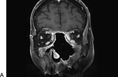


FIGURE 23.7 A–C. Postoperative three-plane contrast-enhanced MR imaging demonstrates complete absence of the tumor. D. Postoperative clinical appearance.
Case 2: Adenoid Cystic Carcinoma.
The medical history of this 40-year-old female patient in the 6th week of pregnancy began with chronic nasal obstruction, hypernasality, slight swelling of the right eyelid with displacement of the globe, and mild pain in the upper jaw. Due to increasing complaints, 3 weeks later, an endoscopic maxillary sinus puncture and drainage of a presumed mucocele was carried out under local anesthesia without preoperative imaging. However, the drainage was suspicious for the presence of a low-grade adenocarcinoma. Therefore, the question whether or not this patient should undergo an MR examination during pregnancy week 13 did not arise; however, intravenous gadolinium was not used. The investigation revealed an extensive tumor mass in the right nasal cavity and paranasal sinuses up to the skull base, with possible erosion of the bony orbit and palate, as well as involvement of the globe (Fig. 23.8A and B). Despite the fact that the level of diagnostic radiation is too low to harm the fetus, a high-resolution thin-section CT scan to clarify the bony aspects was not performed at that time, leaving this important question open. The neuro-ophthalmologic examination revealed a slight asymmetry of the position of the globe with normal eye movement and no diplopia. The open biopsy under general anesthesia revealed an adenoid cystic carcinoma.
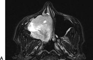

FIGURE 23.8 A and B. Adenoid cystic carcinoma. A and B. Preoperative MR examination during pregnancy (week 13) demonstrating an extensive tumor mass involving the right nasal cavity and paranasal sinuses up to the skull base, infiltrating the orbit and palate.
Diagnosis of cancer during pregnancy is rare, and the conflict between the diagnostic and therapeutic benefits against their risk to the unborn baby must be carefully weighed. Cancer rarely affects the fetus directly, but some cancer treatments may harm it. Following careful interdisciplinary consideration to optimize the safety of the mother and the unborn baby, our treatment planning at this stage of pregnancy and cancer stage T4BN0M0 according to AJCC-UICC was as follows:
· Delayed surgery in pregnancy week 20, posing little hazard to the fetus, yet with a residual risk of miscarriage
· Cesarean section after pregnancy week 32
· Proton therapy after delivery
· No breast-feeding
The management of involvement of the orbit is controversial. Although tumor infiltration of the periorbita and orbital adipose tissue can rarely be controlled by simple excision or irradiation, total orbital exenteration must be considered as a disfiguring and mutilating procedure that may only be performed with curative intention and preoperative agreement with the patient. Restoration of the defect always represents a challenge, and in more extensive cases, the boundaries between the empty orbit and adjacent resection cavities must be reestablished with free tissue transfer. Considering this psychologically and ethically most extreme scenario for the pregnant patient, her family, and the medical staff, I decided to omit both a maxillectomy and orbital exenteration at this first stage.
The extended anterior subcranial approach was done as described in case 1 (Fig. 23.9A and B). In order to gain control of the inferior tumor extension of the nasal cavity and maxilla, an additional modified midfacial degloving approach was selected in the same procedure. Following intraoral infiltration with an epinephrine-containing local anesthetics, a gingivobuccal incision was done and the anterior maxillary wall with the piriform aperture exposed. A full transfixion incision with vestibular extensions allows degloving of the midface and nose. It is now possible to gain access to the inferior aspect of the tumor both in the inferior aspect of the nasal cavity and the maxilla. The tumor is dissected by following the involved bony structures, and free margins are achieved avoiding hemimaxillectomy, with preservation of the tooth-supporting alveolar ridge (Fig. 23.10A and B). En bloc removal of the tumor is now possible via the subcranial route.


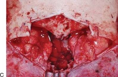
FIGURE 23.9 A–C. Anterior view of the classical subcranial exposure offering direct vision of the superior aspect of the tumor. A. Schematic drawing (Adapted from an illustration courtesy of Professor Dan M. Fliss.). B. Intraoperative view before resection of the tumor. C. Subcranial exposure following resection of the tumor with preservation of the globe and minimal intracranial exposure along the cribriform plate.


FIGURE 23.10 A and B. Additional modified midfacial degloving. A. Schematic drawing. (Adapted from an illustration courtesy of Professor Dan M. Fliss.) B. Intraoperative view following completed resection.
Four days after surgery, the patient was transferred from the intensive care unit to the maternity clinic, where 2 months later, she delivered a healthy girl by cesarean section in pregnancy week 33. Proton radiation therapy started 2 months later. The total radiation dose was 76 Gy at the critical locations, transferred in 30 fractions. High-resolution CT and three-plane contrast-enhanced MR imaging follow-up confirmed a tumor-free status (Fig. 23.11A and B).

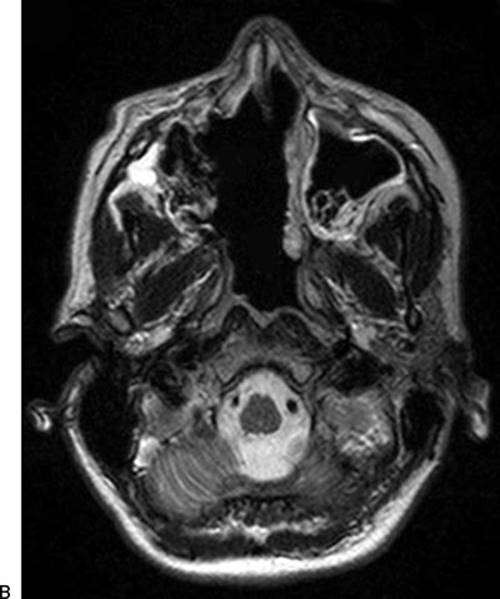
FIGURE 23.11 A and B. Postoperative contrast-enhanced MR imaging confirming tumor-free status.
Despite regular professional nasal care and repeated antibiotic drugs, chronic episodes of rhinosinusitis and intermittent inflammatory periorbital swelling appeared. Later, crystalline particles of the polymer were removed endonasally, and the clinical situation improved. This local foreign body reaction probably occurred because of the decreased vascularity of the recipient site secondary to radiation and exposure of the implant in the nasal cavity. Two years after the initial treatment, a follow-up positron emission tomography–CT detected a hypermetabolic region in the anterior skull base fossa, but further imaging procedures and endoscopic biopsies were negative.
POSTOPERATIVE MANAGEMENT
The patient is monitored postoperatively in the intensive care unit for one night. Antibiotics are given for up to 10 days. Immediately following removal of the nasal packing at postoperative day 8, high-resolution CT and three-plane contrast-enhanced MR imaging are performed to assess the completeness of resection. For local tumor control, surgery is often supplemented with robotic and image-guided fractionated IMRT (Novalis Tx). Unpleasant nasal crusting along with a constant foul-smelling odor is frequently encountered following treatment of this sort. This condition is easily managed with repeated nasal salt water irrigation, to wash out the crusts. Daily nasal hygiene with mechanical loosening of the crusts and application of oily nose drops, emulsions, or ointments are essential.
COMPLICATIONS
Even though the majority of the tumors reported in the above series were extensive, the complication rates were low. Major damage to intracranial nerves was not encountered, only a mild impairment of vision in two cases.
The early and late complications related to the extended subcranial approach are listed as follows: pneumocranium (4.9%) or immediate CSF leakage (3.2%) mostly developed after accidental removal of the nasal packing in the early postoperative stage. These leaks were successfully controlled by a lumbar drain. In three patients with inadequate primary fascia lata alignment, revision surgery was necessary. Enophthalmos (11.4%) was most probably caused by secondary resorption of orbital adipose tissue, whereas telecanthus (6.5%) developed after collapse of the nasal framework. Both were more aesthetic than functional problems. Two of three cases with mucocele formation (2.4%) needed revision surgery several years later. In contrast to earlier reports, partial resorption of orbitomaxillary bone grafts (8.1%) and necrosis of the nasofrontal segment (2.4%) were observed more frequently.
With one exception, all of these patients underwent postoperative radiotherapy. Two patients with near-total segment necrosis developed a nasofrontal fistula, which made a local repair necessary. The collapsed frontonasal segments were successfully reconstructed in three cases. A patient with major cosmetic disfiguring needed difficult free flap reconstruction.
RESULTS
Due to the lack of a uniform international clinical classification and the heterogeneity in the reporting of the tumor extension—mainly for intraorbital and intracranial extension—the treatment outcome differs substantially among different publications in the literature. A combination of benign and malignant tumors within the same statistics is not acceptable. Only a proper histopathologic classification and cancer staging allow a correct and reliable evaluation, interpretation, and comparison of the results. However, the detailed references to the state of disease and overall survival rate related to the extended subcranial Raveh approach are beyond the scope of this article and are stated elsewhere.
The latest retrospective evaluation containing our material compared the surgical approaches—open vs. endoscopic—in 123 patients treated from 1992 to 2008, including information on tumor stage, histology, treatment, and follow-up. Squamous cell carcinoma was the most common histopathologic entity, diagnosed in 30.9%, melanoma was found in 19.5%, and adenocarcinoma in 17.1%. Following, in descending order of prevalence, were esthesioneuroblastoma at 8.9%, lymphoma at 5.7%, undifferentiated carcinoma at 4.9%, adenoid cystic carcinoma at 4.9%, plasmacytoma at 3.3%, metastatic disease at 1.6%, fibrosarcoma at 1.6%, and leiomyosarcoma at 1.6%. Open surgery was performed in 45% and endoscopic resection in 23% of the cases. Nineteen patients were treated with primary radiation therapy, 15 had primary chemoradiation, and four underwent primary chemotherapy. Two patients died prior to the onset of therapy. Adjuvant radiation therapy followed surgery in 64% of the open approach patients and in 68% of the endoscopic resection patients.
The 5-year and 10-year disease-specific survival rates were 63% and 59%, respectively, and the recurrence-free survival rates 49% and 35%, respectively. The comparison of the survival rates and recurrence-free time did not show a significant difference between the treatment groups. However, since most patients with advanced T3 and T4 tumor stages were treated with an open approach, it is likely that the difference, even if not statistically significant, is biased by patient selection. Not surprisingly, patients who underwent endoscopic resection had significantly fewer postoperative complications. Most accepted unfavorable prognostic factors are the involvement of the dura and the intradural spread, extension into the orbits—particularly the apex—as well as invasion of the infratemporal fossa and the skin.
PEARLS
· Preoperative imaging including both three-plane high-resolution CT and contrast-enhanced MRI is essential for exact tumor staging.
· The outline of the nasofrontal segment depends upon the anatomy of the frontal sinus, the tumor size, and location in regard to the intracranial extension.
· Type A osteotomy leaves the posterior wall of the frontal sinus intact to be removed in a second step, whereas type B osteotomy includes the posterior wall in a one-step procedure.
· Special care is taken to preserve a small bridge of bone over the anterior part of the nasal bone, which facilitates later reconstruction of the nasal dorsum.
· If the tumor is unilateral, preservation of the olfactory filaments on the contralateral side may be possible.
· The osteotomies are performed using the thin blade of a high-speed oscillating saw under water irrigation in an oblique manner, providing sufficient bone contact for optimal healing.
· Combinations with classical approaches such as pterional, orbitozygomatic, or Le Fort I osteotomy enable additional exposure to the parasellar region and infratemporal and sphenopalatine fossa, providing en bloc rather than a piecemeal resection, while avoiding injury to vital adjacent structures.
· For dural repair, the fascia must be prepared, so that all adipose tissue and excess tissue is removed to optimize tissue adherence.
· Fibrin sealants are not absolutely necessary, but they do assist as an adjunct to prevent leaking and hemorrhage.
· The frontal sinus is cranialized by total removal of the posterior wall.
· Wrapping of the nasofronto-orbital segment with the pericranial flap should prevent resorption and osteoradionecrosis.
· The extended subcranial approach as developed by Raveh and endoscopy are not two fundamentally opposing principals: in fact, application of endoscope-assisted minimally invasive techniques can be helpful by achieving the goal of a safe and radical resection.
PITFALLS
· Plating and bone grafts along the skull base are contraindicated.
· Free bone grafts exposed to the sinonasal or pharyngeal cavity are vulnerable to infection and necrosis. Covering the grafts with vascularized tissue should reduce these complications.
· Decreased vascularity of the recipient site secondary to preexisting scar formation or high radiation doses compromises a normal healing process and acceptance of any biomaterial.
· The extensive use of expensive alloplastic materials is only justified if the donor site morbidity and operation time are significantly reduced and relevant complications can be avoided.
INSTRUMENTS TO HAVE AVAILABLE
· Neurosurgical tray
· Colorado microdissection needle
· Raney clips
· Bipolar forceps
· Orbital retractors
· Brain spatulas
· Osteotomes and Mallets
· Rongeurs (Kerrison, Blakesley)
· Awl
· High-speed pneumatic drill
· Oscillating saw
· Plating systems
· Endoscopes
· Surgical microscope
SUGGESTED READING
Sekhar LN, Nanda A, Sen CN, et al. The extended frontal approach to tumors of the anterior, middle, and posterior skull base. J Neurosurg 1992;76:198–206.
Raveh J, Laedrach K, Iizuka T, et al. Subcranial anterior approach for skull base tumors: surgical procedure and reconstruction. In: Donald PJ, ed. Surgery of the skull base. Philadelphia, PA: Lippincott-Raven Publisher, 1998:239–261.
Fliss DM, Zucker G, Amir A, Gatot A, Cohen JT, Spektor S. The subcranial approach for anterior skull base tumors. Operative Techniques in Otolaryngology-Head and Neck Surgery. 2000;11(4):238–253.
Laedrach K, Lukes A, Raveh J. Reconstruction of skull base and fronto-orbital defects following tumor resection. Skull Base 2007;17:59–72.
Arnold AM, Ziglinas P, Ochs K, et al. Therapy options and long-term results of sinonasal malignancies. Oral Oncol 2012;48(10):1031–1037.