
Paul A. Gardner and Carl H. Snyderman
INTRODUCTION
Transsphenoidal approaches to the sella have been performed for over a century with progressive improvement in outcomes aided by advances in technology such as the operating microscope and fluoroscopy. However, it was the addition of the endoscope that allowed the expansion of endonasal approaches outside of the sphenoid sinus, especially laterally into the “coronal plane,” which simply could not be visualized with the microscope. Endoscopic, transnasal transsphenoidal approaches initially allowed access to the cavernous sinus (previously felt to be inoperable) by allowing visualization of the entire sphenoid. Surgical teams including both otolaryngology and neurosurgery discovered that the addition of a transpterygoid approach, via a maxillary antrostomy, allowed access to Meckel's cave and even the middle fossa in select cases.
The challenge of a medial approach to Meckel's cave and the middle cranial fossa is proper case selection, respecting the key principle of minimizing neurovascular manipulation. The advantage of an endoscopic endonasal approach (EEA) to this region is that it completely avoids any retraction of the temporal lobe and provides direct access to tumors that originate or extend into the paranasal sinuses or infratemporal fossa.
HISTORY
Lesions that involve Meckel's cave and the middle cranial fossa can have variable presentations including pain, facial numbness, and incidental discovery. Schwannomas and other similar benign tumors involving the trigeminal nerve typically cause trigeminal dysfunction late in their course. However, tumors or inflammation of Meckel's cave often present with ipsilateral headache or retro-orbital pain. Other cranial neuropathies such as palsies of the abducens or oculomotor nerves are also late findings and are often an indication of atypical or aggressive pathology. Indeed, meningiomas rarely cause any cranial nerve dysfunction, and the presence of diplopia, especially acute onset, should raise concern. Perineural spread of a sinonasal malignancy with Meckel's cave involvement, on the other hand, always has neuropathy in the form of numbness with or without pain. Sinus symptoms such as congestion, anosmia, or epistaxis should also be noted as they could direct diagnosis.
Extension to the middle cranial fossa may lead to irritation and/or edema of the mesial temporal lobe resulting in seizures. Very large tumors can create enough mass effect to cause aphasia, though these would be unlikely to be amenable to endonasal resection.
PHYSICAL EXAMINATION
A complete examination of the cranial nerves is critical, focused on the 3rd to 6th cranial nerves, as these are most likely to be affected and ophthalmoplegia indicates cavernous sinus extension. Facial sensation should be tested in all three distributions with light touch, pinprick, and temperature and compared for symmetry with the contralateral side. Rarely, large tumors could affect facial nerve function by direct compression; perineural spread can occur through anastomoses between the trigeminal and facial nerves. If there is any question of compromise of orbital function, a complete neuro-ophthalmologic evaluation is mandatory.
If there is suspicion of a sinonasal tumor, nasal endoscopy should be performed to look for obvious masses and consider preoperative biopsy. Perineural invasion with involvement of multiple branches of the trigeminal nerve can result from a barely perceptible mucosal lesion. Care should be taken to examine all mucosal surfaces including the fossa of Rosenmüller in the nasopharynx. In addition, a complete examination of the neck for evidence of regional spread is critical as this can significantly alter treatment options.
Assessment of memory and speech is important to identify gross deficits in cognitive function caused by middle fossa tumors, though these are usually quite subtle if present at all.
INDICATIONS
EEAs to Meckel's cave and the middle fossa should be limited to those tumors that present directly to the lateral recess of the sphenoid sinus or base of the pterygoid since these are situations that allow for direct access to the lesion with little or no neural dissection or manipulation. The simplest example is a middle fossa meningocele that herniates into the lateral recess of the sphenoid. An open approach requires unnecessary temporal lobe retraction to access the medial defect whereas an EEA gives direct access to the lesion via the sphenoid sinus. Similarly, many schwannomas or meningiomas of Meckel's cave are adjacent to the sinus, providing a natural corridor to the tumor with no retraction of the brain, and the only neural manipulation is related to direct tumor involvement (Fig. 27.1).
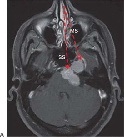
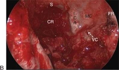
FIGURE 27.1 A. Preoperative, T1-weighted postcontrast axial MRI showing the direct relationship of a tumor of Meckel's cave (arrows) with the sphenoid (SS) and maxillary sinuses (MS). B. Intraoperative endoscopic endonasal view showing tumor within the left Meckel's cave (MC) presenting directly to the surface of the sinus. (CR, clival recess; FR, foramen rotundum; ICA, internal carotid artery [paraclival/vertical petrous]; S, sella; VC, vidian canal.)
Perineural spread of sinonasal carcinoma to Meckel's cave generally carries a poor prognosis, but, in the absence of radiographic or clinical evidence of cavernous sinus invasion, debulking, and/or removal of recurrent disease in this location, followed by focal irradiation for residual microscopic disease, can improve local control or palliation of neuropathic pain.
Rarely, tumors with a medial origin will extend to Meckel's cave. Pituitary adenomas, nasopharyngeal angiofibromas, chordomas, and chondrosarcomas can all be followed into Meckel's cave with relatively low risk, as they tend to displace the neural contents laterally without direct invasion of the nerve.
Rarely, inflammatory diseases (e.g., sarcoidosis), infections (e.g., tuberculosis), or metastatic disease (e.g., meningeal carcinomatosis) primarily involve the trigeminal nerve and/or Meckel's cave. Their diagnosis can often be made by other means such as CSF analysis or inferred from other tests, but sometimes they do require biopsy. These biopsies can be performed endonasally with minimal morbidity or delay in subsequent treatment.
CONTRAINDICATIONS
The EEA is contraindicated for tumors that originate and are primarily growing in the posterior or lateral middle fossa with minimal extension into Meckel's cave or the anteromedial middle fossa. Attempting to resect such tumors would require transection or extensive, unnecessary manipulation of the Gasserian ganglion.
Sinus infection is a relative contraindication for intradural endonasal surgery that requires treatment with antibiotics alone or in combination with drainage. Once the infection has resolved, intradural resection can proceed. Biopsy of the maxillary branch of the trigeminal nerve, peripheral to Meckel's cave, is generally safe even in the setting of infection.
Neoplastic involvement of the petrous internal carotid artery (ICA) is a relative contraindication depending on the goals of surgery and the experience of the surgical team. If proximal control of the ICA is desired, an infratemporal skull base approach may be considered.
PREOPERATIVE PLANNING
Given the complexity and variability of pathologies, both magnetic resonance imaging (MRI) and computed tomography (CT) angiography are generally recommended for evaluation and operative planning. FIESTA or fine-cut T2 MRI sequences can help to determine the relationship of the proximal trigeminal nerve to the tumor. Coronal, postcontrast studies reveal the degree of involvement of the branches of the trigeminal nerve as they exit their neural foramina and can also reveal if there is tumor extension to the cavernous sinus. These factors should also be evaluated on fine-cut (SPGR) postcontrast axial images as they can significantly change the role, extent, or goals of surgery. It is often impossible to know the relationship of the trigeminal nerves and ganglion to the lesion, though this could prove critical for planning of the surgical approach. In the future, high-definition fiber tracking techniques may help to better define these types of relationships.
CT angiography is important to evaluate both bony involvement or erosion and vascular involvement and displacement, both of which are common with Meckel's cave and middle fossa pathology. Enlargement of the foramen ovale and/or rotundum, erosion of the floor of the temporal fossa, tumor-related hyperostosis, and degree of pneumatization of the lateral recess of the sphenoid sinus all play a role in either surgical planning or differential diagnosis. Tumors such as schwannomas and meningiomas that enlarge Meckel's cave over time can significantly displace the horizontal petrous, paraclival, and cavernous segments of the ICA. The position of the artery can affect accessibility as well as safety of resection and/or biopsy. Narrowing of the artery is a sign of significant involvement of its wall, and consideration should be given to balloon test occlusion, depending on the goals of surgery.
Both CT and MRI should be evaluated for signs of sinusitis so that it can be treated preemptively.
SURGICAL TECHNIQUE (VIDEOS 27.1 AND 27.2)
A team consisting of an otolaryngologist and a neurosurgeon working in tandem performs all surgeries. Endoscopic endonasal surgery (EES) lateral to the paraclival and cavernous ICA requires a team with significant experience working in the sellar and parasellar regions. ICA exposure and manipulation are likely to be necessary, and comfort with possible management strategies in the event of an injury is critical before attempting these intradural resections.
Patients are placed in three-pin Mayfield head fixation with their head in slight extension, rotated approximately 15 to 20 degrees to the right and with slight lateral flexion of the vertex to the left. Any head positioning should be tempered by concern for cervical spine immobility or stenosis. The patient is placed in reverse Trendelenburg position to decrease venous hypertension and blood loss. Decongestion of the nasal cavity is achieved by placement of oxymetazoline-soaked pledgets. Image guidance is registered, and then the midface and abdomen are prepped with Betadine and draped. Neurophysiologic monitoring includes monitoring of cerebral function with somatosensory evoked potentials (given the potential for ICA manipulation) and electromyography (EMG) of the motor branch (mandibular/V3) of the trigeminal nerve, third, fourth, and sixth nerves to help with their identification and preservation.
A binaural approach to Meckel's cave is preferred since this improves visualization and access. The need for a vascularized flap for coverage of an exposed ICA or closure of a dural defect is anticipated. A contralateral septal mucosal flap pedicled on the posterior septal branch of the sphenopalatine artery is elevated and stored in the nasopharynx or maxillary sinus (ipsilateral to the flap) until needed. The posterior septum is detached from the sphenoid rostrum, and the bone of the rostrum is removed. Bilateral wide sphenoidotomies are performed. Resection of approximately 1 cm of the posterior septum improves the exposure.
A maxillary antrostomy is performed on the same side as the lesion. The sphenopalatine artery is sacrificed, and the sphenopalatine foramen is enlarged with a 1 mm Kerrison rongeur. The bone of the posterior wall of the maxillary sinus is removed to fully expose the contents of the pterygopalatine space. Within the sinus, the infraorbital nerve (branch of the maxillary nerve) is identified along the floor of the orbit as it courses medially toward the foramen rotundum.
The vidian nerve and its canal are a key anatomic landmark for this approach. The pterygopalatine contents should be carefully retracted laterally to identify the vidian nerve as it enters the bony canal within the pterygoid base (Fig. 27.2). The palatosphenoidal (also known as palatovaginal) canal can also be followed from the nasopharynx along the inferomedial aspect of the pterygoid “wedge” to locate the vidian canal. The vidian canal angles posterolaterally toward the anterior genu of the petrous ICA. The vidian nerve crosses over the petrous ICA lateral to the genu to join with the greater superficial petrosal nerve on the floor of the middle fossa. The pterygoid base must be drilled to gain access to the middle fossa and Meckel's cave, and the pterygoid/vidian canal serves as a good guide during this drilling, especially if the lateral recess of the sphenoid is poorly pneumatized. Attempts to preserve this nerve while accessing Meckel's cave will limit access to the even more critical neurovascular structures in the depth. Anatomic preservation is likely to require excessive manipulation with loss of function.
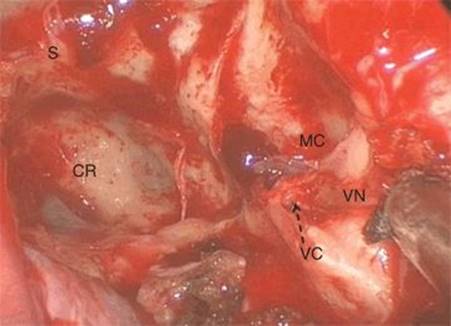
FIGURE 27.2 Intraoperative, endoscopic endonasal view of the left vidian nerve (VN) entering the vidian (pterygoid) canal (VC) (arrow).(CR, clival recess; MC, Meckel's cave behind the lateral recess of the sphenoid; S, sella.)
The foramen rotundum should be identified superolateral to the vidian canal by retracting the contents of the inferior orbital fissure and following the edge of the sphenoidotomy (lateral recess) to the foramen. Some of the bone overlying the inferior orbital fissure just below the orbital floor can be removed to facilitate this, taking care not to enter the orbit. Drilling of the bone of the pterygoid base, between the foramen rotundum and the vidian canal, will lead to complete exposure of Meckel's cave and, laterally, the middle fossa. Additional exposure can be achieved by mobilizing the greater palatine neurovascular bundle from its canal. The pterygoid bone posterior to the canal can be drilled with preservation of the nerve. Further inferior drilling of the pterygoid bone (lateral to the vidian canal) can be done to access the foramen ovale that guards the lateral aspect of the horizontal segment of the ICA. The bone over Meckel's cave can be removed in the same fashion as the bone of the sella, though the angle of the bone surface is unfavorable for the use of a Kerrison rongeur. Once the bone overlying the maxillary branch (V2) and mandibular branch (V3) of the trigeminal nerve have been removed, complete access to Meckel's cave has been achieved. The bony opening can be extending superiorly to the superior orbital fissure and lateral cavernous sinus, though tumor resection in these regions is usually limited due to unwarranted risk to the oculomotor nerves. Extension of the approach lateral to V3 is limited superiorly by the orbital apex and maxillary nerve but can provide access to the floor of the middle fossa and temporal lobe. Medially, the vertical petrous (paraclival) ICA can be uncovered by carefully drilling the bone overlying and medial to it and then dissecting it free from the dura that protects it. This will allow for safe medial retraction of the ICA during tumor resection but may not be necessary in the setting of biopsy.
Biopsy of primary tumors of Meckel's cave or perineural spread of sinonasal cancers can be done with relative ease as long as the ICA is properly identified. In addition to relying on anatomical landmarks in conjunction with image guidance, a thin, long ultrasound probe is invaluable. Approaching lesions at or below the level of the foramen rotundum will avoid accidental entry into the adjacent and contiguous lateral cavernous sinus. Opening of the dura should be done parallel to the closest trigeminal branch, keeping in mind that V2 and V3 form a 45-degree angle with each other as they enter Meckel's cave (Fig. 27.3). Opening in this manner will help to merely split rather than sever the nerve if it is displaced medial to the lesion. Dissection is performed with a Kartush nerve stimulating dissector to identify and preserve the cranial nerves in and around Meckel's cave. Resection of tumors such as schwannomas proceeds with internal debulking followed by extracapsular dissection, recapitulating standard microdissection techniques. Trigeminal schwannomas respect the adjacent oculomotor nerves, displacing them superiorly while tumors such as meningiomas and sinonasal carcinomas tend to be much more invasive, increasing the risk to these nerves. Pituitary adenomas and chondroid tumors are variable but tend to displace the dura of Meckel's cave as well as its contents.
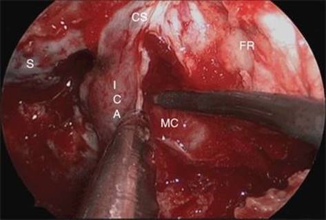
FIGURE 27.3 Intraoperative, endoscopic endonasal view during dissection of the left Meckel's cave portion of the tumor shown in Figure 27.1. A Kartush electrical nerve stimulator is being used to identify motor nerves in Meckel's cave (MC) and the adjacent cavernous sinus (CS). (FR, foramen rotundum; ICA, internal carotid artery [paraclival/vertical petrous]; S, sella.)
The limits of dissection are anatomical and include the lateral cavernous sinus superiorly (unless the nerves therein are already affected), the paraclival ICA medially, horizontal petrous ICA inferiorly, and trigeminal ganglion, middle fossa dura, and temporal lobe laterally (Fig. 27.4). Dissection and resection can extend to the middle fossa if the tumor has created a corridor through which to access it (Fig. 27.5). In the depth, the entrance to the posterior fossa from Meckel's cave can be dilated, allowing for greater access to this region from an anterior approach, but this can prove to be a limitation as well, and dissection should not proceed blindly past this point.
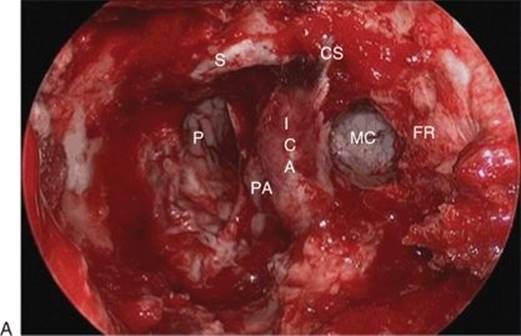
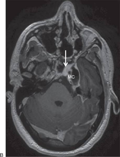
FIGURE 27.4 A. Intraoperative, endoscopic endonasal view following complete removal of the tumor shown in Figure 27.1. (CS, cavernous sinus; FR, foramen rotundum; ICA, internal carotid artery [paraclival/vertical petrous]; P, pons; PA, petrous apex; S, sella.) B. Postoperative, T1-weighted, postcontrast axial MRI showing complete tumor removal of Meckel's cave tumor (MC) shown in Figure 27.1. The nasoseptal flap used for reconstruction enhances brightly (arrow).
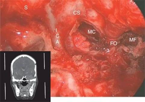
FIGURE 27.5 Intraoperative, endoscopic view following resection of a recurrent meningioma (coronal MRI inset) that extended from Meckel's cave (MC) to the middle fossa (MF). Resection was performed by drilling the base of the pterygoid to expose the mandibular branch of the trigeminal nerve (V3) within the foramen ovale (FO). (CS, cavernous sinus; ICA, internal carotid artery [paraclival/vertical petrous]; S, sella.)
Reconstruction depends on the extent of exposure to CSF. Occasionally, Meckel's cave has an arachnoid diverticulum that can lead to CSF leak during even simple biopsy. Normally, unless dissection extends to the proximal Gasserian ganglion, there is little risk of intraoperative CSF leak. In the absence of a leak or ICA exposure, application of fibrin sealant or a free mucosal graft is adequate. Otherwise, vascularized flaps such as the nasal septal flap (see Chapter 42) are used to cover the opening into Meckel's cave or the middle fossa as well as exposed segments of the ICA.
POSTOPERATIVE MANAGEMENT
Lumbar drainage is used only in the setting of high-flow intraoperative CSF leaks (dissection in an arachnoid cistern). Intravenous, broad-spectrum antibiotics such as a third or fourth generation cephalosporin are administered for 24 to 48 hours postoperatively and then converted to a similar oral regimen until nasal packing is removed. Corticosteroids are used in cases with high risk or evidence of cranial nerve injury.
In the absence of a CSF leak, nasal packing can be removed in 24 to 48 hours. If a vascularized septal flap is used to repair a dural defect, packing is left in place for 5 to 7 days. Silastic nasal splints are removed at 1 week if a septal flap is not used and at 3 weeks if one is used. The Silastic splints maintain humidification and improve mucosalization of exposed septal cartilage. Patients are encouraged to use saline nasal spray throughout the day. Saline flushes of the nasal cavity are instituted at 3 weeks. Patients are advised to avoid activities that increase CSF pressure for at 4 weeks if a dural reconstruction is performed. If a patient requires continuous positive airway pressure (CPAP) for treatment of obstructive sleep apnea, it is generally safe to resume 1 week following surgery (after nasal packing is removed).
COMPLICATIONS
CSF leak is the most common complication after EES in general but has decreased significantly with the use of vascularized flaps. Dry eye is an inevitable consequence of vidian nerve sacrifice, though most patients are not aware of nor symptomatic from this loss of emotional tearing. Care must be taken to avoid both vidian nerve and V1 (ophthalmic branch of trigeminal nerve) dysfunction, as this creates a dry, insensate eye that may be destined for corneal keratopathy. Corneal sensation should be tested and appropriate precautions and care provided in case of its absence. Diplopia can be transient and unless a nerve is severed, allow 3 to 6 months for recovery. Intraoperative injury of the ICA is rare, but respect for the significant learning curve associated with EES will allow for appropriate management. Any surgery lateral to the ICA is considered high risk and should only be performed following extensive experience with sellar and parasellar lesions as well as planning for management of intraoperative vascular injury. Advanced skull base surgery requires endovascular support for immediate angiography and potential sacrifice, coiling, or stenting. Injury to the motor branch of the trigeminal nerve results in muscle atrophy with attendant cosmetic defect, decreased chewing strength, and jaw drift with opening. Temporary trismus is common if there is dissection of the pterygoid muscles.
RESULTS
EEAs are widely accepted for the treatment of middle fossa meningoencephaloceles extending into the lateral recess of the sphenoid sinus. In addition, they have been used successfully for biopsy and resection of benign and malignant pathologies involving Meckel's cave and the middle fossa, including meningiomas (typical and atypical), schwannomas, and sinonasal carcinomas (such as adenoid cystic carcinoma and squamous cell carcinoma).
The most consistent and classic pathology of Meckel's cave is trigeminal schwannoma. Between 2003 and 2009, 11 trigeminal schwannomas were resected by EEA at our institution. They all had some degree of Meckel's cave involvement but were primarily growing in the following areas: four in Meckel's cave, two in the middle fossa, two in the orbital apex, and three in the infratemporal fossa. Their average diameter was 3.5 cm, and two patients underwent EEA combined with a retromastoid craniectomy. Seven patients (64%) underwent gross total resection, three (27%) near total resection (Fig. 27.6), and one (9%) subtotal resection. Out of 11 trigeminal schwannoma patients, two patients (18%) developed a sensory deficit only, two patients (18%) developed motor and sensory deficits, and one patient (9%) developed a motor deficit only after EEA. Overall, four patients (36%) developed new sensory deficits and three patients (27%) developed new motor deficits postoperatively. There were two new postoperative cranial neuropathies (III and VI nerve palsies) that were improved at last follow-up (>1 year). On the other hand, three patients had improvement in trigeminal (sensory) function and three of four patients with preoperative abducens palsies recovered. One preoperative oculomotor palsy improved. Importantly, there were no CSF leaks following EES for trigeminal schwannoma. All of these rates compare favorably with conventional or open skull base approaches.
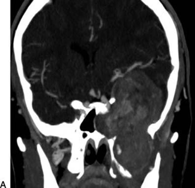
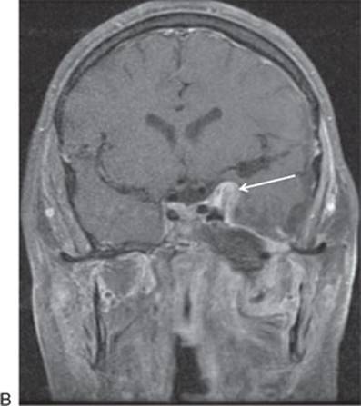
FIGURE 27.6 Preoperative (A) and postoperative (B) T1-weighted, coronal postcontrast MRI showing near-total resection of a giant V3 schwannoma treated endoscopically in two stages. A small area of residual tumor (arrow) was left adjacent to the cavernous sinus to ensure preservation of oculomotor function.
PEARLS
· Drilling of the pterygoid bone between the foramen rotundum and the vidian canal provides direct endonasal access to Meckel's cave. They converge posteriorly on Meckel's cave, and the distance between them depends on the pneumatization of the lateral recess of the sphenoid.
· The vidian nerve has to be sacrificed to achieve full, safe access to Meckel's cave.
· Dissecting at or below the level of V2 should prevent damage to the oculomotor nerves.
PITFALLS
· Preoperative imaging is used to determine the relationship of the trigeminal nerve to a tumor in Meckel's cave. A different approach should be considered if the nerve is displaced medially by tumor.
· The vidian nerve provides a good landmark for access to Meckel's cave, but does NOT lead directly to the anterior genu of the ICA but rather turns laterally to cross over the horizontal petrous ICA.
· Invasion of and attachment to cranial nerves varies by tumor type and should be taken into consideration when planning resection.
· Drilling medial to the vidian canal can still result in injury to the ICA.
· Sacrifice of the vidian nerve can result in a dry eye in elderly patients and should be avoided if possible in patients with absence of corneal sensation (V1).
INSTRUMENTS TO HAVE AVAILABLE
· Zero- and 45-degree rod lens endoscopes (Storz)
· Extended tip micro-Doppler probe (for ICA identification)
· Standard sinus instruments
· Monopolar needle tip and suction electrocautery tips
· High-speed electric drill with extended tip and extended, coarse diamond drill bit (Stryker)
· Extended tip neurodissectors (KLS Martin)
· Pistol-grip Kurze microscissors (straight, curved left and right, and rotatable) (Storz)
· Pistol-grip bipolar electrocautery (side angle, “up-toe,” and fine, straight tip) (Storz)
· Kartush stimulating dissector to be used with EMG
SUGGESTED READING
Vescan A, Snyderman C, Carrau R, et al. Vidian canal: analysis and relationship to the internal carotid artery. Skull Base 2007;17(Suppl 2):118.
Zanation AM, Al-Sheibaini S, Carrau R, et al. Endoscopic endonasal transpterygoid nasopharyngectomy. Skull Base 2008;18(Suppl 1):28.
Kassam AB, Prevedello DM, Carrau RL, et al. The front door to Meckel's cave: an anteromedial corridor via expanded endoscopic endonasal approach—technical considerations and clinical series. Neurosurgery 2009;64(3 Suppl):71–82; discussion 82–83.
Zanation AM, Carrau RL, Snyderman CH, et al. Nasoseptal flap reconstruction of high flow intraoperative cerebral spinal fluid leaks during endoscopic skull base surgery. Am J Rhinol Allergy 2009;23(5):518–521.
Hofstetter CP, Singh A, Anand VK, et al. The endoscopic, endonasal, transmaxillary transpterygoid approach to the pterygopalatine fossa, infratemporal fossa, petrous apex, and Meckel's cave. J Neurosurg 2010;113(5):967–974.
Prevedello DM, Pinheiro-Neto CD, Fernandez-Miranda JC, et al. Vidian nerve transposition for endoscopic endonasal middle fossa approaches. Neurosurgery 2010;67(2 Suppl Operative):478–484.
Fernandez-Miranda J, Pinheiro-Neto C, Vaezi A, et al. Transposition of the pterygopalatine fossa during endonasal endoscopic transpterygoid approaches. Skull Base 2011;21:62.
Pinheiro-Neto CD, Fernandez-Miranda JC, Rivera Serrano CM, et al. Endoscopic anatomy of the palatovaginal canal (palatosphenoidal canal): a landmark for dissection of the vidian nerve during endonasal transpterygoid approaches. Laryngoscope 2012;122(1):6–12.