
Paul A. Gardner and Carl H. Snyderman
INTRODUCTION
Transsphenoidal approaches to the sella have been performed for over a century with progressive improvement in outcomes associated with the addition of technologies such as the operating microscope, fluoroscopy, intraoperative image guidance, and modified specula. However, these approaches remain limited in the paramedian regions of the skull base requiring alternate approaches such as anterior transmaxillary approaches that themselves have significant limitations below the sphenoid sinus. The introduction of the endoscope allows expansion of endonasal approaches outside of the sphenoid sinus, especially laterally into the “coronal plane” and inferiorly into the nasopharynx and parapharyngeal space. The addition of a transpterygoid approach, via a medial maxillotomy, provides access to the pterygoid base and foramen lacerum. Combining these caudal and paramedian approaches has made it possible to resect lesions that extend to or originate from the jugular tubercle, jugular foramen, and medial occipital condyle in select cases.
Proper case selection must respect the basic principle of endoscopic endonasal surgery (EES), which is to minimize the manipulation of normal neural and vascular structures. The advantage of an endoscopic endonasal approach (EEA) to this region is that it completely avoids an external incision and associated trauma to normal tissues, requires no cerebellar retraction or manipulation of the vertebral artery or lower cranial nerves, and provides direct access to tumors that originate or extend into the clivus, paranasal sinuses, or parapharyngeal space.
HISTORY
Lesions involving the jugular foramen would logically affect the lower cranial nerves, often presenting with dysphagia or hoarseness. Those that arise medially or caudally may be more likely to affect the hypoglossal nerve first, with dysarthria as a primary complaint. All of these may be subtle at first, with only occasional coughing or choking on liquids and voice changes that may be missed if not specifically questioned. Patients adapt very well to slowly progressive deficits and may present with large tumors by the time overt symptoms have developed.
Occipital headache can be specific to lesions in the clivus or jugular foramen/tubercle. Involvement of the occipital condyle can lead to instability with mechanical neck or head pain. Bone tumors classically present with pain that is worse at night and relieved by nonsteroidal anti-inflammatory drugs. Large masses with brainstem or cerebellar compression may present with ataxia or even quadriparesis.
PHYSICAL EXAMINATION
A detailed examination of the lower cranial nerves is critical for the evaluation of tumors of the jugular foramen region. This should include observation of palatal function with gag testing, and assessment of trapezius muscle strength (shoulder shrug, head turning) and tongue function (atrophy or fasciculations, weakness, and deviation to the side of the lesion with protrusion). Laryngoscopy should be performed to observe vocal cord mobility and assess the degree of aspiration. A full examination of the head and neck with palpation of the soft tissues is important to identify soft tissue masses or associated lymphadenopathy. A large parapharyngeal space mass may displace the tonsil fossa medially.
A full neurologic examination should be performed to include testing of gait and dysmetria. Long tract signs and proprioception can be affected in cases with compression of the brainstem compression and other sensations, including light touch and pinprick in the face, trunk, and extremities, may also be abnormal; the pattern of its loss may localize to the lateral medulla.
INDICATIONS
The indications for a medial EEA to the jugular foramen are limited. Many jugular foramen tumors (e.g., paragangliomas) originate or are primarily based lateral to the pars nervosa (which is medial to the pars venosum), placing the lower cranial nerves between the surgeon and the tumor with an endonasal approach. However, there are tumors that originate in the midline or paramedian skull base that extend out to the jugular foramen. Chordomas, chondrosarcomas, and some petroclival meningiomas can all be approached endonasally and often extend out toward the jugular foramen and/or occipital condyle. The primary limitation of an infrapetrous approach is the horizontal petrous segment of the internal carotid artery (ICA) and foramen lacerum superiorly and the hypoglossal nerve inferolaterally.
There are occasional small meningiomas that arise from the jugular tubercle, medial to the lower cranial nerves that are ideal for EES (Fig. 28.1). These can be completely resected with no manipulation of the nerves, something that is impossible with any transcranial approach. Some cholesterol granulomas of the petrous apex do not extend medial to the paraclival (vertical petrous) ICA and therefore have to be drained through an infrapetrous approach.
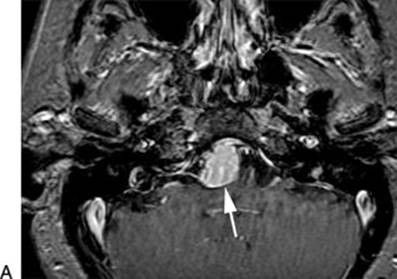
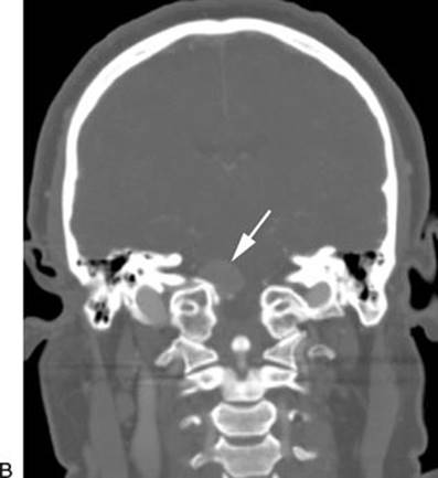
FIGURE 28.1 T1-weighted, postcontrast axial MRI (A) and coronal CT angiogram (B) showing a small medial jugular tubercle meningioma (arrows). The tumor originates medial to the lower cranial nerves, making it ideal for a medial (endonasal) approach.
Malignancies of the nasopharynx are usually advanced at the time of presentation and are treated primarily with radiation therapy. The primary role of surgery is biopsy for diagnosis and debulking of tumor to relieve symptoms prior to radiation therapy. Exceptions include small tumors that can be completely resected with adequate margins. For adenoid cystic carcinoma, the goal of surgery is maximal removal with minimal morbidity, followed by radiation therapy. It is not possible to achieve clear resection margins with adenoid cystic carcinoma of the skull base due to perineural spread, and the extent of surgery is limited by the surrounding neural and vascular structures. Surgical salvage of residual tumor following radiation therapy is of potential value for local control and should be considered based on posttreatment functional imaging with PET–CT or the results of biopsies. Understanding the paramedian anatomy below the petrous ICA is critical for safely expanding the above indications.
CONTRAINDICATIONS
There are no absolute contraindications to an endonasal approach as long as the tumor is medial to the lower cranial nerves. Occlusion of the contralateral ICA warrants consideration of a posterolateral approach that would avoid any potential risk to the patent ICA. Extensive tumor (benign or malignant) with encasement of the ICA limits the goals of surgery, and complete resection is not possible without sacrifice of the ICA. Depending on the surgeon's experience and available resources for dealing with an injury to the ICA, an open approach with better proximal and distal control of the ICA may be preferred. Any sinus infection would need to be properly treated before intradural surgery is performed, but sinusitis can usually be cleared within a week or two with antibiotics, drainage, or a combination of therapies.
PREOPERATIVE PLANNING
This region is difficult to image due to the dense bone and closely associated mucosal surfaces, muscles, vessels, and nerves. As a result, MRI and CT are complementary imaging modalities, and often both are needed to establish a differential diagnosis and determine extent of disease. Fine-cut T2-weighted MRI or FIESTA sequences can be critical in determining the relationship of the lower cranial nerves to any lesion in the infrapetrous space. Postcontrast T1-weighted images can help to reveal the vascularity of a tumor or the presence of a dural tail. CT is critical, especially for small lesions of the petrous bone that can be quite heterogeneous on MRI. Even asymmetric petrous pneumatization can be deceiving until evaluation with CT. The addition of CT angiography is important to define the vascular relationships and involvement, especially of the ipsilateral ICA and vertebral artery. Combined PET–CT scan imaging is helpful in differentiating cancer from radiation changes in patients with recurrent or residual sinonasal malignancy.
A full swallowing evaluation is necessary to identify cranial nerve deficits and assess the risk of perioperative complications related to airway obstruction or aspiration. Complete vocal cord paralysis or significant aspiration may require prophylactic tracheostomy to prevent aspiration in the perioperative period. The detection of cranial nerve dysfunction by physical examination or electromyography can provide prognostic information regarding the potential for additional nerve injury or recovery.
SURGICAL TECHNIQUE (VIDEO 28.1)
All EES is best performed by a team of two surgeons, composed of otolaryngology and neurosurgery specialties. Patients are positioned supine and preferably in head pin fixation with the head in slight extension and laterally flexed to point the chin at the surgeons, both of whom stand on the patient's right side (right-handed surgeons). Oxymetazoline (0.05%)-soaked pledgets are placed in the nose, image guidance is registered, and the midface and abdomen are prepped and draped. Antiseptics are not used intranasally except for the nasal vestibule.
Intradural cases or those with significant ICA exposure should have a vascularized nasal septal flap (see Chapter 42) harvested from the side contralateral to the infrapetrous approach at the beginning of the operation. The right middle turbinate is frequently resected to allow working room for the endoscope, and the sphenoid sinus is opened widely, fully exposing the lateral recess on the operative side. The key to accessing the infrapetrous region is a medial maxillary antrostomy and transpterygoid approach.
A maxillary antrostomy is performed on the same side as the lesion. The sphenopalatine artery is sacrificed, and the sphenopalatine foramen is enlarged with a 1-mm Kerrison rongeur. The bone of the posterior wall of the maxillary sinus is removed to fully expose the contents of the pterygopalatine space. Within the sinus, the infraorbital nerve (branch of the maxillary nerve) is identified along the floor of the orbit as it courses medially toward the foramen rotundum. Medially, at the inferior margin of the sphenoidotomy, the palatosphenoidal (palatovaginal) vessel is sacrificed, and the soft tissues of the pterygopalatine space are elevated from the underlying bone to identify the vidian canal within the base of the pterygoid bone. It is difficult to identify the canal without first sacrificing the terminal branches of the internal maxillary artery.
The vidian nerve and its canal are a key anatomic landmark for this approach. The pterygopalatine contents should be carefully retracted laterally to identify the vidian nerve as it enters the bony canal within the pterygoid base. The vidian canal angles posterolaterally toward the anterior genu of the petrous ICA. However, the vidian nerve crosses over the petrous ICA lateral to the genu to originate from the greater superficial petrosal nerve on the floor of the middle fossa. The pterygoid base must be drilled to define the course of the petrous ICA and provide localization of the anterior genu of the carotid and foramen lacerum (Table 28.1). The pterygoid/vidian canal serves as a good guide during this drilling, especially if the lateral recess of the sphenoid is poorly pneumatized. Drilling the bone of the “pterygoid wedge” (the medial extension of the pterygoid base onto the sphenoid floor) along the path of the vidian nerve will lead to the ICA genu (Figs. 28.2 and 28.3), but it is critical to understand that the vidian nerve crosses the ICA immediately lateral to the genu. Lateral exposure is limited by the vidian nerve and descending palatine branch of the second division of the trigeminal nerve. Both nerves can be skeletonized with careful drilling and released from their respective canals with an attempt at preservation of function. Greater exposure of the ICA, however, usually requires sacrifice of the vidian nerve.
Table 28.1 Segments of the Cranial Base Internal Carotid Artery and Associated Endonasal Anatomic Landmarks for Localization
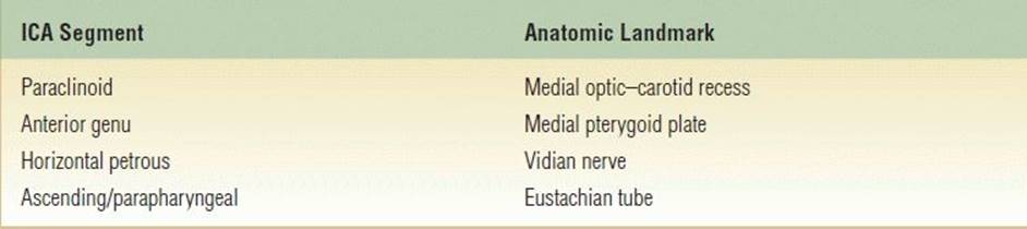
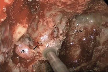
FIGURE 28.2 Intraoperative, endoscopic endonasal view of the right vidian neurovascular bundle (VN) entering the vidian canal (VC), which has been drilled to the anterior genu of the petrous internal carotid artery (ICA). Note that the vidian nerve runs lateral to the genu (just deep to the drill in this image), which lies just superior to the foramen lacerum (FL). (CR, clival recess.)
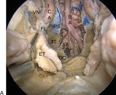

FIGURE 28.3 A. Endoscopic endonasal view of a cadaveric dissection showing transection of the right eustachian tube (ET) attachment to foramen lacerum (FL). The hypoglossal nerve (XII) enters the hypoglossal canal just deep to the ET and separates the occipital condyle (OC) and the jugular tubercle (JT). (BA, basilar artery; ICA, internal carotid artery [paraclival segment]; IPS, inferior petrosal sinus; VN, vidian nerve.) B. Endoscopic endonasal view of cadaveric dissection showing the parapharyngeal internal carotid artery (ICA) and jugular foramen (JF) following transection and removal of the eustachian tube. (BA, basilar artery; IPS, inferior petrosal sinus; FL, foramen lacerum; JT, jugular tubercle; OC, occipital condyle; XII, hypoglossal nerve.)
The second key anatomic landmark is the eustachian tube (ET) and its attachment to foramen lacerum. The ET can be easily identified as the lateral boundary of the nasopharynx (Fig. 28.3). The nasopharyngeal mucosa should be dissected from the floor of the sphenoid along the medial pterygoid plate just above the ET. This region is the epicenter of the exposure, and drilling this bone will lead directly to the cartilage of foramen lacerum that is contiguous with the ET cartilage. This cartilage is attached firmly to the inferior aspect of the ICA genu and cannot be easily dissected from the ICA. Careful removal of the majority of this cartilage with a “thru-cut” instrument can expand the superior access to the petroclival junction but must be done with extreme care given the proximity and high risk of injury to the ICA. Further dissection of the ET laterally to the junction of the cartilaginous and bony ET leads to the skull base adjacent to the entrance of the parapharyngeal ICA into the petrous bone. Dissection of soft tissues in this region must be done very carefully since the parapharyngeal ICA can be very tortuous and does not have reliable landmarks. A micro-Doppler probe can be used to explore the soft tissues to identify the ICA, but this may provide a false sense of security given the precise placement required for accurate sonography.
The final key anatomic point is the supracondylar groove (Fig. 28.4). This paramedian osseous landmark provides the common attachment region of the capsule joint, the atlanto-occipital membrane as well as the rectus capitis anterior muscle and accurately estimates the position of the hypoglossal canal. In fact, drilling over this region will expose the anterior cortical bone of the hypoglossal canal, which divides the inferolateral area of the clivus into two compartments: superior (jugular tubercle) and inferior (occipital condyle). By resecting the overlying mucosa and musculature with a needle tip monopolar cautery (with caution, depending on the proximity of the parapharyngeal ICA) or straight and angled thru-cutting rongeurs, these landmarks and structures can be carefully but reliably exposed.
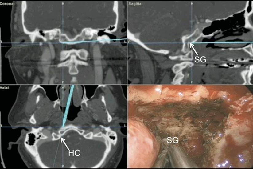
FIGURE 28.4 Intraoperative image-guided view (endoscopic endonasal view lower right) showing the supracondylar groove (SG) and its direct relationship with the hypoglossal canal (HC).
Tumors that originate from or extend into these paramedian structures, displacing the lower cranial nerves laterally, can generally be safely accessed by removing the bone of the medial jugular tubercle and occipital condyle with a high-speed electric drill and cutting diamond burr. When drilling, care must be taken to preserve the dural sheath around the hypoglossal nerve. Often, this can be ensured by leaving the inner cortical bone of the hypoglossal canal intact.
The majority of the surgery is performed with a 0-degree endoscope. Angled endoscopes are needed to resect lateral extension of the tumor into the petroclival synchondrosis or medial jugular tubercle and occipital condyle. Similarly, angled instruments are used in these regions to safely resect tumor. Care should be taken to ensure that injury to the parapharyngeal and petrous ICA does not occur when resecting tumor deep to them. If there is extensive involvement of the ICA in these regions, exposure through the neck for proximal control should be considered.
Further lateral resection is generally limited by neurovascular structures; in this case, the parapharyngeal ICA and lower cranial nerves. Removal of bone laterally in the jugular tubercle leads to the inferior petrosal sinus, which lies immediately medial to the pars nervosa. Dissection should not proceed lateral to this to avoid injury to these nerves. There can be significant venous bleeding from the inferior petrosal sinus, but this can be controlled by packing with morselized, flowable Gelfoam (Surgifoam, Surgiflo, Floseal). When resecting the condyle, the joint capsule should be preserved when possible. Based on our experience and recent biomechanical data, the medial half of the condyle can be resected without overt instability, as long as the joint capsule is not disrupted or the condyle disconnected from the skull base.
Resection of intradural tumors in these regions should proceed similarly, from medial to lateral, with care to identify the relevant intradural structures: the vertebral artery with hypoglossal nerve lying immediately dorsal to it as it exits into its canal and the glossopharyngeal and vagal nerves just below the foramen lacerum (Fig. 28.5). Resection of the lateral margins should be performed or confirmed with angled endoscopy and often requires angled instruments. This should be done with care and cognizance of the location of the parapharyngeal ICA to prevent inadvertent injury to its deep surface. Similarly, the horizontal petrous ICA and lacerum segment should be well localized superiorly. Often, the ICA in both locations can be located with a long Doppler probe. Confirmation of location with ultrasound is reassuring, but lack of localization is not, given the fickle nature of the directional tip of the device.
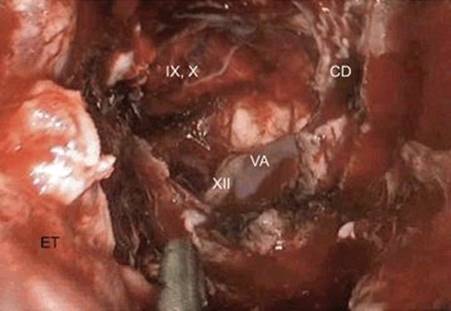
FIGURE 28.5 Intraoperative, endoscopic endonasal view following resection of the medial jugular tubercle meningioma shown in Figure 28.1. Note that the hypoglossal nerve passes deep to the vertebral artery. (CD, clival dura; ET, eustachian tube; VA, vertebral artery; IX, X, 9th and 10th cranial nerves extending to the jugular foramen; XII, hypoglossal nerve.)
Reconstruction of the dural defect is undertaken with an inlay graft of DuraGen or other collagen allograft or dural substitute. This is usually done to replace or reinforce the arachnoid layer. The dura can be reconstructed primarily with a vascularized nasal septal flap (see Chapter 42). A wider flap is harvested that includes the mucosa of the nasal floor. This allows simultaneous reconstruction of the dural defect and coverage of the ICA. A deep clival defect may require augmentation with an adipose tissue graft placed between the dural graft and the mucosal flap. In either technique, care must be taken to ensure that the flap is in contact with bone or dura circumferentially. There should not be mucosa deep to the flap, and its pedicle must also be in contact with bone or other tissue across its entire length to prevent retraction of the pedicle and subsequent flap displacement. Surgicel, tissue glue, and Gelfoam packing then follow, held in place with either a Foley catheter balloon or Merocel tampons, depending upon the size and shape of the defect.
POSTOPERATIVE MANAGEMENT
Packing is maintained for 5 to 7 days when there is a dural defect (depending on size, extent, and quality of reconstruction). Patients are kept on antibiotics while packing is in place (intravenous for 24 to 48 hours, then oral, broad-spectrum cephalosporin or equivalent). The use of perioperative lumbar drainage has not been well studied, but it does reduce cerebrospinal fluid (CSF) pressure during the initial period of healing, and it is our current practice to maintain drainage for 3 postoperative days in cases of large clival or paraclival defects, especially with extensive arachnoid dissection.
After the nasal packing is removed, patients should be observed closely for signs of a CSF leak. With low paraclival defects, this may present as postnasal, pharyngeal drainage with frequent coughing. Patients should be questioned about this and checked for clear nasal drainage. It may be more difficult to detect leaks in this location, and bedside endoscopy should be performed if there is any question. Patients are started on saline nasal spray QID and PRN postoperatively. They are cautioned to avoid activities that increase intracranial pressure and stress the repair. After 3 weeks, saline irrigations are started for more effective cleaning of the nasal cavity. Nasal endoscopy with gentle debridement is performed every few weeks until healing is complete. Silastic nasal splints are removed at 3 weeks if a nasoseptal flap has been used and at 1 to 2 weeks if not.
Lower cranial nerve function should be assessed in the postoperative period prior to allowing oral intake. If suspicion of injury is low, this can consist merely of bedside observation of intake of fluids with varied consistency. Otherwise, a formal evaluation, often including fiberoptic endoscopic evaluation, may be necessary.
Any patient with tumor resection involving the condyle should have ongoing evaluation to ensure they do not develop instability. This consists of evaluation of neck pain, range of motion, and flexion/extension radiographs. If there is new or increasing mechanical neck pain or radiographic evidence of listhesis or other instability, CT and MRI should be performed to assess the craniocervical junction and evaluate the need for fixation.
COMPLICATIONS
The close anatomical relationships and potential tumor involvement of the petrous and parapharyngeal segments of the ICA place these structures at greater risk with the infrapetrous approach. The lack of reliable landmarks outside the skull base and the difficulty of dissecting around the foramen lacerum further increase the risk. Proximal control of the ICA is a challenge, and balloon test occlusion and neck dissection for proximal control should be considered as needed. Currently, there are not good techniques for suturing endonasally, leaving only clip reconstruction or packing for repair of a vessel. Any injury should be evaluated as soon as possible with formal digital subtraction angiography to evaluate for active extravasation, pseudoaneurysm, thromboembolus, dissection, or occlusion. As a general rule, extensive tumor resection should not proceed following such an injury. It is important that the surgical team be prepared to handle such an emergency, and simulation training is recommended.
Lower cranial neuropathy can have a devastating effect on patient quality of life and should be avoided when feasible, even if this means choosing treatment modalities other than complete resection. As a result, resection lateral to the inferior petrosal sinus should generally be avoided.
CSF leak is a potentially challenging complication, regardless of approach. Caudal, paramedian dural defects are at the limit of coverage for vascularized flaps such as the nasoseptal flap but can often be successfully managed with inlay and onlay allo- or autografts with an overlay of adipose tissue grafts deep to the flap. If a leak is suspected, it should be evaluated efficiently, even if this requires reexploration under anesthesia to confirm healing. If a secondary repair is performed, a lumbar spinal drain is placed at the same time.
RESULTS
The infrapetrous approach has been used with great success for complete removal of meningiomas and chondrosarcomas of the medial jugular tubercle and petroclival meningiomas. An understanding of the inferior and lateral extension of a transclival approach is critical for achieving maximal resection of tumors of midline or paramedian origin. This is demonstrated by the learning curve for clival chordoma. In our first 60 chordoma cases, the midclival resection rate was 100%, while the inferior clivus was only 47.6%; midline resection rates were 76.7% while lateralized tumor had only 56.7% of their volume resected. Gaining an understanding of the anatomy and technique of the infrapetrous approach is a key part of the learning curve, demonstrated by a sequential increase in gross total resection rates from 36.4% in early years to 88.9% in the last 15 cases.
PEARLS
· Dynamic endoscopy and an experienced surgical team (two surgeons, three or four hands) are critical for paramedian, “coronal plane” approaches.
· Proximal ICA control may require a small incision in the neck.
· The supracondylar groove, an important point for muscle and capsule attachment, lies at the precise level of the hypoglossal canal and, as such, provides an important anatomic landmark.
· Paramedian and caudal CSF leaks can present as nasopharyngeal drainage, rather than nasal dripping. This requires close patient questioning and often presents as excessive secretions when supine.
PITFALLS
· The vidian nerve crosses the petrous ICA lateral to the genu of the ICA at foramen lacerum and does not accurately predict the medial margin of the genu.
· Dissection lateral to the ET should be done with caution to avoid injury to the parapharyngeal ICA. Preoperative radiographs should be reviewed to identify a tortuous ICA that projects medially.
· Bleeding from the inferior petrosal sinus marks the lateral extent of resection of a “far medial” or infrapetrous approach; extension beyond this requires dissection into the pars nervosa of the jugular foramen with attendant lower cranial neuropathy.
INSTRUMENTS TO HAVE AVAILABLE
In addition to the standard endoscopic sinus surgery instruments, it is helpful to have the following instruments:
· Angled endoscopes
· Angled thru-cutting instruments
· Malleable suction tip
· Extendable, endoscopic dissection instruments (KLS Martin)
· Angled, Fisch dissectors
· 4-mm coarse diamond bit for drill
· Pistol grip, endoscopic bipolar electrocautery
· Micro-Doppler probe
· Nerve stimulation probe (Kartush)/insulated dissector tip
· Aneurysm clips (available) and pistol grip applier.
SUGGESTED READING
Kassam AB, Vescan AD, Carrau RL, et al. Expanded endonasal approach: vidian canal as a landmark to the petrous internal carotid artery. J Neurosurg 2008;108(1):177–183.
Morera VA, Fernandez-Miranda JC, Prevedello DM, et al. “Far-medial” expanded endonasal approach to the inferior third of the clivus: the transcondylar and transjugular tubercle approaches. Neurosurgery 2010;66(6 Suppl Operative):211–219, discussion 219–220.
Koutourousiou M, Gardner PA, Tormenti MJ, et al. Endoscopic endonasal approach for resection of skull base chordomas: outcomes and learning curve. Neurosurgery 2012;71(3):614–625.
Ozturk K, Snyderman CH, Gardner PA, et al. The anatomical relationship between the Eustachian tube and petrous internal carotid artery. Laryngoscope 2012;122(12):2658–2662.
Scopel TF, Fernandez-Miranda JC, Pinheiro-Neto CD, et al. Petrous apex cholesterol granulomas: endonasal vs. infracochlear approach. Laryngoscope 2012;122(4):751–761.