
Chandranath Sen
INTRODUCTION
This is an approach that permits access to extraaxial intradural and extradural lesions primarily based in the middle cranial fossa, the parasellar region, the petrous apex, and the upper clivus. Extensions into the infratemporal fossa can be accessed by extending the approach inferiorly by unroofing the entire petrous segment of the internal carotid artery (ICA) and displacing it laterally. Knowledge of the surgical anatomy of the petrous temporal bone and the sphenoid bone from a lateral perspective is essential (Fig. 32.1) for the approach. The main advantage of this approach in comparison with a suboccipital approach is that it provides an anterolateral access to the tumor located in front of the brain stem and, with the exception of the trigeminal nerve and ganglion, it allows the surgeon to work anterior to most of the cranial nerves.
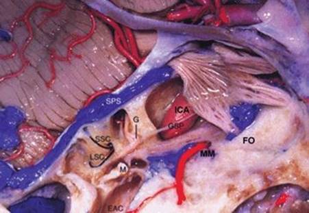
Figure 32.1 Anatomical dissection of the right side. The tentorium and dura covering the middle cranial fossa have been removed revealing the superior petrosal sinus (SPS). The bone has been drilled away to reveal the internal auditory canal, the superior and lateral semicircular canals (LSC, SSC), the middle (M) and external ear (EAC), the horizontal petrous ICA (ICA), and the middle meningeal artery (MM). (FO, foramen ovale; G, geniculate ganglion.)
HISTORY
Since many of these tumors are slow growing, there is paucity of signs and symptoms in most patients. Headache may be the only problem they are experiencing. Cranial nerve dysfunction of the 3rd, 4th, 5th and 6th nerves can produce diplopia, facial pain, and facial numbness. Compression of the brain stem can lead to difficulty with balance and incoordination. Contralateral weakness or hemiparesis can be seen in large tumors.
PHYSICAL EXAMINATION
The location and size of a tumor can be inferred from the physical examination. Common findings are deficits of cranial nerves. The physical examination should include a complete ophthalmologic assessment including visual acuity and ocular motility. A palsy of the 6th (abducens) cranial nerve is most frequent, probably a reflection of its long course. The 6th cranial nerve may be involved at the brain stem, at the petrous apex (Dorello’s canal), or in the superior orbital fissure. Deficits of multiple cranial nerves (III, IV, and VI) suggest tumor involvement of the superior orbital fissure. Evaluation of trigeminal nerve function includes an assessment of sensory and motor function of all three branches. Decreased motor function of the third division is evidenced by decreased muscle contraction by palpation, facial asymmetry due to muscle wasting, and drift of the mandible with opening the mouth. Involvement of multiple branches suggests tumor involvement of Meckel’s cave or the proximal nerve trunk.
INDICATIONS
This approach is useful for a variety of tumors that can be found here that include intradural tumors such as meningioma, trigeminal schwannoma, and epidermoid tumors. Extradural tumors that may also have intradural extension include chordoma, chondrosarcoma, and cholesterol granuloma. The approach allows access to a very specific area of the skull base, and the surgeon should carefully evaluate the preoperative imaging studies to ascertain the adequacy of the approach to the lesion at hand. It is suited for tumors in the middle cranial fossa that spill over the petrous apex and upper clivus into the posterior fossa, usually on the ipsilateral side of the midline. The inferior most reach of the approach is the level of the internal auditory canal.
CONTRAINDICATIONS
There are no absolute contraindications. A relative contraindication is a lesion that has eroded through the clival bone and has a significant component in the sphenoid sinus. Dural repair after removing such a tumor will be quite difficult, resulting in a high risk for developing a cerebrospinal fluid fistula.
PREOPERATIVE PLANNING
MRI scans before and after administration of gadolinium are the essential diagnostic as well as surgical planning tool. Thin-slice scans of the posterior fossa in the axial and coronal planes are performed and also a CISS (constructive interference in steady state) sequence that allows better visualization of the relationship of the tumor to the surrounding neurovascular structures. Meningiomas are dural based while schwannomas are found along the course of the cranial nerves, most commonly the trigeminal nerve. Both of them enhance vividly with gadolinium. Epidermoids do not enhance with contrast and may be difficult to differentiate from cerebrospinal fluid signal. They are best seen in the diffusion-weighted sequence of the MRI. The marrow signal inside the petrous bone and clivus may be altered in chordomas and chondrosarcomas. These tumors have a mixed signal characteristic but are mostly hyperintense on T2-weighted images. They enhance to a variable extent with contrast. CT scan performed in thin axial slices provides information regarding the relationship of the tumor to the bony anatomy at the skull base. Irregular bone destruction is the hallmark of chordomas and chondrosarcomas, whereas schwannomas will produce smooth bone remodeling along the course of the nerve.
SURGICAL TECHNIQUE
Anesthesia, Intraoperative Neurophysiologic Monitoring and Positioning
The patient is operated under general anesthesia. Sequential compression stockings are applied to the lower extremities for prevention of deep vein thrombosis, and the patient is given steroids and broad-spectrum antibiotics. Intraoperative neurophysiology is used in the majority of instances in order to monitor cranial nerve and brain stem functions. The electrodes are applied for continuous somatosensory and transcranial motor evoked potential monitoring. Since the geniculate ganglion of the facial nerve is in the path of the approach, direct facial nerve stimulation as well as transcranial facial nerve stimulation and responses are monitored. Thus, muscle relaxation is avoided during these procedures.
The patient lies supine, and the head is positioned rotated 60 degrees to the side opposite the tumor and the vertex is turned slightly down to the floor and fixed in a three-point pin fixation (Fig. 32.2A and B). The body of the patient is adequately padded and secured with multiple straps to the operating table. This is important because the table is often rotated side to side during the operation to facilitate the exposure and visualization.

FIGURE 2.12 A and B.Position for surgery. The head is rotated 60 degrees to the side opposite to the side of the tumor, and the vertex is dropped slightly and stabilized in a three-point pin fixation.
Incision and Soft Tissue Exposures and Craniotomy
A question mark incision is made starting immediately anterior to the external auditory canal, curving upward, above the ear and towards the hairline (Fig. 32.3). The scalp flap is raised using a subfascial dissection plane (to protect the frontalis branch of the facial nerve) above the temporalis muscle, exposing the zygomatic arch and lateral rim of the orbit. The temporalis muscle is elevated from the side of the head. Removal of the zygomatic arch allows the temporalis muscle to be fully depressed below the working angle of the surgeon. The zygomatic osteotomy is made by making a cut with the reciprocating saw at the root of the zygoma, staying anterior to the temporomandibular joint and another one at the junction of the zygoma with the lateral rim of the orbit. The zygomatic bone segment is left attached to the temporalis and the masseter muscles and the entire complex is retracted inferiorly. Two burr holes are now made: One of them is located at the “keyhole” (behind the frontozygomatic suture, where the burr hole would straddle the floor of the frontal fossa and the roof of the orbit), and the other one is at the root of the zygoma above the external auditory canal. A frontotemporal bone flap is now raised with a high-speed drill.
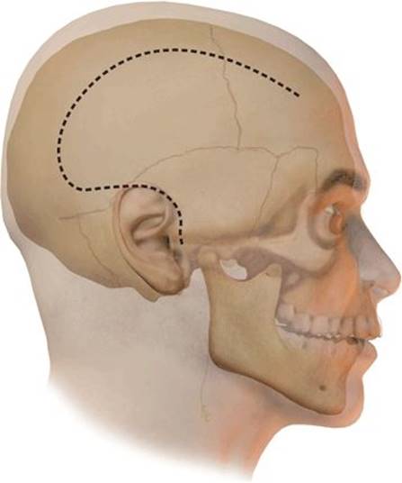
Figure 32.3 The incision is shown by the dotted line. The posterior extension of the incision above the ear allows the surgeon to have visualization along the petrous ridge. The sphenoid ridge is the anterior limit.
Extradural Dissection and Elevation of the Temporal Lobe
A substantial amount of extradural dissection is performed to elevate the temporal lobe from the sphenoid wing anteriorly to the petrous ridge posteriorly (Fig. 32.4A). It is important for the brain to be relaxed with either a lumbar spinal drain that is placed after the patient is anesthetized or administration of mannitol, in order to facilitate this dissection. The floor of the middle cranial fossa is drilled down along with the sphenoid wing to provide a flat and low approach. The dura is elevated systematically starting posteriorly over the petrous ridge of the temporal bone and then progressing anteriorly. The posterior petrous ridge and the arcuate eminence are identified. Immediately anterior to the arcuate eminence is the recess in the bone through which the greater superficial petrosal nerve (GSPN) emerges after originating from the geniculate ganglion of the facial nerve. The foramen spinosum and foramen ovale are seen transmitting the middle meningeal artery and the mandibular branch of the trigeminal nerve respectively. Further anteriorly the foramen rotundum is seen, through which the maxillary branch travels to the pterygopalatine fossa.

FIGURE .32.4 A. The temporal lobe is elevated extradurally, and the middle meningeal artery is exposed in the foramen spinosum, coagulated, and divided. The GSPN has also been exposed and divided to facilitate identification of the horizontal segment of the petrous ICA. In most situations, it is not necessary to divide the nerve. It can be preserved by leaving the outer layer of the temporal dura along with the nerve and elevating the temporal lobe without putting traction on the nerve. (V2, foramen rotundum; V3, foramen ovale; MM, middle meningeal artery; ICA, horizontal petrous internal carotid artery.) B. The outer layer of the dura that forms the lateral wall of the Meckel’s cave has been peeled away exposing the trigeminal ganglion and the three divisions. The dura posteriorly is incised to follow the trigeminal root. The petrous apex medial to the petrous ICA has been drilled away to expose the posterior fossa dura (arrowheads). (GG, gasserian ganglion.)
Using a diamond burr on a high-speed drill, the foramen spinosum, ovale, and rotundum are unroofed. The middle meningeal artery is coagulated and divided. Under high magnification, the GSPN is followed anteriorly along its course as it travels between the two layers of the dura under the temporal lobe. The periosteal layer of dura is sharply incised in a direction parallel to the course of the nerve. This allows the temporal lobe dura to be elevated leaving the nerve traveling along the bony floor medial and deep to the foramen ovale. Thus, traction on the nerve, which can produce facial paralysis, is avoided. The GSPN closely follows the course of the horizontal segment of the petrous ICA. The artery is exposed by unroofing it with a small (3 mm) diamond drill. If only a limited exposure of the ICA is needed, the GSPN can be left in place and the drilling is done just lateral to it. If the vessel needs to be fully unroofed, the GSPN is divided at a distance from the geniculate ganglion to avoid traction. The bony eustachian tube is located immediately lateral to the artery, separated from the vessel with a very thin layer of bone. While drilling lateral to the ICA, one has to be careful not to open into the eustachian tube. The tensor tympani muscle travels on the superior surface of the eustachian tube and is usually exposed first before opening into the actual eustachian tube. Inadvertently opening this with the drill should be avoided; otherwise, a cerebrospinal fluid fistula can result. It must be recognized and dealt with by opening it sufficiently and packing it thoroughly with a plug of muscle.
Further dissection varies according to the nature and location of the tumor, whether extra- or intradural.
Extradural Tumors
The usual extradural tumors are chondrosarcomas, chordomas, and cholesterol granulomas. The superoinferior extension as well as the medial–lateral extension of the tumor must be carefully assessed on the preoperative images (CT and MRI). The soft tissue extension, the area of bone destruction, and the relation to the foramina at the skull base and the petrous carotid artery are noted. Based on these assessments, the area and size of the exposure at the skull base is determined. The horizontal segment of the petrous ICA is unroofed using a diamond drill after dividing the GSPN as described above. The artery is unroofed from the posterior genu of the petrous segment up to the precavernous segment as it turns upward deep to the mandibular nerve. An incision is carefully made in the subtemporal dura immediately lateral to the point of exit of the mandibular and maxillary divisions under the temporal lobe. The temporal dura is bluntly dissected away from the lateral surface of the trigeminal ganglion, thus exposing the trigeminal ganglion and the three branches arising from it (Fig. 32.4B). The ganglion is covered by a thin arachnoid layer. This dural incision is extended posteriorly to expose a small portion of the trigeminal root behind the ganglion. Prior to drilling the petrous apex, it is important to be aware of the location of the cochlea, geniculate ganglion, and the internal auditory canal (Fig. 32.1). The geniculate ganglion is immediately posterior to the facial hiatus (emergence of the GSPN). There is a thin layer of bone that separates the ganglion from the subtemporal dura, and on occasion, it is not covered by any bone. The cochlea is located just posterior to the genu of the ICA, where the vertical segment of the petrous ICA turns into the horizontal segment. The internal auditory canal is located along a line bisecting the angle sustained by the junction of the arcuate eminence and the course of the GSPN. The bone medial to the horizontal petrous ICA and posterior to the trigeminal ganglion is gradually drilled down with a diamond burr. This bone is drilled in a quadrangular area with the petrous ridge behind, the trigeminal root superiorly, the trigeminal ganglion anteriorly, and the horizontal petrous ICA laterally. There may be some air cells that are unroofed in the process. The dura in front of the brain stem is thus exposed. The bone drilling can be carried out laterally in the temporal bone, to unroof the internal auditory canal. This marks the posterior limit of the exposure. Bone drilling in the vicinity of these structures needs to be done carefully and with neurophysiologic monitoring of the facial nerve.
Tumor Removal
Chondrosarcomas and chordomas are extradural tumors, and are usually encountered by the time the petrous apex is being drilled (Fig. 32.5A and B). These tumors are usually gray and gelatinous and soft with areas of calcification. Small ring curettes and suction are used to remove the tumor in a systematic manner clearing out all extensions of the tumor. The carotid artery is carefully protected while drilling out additional bone, until normal bone is encountered. Since the majority of these tumors are extradural, the posterior fossa dura in front of the brain stem and the dura inferior to the trigeminal ganglion and root are the limits of the tumor resection. If the approach is used for a cholesterol granuloma, the cyst wall is usually visible and the cyst contents can be drained. The cyst wall is then carefully dissected free from the surrounding bone and removed in a piecemeal manner.
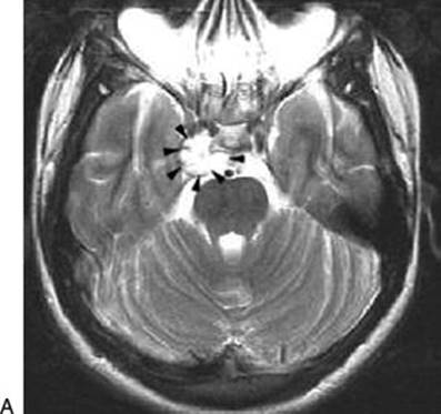
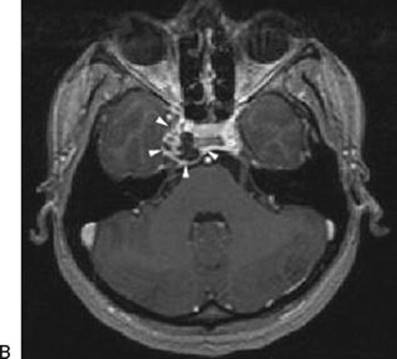
FIGURE 32.5 A. Axial T2-weighted image of a low-grade chondrosarcoma of the right petrous apex that has a high signal intensity in this sequence (arrowheads). B. Axial MRI T1 with contrast shows that the tumor has irregular enhancement with areas of low intensity corresponding to calcifications (arrowheads).
Intradural Tumors
Intradural tumors located in the middle cranial fossa, the anterior portion of the tentorium and incisura, and the anterolateral portion of the clivus (ipsilateral to the side of the exposure) are accessible by this approach. These include meningiomas (Fig. 32.6A and B; Fig. 32.7A and B), trigeminal nerve schwannomas (Fig. 32.8A and B), and epidermoid tumors. Although an excellent exposure of the middle cranial fossa is achieved by this approach, the posterior fossa exposure is limited to a specific anatomical region. This region is defined by the bony window into the posterior fossa: superiorly, the posterior clinoid process and inferiorly, the internal auditory canal and the horizontal segment of the petrous ICA. This area can also be accessed through a retrosigmoid approach or a presigmoid retrolabyrinthine approach, but the main advantage of using the anterior transpetrosal approach is that it allows the surgeon a direct route to the lesion and to work anterior to the VII and VIII cranial nerves, instead of across them.
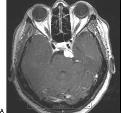
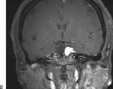
FIGURE 32.6 A and B. Axial MRI scan with gadolinium showing a meningioma in the posterior fossa located medial to the trigeminal root. B. The vertical extent is shown on the coronal images. The inferior extent of the tumor is above the level of the internal auditory canal (arrowhead).
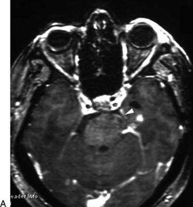

FIGURE 32.7 A. Axial MRI with gadolinium after resection of the meningioma in Figure 32.6 (arrowheads). B. T2-weighted image showing the temporal lobe after surgery with minimal retraction changes.
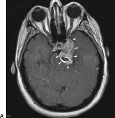
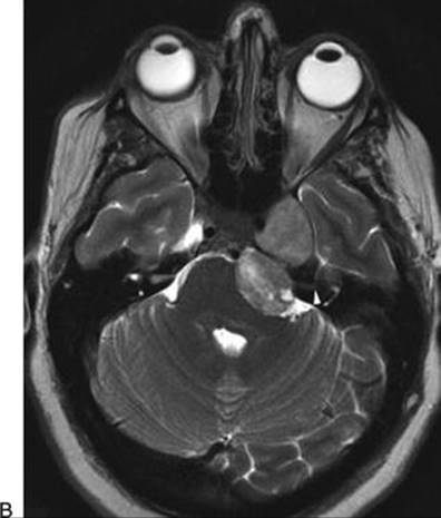
FIGURE 32.8 A. Axial MRI scan with gadolinium as well as a T2-weighted image showing a dumbbell-shaped trigeminal schwannoma occupying Meckel’s cave and extending into the posterior fossa. Since the petrous apex is partially eroded by the tumor (arrowheads), it is ideally approached by the anterior transpetrosal approach. B. The T2-weighted image shows that the tumor extends down to the level of the internal auditory canal (arrowhead).
The temporal dura is elevated from the petrous ridge posteriorly to the lesser wing of sphenoid, anteriorly. The GSPN is isolated and divided to allow further elevation of the subtemporal dura. Meckel’s cave and the trigeminal ganglion are exposed by incising the dura lateral to the maxillary and mandibular divisions and peeling away the dura of the lateral wall (Fig. 32.9A and B). The horizontal petrous ICA is exposed from its genu posteriorly to its precavernous segment under the trigeminal ganglion. The bone medial to the petrous ICA is drilled away with a diamond burr (3 or 4 mm) thus exposing the dura ventral to the brain stem and inferior to the trigeminal root (Fig. 32.9C). The posterior and inferior limit of the bone drilling is the internal auditory canal. The subtemporal dura is incised posteriorly along the trigeminal root until the superior petrosal sinus. The superior petrosal sinus runs at the junction of the subtemporal dura with the prepontine dura and marks the attachment of the tentorium. The incision of the subtemporal dura is carried posteriorly parallel to and above the superior petrosal sinus, and another incision is made below the superior petrosal sinus in the dura ventral to the brain stem. The sinus is then thoroughly coagulated with the bipolar and divided. The temporal lobe is elevated, and the tentorium is incised to the incisura (Fig. 32.10A and B). The prepontine dura is incised inferiorly as far as the bony opening permits, to visualize the caudal pole of the tumor.
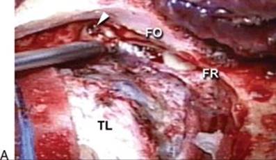
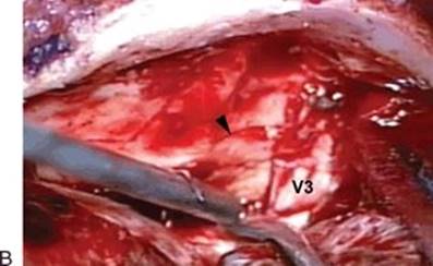
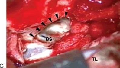
FIGURE 32.9 A. Intraoperative photo of meningioma shown in Figure 32.6. The patient’s vertex is downward, and the nose is to the reader’s right. Left-sided extradural subtemporal dissection shows that the middle meningeal artery has been divided at the foramen spinosum (arrowhead). Foramen ovale (FO) and foramen rotundum (FR) are exposed where the V3 and V2 trigeminal branches are exiting. (TL, temporal lobe dura.) B. The dura of the lateral wall of Meckel’s cave has been incised and peeled away showing the nerve fascicles of V3. The horizontal segment of the petrous ICA is exposed (arrowhead). C. The left petrous apex has been drilled medial to the petrous ICA (arrowheads). The dura anterior to the brain stem has been exposed (BS).
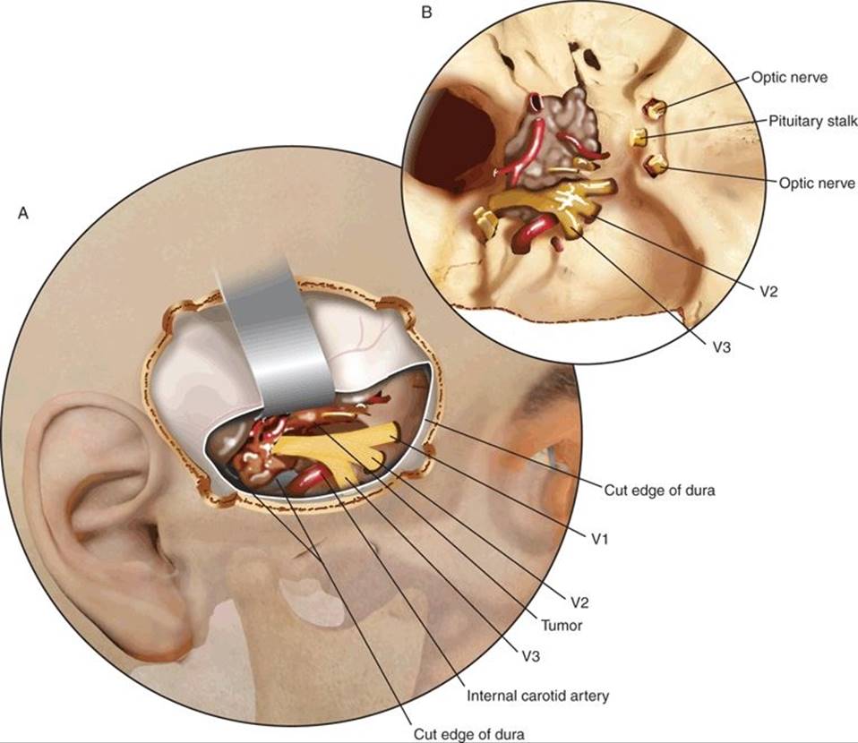
FIGURE 32.10 A. Tumor (T) is exposed in this area after the tentorium has been incised (arrowheads). B. An axial diagram shows the location of the tumor relative to the surgical approach and its relation to the surrounding structures. The lower limit of the tumor that is accessible by this approach is the level of the internal auditory canal (arrowhead). (V2, maxillary nerve; V3, mandibular nerve.)
The tumor is visualized and progressively debulked. It is then dissected free from the surrounding cranial nerves, brain stem, and vascular structures (Fig. 32.11A and B).

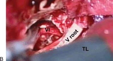
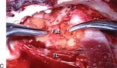
FIGURE 32.11 A.Intraoperative image of the case of the meningioma shown in Figure 32.6. Vertex of the patient is downward and the nose is to the right. After dural opening and division of the tentorium, the entire trigeminal root (V root) has been exposed up to the brain stem and the tumor (Tu) medial to it is seen. B. After removal of the tumor, the 6th nerve (VI) is seen ventral to the brain stem and the 5th nerve (V root) root is seen above it. (TL, left temporal lobe.) C. For closure of the defect in the posterior fossa dura, a dural substitute is laid over the defect and fat graft (Fat) is laid on top of it. Fibrin glue is poured around the edges. (TL, temporal lobe.)
Closure and Reconstruction
The floor of the middle cranial fossa is carefully inspected under the microscope for any breaches into air cells. These are carefully occluded with bone wax. Dural closure is achieved by placing a piece of dural substitute over the dural defect and is secured to the surrounding dura with a few sutures in order to prevent it from migrating. Fibrin glue is used along the edges to further secure it. A small adipose tissue graft harvested from the abdomen is placed on top of the dural repair to bolster it (Fig. 32.11C). The bone flap is secured with miniplates. If there is a substantial gap in the bone at the inferior edge of the craniotomy where the bone was drilled away, a small cranioplasty is performed with titanium mesh to minimize any unsightly indentations. The temporalis muscle is secured back into place by resuspending it with sutures passed through small drill holes in the bone. The zygomatic arch is secured with miniplates, and a subgaleal drain is left in place; brought out through a separate stab wound. This is not connected to a suction bulb but is instead attached to a gravity drainage bag. The drain is used routinely because there is always a subcutaneous CSF collection that accumulates and by providing a temporary drainage system, it allows the scalp and the muscle to stick down and minimizes the possibility of a pseudomeningocele formation.
POSTOPERATIVE MANAGEMENT
The patient is maintained in the neurosurgical intensive care unit to watch for delayed temporal lobe swelling or bleeding. Since seizures can occur from temporal lobe retraction or contusion, the patients are routinely placed on anticonvulsant prophylaxis. A CT scan without contrast is performed the day after the operation to check for hematoma and brain edema.
COMPLICATIONS
1. Cranial nerve problems: The cranial nerves that are exposed to risk are the 3rd to the 6th on the side of the approach. Although the inferior limit of the approach is at the level of the internal auditory canal, if there is any significant level of 7th and 8th cranial nerve involvement or extension into the internal auditory canal, this is not a preferred approach for such a tumor. The approach is performed by elevating the dura covering the middle fossa floor and dividing the tentorium posteriorly. This allows the cranial nerves to be exposed from their origin at the brain stem to the cavernous sinus and parasellar region. Since the trigeminal nerve is entirely within the field of the exposure, it can be easily traumatized. Impairment of the trigeminal nerve is especially important because of the corneal anesthesia that can result and jeopardize the eye. Careful vigilance is required to detect and treat trophic ulcers.
2. Brain stem problems: Preoperative evaluation of the MRI scan is important in anticipating potential brain stem problems. When the T2-weighted and FLAIR sequences show brain stem edema, it is a poor prognosticator. It indicates that the tumor has violated the arachnoid and pial membranes and will have no clear plane to allow separation of the tumor from the brain stem. There is a high potential for injuring the brain stem in such a situation, and it is usually advisable to leave a small residue of tumor against the brain stem to avoid the devastating complication of a brain stem injury.
3. Arterial injury: These tumors often are intimately related to the basilar artery and its branches. If more than 180 degrees of the circumference of the vessel is surrounded by the tumor, there is a substantial risk of injuring the main vessel or its perforating branches if the tumor is found to be adherent and aggressive attempts are made at separating it from the vessel. The usual mechanism of injury is from avulsion of perforators at the origin from the main vessel. The bleeding can be controlled by accurately coagulating the site with a bipolar or sometimes may require placing a suture such as 8-0 nylon. The most severe consequence of an arterial or perforator injury leading to a brain stem stroke is devastating and is best avoided by meticulous technique and careful surgical judgment.
4. Temporal lobe problems: Excessive and prolonged retraction is the common reason for temporal lobe contusion and edema, and occasionally a hematoma can result. Aggressive drilling of the middle fossa floor during the opening allows the surgeon to angle the microscope to visualize the top of the tumor instead of retracting the temporal lobe. When the portion of the tumor that is in the posterior fossa is being accessed, cutting the tentorium and drilling the petrous apex are important maneuvers to minimize temporal lobe retraction.
RESULTS
The approach provides excellent access to selected tumors in this area. Although complete tumor removal is the goal, the degree of resection is usually determined by the biologic behavior of the tumor such as its invasiveness into neurovascular structures. Temporary cranial nerve palsies can result from their manipulation, as described below.
PEARLS
· The preoperative imaging studies must be carefully evaluated to determine if the anatomical access provided by the approach is adequate.
· The FLAIR and T2 sequence must be checked to see if there is hyperintense signal in the brain stem indicating pial invasion. This indicates that the tumor will not be safely separable from the brain stem.
PITFALLS
· While drilling the bone of the petrous apex, the air cells must be carefully waxed to prevent cerebrospinal fluid fistula formation.
· During the elevation of the temporal lobe from the floor of the middle cranial fossa, care must be taken not to injure the geniculate ganglion that may be devoid of bony cover.
· Traction on the GSPN must be avoided to prevent facial palsy.
INSTRUMENTS TO HAVE AVAILABLE
· High-speed drill with assorted cutting and diamond burrs with irrigation
· Microsurgical instruments
· Nerve integrity monitor for facial nerve stimulation
SUGGESTED READING
Kawase T, Shiobara R, Toya S. Middle fossa transpetrosal transtentorial approaches for petroclival meningiomas. Selective pyramid resection and radicality. Acta Neurochir (Wien) 1994;129:113–120.
Ichimura S, Kawase T, Onozuka S, et al. Four subtypes of petroclival meningiomas: differences in symptoms and operative findings using the anterior transpetrosal approach. Acta Neurochir (Wien) 2008;150:637–645.
Muto J, Kawase T, Yoshida K. Meckel’s cave tumors: relation to the meninges and minimally invasive approaches for surgery: anatomic and clinical studies. Neurosurgery 2010;67(Suppl 1):ONS291–299.