
Ziv Gil
INTRODUCTION
The infratemporal fossa (ITF) is a deep space located below the lateral skull base. It is bordered anteriorly by the pterygomaxillary space, posteriorly by the carotid space, inferiorly by the masticatory space, and medially by the parapharyngeal space (Fig. 31.1). The medial pterygoid muscle attached to the mandible delineates its inferior border. Its other boundaries are anterior—infratemporal surface of the maxilla and the ridge that descends from its zygomatic process; posterior—temporal bone (articular tubercle) and the sphenoid bone (spinal angularis); medial—the lateral pterygoid plate, and lateral—ramus of mandible and the zygomatic arch. The ITF contains many important anatomical structures including the third division of the trigeminal nerve (transmitted through the foramen ovale), the middle meningeal artery (transmitted through the foramen spinosum), the internal maxillary artery, and the pterygoid venous plexus. Other nerves located in the ITF are the lingual and buccal nerves, the chorda tympani, and the otic ganglion. The temporalis muscle and the lateral and medial pterygoid muscles are also contained within the ITF.


Figure 31.1 Anatomical description of the right ITF. A. Lateral view of the skull. B. Anterolateral view of the soft tissue structures.
Less than 1% of all head and neck neoplasms originate in the ITF. These can be either primary tumors or secondary tumors that invade or metastasize to the ITF. Primary ITF tumors can originate from any tissue within this compartment: nerve, vessels, muscle, bone, cartilage or connective tissue. These tumors may go undetected for a long period of time, after which they usually present as a symptomatic mass in the temporal area. Primary benign tumors that may involve the ITF include meningioma, juvenile angiofibroma, neurofibroma, schwannoma, fibroma, osteoma, and fibrous dysplasia. Malignant tumors include chordoma, soft tissue sarcomas, nasopharyngeal carcinoma, and sinonasal carcinomas. Tumors that may invade this space include carcinomas of the skin, sinonasal mucosa, or minor salivary glands.
The complex anatomy of the ITF accounts for the diversity of surgical approaches used to remove tumors from this anatomic area. The surgical approach is tailored according to the anatomical extent of the tumor, its histologic type (benign or malignant), and the patient’s past medical history (previous surgery or radiation treatment).
Large tumors that involve the ITF can be approached extracranially and intracranially depending on the primary origin of the tumor and its extensions. The contemporary surgical technique for resection of tumors arising in the ITF is based on the classical approach developed by Fisch and Mattox that involved a retroauricular incision and transposition of the entire facial nerve. The classical type-C Fisch ITF approach provides access from the sigmoid sinus to the parasellar region, cavernous sinus, and Meckel’s cave. The main limitation of this approach is that it imminently leads to conductive hearing loss and facial nerve neuropraxia, which are rarely encountered in the modern approach. Other variations of the Fisch approach are the lateral facial and lateral transtemporal sphenoid approaches, which allow limited superior access to the middle cranial fossa. The lateral facial approach provides access to small tumors of the middle fossa and is limited by the inferior displacement of the zygomatic arch and temporalis muscle (Fig. 31.2). Removal of the zygomatic arch in the lateral temporal sphenoid approach allows wider exposure of the subtemporal region and allows intracranial and extracranial removal of the tumor without rerouting the facial nerve.


Figure 31.2 Lateral facial approach to the cranial base. This approach allows limited access to the subtemporal area without removal of the zygoma. A. A question mark incision is used to expose the temporalis muscle and the lateral orbit (ribbon retractor). B. A pterional craniotomy is performed. Retraction of the temporal lobe dura reveals branches of the trigeminal nerve and attachment of the pterygoid plate at the base of the sphenoid bone.
The more common subtemporal–preauricular infratemporal fossa approach developed by Sekhar, Janecka, and Schramm is a combination of the lateral transtemporal sphenoid approach and the transparotid approach to the parapharyngeal space, in addition to temporal craniotomy (Fig. 31.3). It is also known as the lateral transtemporal infratemporal fossa approach. This approach affords wide exposure to the structures of the middle cranial base using a combination of transparotid, lateral transtemporal sphenoid, and temporal craniotomy approaches. This approach allows access to various anatomical areas including the temporal and infratemporal fossa, temporal lobe, parasellar region, retro-orbital region, nasopharynx, retromaxillary space, pterygopalatine space, masticatory space, retromaxillary fissure and pterygoid plates, sphenoid ridge, and trigeminal nerve. The main advantages of this technique are excellent exposure of the middle cranial fossa and infratemporal compartments and minimal sensory–motor morbidity. This chapter describes the technique of the preauricular ITF approach and its combination with various other important techniques used in surgery of the cranial base.
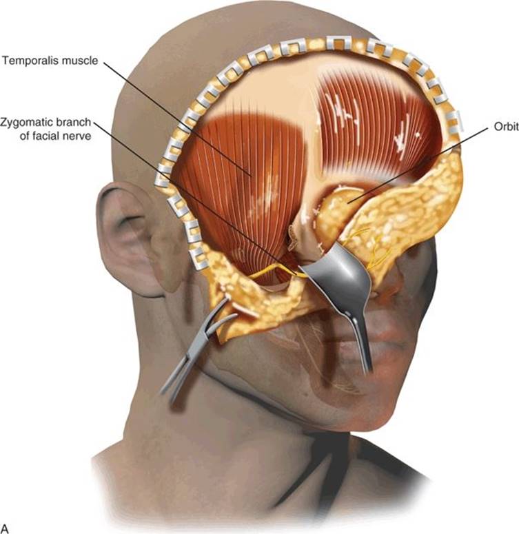
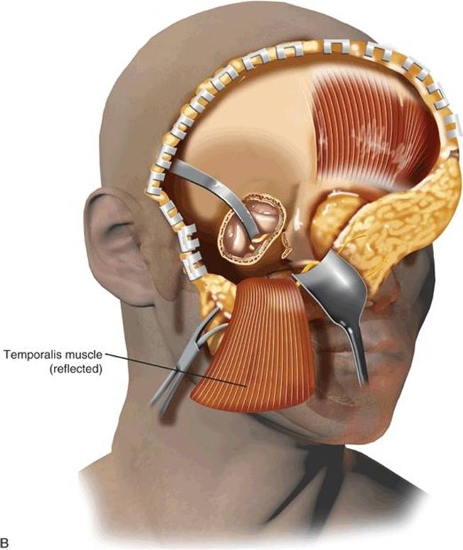
Figure 31.3 Subtemporal–preauricular and infratemporal approach to the cranial base as described by Sekhar and Janecka. A. Initial exposure is similar to the lateral facial approach shown in Figure 31.2. The caudal exposure is extended by removal of the zygomatic arch. Figure 31.3(Continued ) B. The temporalis muscle has been reflected inferiorly after removing the zygomatic arch, the mandibular condyle has been resected, and a subtemporal craniectomy has been performed.
HISTORY
The clinical presentation of ITF tumors varies greatly and relates directly to the location and rate of growth of the lesion. A thorough history of the present illness as well as past medical and surgical histories are obtained. In addition to the relevant risk factors, the patient is questioned regarding symptoms of cranial nerve dysfunction, head and ear aches, trismus, bleeding from the ear, and otorrhea. Signs and symptoms can often be misleading and may initially be interpreted as being infectious and benign diseases. Common symptoms and signs suggestive of tumors of the ITF include the following: (1) facial swelling and asymmetry that can result from tissue destruction and advancement of the tumor into the soft tissues of the head and neck; (2) palpable metastatic adenopathy, a possible indication of advanced disease; (3) hearing loss usually resulting from extension of the tumor and obstruction of the eustachian tube or middle or inner ear, (4) numbness of the face or pain as a manifestation of tumor invasion into various branches of the trigeminal nerve; and (5) neurologic or ocular manifestations, such as diplopia, exophthalmos, and multiple cranial nerve palsies, that suggest invasion of the nerves at the skull base or cavernous sinus. Pain is a significant adverse symptom suggestive of a malignant tumor. The classical triad of signs of cancer of the temporal bone includes otorrhea, pain, and bleeding. The usual comorbidities should be identified in addition to mental disorders and active alcohol, drug, or tobacco abuse. In addition to the above criteria, potential candidates must be well-informed about surgery, must be motivated, and should be prepared to accept long-term postsurgical follow-up.
PHYSICAL EXAMINATION
Physical examination should include a meticulous survey of the temporal area and auricular and preauricular skin. The neck and parotid gland are palpated in search of lymph node metastases. The function of the facial and other cranial nerves is evaluated. Otoscopy is used to define the extent of the tumor in the external auditory canal. If the tympanic membrane can be seen, its integrity is verified. Jaw movement and trismus are examined as a potential sign of extension of the tumor into the temporomandibular joint (TMJ). Cranial nerve function is evaluated.
The comprehensive physical examination should always include an endoscopic evaluation of the nose, sinuses, nasopharynx, and oropharynx. Masses should be examined for friability, vascularity, bleeding, and signs of necrosis, possibly suggesting malignancy and necessitating additional investigation. Different entities may display specific and pathognomonic findings on physical examination, for example a vascular mass arising from the hypotympanum on otoscopic examination. However, most tumors are similar in their initial and late symptoms and demand a high level of suspicion in order to reach the correct final diagnosis. This type of tumor will silently advance in size and extent until it has infiltrated a cranial nerve or grown sufficiently to obstruct the nasopharynx. The most common findings on physical examination include the following: (1) nasal, paranasal, or nasopharyngeal mass. (2) Proptosis, the mild protrusion of the eye, may be consistent with tumor compression of the periorbita without frank invasion. (3) Cranial nerve deficits are always considered to be indicators of advanced disease and a poor prognosis. For example, invasion of the cavernous sinus by cancer, as can be suspected from signs of involvement of the abducens nerve, can be an absolute contraindication for surgery. (4) Any abnormal findings on the neurologic examination should raise the suspicion of involvement of the dura and brain. (5) A suspicious mass in the neck suggests the presence of a malignant tumor that has already undergone regional lymph node metastasis. (6) Unilateral middle ear effusion is an uncommon finding that mandates a thorough examination of the nasopharynx to rule out eustachian tube obstruction. Fiberoptic evaluation of the upper aerodigestive tract is an integral part of the physical examination. Sites of potential donor tissue to be used for reconstruction are examined.
INDICATIONS
Indications for surgery include extirpation of benign and malignant tumors that originate in the ITF or that invade this compartment from neighboring structures. Rarely, this approach is performed for treatment of infectious diseases including local abscess or necrotizing fasciitis. Tailored treatment should take into consideration the fact that survival after recurrent or residual disease, as well as prior radiotherapy, is lower compared to primary treatment, and that the main cause of death is local recurrence rather than regional or distant metastases. For these reasons, an aggressive effort toward complete extirpation should be made whenever cure is the goal of the treatment or for palliation of intractable pain. Cosmesis and function are addressed by an adequate reconstruction plan.
When contemplating treatment of a specific patient, the following key questions need to be addressed: What is the goal of the treatment? What is the appropriate extent and nature of surgical resection? Is postoperative adjuvant therapy with radiation or chemoradiation anticipated? And finally, what reconstruction techniques will be employed?
CONTRAINDICATIONS
Contraindications for surgery include the diagnosis of tumors amenable to chemoradiation therapy (lymphoma, carcinomas of oropharyngeal or nasopharyngeal origin) and evidence of distant metastases. An exception is adenoid cystic carcinoma or single metastases of specific tumors (e.g., melanoma and sarcomas) that are amenable to excision. Cancer invasion of the prevertebral fascia, encasement of the carotid artery, invasion of the cavernous sinus, and considerable brain involvement are also considered contraindications for surgery. Stable small paragangliomas or schwannomas can be followed radiographically with magnetic resonance imaging (MRI). Severe comorbidities, marked debilitated status, or demented patients are considered to be potential contraindications for surgery.
PREOPERATIVE PLANNING
Imaging Studies
Imaging should always be used prior to surgery since it can influence decision making. A contrast computerized tomographic (CT) scan and a basic MRI study with fat suppression should be used for preoperative evaluation and decision making regarding treatment. A T2-weighted MRI study with fat suppression is usually added (Fig. 31.4). Visualization of a vascular flow void on an MRI study is usually sufficient for the diagnosis of a vascular tumor such as a paraganglioma, but magnetic resonance angiography may be added for a more precise diagnosis. Malignant tumors usually demonstrate a soft tissue mass invading intracranial or adjacent spaces, accompanied by bone destruction. The CT may show mild enhancement, and T1-weighted MRI may demonstrate strong postcontrast enhancement. Application of a fat suppression technique, such as short tau inversion recovery and frequency-selected fat suppression, eliminates strong signals from adipose tissue. In addition, the definition of normal anatomic structures is significantly improved, enhancing lesions become clearer, and the margins of a lesion are better defined when suppression of fat is used in combination with contrast enhancement. MRI 3D reconstruction enables multiplanar imaging of tumor extension and vessel encasement. These features can be established in tumors as small as 10 mm in diameter. If a malignant tumor is suspected, radiologic staging of the patients is completed by using a positron emission tomography–CT hybrid for assessing the presence of regional and distant metastases. Most of the malignant tumors arising in the ITF can invade the intracranial compartment, maxilla, mandible or TMJ, orbit and auditory canal, or skin. Other routes of spread include hematogenous, lymphatic, or perineural invasion along cranial nerves. Perineural invasion is common in adenoid cystic carcinoma and is the pathway to the intracranial space so that imaging studies should be directed toward the foramen ovale and rotundum, the cavernous sinus, orbital apex, trigeminal ganglion, and dura. In addition, the radiologic evaluation must include the neck in any case of suspected malignancy to evaluate for lymph node metastasis. Patients with suspected paragangliomas will require preoperative embolization a few days prior to surgery.
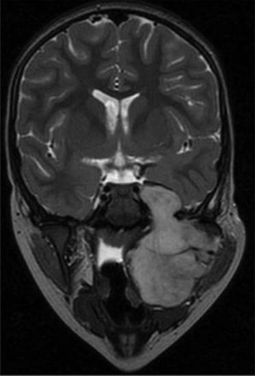
Figure 31.4 Magnetic resonance imaging of the ITF. A coronal T2 MR image of a child with a large rhabdomyosarcoma of the ITF.
Tissue Diagnosis
Tissue diagnosis should be an integral part of preoperative evaluation. Although radiologic imaging could provide indications regarding the type of neoplasm, tissue diagnosis is sensitive to differences between benign and malignant lesions. Fine needle aspiration (FNA) biopsy can be performed via ultrasound but frequently will require CT-guided FNA, which is technically challenging. Radiologic imaging should precede FNA if a vascular tumor is suspected to avoid potential bleeding. Some exceptions to preoperative biopsy include juvenile angiofibroma and paragangliomas.
SURGICAL TECHNIQUE
The patient is placed in a supine position without shaving the hair at the surgical site. Tracheostomy usually is not performed. A lumbar spine catheter is inserted for a period of 3 days for CSF drainage when dural resection is expected, to reduce the risk of postoperative CSF leak. I do not recommend routine administration of prophylactic antibiotics in clean operations. First-generation cephalosporins and metronidazole are routinely used if the operation includes dissection in the oral cavity or pharynx. Surgery is performed under general anesthesia without muscle relaxants. This allows for monitoring of the spinal accessory, hypoglossal, phrenic, and marginal mandibular nerves and brachial plexus, during dissection with electrocautery. Monitoring of facial nerve integrity is used when preservation of the nerve is a goal.
Various surgical approaches for the resection of ITF tumors are used. In this chapter, I describe the preauricular approach that is the most acceptable contemporary technique for resection of neoplasms of the ITF.
Description of the Technique
After the induction of anesthesia, an oral endotracheal tube is inserted and secured contralateral to the surgical site. The patient is prepped and draped with the hemiface and calvarium exposed. Ointment is placed in both eyes, and the eyelids contralateral to the surgical site are taped shut. The patient’s hair is shampooed vigorously with 4% w/v chlorhexidine (Septal Scrub), parted with a sterile comb along the proposed incision line, and tied in tufts with rubber bands. The head is stabilized with a soft donut holder, and then the entire operating table is tilted at an angle with the head up in order to minimize bleeding. The operative field is scrubbed with surgical sponges containing chlorhexidine solution (0.05% w/v) and draped with sterile towels that were held in place with 2-0 silk sutures and surgical staples.
Skin Incision
The preauricular ITF approach combines three common incisions: hemicoronal or question mark scalp incision, preauricular modified Blair incision, and apron incision (Fig. 31.5). Any variation of these can be used according to tumor histology and location. Marking the incision is preferably performed with the neck slightly flexed in order to identify the lines of relaxed skin tension. The incision normally extends from the mastoid tip to the cricoid arch, 2 to 3 fingers below the ramus of the mandible along a transverse skin crease in the lower neck. After marking the incision, a roll is placed under the shoulders to hyperextend the neck, and the head is rotated toward the contralateral side. The skin is incised with a no. 15 blade, and subsequent dissection of the subcutaneous tissue and the platysma is carried out with an electrocautery instrument at the lowest effective setting.
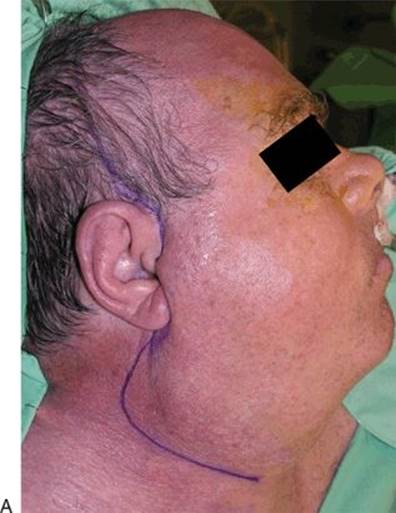
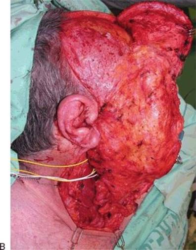
Figure 31.5 Elevation of superior and inferior platysmal flaps. A. The ITF approach combines three common incisions: hemicoronal or question mark scalp incision, preauricular modified Blair incision, and apron incision. B. The skin is incised with a no. 15 blade, and subsequent dissection of the subcutaneous tissue and the platysma is carried out with an electrocautery at the lowest effective setting. Subplatysmal and subgaleal flaps are elevated superiorly and inferiorly.
Exposure of the Neck and Vascular Control
Tumors located close to the internal or external carotid arteries or adjacent to the jugular foramen require control of the large vessels, prior to resection of the tumor. Similarly, I advise exposure and identification of cranial nerves that are at risk of injury during the operation. These include the hypoglossal, spinal accessory, and facial nerves. In order to achieve identification and preservation of these neurovascular structures, I recommend beginning this procedure with a transcervical approach (Fig. 31.5B). If a neck dissection is indicated or if a free flap reconstruction is anticipated, the neck dissection would serve the goal of both removing lymph node metastases and exposing the blood vessels for anastomosis. The detailed surgical technique of neck dissection is not included in the scope of this chapter.
Elevation of the Scalp Flap and Exposure of the Zygoma
A routine superficial parotidectomy is performed if indicated with dissection of all facial branches, and the various branches are then sharply dissected away from the deep lobe of the parotid. If the facial nerve is infiltrated by the tumor, a total parotidectomy is performed and the facial nerve is sacrificed. If the tumor grossly extends toward the skull base, the posterior belly of the digastric muscle can be retracted cephalad, or transected exposing the tumor at the base of the skull. The stylohyoid muscle and stylomandibular ligament may be also divided to allow a wider approach to the ITF from below. This maneuver could also prevent accidental injury to the internal jugular vein at the jugular foramen, which would be difficult to control and hemostasis would require ligating the vein.
Elevation of the scalp flap is continued down to the level of the adipose tissue pad overlying the zygoma, above the temporalis fascia. On the ipsilateral side, the remainder of the dissection dips below the level of the temporalis fascia and continues as a fasciocutaneous flap.
It is important to recognize the anatomy of the structures below the temporalis line. The temporal adipose tissue pad lies lateral to the temporalis muscle (Fig. 31.6). Above the zygoma the superficial temporal fascia splits to envelop the superficial adipose tissue pad. The frontal branches of the facial nerve run lateral to the superficial adipose tissue pad. In order to preserve these branches, dissection below the temporal line must continue along the deeplayer of the temporalis muscle fascia, and deep to the superficial temporal adipose tissue pad. At this level, the sharp dissection is performed with a scalpel and Freer dissector until the superior edge of the zygomatic bone is exposed. The superficial temporal adipose tissue pad and the zygomatic branches of the facial nerve are preserved and carefully elevated with the galeal layer. Next, the zygomatic bone and the lateral and superior walls of the orbit are exposed and skeletonized (Fig. 31.7). The temporalis muscle is freed from the medial surface of the zygomatic arch and bone.

Figure 31.6 Anatomy of the temporalis muscle and its fascia. Superior to the zygoma, the superficial temporal fascia splits to envelope the superficial adipose tissue pad. Lateral to the superficial adipose tissue pad run the frontal branches of the facial nerve. In order to preserve these branches, the dissection inferior to the temporal line must continue along the deep layer of the temporalis muscle fascia, deep to the superficial temporal adipose tissue pad. (SMAS, superficial muscular aponeurotic system.)
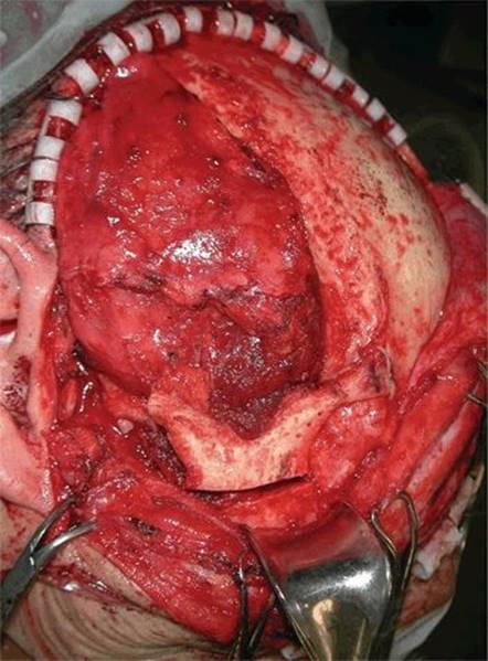
Figure 31.7 Osteotomy of the zygomatic arch. The anterior osteotomy is performed at the zygomatic process of the frontal bone, and the posterior osteotomy is performed anterior to the auricular tubercle in the posterior border of the arch. The medial attachment of the zygomatic bone to the lateral wall of the orbit is also cut in order to complete the separation of the bone.
Osteotomy of the Zygomatic Arch
After the zygomatic arch and bone are exposed, the osteotomies are performed. Prior to the osteotomies, titanium mini plates are fixed and pre-bent to allow accurate repositioning of the bone. The anterior osteotomy is performed at the zygomatic process of the frontal bone, and the posterior osteotomy is performed anterior to the auricular tubercle in the posterior border of the arch (Figs. 31.7 and 31.8). The medial attachment of the zygomatic bone to the lateral wall of the orbit is also cut in order to complete separation of the bone. Here the osteotomy is performed medial to the zygomatic bone along the zygomatic and frontal process of the lateral orbital wall. Following the osteotomies, the bone is gently elevated and separated from its medial attachments to the temporalis muscle fascia. The bone segment is then stored in saline during the length of the operation.


Figure 31.8 Exposure of the ITF. After the zygomatic process and arch are removed, (A) the temporalis muscle is reflected down (B) exposing the greater wing of the sphenoid bone and squamous portion of the temporal bone.
Exposure of the ITF
After the zygomatic process and arch are removed, the temporalis muscle is reflected downward, exposing the greater wing of the sphenoid bone and squamosa of the temporal bone (Fig. 31.8). Electrocautery is used here to reduce blood loss. The extracranial content of the ITF including the lateral pterygoid plate, the styloid process and its muscles, the lateral pterygoid muscles, the internal maxillary artery and its branches, and the branches of the mandibular nerve (V3) are now exposed. The condyle of the mandible may be retracted, or resected if indicated, to expose the spine of the sphenoid bone, the foramen ovale and V3, the middle meningeal artery, and the ICA at its entrance into the skull. If involved by the tumor, the sphenoid or maxillary sinuses are opened. The lateral wall of the sphenoid sinus is located between the maxillary nerves V2 and V3 and the base of the pterygoid plates.
Craniotomy and Exposure of the Middle Fossa
When the pterional area, the lateral orbital wall and orbital roof are exposed, care is taken to perform the craniotomy, which is tailored according to the intracranial and extracranial extension of the tumor. For tumors confined to Meckel’s cave, a pterional craniotomy is adequate. For tumors situated more anteriorly that involve the petrous apex, and when exposure of the superior orbital fissure and cavernous sinus are indicated, an orbitozygomatic craniotomy is required (Fig. 31.9). A burr hole is placed beneath the temporalis muscle, and a curvilinear craniotomy is performed. The bone flap is extended down toward the base of the middle cranial fossa. After the osteotomy, the bone flap is elevated and carefully detached from the dura overlying the middle cranial fossa with a bone dissector. If not involved by the tumor, the bone segment is stored in saline for the duration of the operation. Gentle retraction of the dura superiorly offers exposure of the entire middle fossa including the greater sphenopalatine nerve (GSPN), the middle meningeal artery, and cranial nerve divisions of the trigeminal nerve (Fig. 31.10). Using the dissecting microscope, the divisions of V3 and V2 are unroofed by drilling out the greater wing of the sphenoid. The superior orbital fissure and its neurovascular structures may be exposed in the same manner. Rarely, exposure of the intrapetrous part of the internal carotid artery is also indicated. This can be achieved by first dividing the middle meningeal artery and the GSPN and then removing the anterior clinoid process and the surrounding bone. Next, careful drilling along the external opening of the carotid canal may be performed to complete the approach.


FIGURE 31.9 Orbitozygomatic (A) and pterional (B) craniotomies. For tumors confined to Meckel’s cave, a pterional craniotomy is adequate. For more anterior tumors that involve the petrous apex and when exposure of the superior orbital fissure and cavernous sinus are indicated, an orbitozygomatic craniotomy is needed.


Figure 31.10 Exposure of the middle fossa. A. Gentle retraction of the dura superiorly provides exposure of the entire ITF and temporal fossa. B. Exposure of the foramen ovale and V3 (indicated by the arrowhead).
Extensions of the ITF Approach
The ITF approach can be combined with other approaches according to the anatomical area and histology of the tumor. Table 31.1 indicates the different combined approaches and their indications.
Table 31.1 The Various Approaches to the ITF, Their Indications, and Limitations

ITF, infratemporal fossa.
Combined Transcervical ITF Approach
The combined transcervical ITF approach is indicated for benign tumors involving the parapharyngeal space that extends to ITF. This approach is most frequently used for large schwannomas and pleomorphic adenomas in this area. The transcervical approach is extended by dividing the posterior belly of the digastric muscle and the stylohyoid and stylomastoid muscles. The stylomandibular ligament is also divided, allowing anterior retraction of the mandible. The submandibular gland is dissected out and retracted anteriorly. The tumor is now identified, and its association with the cranial nerves should be explored. The tumor can now be separated from the surrounding tissue, and here it is advisable to use meticulous dissection with a thin hemostat in order to prevent injury to the great vessels and cranial nerves. Care is taken to prevent accidental injury to the internal jugular vein at the jugular foramen. Bleeding in this area may be difficult to control, and hemostasis may require tying off the vein. In large ITF tumors that extend to the neck, removal of the submandibular gland can improve exposure and facilitate finger dissection in this area.
Once the specimen is completely freed from its surrounding tissue, its proximal and distal margins are clamped and tied with a 3-0 silk suture, and the tumor is then removed.
Combined Transmandibular ITF Approach
This approach is suitable for patients with extremely large benign tumors, malignant tumors, and for highly vascular lesions. There are two options for this approach: (1) a lateral approach using a segmental mandibulectomy and (2) a mandibulotomy using a lip split incision. The first approach is indicated as part of a composite resection involving the ramus of the mandible (Fig. 31.11). The second is indicated when the mandible is not involved.


Figure 31.11 Combined transmandibular ITF approach. A patient with high-grade mucoepidermoid carcinoma. The composite resection included preauricular ITF approach, radical parotidectomy, and segmental mandibulectomy. A. Marking the incision. B. The tumor was resected, and reconstruction was performed with an anterolateral thigh free flap.
Combined Transtemporal ITF Approach
For malignant tumors extending posterior to the ITF, a combined lateral temporal bone resection is the foundation of the treatment. This is usually accompanied by a segmental resection of the ramus of the mandible and its coronoid and condylar processes along with en bloc removal of the mandibular fossa, auditory canal, and mastoid portion of the temporal bone. The extent of the excision may also involve the auricle, adjacent skin, and adjacent structures according to the specific tumor and patient’s characteristics (Fig. 31.12). Most commonly, this approach would be part of a more comprehensive surgery that may include parotidectomy and neck dissection.


Figure 31.12 Combined transtemporal ITF approach. A patient with adenoid cystic carcinoma of the parotid and long-standing facial nerve paralysis. The composite resection included preauricular ITF approach, segmental mandibulectomy, radical parotidectomy, and lateral temporal bone resection. A. The main tumor resection was performed via the ITF approach and pterional craniotomy. B. The lateral temporal bone resection was performed through a posterior occipitotemporal flap.
If the auricle or parts of it are to be preserved, considerations for the blood supply must be a part of planning of the skin incision. Here, skin incisions and soft tissue approach are similar to those described above for the ITF. A temporal extension of the approach is achieved by elevation of the posterior retroauricular flap that includes the auricle. The flap is elevated posteriorly over the mastoid process and occipital bone (Fig. 31.12). A retroauricular skin incision may be used as an alternative. However, this incision puts the blood supply to the auricle at risk, especially in previously irradiated patients or if a previous preauricular incision was performed. Skin incisions should also accommodate reconstructive efforts, allowing access to local flaps as required. For tumors involving the external auditory meatus, a circular area of auricle engulfing the meatus is incorporated with the specimen. Bone work commences with a complete canal wall up mastoidectomy, and decortication is extended toward the mandibular fossa and includes the entire mastoid tip. A middle fossa craniotomy is necessary to separate the specimen in en bloc fashion. Eventually, the temporal fossa is reached, and the dissection is continued in the space between the annulus and the jugular bulb. If a segmental mandibulectomy is indicated, the TMJ is included along with the specimen. At this stage, the residual attachments of the specimen are connected to the posterior aspect of the TMJ capsule and adjacent bone. The bone can be disrupted by anterior fracturing of the specimen or with the aid of an osteotome, passed from the mastoid through the facial recess.
Combined Transfacial ITF Approach
Although the ITF approach permits complete tumor resection in the majority of cases, there could be situations in which the anterior aspects of the tumor are not adequately exposed. These include neoplasms that extend to the hard palate, orbit, nasopharynx, paranasal sinuses, and anterior skull base. Such cases require a combination of the ITF approach with a standard transfacial approach, to allow proper exposure and tumor extirpation. These combined approaches require additional incisions and osteotomies according to type and extent of the tumor.
The question mark incision is extended with a coronal incision, and the skin flap elevation continues down to the level of the adipose tissue pad overlying the zygoma, above the temporalis fascia. The ITF approach is performed as described above. The next stage includes osteotomies in both the frontal and pterional regions. The standard frontal osteotomy is modified by its lateral extension to include a portion of the orbital roof and temporal bone. A lateral rhinotomy or Weber-Ferguson incision is performed and a maxillectomy with or without orbital exenteration is achieved via the transfacial approach (Fig. 31.13).


Figure 31.13 The combined ITF craniofacial approach. This is a patient with a high-grade mucoepidermoid carcinoma of the paranasal sinuses invading the ITF, hard palate, and orbit. The composite resection included ITF approach, extended maxillectomy, and orbital exenteration. Reconstruction was performed with an anterolateral thigh free flap. A. Lateral view of the resection showing the preauricular ITF approach and exposure of the orbit. B. Anterior view of the resection showing transfacial approach and extended maxillectomy via upper cheek flap incision.
Combined ITF–Transorbital Approach
A combined ITF–transorbital approach is used for malignant tumors that penetrate the bone of the orbit and periostium, and infiltrate the anterior orbital contents or orbital apex. The skin flap is performed as described above in the ITF approach. The superior and lateral walls of the orbit are exposed and the periosteum stripped from the bone. If the skin is not involved by the tumor, the upper and lower lids may be spared, allowing for future insertion of an orbital implant and improved cosmetic results. For advanced skin cancers, the exenteration can involve the eyelids, scalp, and skin overlying the temporal region. In this case, a circular skin incision is performed along the superior and inferior orbital rims, and the skin of the lids is left on the main specimen (Fig. 31.14). Usually, this approach involves composite resection of the skin, zygomatic bone and arch, and the lateral orbital wall along with the orbital contents (Fig. 31.15).
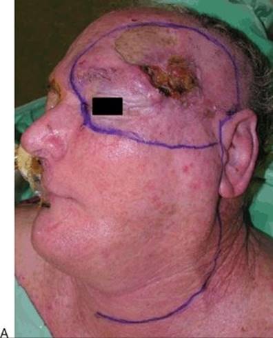

Figure 31.14 Combined ITF transorbital approach—I. This patient had a recurrent squamous cell carcinoma of the skin with invasion of the orbit. A. Planning of the incisions. B. Superficial parotidectomy with preservation of the lower branches of the facial nerve and neck dissection.


Figure 31.15 Combined ITF transorbital approach—II. Continuation of the resection including preauricular ITF approach and temporal craniotomy. Reconstruction was performed with an anterolateral thigh free flap (not shown). A. Before removal of the specimen. B. After tumor extirpation and temporal craniotomy.
Reconstructive Considerations and Wound Closure
The specimen is oriented and submitted separately for analysis to the pathology laboratory. The wound is copiously irrigated and inspected. Systolic blood pressure should be above 120 mm Hg to allow hemostasis. Any communication between the upper aerodigestive tract and the intracranial compartment should be closed. In case of dural resection, I prefer using double-layer fascia lata free graft, sutured to the edge of the resected dura with a continuous nonabsorbable suture. The eustachian tube should be plugged with a strip of fascia to prevent CSF leak. Any opening to the sphenoid or maxillary sinuses or to the nasopharynx should be eliminated. This is most commonly achieved by a free tissue transfer or temporalis muscle rotational flap. If exposed, the internal carotid artery should be covered with vascularized tissue to prevent carotid blowout. I do not recommend the use of nonvascularized free muscle graft for any reconstruction purpose, since it completely disintegrates within a few days after surgery. Primary closure of the surgical defect should be done when possible. When the facial nerve is sacrificed, a reconstructive procedure is required, and in such cases, an interposition sural nerve graft is used during the same procedure. Larger defects require regional flaps (temporalis muscle flap) or free flaps (radial forearm or anterolateral thigh free flaps). Finally, the previously removed zygomatic bone and the temporal bone segments, if not involved by tumor, are repositioned and secured with titanium mini plates and craniofix screws, respectively. If the temporalis muscle has been preserved, it is repositioned and sutured to the pericranium. Two No. 7 Jackson-Pratt drains are placed into the neck and ITF compartments.
POSTOPERATIVE MANAGEMENT
After the operation, the patient is extubated and immediately transferred to the postsurgery care unit for monitoring before transfer to the wards. The wound is kept clean by saline rinsing three times a day, and covered with antibiotic ointment after each cleansing. The drains are removed either 3 days after the operation or when the drainage is less than 20 to 30 mL in 24 hours. Prophylactic antibiotic treatment is not indicated in the postoperative period. For pain control, patients are treated with nonsteroidal anti-inflammatory drugs (diclofenac 75 mg intramuscularly or orally) once daily, or with tramadol 40 to 100 mg if requested by the patient or considered necessary by the nurses. If a free flap is also performed, selective COX-2 inhibitors should be avoided. CSF drainage is indicated after large dural resections. A lumbar drain is inserted prior to surgery and left for 3 days, while draining 5 to 10 mL of CSF per hour. In such cases we usually administer postoperative broad antibiotic therapy until the drain is removed.
COMPLICATIONS
Surgery in this area is dangerous and requires experience in skull base and head and neck techniques. Major morbidity and mortality may be associated with tumor extirpation in this area, mainly due to the potential injury to neurovascular structures. Complications of the various surgical approaches to the ITF are similar and are listed in Table 31.2. The most common perioperative complication is injury to the cranial nerves. Temporary injury to the facial nerve will occur in 10% to 20% of the patients. Most cranial nerve injuries are temporary, and recovery can be expected within 6 months. Wound infection and meningitis may occur in up to 10% of the patients. CSF leak is rare but may occur through a defect in the dura and/or in the maxillary or sphenoid sinuses or the eustachian tube. Other significant potential complications include thrombosis of the jugular vein or sigmoid sinus.
Table 31.2 Complications Associated with the ITF Approach
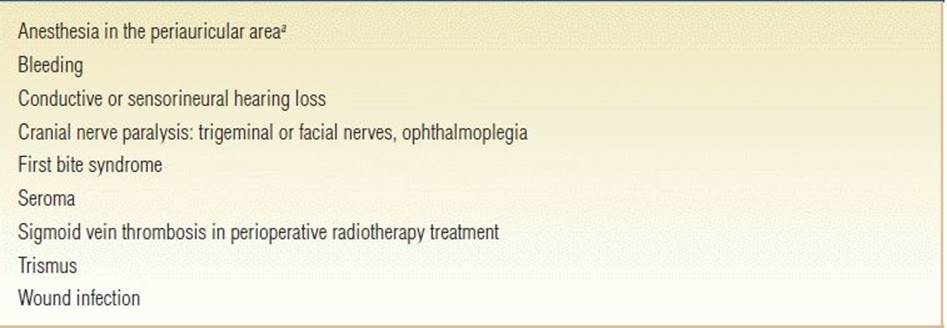
aA direct consequence of surgery rather than a complication.
RESULTS
The overall 5-year survival of patients with malignant tumors of the anterior skull base is 50%. Their prognosis depends above all on histology and margin status. Other factors having an impact on survival are staging and invasion of the orbit, dura, or brain. Most patients with tumor recurrence die of local tumor recurrence followed by distant metastases. Consequently adjuvant postoperative radiation therapy is recommended for malignant tumors of the ITF.
PEARLS
· The operation is performed without muscle relaxation in order to monitor the cranial nerves.
· Frontal branches of the facial nerve are preserved by dissecting along the deep layer of the temporalis muscle fascia.
· Meticulous reconstruction of the dura and plugging of the eustachian tube is required to prevent CSF leak and meningitis.
· Immediate extubation is required to allow continuous neurologic monitoring.
· Early reinstatement of TMJ and jaw physiotherapy should be used to prevent trismus.
PITFALLS
· Exposure of the zygomatic arch from its lateral border may lead to permanent injury of the facial nerve.
· Removal of a small segment of the zygoma will limit exposure of the ITF.
· A small or misplaced craniotomy will make access to the middle fossa difficult.
· Failure to plug the eustachian tube may lead to a CSF leak.
INSTRUMENTS TO HAVE AVAILABLE
· Navigation system
· Dissecting microscope
· Nerve monitor
· Skull pin frame
· Bovie set including monopolar and bipolar electrocautery
· Electrical drill and saw
· Micro air drill
· Craniofacial tray
· Craniotomy tray
· Basic neuro tray
· Micro neuro tray
· Craniotome tray
· Malleable suctions tray
· Kerrisons tray
· Rongeurs tray
· Neuro curettes
· Basic neck dissection tray
· Basic plastic tray (for fascia lata harvesting)
· Microvascular reconstruction tray (for free flap reconstruction)
· Surgicel
· Gelfoam size 100
· Neurosurgical patties
· Vascular clips—small and medium size
· Dural substitution
ACKNOWLEDGMENT
The author would like to acknowledge Dan M. Fliss, MD; Nevo Margalit, MD; Ophir Handzel; and Arick Zaretsky, MD, who participated in some of the operations.
SUGGESTED READING
Al-Mefty O, Fox JL, Rifai A, et al. A combined infratemporal and posterior fossa approach for the removal of giant glomus tumors and chondrosarcomas. Surg Neurol 1987;28(6):423–431.
Sekhar LN, Schramm VL Jr, Jones NF. Subtemporal-preauricular infratemporal fossa approach to large lateral and posterior cranial base neoplasms. J Neurosurg 1987;67(4):488–499.
Mansour OI, Carrau RL, Snyderman CH, et al. Preauricular infratemporal fossa surgical approach: modifications of the technique and surgical indications. Skull Base 2004;14(3):143–151.
Donald PJ. Infratemporal/middle fossa tumors. In: Hanna EY, DeMonte F, eds. Comprehensive management of skull base tumors. New York: Informa Healthcare, 2009;20:305–330.
Gil Z, Fliss DM, eds. Tumors of the skull base and paranasal sinuses. Delhi: Byword Books, 2012.