
Kris S. Moe and Richard G. Ellenbogen
INTRODUCTION
There are numerous effective and safe open surgical approaches to the middle cranial fossa (MCF). These include, but are not restricted to, the pterional, orbitozygomatic, frontotemporal, and temporal approaches. These approaches are very effective, offering ample exposure with a high degree of safety. However, there are some drawbacks to each of these procedures. The commonly cited and recognized drawbacks or risks include morbidity related to a large scalp incision, atrophy of the temporalis muscle, damage to the frontal branch of the facial nerve, and postoperative cerebral edema that occurs as a result of retraction of the brain required for access to the lesion.
The same surgical interests that have inspired and accelerated the development of endoscopic approaches to the anterior cranial fossa (ACF) have pushed forward research in minimally disruptive approaches to the MCF. Transnasal approaches to the medial MCF (parasellar region and medial aspect of the cavernous sinus) have been described and are effective. Extended endoscopic approaches to the anterior base of the MCF have also been developed and have been evolving in clinical practice. Admittedly, extension laterally and superiorly beyond these regions is more challenging. The challenge to develop safe and effective minimally invasive endoscopic approaches to the MCF is, in part, a result of the significant length of the surgical pathway and surgical obstacles such as the orbital contents and the optic nerve.
As a means of circumventing these obstacles, while providing short, direct pathways to lesions in the MCF, we developed a system of transorbital endoscopic approaches that is unique but effective in treating lesions in this region. The surgical trajectory through the orbit is based on an orbital quadrant system, as discussed in Chapter 15 (Fig. 34.1). This system divides the orbit into distinct approach quadrants based on common target regions adjacent to and beyond the orbit, as well as anatomic structures related to the orbit that should not be disturbed. These include the superior orbitotomy through a superior lid crease (SLC) incision and medial orbitotomy through a precaruncular (PC) incision to the ACF (see Chapter 16). Access to the MCF is available through a lateral orbitotomy using a lateral retrocanthal (LRC) approach, an inferior orbitotomy through an inferior transconjunctival (ITC) approach (Fig. 34.2), and a medial orbitotomy through the contralateral orbit using a contralateral precaruncular (CPC) approach (see Chapter 15 and below). These approaches are in common use for the repair of orbital trauma and treating orbital and intracranial complications of sinogenic abscesses. We have reported on the safety and efficacy of the use of these techniques in a large series of cases. These approaches can be used alone in monoportal technique or combined with contralateral transorbital, transnasal, or transmaxillary approaches for multiportal access.

Figure 34.1 The four quadrants of the right orbit. The surgical approach is chosen by the quadrant of the orbit that is most involved by the underlying pathology. The superior quadrant is accessed by the SLC approach (see Chapter 15); the medial quadrant is entered through a PC incision; the inferior quadrant approach is made through an ITC incision; and the lateral quadrant is reached through an LRC portal. (SLC, superior lid crease; PC, precaruncular; ITC, inferior transconjunctival; LRC, lateral retrocanthal.)

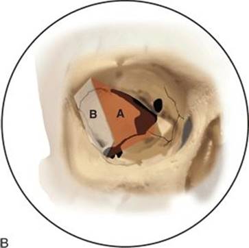


FIGURE 34.2 A. Surgical anatomy of the right orbit. 1, frontozygomatic suture; 2, sphenozygomatic suture; 3, superior orbital fissure; 4, optic canal; 5, inferior orbital fissure; 6, foramen of infraorbital nerve. B. Outline of bone removed through lateral orbitotomy: A, approach to MCF through greater wing of sphenoid bone; B, approach to infratemporal fossa. C. Region of approach through inferior orbitotomy. (MCF, middle cranial fossa.)Figure 34.2(Continued ) D. Arrow on foramen rotundum seen through inferior orbital fissure.
The choice of pathway and number of portals is made with a preoperative computerized approach analysis using a surgical navigation system and includes a full 360-degree evaluation of the anatomy involved in and adjacent to the pathology, as well as the anatomic structures that are displaced or traversed by the potential endoscopic pathways to the target. The pathway(s) are chosen to provide the most efficacious and least morbid approach. The goal of surgery is to achieve the best possible angle(s) for the endoscopic treatment of the pathology and ultimately to lessen the morbidity by avoiding an open cranial microsurgical approach (Fig. 34.3).

Figure 34.3 Example of preoperative computer analysis of possible endoscopic pathways to right cavernous sinus, superior view, CT scan. Lateral (red), inferior (blue) contralateral PC (green) approaches. MCF and adjacent sphenoethmoid regions are highlighted in yellow. (PC, precaruncular; MCF, middle cranial fossa.)
While the LRC approach provides retro-orbital access to regions of the MCF that cannot be safely achieved transnasally, the ITC and CPC portals provide access that can also be obtained through transnasal or transmaxillary approaches. The angles of approach that they provide to a target are quite different; the distances may be significantly shorter than transnasal approaches, and as such, the portals can be complementary. At times, however, pathology may involve the floor of the orbit (e.g., metastatic tumor) or a portion of the orbit that would be resected with the pathology. In these situations, a transorbital portal can provide a clearly advantageous surgical visualization of the orbit, particularly if orbital reconstruction will be required. As experience with all of these approaches grows, the indications and contraindications to the use of each portal will be further delineated.
This chapter provides a discussion of the transorbital endoscopic approaches that we use for access to the MCF, including the LRC, ITC, and CPC portals. Though our approach for sellar and parasellar pathology is most commonly performed through transnasal portals, a description of these is beyond the scope of this discussion.
HISTORY
The presentation and history of patients with MCF lesions are highly variable, depending on the location, type, and stage of the underlying pathology. Tumors in this region are often silent until they invade and travel along nerves, at which time they may cause facial numbness or pain. Progression within the brain may cause symptoms depending on the region involved. Tumor involvement in the infratemporal or pterygopalatine fossae may cause interference or pain with mastication. Spread anteriorly with the orbit may cause diplopia, proptosis, and progressive loss of vision.
PHYSICAL EXAMINATION
Any patient presenting with symptoms consistent with pathology involving the MCF should have a complete examination of the head and neck, as well as a thorough neurologic evaluation including all of the cranial nerves. Evaluation of facial sensation in all branches of the trigeminal nerves is important. Nasal and pharyngeal endoscopy should be performed to evaluate involvement of the medial orbit, interorbital skull base and, sphenoid and clival regions.
Imaging is then performed based on the history and physical examination, with computerized tomographic (CT) scans and magnetic resonance imaging (MRI) being the most frequently indicated studies alone or as complementary evaluations. Angiography may be indicated for examination of vascular involvement or for preoperative embolization to make surgery safer.
INDICATIONS
A wide range of pathology can involve the MCF as a primary site, through metastasis from a distant site, or through local extension from an adjacent structure. Tumors arising within the MCF are usually benign and include pituitary adenoma, meningioma, and trigeminal schwannoma. (Temporal bone pathology is excluded from this chapter.) Malignancies arising in this area include chordoma, chondrosarcoma, and osteosarcoma; perineural invasion from squamous cell carcinoma or adenoid cystic carcinoma. Local invasion and hematologic metastases are also common.
The most common open procedures to access these regions are the pterional, orbitopterional, orbitozygomatic, and transzygomatic/infratemporal fossa approaches. The open microvascular procedures, although effective, can be morbid due to brain retraction, cerebral edema, and disruption of normal structures associated with large, potentially visible scars. Furthermore, open procedures through the frontotemporal regions require displacement of the temporalis muscle that may cause significant visible hollowing of the temporal contour. In addition, the frontal branch of the facial nerve is at risk for injury during the creation of these surgical pathways. Though paralysis of this nerve is not common, when it does occur, it has a significant negative impact on a patient’s appearance and may cause blockage of the superior visual field. Removal of a large cranial bone flap also carries the risk of infection of the flap and added recovery time for the patient.
For reasons of safety, better visualization of the pathology and improved healing time, transnasal endoscopic approaches have been described and investigated for access to the parasellar regions, petrous apex, and medial aspect of the cavernous sinus. These approaches offer excellent access through a natural corridor. There are times, however, when an additional access portal to these regions may be beneficial to improve the angles of visualization of the pathology or to provide increased working space between the surgeons’ hands or instruments.
Transnasal approaches may also be limited in their ability to provide visualization and endoscopic access to the lateral aspect of the cavernous sinus/parasellar regions and locations lateral to the trigeminal ganglion, and when these regions can be reached, this endoscopic route may be at the upper limits of their effective range.
Another region of limited access transnasally is the superior infratemporal fossa and the anterior–superior MCF. These regions are within the orbital “shadow effect” of a transnasal approach, making these areas challenging to access without endangering critical neurovascular structures.
Transorbital approaches can be used to access these areas that are either difficult or dangerous to reach through the nose or to provide multiportal corridors in addition to transnasal approaches. The indications for these approaches are similar to those for transnasal portals and are expanding with growing familiarity of the anatomic perspectives and technical demands. Transorbital endoscopic approaches to the MCF are currently indicated for benign and localized malignancies involving the ventral aspects of the MCF along the inferior, medial, lateral, and anterior borders. The primary transorbital approaches to these regions are the LRC, ITC, and the CPC portals as described above.
The indications and applications of these approaches are continuing to expand and will likely further develop as advanced instrumentation becomes available. The lateral orbitotomy (LRC approach) provides access to the infratemporal fossa, the greater wing of the sphenoid, and adjacent regions of the MCF. Its applications include trauma and CSF leak repair, and resection of tumors. This pathway can be used for access to the lateral aspect of the cavernous sinus and trigeminal ganglion, with a relatively short intracranial component (Fig. 34.4). There is considerable flexibility in the placement of the entry portal in the coronal plane, and the choice of entry is made by vector analysis with regard to the surgical target.


FIGURE 33.4 A. Surgical planning view, LRC approach. B. Enlargement demonstrating trajectory of LRC approach. C. Access to lateral MCF. Note proximity of infratemporal fossa that can easily be entered as needed. (LRC, lateral retrocanthal; MCF, middle cranial fossa.)
The ITC approach can be used for targets at the inferior anterior aspect of the MCF, such as the region of the foramen rotundum (Fig. 34.5). It also offers a potential pathway to the inferior lateral aspect of the cavernous sinus following a trajectory inferior to the optic nerve. This approach is particularly useful for pathology that involves the inferior orbit and maxilla, such as tumors in the infraorbital nerve.
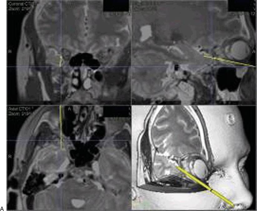


Figure 34.5 ITC approach. A. Preoperative pathway analysis with vector from entry portal to the surgical target. B. Enlarged parasagittal view demonstrating pathway inferior to the globe, leaving the orbit posteriorly, where the orbital floor rises, to enter the MCF. C. ITC approach to foramen rotundum with CPC approach (green) to lateral sphenoid. (ITC, inferior transconjunctival; MCF, middle cranial fossa; CPC, contralateral precaruncular.)
The CPC approach is indicated for pathology in the medial aspect of the cavernous sinus, optic nerve, and selected sellar and suprasellar lesions (Fig. 34.6). Pathology of the optic nerve and chiasm as well as the resection of encephaloceles are strong indications for inclusion of this portal in the surgical plan.

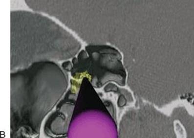
FIGURE 34.6 A. Left CPC approach to right MCF (Sternberg’s) encephalocele (yellow). B. Magnified segment of (A), with virtual endoscopy demonstrating favorable approach for viewing and instrumentation. (CPC, contralateral precaruncular; MCF, middle cranial fossa.)
CONTRAINDICATIONS
As for the transorbital approaches to the ACF (see Chapter 16), the primary contraindication to transorbital approaches to the MCF appears to be a recent history of severe orbital trauma. A history of LASIK surgery, though not a contraindication to surgery, should caution the surgeon to treat the cornea with great care and follow the patient closely postoperatively for any corneal complications (see Complications section). These patients may have partial corneal anesthesia and may be at higher risk for corneal ulceration or exposure damage. Patients with primary orbital pathology, particularly those with corneal anesthesia, should be evaluated preoperatively by an ophthalmologist and receive joint follow-up postoperatively.
The pathology must be of a suitable extent, location, and character for resection with endoscopic visualization and the instrumentation at the surgeon’s disposal. Care should be taken to ensure that the lesion is a solitary lesion in cases of metastatic disease.
Surgeons should receive adequate training and/or mentoring in these approaches before using them clinically. Each patient’s pathology should be evaluated individually with preoperative CT and MRI analysis to be certain that an endoscopic approach of any type is appropriate and to determine which type of monoportal or multiportal surgical approach will be optimal.
PREOPERATIVE PLANNING
As for any complex surgical procedure, transorbital endoscopic approaches to the MCF should be considered only after comprehensive evaluation of the patient including comprehensive imaging and consultation with all of the members of the skull base team who will be involved. When possible, it is optimal for the patients to be seen by the treating physicians synchronously to provide a comprehensive evaluation. Tumor cases should be presented and discussed at a multidisciplinary skull base tumor board involving skull base surgeons from the fields of neurologic surgery and otolaryngology as well as neuroradiologists, neuropathologists, medical oncologists, and radiation oncologists.
Approaches to the MCF are planned in the same manner as described in Chapter 16 for the ACF. Suitability of the pathology for an endoscopic approach must be considered, and the ability to successfully treat the surgical target with available instrumentation should be confirmed.
Preoperative computer analysis should be undertaken to determine the optimal surgical approach or approaches. This is performed on a planning station, using uploaded CT and/or MRI images (Fig. 34.3). The target is highlighted (Fig. 34.7), and the three-dimensional image is examined circumferentially to familiarize the surgeon with the structures that are involved with or adjacent to the pathology. The potential pathway vectors under consideration are then diagrammed and analyzed with respect to the following:


Figure 34.7 Preoperative approach planning. Lesion within bone of left greater wing of the sphenoid, segmented yellow. LRC (green) approach is shortest, most direct, and requires less adjacent tissue retraction. A. Possible entry points (inferior orbitotomy, red; lateral orbitotomy, green). B. Approaches with surgical target segmented. C. Lateral monoportal approach is able to access entire lesion. Approaches and target are evaluated circumferentially. (LRC, lateral retrocanthal.)
· The pathway should not cross critical neurovascular structures.
· Creation of the pathway must be technically feasible.
· Reconstruction of any portion of the pathway, if necessary, must be within the skills of the surgeon.
· The pathway should be short and direct to minimize collateral damage.
· The view of the target provided through the pathway should be unobstructed by tissue or instruments.
· The angle of manipulation of the pathology provided by the endoscopic pathway should be adequate to complete all surgical tasks.
· Until the advent of flexible endoscopic surgery, the pathway must be linear, and direct virtual endoscopy can be performed to evaluate the structures involved with and encountered along the proposed surgical pathway. Adequacy of visualization of the target can be similarly analyzed, and mock instruments can be placed in multiportal fashion to determine if their approach angle is adequate and removed from the viewing trajectory of the endoscope.
As described in Chapter 16, the choice of the orbital region for the surgical approach is based on a division of the orbit into four quadrants (superior, medial, inferior, and lateral; Fig. 34.1). The quadrant that is directly involved by the pathology or is within the chosen surgical pathway is employed. The primary endoscopic transorbital approaches to the MCF are the LRC, ITC, and CPC. The ipsilateral PC approach to the medial quadrant and the transpalpebral SLC approach to the superior quadrant are described in Chapter 16, on transorbital approaches to the ACF.
The inferior quadrant is bounded medially by the lamina papyracea and laterally by the inferior orbital fissure and its anterior extension toward the orbital rim. The entry portal is through an ITC preseptal or inferior fornix incision. There is no medial anatomic boundary of this incision as it can be continued into a PC incision. Likewise, there is no lateral anatomical boundary to the incision as it can be extended into a LRC incision.
The lateral quadrant is bounded superiorly by the superior orbital fissure and its anterior extension to the orbital rim and inferiorly by the inferior orbital fissure and an anterior extension to the orbital rim. The portal of entry is through the LRC approach. The superior limit of the incision is the lateral horn of the levator aponeurosis and muscle. There is no inferior limit to this incision since it can be continued inferomedially into an IT incision.
As noted above, the PC, ITC, and LRC approaches can all be combined as they are transconjunctival, and the incisions do not cross anatomic structures. The SLC incision is transcutaneous and, as such, is not typically connected with transconjunctival incisions. The use of an adjacent transcutaneous and transconjunctival portal would be possible, as would joining the incisions laterally through the lateral canthus, but this is rarely necessary due to the ample surgical pathways that can be created by each of these independently.
The medial quadrant boundaries include the ethmoid arteries superiorly and the junction of the lamina papyracea and orbital floor inferiorly. Entry to this quadrant is achieved through the PC transconjunctival portal (see Chapter 15). Superiorly, the incision can extend up to the medial horn of the levator aponeurosis. Inferiorly, the incision and approach can be extended into an IT incision through the conjunctiva of the lower eyelid.
Preoperative approach analysis is essential for these surgical procedures and should be performed with a detailed study of the anatomic structures that involve or are adjacent to the surgical target. A surgical plan is formulated, including the choice of monoportal versus multiportal technique. The entry portals are then chosen based on the criteria described above. This evaluation should be done preoperatively so that the possible surgical routes can be discussed with the patient, bringing the patient’s desires into the decision making and enabling a detailed informed consent process. A detailed surgical plan should be made, including the planned method of any reconstruction that might be required. As these procedures are technology intensive, the nursing staff must know ahead of time which powered and manual instruments and materials will be required to avoid intraoperative delays.
SURGICAL TECHNIQUE
As described in Chapter 16 in detail, the patient is placed in the supine position on the operating table and general anesthetic is administered. A lumbar drain is then placed if desired, and the bed is rotated 180 degrees with the foot toward the anesthesiology equipment. A circular gel headrest is used, or the patient’s head may be placed in pin fixation. The patient’s head is retroflexed 15 degrees to allow relaxation of the brain away from the skull base to minimize the need for brain retraction during the procedure. The head of the bed is elevated slightly to improve the intraoperative hemostasis; a small amount of local 1% lidocaine with epinephrine 1:100,000 (0.1 mL) is infiltrated into the planned conjunctival incision sites. The pupils are checked for symmetry before anesthetic injection as the local anesthetic may cause dilation of the pupil.
The surgical navigation system of choice is used, registration is performed, and the accuracy is confirmed (applying the navigation probe to the occlusal surface of the central upper incisors is an effective point to check). Using the navigation probe, a final check of the approach vector is made to confirm the choice and precise location of the surgical portal. The patient’s face is then prepared and draped in the usual sterile fashion. If an optical navigation system is used, a colorless preparation solution should be used to avoid system malfunction, and care must be taken to avoid covering the infrared light-emitting diodes with the drapes.
Three primary transorbital approaches are used to access the MCF: the LRC, ITC, and CPC. These are described below. The SLC and PC approaches are described in Chapter 16. Though the ipsilateral PC approach can be used to access the medial aspect of the cavernous sinus, access to targets lateral to this is obstructed by the orbit and optic nerve.
Orbital endoscopy is carried out in a similar fashion to that of sinus surgery, with standard 4-mm endoscopes with 0- and 30-degree lenses. An irrigating system may be used, but there is often less blood and secretions than experienced with transnasal portals, and this may be unnecessary. To create an optical cavity, malleable ribbon retractors are used to gently retract the orbital contents or brain as needed. Minimal pressure is exerted against the globe, and the pupil is regularly checked. If the pupil begins to dilate relative to the contralateral side, all instruments and retractors are removed until symmetry returns. Creation of a pathway within the orbit occurs between the orbital bone and periorbita (periosteum). The plane is dissected with the aid of a Freer suction elevator. The elevator is attached to a soft suction extender rather than directly to the relatively stiff standard suction tubing.
Our preference for creation of the pathway is for the surgeon to hold the endoscope and dissecting instrument; the assistant holds the malleable retractor in one hand and a small skin retractor in the other hand to hold open the portal (Chapter 16, Fig. 16.10). When the target has been reached, the surgeon may wish to operate with bimanual microsurgical technique as described elsewhere (see Suggested Reading).
The technique for creation of the endoscopic transorbital craniectomy depends on the characteristic of the bone, particularly its thickness. For the thin bone of the orbit, a periosteal elevator may be used to gently fracture the bone and then remove the fragments as needed. For thicker bone, such as that of the greater wing of the sphenoid, a drill or ultrasonic bone aspirator is used. We prefer the latter, as the bone aspirator irrigates, ablates bone, and aspirates with one instrument. Furthermore, the tip of the instrument has no tendency to skip off the bone and injure adjacent structures. In addition, it is our impression that the ultrasonic aspirator is less damaging to the underlying dura if the bone is directly transgressed. Our practice is to thin the bone to the point of eggshell thickness, then gently fracture and excise the elements remaining on the dura.
Medial Quadrant: Contralateral Precaruncular Approach
The PC approach may be used to access the ipsilateral ACF, the contralateral ACF, and the contralateral MCF. There are times when a CPC approach to the target is preferred, to optimize the angle of target approach for improved visualization and efficacy of manipulation.
This may be the case for lesions involving the cavernous sinus, lateral wall of the sphenoid, optic nerve, and suprasellar regions (e.g., craniopharyngioma). An example of this is shown in Figure 34.6: a meningoencephalocele of the anterolateral sphenoid (Sternberg canal) was treated through a contralateral PC approach when preoperative navigation analysis and virtual endoscopy demonstrated the excellent working and visualization angles of the approach.
The CPC approach is undertaken with the same technique as a PC approach, as described in Chapter 16 (Fig. 34.8). In brief, a PC approach is created in the contralateral orbit. The bone of the medial wall of the orbit is removed by ultrasonic aspiration or other technique at the site determined by navigation. The extent of bone removal is dictated by the instrumentation that will be used, creating the smallest necessary corridor. The pathway is then continued through the ethmoid cells crossing posteriorly to the sphenoid sinus adjacent to the pathology. A sphenoidotomy is created, providing access to the appropriate region of the MCF.


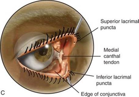
Figure 34.8 Contralateral PC approach. A. Primary support system of eyelids: medial canthal tendon, lateral canthal tendon, levator aponeurosis and muscle, lower lid retractor. B. Lacrimal probes in place, incision between apex of medial canthus and caruncle. C. Dissection follows posterior limb of medial canthal tendon to lamina papyracea. (PC, precaruncular.)
Lateral Quadrant: Lateral Retrocanthal Approach
Access to the lateral orbit, lateral ACF, anterior MCF, and infratemporal fossa can be obtained through the LRC approach (Figs. 34.2B, 34.4, 34.8, and 34.9). While numerous techniques of lateral orbitotomy have been described with excision of the lateral orbital rim, the LRC orbitotomy preserves the bone of the orbital rim, and thus does not require extended incisions. Furthermore, though a lateral canthotomy/cantholysis may be performed if the surgeon prefers, no skin incisions are required, and the functional integrity of the lateral canthus is maintained.


Figure 34.9 LRC approach. A. Highlighted area shows region of approach. B. Conjunctival incision (dotted line) contacting bone posterior to insertion of lateral canthus. C. Bone of the lateral orbit exposed. D. LRC approach, lateral canthus retracted, malleable retractor protecting orbital contents. (LRC, lateral retrocanthal.)
A lubricated corneal protector is placed, and the lateral canthus is retracted laterally with a small retractor. An incision is made through the conjunctiva adjacent to the lateral orbital rim (Fig. 34.9). The dissection then follows the posterior aspect of the lateral canthal tendon to its insertion on the medial face of the lateral orbital wall. The incision is extended superiorly as needed, dissecting between the bone and periorbita as described above. In doing so, the lacrimal gland and orbital contents are retracted medially. The incision can be extended as far inferiorly as needed and extended into an IT orbitotomy if desired. The periorbita is lifted off the entire orbital wall under endoscopic and navigation guidance, dissecting posteriorly until the superior and inferior orbital fissures are encountered (Fig. 34.2B). The optic nerve is medial to the confluence of these structures at the orbital apex and will not be visualized unless the contents of the superior fissure are transgressed.
The sphenofrontal suture can be visualized at the superior aspect of the lateral orbital wall. For lateral ACF targets, the craniectomy will be created above this line, as described in Chapter 16. For MCF targets, the craniectomy will be located below the suture. The entire section of greater wing of the sphenoid can be removed between the superior and inferior sutures as needed for access (Fig. 34.2B).
For access to the infratemporal fossa, the thin bone lateral to the sphenozygomatic suture, posterior to the lateral orbital rim is removed (Fig. 34.2B). Navigation is then used to direct the dissection, which occurs between the temporalis muscle and underlying bone.
Inferior Quadrant: Inferior Transconjunctival Approach
Access to the inferior orbit is obtained through an ITC approach with the same technique that is used for repair of a fracture of the orbital floor (Figs. 34.5 and 34.9). The approach can be extended laterally into an LRC or medially into a PC portal as needed.
The preseptal IT approach is advantageous in that, by preserving the orbital septum, there tends to be less prolapse of orbital fat into the surgical path. The deep fornix ITC approach has the benefit of leaving a small amount of fat on the lower eyelid and posterior aspect of the septum that may provide a protective layer that shields the lower eyelid. Either approach is effective, though we recommend the latter for less experienced surgeons.
The procedure is begun by placing a lubricated corneal protector. For a preseptal approach, an incision is made 2 to 3 mm inferior to the tarsus on the conjunctival surface of the lower eyelid (6 to 8 mm inferior to the eyelid margin) (Fig. 34.10D). The orbicularis oculi muscle will then be visible. Dissection continues inferiorly between the orbicularis and the septum until the inferior orbital rim is reached. The septum itself is quite thin and difficult to recognize—confirmation of the appropriate dissection plane is obtained by dissecting immediately deep to the orbicularis, and superficial to the orbital fat that is retained behind the septum. To perform an inferior fornix incision, the lower eyelid is retracted anteriorly, and the inferior orbital rim is palpated through the conjunctiva with a periosteal elevator or similar instrument. An incision is then made directly through the conjunctiva onto the orbital rim. This can be done with a scalpel, or a needle-tip Bovie cautery on a low setting.
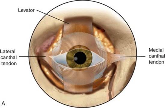
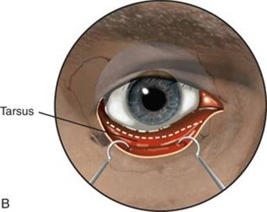

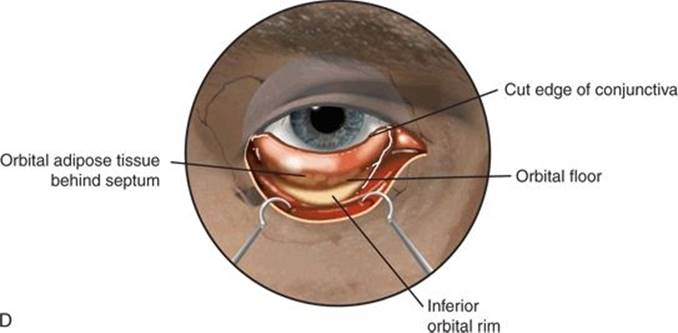
Figure 34.10 Inferior transconjunctival approach. A. supporting structures of the lower eyelid, skin, and orbicularis oculi muscle removed. B. Lower eyelid retracted anteriorly. C. Preseptal approach to inferior orbital rim. A direct inferior fornix approach (posterior to septum can also be used). D.Conjunctiva retracted, bone of inferior orbital rim exposed. Periosteum is incised, and periorbita is then elevated off the orbit floor.
When the orbital rim has been reached, a retaining suture is placed through the edge of the inferior conjunctival flap. This flap is then retracted superiorly over the corneal protector. The periosteum is then incised at the superior aspect of the inferior orbital rim, and a periosteal flap is raised posteriorly. This plane is then dissected posteriorly, and the orbit is entered. Dissection is continued posteriorly, lifting the periorbita off the orbital floor. When a suitable optical cavity has been developed, a 4-mm 0-degree endoscope is brought into the field and the rest of the dissection is performed under endoscopic visualization. The orbital contents are gently displaced superiorly with a malleable brain elevator. The infraorbital nerve is visualized running through a canal in the orbital floor; there may be thin fascial attachments between the nerve and the overlying orbital contents that are sharply severed. Dissection continues to the orbital apex, bordered laterally by the inferior orbital fissure and medially by the lamina papyracea. To develop the path beyond the orbit to the point of the craniectomy, the orbital bone is removed in the location indicated by navigating along the chosen approach vector to the surgical target. The bone can be removed by gentle down-fracturing if it is appropriately thin. For regions with thicker bone, we use an ultrasonic bone aspirator. Alternatively a fine diamond drill can be used, but care must be taken not to damage adjacent tissue. Dissection then continues along the indicated trajectory to the MCF; the craniectomy is then performed at the appropriate point indicated by surgical navigation. To perform the craniectomy, we use ultrasonic aspiration, thinning the cranial bone to the point of transparency. The bone is then gently fractured and lifted off the subjacent dura. The target is then approached in an intracranial-subdural plane, or with intradural dissection according to the operative plan. The intracranial pathway is often quite short, measuring 3 cm or less. Endoscopic surgical treatment of the pathology is then undertaken always tracking the surgical relationship to the globe and brain.
Reconstruction
The need for reconstruction of the surgical pathway depends on the extent of orbital bone that has been removed, which in turn depends on the location and extent of the underlying pathology. For the lateral approach, the bone that is removed has little role in supporting the globe. If an infratemporal fossa approach is used and temporalis muscle fills the defect, there is no need for reconstruction. If a significant amount of the greater wing of the sphenoid has been removed, the orbital volume may have expanded enough to cause enophthalmos. In this case, a small abdominal adipose tissue graft can be placed within the bone. The bone defect can also be covered with titanium mesh, or a 0.25-mm thick sheet of PDS foil. If the lateral canthus has not been disturbed, we do not close the incision. If a canthotomy and cantholysis has been performed, the canthal tendon is repaired.
If an inferior approach has been used, the defect in the orbital floor is repaired. We typically use a titanium implant manufactured for orbital fracture repair, or PDS foil. If there is no pathology invading the orbital floor, we place and shape the implant to the existing floor before bone is removed so that we precisely match the original contour, then remove the implant until the end of the case. If this is not practical, we complete the reconstruction and then check the position of the implant against the configuration of the original floor using the navigation CT scan. We do not close the incision unless it has been extended into a medial or lateral approach, in which case one or two sutures are placed to align the conjunctiva using 6-0 resorbable suture with inverted technique.
If a contralateral medial orbital approach is used, the medial wall may require reconstruction if the defect is large enough to cause enophthalmos. If this occurs, we reconstruct the defect with a thin titanium fracture implant or PDS foil.
POSTOPERATIVE MANAGEMENT
The same considerations for the postoperative management of patients who have undergone transorbital surgery of the ACF (see Chapter 16) are given to patients who have had surgery of the MCF. The morbidity of the approach itself depends on the length of the procedure and extent of tissue disruption—for the lateral and inferior approaches, it is similar to the repair of an orbital fracture.
The postoperative care is dictated predominately by the treatment of the target pathology rather than the surgical approach itself. Postoperative treatment of the eye is directed at maintaining corneal hydration and minimizing conjunctival edema. Ophthalmologic lubrication is administered at least twice daily for 7 days after surgery. For patients who have had prolonged surgery, we often administer dexamethasone for the first 48 hours to minimize edema. If the procedure was performed for repair of a CSF leak, consideration is given to maintaining a lumbar drain in place if one was used for administration of fluorescein. The head of the bed is typically elevated 15 degrees for 48 hours.
COMPLICATIONS
The transorbital endoscopic approaches have been demonstrated to be safe in a large series of cases (see Suggested Reading). To date, the most significant complication we have experienced has been a corneal ulcer that apparently developed 2 weeks after surgery. The circumstances surrounding this are uncertain, but the patient had had previous LASIK surgery. LASIK surgery has been associated with dry eye and neurotrophic epitheliopathy. It has been demonstrated to cause loss of corneal sensation, which places a patient at greater risk for corneal damage. We have subsequently performed transorbital endoscopic surgery in this setting, but with meticulous use of postoperative lubrication, we have not had this complication recur. We recommend that patients who have had LASIK be examined by an ophthalmologist before undergoing transorbital surgery.
Postoperative eyelid malposition is also a possible complication of these surgical approaches, as it is with orbitotomy in general. Though we have not had this complication occur in this patient population, we take great care to perform incisions precisely and do not use cautery near the margin of the eyelid. We avoid traction on the eyelids. As mentioned, the inferior fornix IT approach appears to be somewhat safer than the PS approach for the lower lid and should be used by less experienced surgeons.
Though none of our patients have suffered visual loss from transorbital endoscopic procedures, the pupil is regularly checked during surgery. If the pupil begins to dilate, the instruments should be removed from the orbit until the pupil returns to symmetry with the contralateral pupil. This typically occurs within several minutes. During surgery, the globe must remain well lubricated, and instruments should be passed in and out of the orbit carefully to protect adjacent structures.
Use of the ultrasonic bone aspirator carries a theoretical risk of thermal damage to the adjacent tissue. This risk appears to be mitigated by the continuous irrigation that is provided by the instrument, as well as the insulating sheath that covers the shaft of the instrument. The surgeon must be certain that the irrigation is sufficient and that the sheath is in place and intact at all times.
RESULTS
Transorbital approaches to the MCF are a recent development with applications that appear to be expanding, particularly with the advent of newer technologies such as ultrasonic bone aspiration. As instruments develop to allow the performance of multiple functions such as ablation, irrigation, and aspiration with a single apparatus, we have begun to move from the concept of “four-handed surgery” to “four-function surgery” and beyond. By empowering each surgeon to perform multiple functions synchronously, it is possible to diminish the number of instruments in each portal, and thereby decrease the cross-sectional area of each pathway. Decreasing the number of instruments in a given portal, each of which is transported to the target and back multiple times, it is also possible to diminish the amount of collateral trauma that occurs within and adjacent to the surgical pathway. The availability of flexible endoscopic surgical systems will be a critical advancement as we continue to strive toward minimally disruptive surgery.
The varied approaches described in this chapter can provide access to a large region of the base of the MCF, extending from the cavernous sinus to the infratemporal fossa. They can be used in a monoportal or multiportal strategy, depending on the location and characteristics of the target as well as the surgeon’s preference. Preoperative surgical planning is critical to assure the safety and success of the procedure.
As described above, appropriate care is taken to prevent injury to the globe and the brain. The transorbital pathways follow along natural planes of dissection within the orbit, between the periorbita and bone. The available trajectories parallel the bone of the orbit; intraorbital dissection is done to allow an optical cavity no greater than necessary for the introduction of a 4-mm endoscope, and the pressure needed to maintain this pathway with a ribbon retractor is minimal. Once the dissection passes beyond the orbit on the path to the pathology, no retraction on the orbital contents is required beyond the volume of the instruments themselves. Meticulous attention during intraoperative navigation keeps us cognizant of our relationship to critical structures of the cerebrum.
The skill set used in transorbital endoscopic surgery is a hybrid of the techniques used in endoscopic sinus surgery, open cranial base surgery, and orbital trauma surgery. Arguably, there are few residencies that currently provide training in all of these areas, the additional techniques can be learned during fellowship training or by individual study and by attending a cadaveric dissection course, depending on the surgeon’s background. Surgical teams should include members skilled in neurologic surgery, facial plastic/head and neck surgery, and ophthalmology. Cases should be presented for discussion at a multidisciplinary skull base tumor conference before and after surgery.
We have had favorable outcomes using these endoscopic routes for pathology ranging from malignant tumors to CSF leaks and fractures. We have not failed to reach the pathology and the access provided has allowed adequate and safe manipulation of the surgical targets. The postoperative recovery time has been rapid, with minimal pain; the scars are not visible. There is no subcutaneous hardware that can become palpable, visible, or painful over time.
PEARLS
· Complete preoperative analysis of the pathology, location, and the extent of disease is critical, including CT and MRI imaging and three-dimensional analysis on a navigation station. A metastatic evaluation may be required to confirm a solitary lesion prior to surgery.
· Pathway planning is completed preoperatively, including the choice of monoportal or multiportal access, location of the surgical portals, and necessary instrumentation to complete the procedure. The method of reconstruction, if any, should be considered.
· A detailed list of the necessary surgical instrumentation should be provided to the OR staff in advance of the operation. This should include all powered instrumentation, skull base, orbital and sinus instruments, adhesives such as fibrin glue, and reconstruction materials such as allografts and implants.
PITFALLS
· These procedures are complex and may involve skill sets that are new to a surgical team. If this is the case, the surgeons should attend an appropriate course or learn the techniques in a cadaver laboratory as needed.
· It is highly beneficial to have a dedicated team of operating room staff who consistently participate in these procedures. The team should understand the steps of the procedure and be familiar with the nomenclature, operation, and troubleshooting of all instruments. We aim to avoid turnover of staff during critical portions of the operation.
· Before performing new approaches, or if uncertain of the access provided by possible approaches to a particular location, the surgeon should perform the procedure in a cadaver laboratory preoperatively.
INSTRUMENTS TO HAVE AVAILABLE
· Complete endoscopic skull base instrument set with malleable suction cannulae, suction Freer elevators, malleable brain retractors, endoscopic bipolar cautery; soft suction extenders to attach to instruments
· Oculoplastic set with retractors, corneal protectors, lacrimal dilator, and probes
· High-quality endoscopes (0 and 30 degrees) with high-resolution monitors, preferably suspended from ceiling in ergonomic positions
· Endoscope irrigation system
· Endoscopic microdebrider
· Drill with diamond burr or, preferably, ultrasonic bone aspirator (Sonopet)
· Radiofrequency soft tissue aspirator (Coblator)
· Surgical navigation system with vector analysis and lesion highlighting (segmentation) software
· Intraoperative CT scanner (useful but not critical)
· Cranial bone drills and microsurgical sets (as back-up but not opened on the table)
SUGGESTED READING
Balakrishnan K, Moe KS. Transorbital endoscopic surgery of the skull base and sinuses. In: Simmen D, Jones N, eds. Manual of endoscopic sinus surgery. New York, NY: Thieme, 2005.
Ciporen JN, Moe KS, Lopez S, et al. Multi-portal endoscopic approaches to the central skull base: a cadaveric study. World Neurosurg 2010;73(6):705–712.
Moe KS, Bergeron CM, Ellenbogen RG. Transorbital neuroendoscopic surgery. Neurosurgery 2010;67(3):16–28.
Balakrishnan K, Moe KS. Applications and outcomes of orbital and transorbital endoscopic surgery. Otolaryngol Head Neck Surg 2011;144(5):815–820.
Moe KS, Kim LJ, Bergeron CM. Transorbital endoscopic repair of complex cerebrospinal fluid leaks. Laryngoscope 2011;121:13–30.
Lim J, Sardesai M, Ferreira M, et al. Transorbital neuroendoscopic management of sinogenic complications involving the frontal sinus, orbit and anterior cranial fossa. J Neurol Surg B Skull Base 2012;73(6):394–400.