
Paul A. Gardner and Carl H. Snyderman
INTRODUCTION
The clivus has traditionally been broken into “thirds,” segmented by neural foramina and requiring different open approaches or variations depending on tumor extension into each third. From an endonasal perspective, the upper third of the clivus includes the bone superior to the floor of the sella and extends from the posterior clinoids and dorsum sellae to the abducens nerve in Dorello’s canal. Traditionally, an orbitozygomatic or anterior transpetrosal or Kawase approach is required to access this region. Obviously, the pituitary gland is the critical structure associated with the upper clivus and must be handled carefully regardless of approach to avoid endocrine dysfunction. From an anterior, endonasal approach, this requires some degree of pituitary “transposition” depending upon extension of the tumor and the patient’s anatomy.
HISTORY
Tumors that involve or are associated with the upper clivus can cause a variety of symptoms or none at all. Diplopia from involvement of Dorello’s canal (abducens nerve) is a common presenting symptom for chondroid tumors (chordomas/chondrosarcomas); lateral extension of tumors such as petroclival meningiomas could also affect the oculomotor nerve. Facial numbness, paresthesias, or pain from involvement of the trigeminal nerve, though rare, can occur. Dural distension can lead to headache or retro-orbital pain. Mass effect upon the brain stem and midbrain from intradural extension can cause symptoms ranging from ataxia, quadriparesis, and dysphagia to decreased consciousness.
Despite its intimate association, most tumors treated with this approach do not present with pituitary dysfunction. The exception to this is the craniopharyngioma, which often presents with some degree of hypopituitarism or diabetes insipidus (DI), even if subtle. Careful questioning for symptoms such as increased urination, fatigue, decreased libido, and weight gain can usually reveal these deficits. Baseline testing of pituitary function is important even in the absence of symptoms. Tumors with significant suprasellar extension can cause vision loss, with a typical bitemporal pattern. Obstruction of the third ventricle outflow or even the foramina of Monroe can result in subacute hydrocephalus with associated symptoms of headache, visual loss, cognitive dysfunction, and ataxia.
PHYSICAL EXAMINATION
The upper cranial nerves should be thoroughly evaluated, especially function of the extraocular muscles. Facial sensation in each division of the trigeminal nerve including both pinprick/temperature and light touch can be tested quickly and easily. Full neuro-ophthalmologic evaluation, including visual field and acuity testing can detect suprasellar extension with optic nerve or tract compression. Examination of the optic fundus can reveal papilledema associated with hydrocephalus; optic atrophy with pallor of the disk is a late finding of optic nerve compression or prolonged papilledema.
Full gait and reflex testing should be performed to pick up long tract signs in large tumors with brain stem compression. Hormonal effects can manifest as skin pallor, hair loss, and rarely galactorrhea.
INDICATIONS
Tumors that can be accessed employing this approach include bony neoplasms such as chordomas and chondrosarcomas, dural tumors with intradural extension such as clival or medial petroclival meningiomas, and intradural tumors such as craniopharyngiomas and granular cell tumors with retroinfundibular growth. Chondroid lesions tend to grow in the bone or interdural space into the upper clivus. Adding an extra- or interdural pituitary transposition to a transclival approach for meningioma will allow access to the superior aspect of tumors, which extend behind or even above the dorsum sellae (Fig. 35.1). Rare infundibular tumors with purely retroinfundibular extension can be accessed via an intradural transposition, though these tumors often have compromised pituitary function pre- or postoperatively due to the intrinsic involvement of the stalk. Sacrifice of the pituitary gland can be considered, especially with preexisting panhypopituitarism as this is very unlikely to recover.
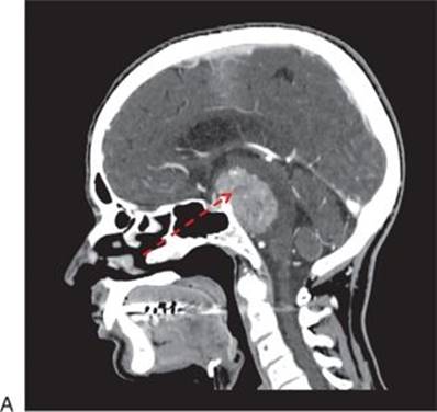
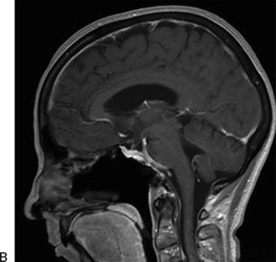
FIGURE 35.1 A. Preoperative CT angiogram sagittal reconstruction showing a petroclival meningioma that requires a pituitary transposition for access to the upper pole of the tumor. The dashed arrowshows the corridor of access achieved following transposition. B. Sagittal MRI following endonasal resection with pituitary transposition.
Tumors that are intimately involved with the posterior cerebral arteries (PCAs) can be difficult to safely dissect without adequate exposure of the capsule of the tumor. Removal of the dorsum sellae or complete gland transposition provides excellent exposure and access to this region. Rare basilar or PCA aneurysms could be accessed as well (Fig. 35.2), though this requires significant experience and very careful patient selection.
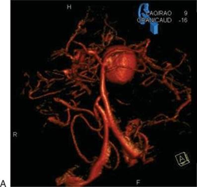
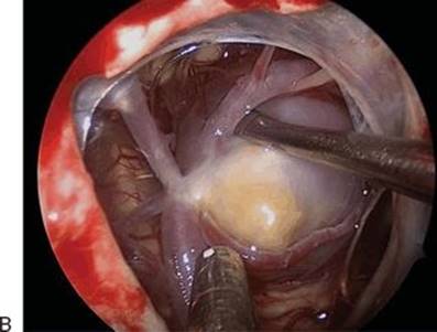
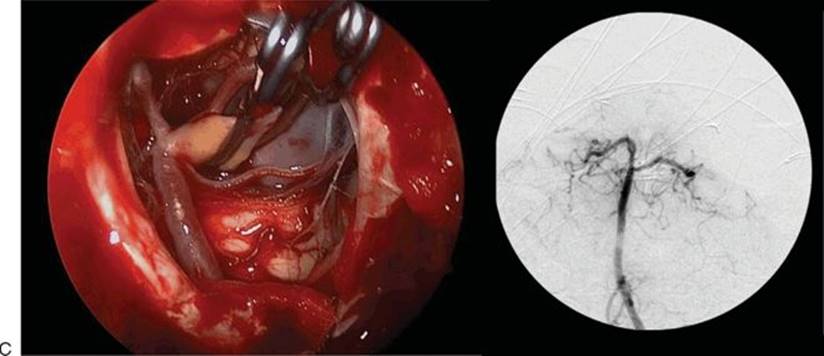
FIGURE 35.2 A. Preoperative CTA showing a three-dimensional reconstruction view of a large PCA aneurysm causing an oculomotor nerve palsy. B. Intraoperative view of the aneurysm before clipping. C. Intraoperative view of the aneurysm after clipping. Intraoperative angiogram showing complete obliteration of the aneurysm and patency of normal basilar apex branches.
CONTRAINDICATIONS
Sinus infection is a contraindication to endonasal intradural (or potentially intradural) tumor resection. However, this can usually be cleared in a short period of time, with antibiotics, surgical drainage, or a combination of therapies. Otherwise, there are no absolute contraindications, though care must be taken to evaluate the relationships of the upper cranial nerves to the mass, especially when it originates laterally such as a petroclival meningioma. If the majority of cranial nerves are displaced medially, a lateral or posterolateral approach is preferable.
Finally, the pituitary transposition approach requires the use of angled endoscopy and a comfort level with management of the cavernous and intercavernous sinuses, basilar venous plexus and the paraclival, and parasellar internal carotid artery (ICA) and its branches. This requires a surgical team with significant prior experience performing endoscopic skull base surgery.
PREOPERATIVE PLANNING
Both magnetic resonance imaging (MRI) and computed tomography angiography (CTA) are important in planning any clival approach. They provide complementary information: MRI shows specific tumor characteristics that may narrow the differential diagnosis and demonstrates the relationships of the nerves to the tumor (especially with FIESTA or fine-cut T2 imaging); CTA reveals bone involvement in the form of erosion or hyperostosis and ICA and basilar artery relationships. It is especially critical to evaluate the size and height of the posterior clinoid processes (PCPs). If there is any sign of calcification of the dural ring or petroclinoidal ligament, dissection behind the ICA can result in potential disaster if this calcified spike is unknowingly manipulated into the artery.
Both MRI and computed tomography (CT) should also be closely examined for signs of sinus infection or other associated sinonasal pathology.
Hormonal evaluation with a full pituitary panel should be performed to document and treat preoperative dysfunction and provide baseline levels for comparison following surgical manipulation of the gland.
Preoperative treatment with corticosteroids is useful for patients with new or progressive neurologic deficits. A full course of antibiotic therapy with careful preoperative reevaluation for clearance of infection is important in patients with identified sinusitis.
SURGICAL TECHNIQUE (SEE VIDEO 35.1)
The superior clivus can be approached endonasally in one of the three ways: extradurally, interdurally, and intradurally. These approaches provide an increasing degree of access to the upper clivus as well as increasing risk of pituitary dysfunction. Both factors should be balanced for optimal tumor management. A purely extradural dissection has almost no risk of permanent pituitary failure but provides limited exposure in cases of extensive clinoid involvement or a “tall” dorsum sellae, whereas an intradural transposition allows complete, unencumbered access to the dorsum, clinoids, and perimesencephalic cistern but may carry a high risk of hypopituitarism from loss of venous drainage and manipulation of the gland.
As with any endoscopic endonasal approach, the surgery is performed by a surgical team consisting of an otolaryngologist and a neurosurgeon. The patient’s head is typically fixed in headpins to prevent inadvertent movement with the head slightly extended and rotated toward the surgeons. The nasal cavity is decongested with oxymetazoline-soaked pledgets immediately following induction. After image guidance is registered, the midface and abdomen (in the rare likelihood of needing an adipose tissue or muscle graft) are prepped and draped. A third- or fourth-generation cephalosporin or equivalent coverage for nasal flora with cerebrospinal fluid (CSF) penetration is given for antibiotic prophylaxis.
Reconstructive needs should be considered prior to the exposure. Reconstruction with a vascularized nasal septal flap must be decided upon and performed prior to a transclival approach as the flap pedicle overlies the sphenoid rostrum, which must be removed to access the midclivus. If there is risk of an intraoperative CSF leak or if there is need for ICA exposure, a flap should be harvested and placed either in the nasopharynx or in the maxillary sinus (if mid or lower clival exposure is needed). The exposure of the upper clivus is essentially identical to a pituitary/sellar exposure with wide sphenoidotomy, extending from the planum/tuberculum to the clival recess and wide opening of the lateral recesses of the sphenoid. In order to safely mobilize the gland superiorly, the entire face and floor of the sella as well as the tuberculum sellae must be removed to prevent compression of the gland when it is elevated. The sellar exposure should extend as far laterally as the medial cavernous sinus to allow adequate mobilization and access to the dura overlying these sinuses. In the case of an interdural or intradural transposition, exposure of the medial ICA should also be performed as the inferior hypophyseal artery will need to be controlled.
For tumors of the clivus with superior extension, the midclivus should generally be drilled prior to proceeding with pituitary transposition. An extradural transposition begins with careful dissection of the dura from the dorsum sella, starting in the midline at the floor of the sella (Fig. 35.3). Depending on the height of the dorsum, an extradural dissection will be adequate to reach the superior edge of the dorsum in the midline, typically the lowest point of the top of the clivus, which extends superiorly out to the PCPs. This area is generally thin and can be removed with a Kerrison rongeur as long as the deep plane with the dura is carefully respected. Inevitable bleeding from the inferior intercavernous sinus and basilar plexus can be controlled with embolization of morselized Gelfoam (Surgifoam, Floseal, Surgiflo, etc.), sometimes repeatedly. The dissection required for a transposition often requires visualization with an angled (45-degree) endoscope. This provides a better view while limiting the degree of gland manipulation.
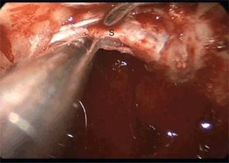
Figure 35.3 Endoscopic endonasal view showing the initial step of extradural pituitary transposition, elevation of the dura from the floor of the sella (S), and dorsum sellae.
As noted, the PCPs are generally slightly superior to the rostral dorsum sellae. In addition, the lateral attachment of the dura of the floor of the sella (the inferior leaf of the inferior intercavernous sinus) to the cavernous sinus takes a slight downturn. This combines to limit access to the tip of the PCP, where the dural ring extending around the ICA attaches. Extreme care must be taken in detaching this dural attachment. If the PCP is shallow and there is no calcification of the dural ring, the PCP can be peeled relatively easily from just behind the ICA without further exposure. However, in many cases, further exposure is necessary for safe removal. This can be accomplished with an interdural dissection (Fig. 35.4). The outer leaf of the inferior intercavernous sinus is opened at its point of attachment to the medial cavernous sinus. Once the venous bleeding is packed off, this triangular space, bounded medially by the wall of the medial cavernous sinus and laterally by the medial parasellar ICA, provides much wider access to the posterior clinoid. The inferior hypophyseal artery, which does not provide critical supply to the gland, crosses the mid portion of the PCP and should either be sacrificed or carefully dissected from the PCP and preserved. Preoperative identification of a calcified dural ring, petroclinoidal ligament, or dural attachment is critical to avoid manipulating this bony spike into the ICA, resulting in injury. If this spike is identified, it should be disconnected (with a Kerrison rongeur, drill, or ultrasonic bone aspirator).
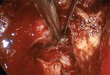
Figure 35.4 Endoscopic endonasal view showing an interdural dissection to expose the posterior clinoid process (PCP). (S, sella; ICA, internal carotid artery; CD, clival dura.)
The exposure provided by the interdural dissection allows careful protection of the ICA with a suction tip while the dural attachments to the PCP are dissected and the process removed either piecemeal or en bloc. This can be done with a Kerrison rongeur, drill, or ultrasonic bone aspirator. If an interdural transposition is performed bilaterally, the entire inferior intercavernous sinus and both medial cavernous sinuses are thrombosed, leaving the superior and lateral cavernous sinus for drainage of the gland. In addition, the inner dural layer, composed of the inner leaf of the inferior intercavernous sinus and the medial wall of the cavernous sinus, provides protection to the adenohypophysis.
By comparison, an intradural transposition (Fig. 35.5) requires sacrifice of both superior and inferior intercavernous sinuses as well as disconnection of the gland laterally from the medial cavernous sinuses and posteriorly from any drainage provided via the dural plexus overlying the dorsum sellae. This, in addition to the mobilization of the stalk and superior hypophyseal arteries, leads to a significant increase in the risk to the function of the adenohypophysis. With any type of transposition, care must be taken not to directly damage the neurohypophysis (posterior pituitary gland).
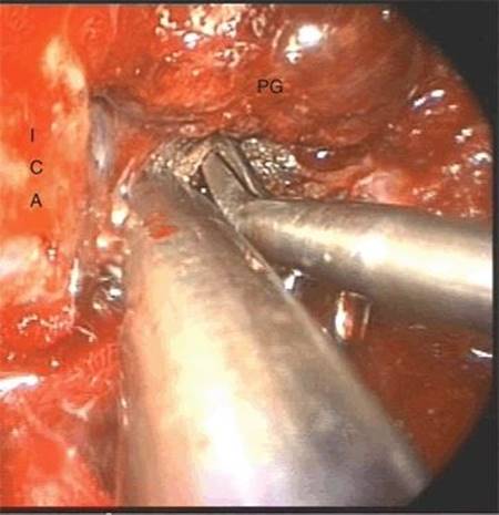
Figure 35.5 Endoscopic endonasal view showing dissection of the dorsum sellae dura following intradural pituitary transposition. (PG, pituitary gland; ICA, internal carotid artery.)
As previously noted, intradural transposition begins with ligation of the superior intercavernous sinus (SIS). The simplest method is to make a small horizontal incision above (suprasellar) and below (over the gland) the sinus and carefully coagulate across it before cutting it. Care should be taken not to inadvertently coagulate a superior hypophyseal artery. Once the SIS is cut, the incision can continue across the diaphragma up to the stalk, completely releasing it from its aperture in the diaphragma. At this point, the lateral gland can be dissected from the medial cavernous wall. Care should be taken to identify the fibrous capsule of the gland and sharply dissect it from the medial cavernous wall by cutting the fibrous bands connecting the two. Inevitably, cavernous bleeding will need to be packed off. The gland is usually relatively easily dissected from the dura on the floor of the sella, leaving only the dorsum dura to dissect before the gland can be lifted in entirety from the sella and placed either on the planum or in the suprasellar space. The opening of the diaphragma to the stalk must be completed to allow full mobilization.
Once the gland is fully mobilized or the dura of the dorsum is opened behind a partially mobilized gland, the intradural view is unparalleled (Fig. 35.6), revealing the mammillary bodies, basilar apex, oculomotor nerves, and deep surface of the stalk. This access allows safe dissection of the upper basilar artery and branches from the deep surface of tumors. In addition, resection of retroinfundibular or hypothalamic tumors can lead to entrance into the third ventricle.
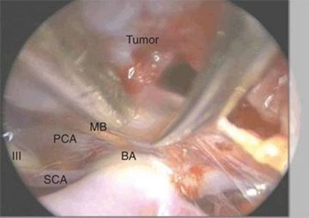
Figure 35.6 Endoscopic endonasal view following pituitary transposition. (BA, basilar apex; PCA, posterior cerebral artery; SCA, superior cerebellar artery; II, oculomotor nerve; MB, mammillary body.)
Once tumor removal is complete, the gland should be replaced into its native position. Even if nonfunctioning, it does provide tissue to help seal a dural defect, and theoretically, the gland has a much better chance of normal function if residing in its “normal” location. Any clival dural defect should be repaired with a vascularized flap whenever possible.
POSTOPERATIVE MANAGEMENT
Naturally, pituitary function must be monitored following pituitary transposition. Even when the dissection is extradural, there is risk for transient DI from manipulation of the gland. If the patient is not on steroids, a cortisol level can be checked each morning to evaluate for adrenal insufficiency (AI). In the absence of DI or AI, other hormone dysfunction is unlikely. Nevertheless, delayed full hormone evaluation should be performed in all patients who undergo a transposition.
When there is a dural defect, packing is left in place for 5 to 7 days while the patient stays on antibiotics (broad-spectrum IV for 48 hours and then oral cephalosporin or equivalent). Upper clival defects carry a high risk of postoperative CSF leak due to significant dural defects associated with arachnoid cistern dissection or even entry into the ventricle. While the role of lumbar drainage is not well studied, these defects should be considered for short-term CSF diversion. As with any endonasal surgery, postoperative precautions such as orders forbidding nasal instrumentation, positive pressure ventilation, and nose blowing should be followed closely. In addition, head of bed elevation and stool softeners (to avoid straining) should be continued even after discharge. Nasal saline irrigation is used in the early postoperative period but should be temporarily discontinued if there is a question of a CSF leak.
COMPLICATIONS
The two major complications of pituitary transposition are pituitary dysfunction and CSF leak. The rates of both for this approach are not well documented and vary depending on degree of transposition and size of defect and extent of dissection, respectively.
The dissection of the ICA required for removal of the PCPs places the ICA at significant risk. Angled endoscopy can improve the view during this dissection, but constant care must be taken to avoid injury. Preoperative identification of a calcified dural ring or attachment to the PCP that could result in ICA puncture is critical to avoid this as a source of injury.
RESULTS
Pituitary transposition is not well studied. Kassam et al. presented the first 10 cases performed with reasonable results, but this was a mixture of intra- and extradural transpositions. This experience has been expanded, and it seems that extradural or interdural transpositions are very effective in achieving significant increase in exposure and access with negligible rates of hypopituitarism. By comparison, intradural transpositions carry a very high rate of pituitary dysfunction, though this is complicated by the fact that this approach is used almost exclusively for tumors that involve the pituitary stalk directly. Nevertheless, we use the intradural transposition very sparingly due to concern for loss of venous drainage and resultant dysfunction.
PEARLS
· The addition of a pituitary transposition is critical for access to areas behind the pituitary gland.
· Removal of the planum and tuberculum prevents compression of the gland during transposition.
· Reconstruction with a vascularized flap decreases the risk of a postoperative CSF leak.
· Use of an angled (30- or 45-degree) endoscope during dissection and dorsum sellae and posterior clinoid resection can increase safety and limit the amount of gland elevation.
PITFALLS
· Intradural transposition should be avoided when possible due to loss of pituitary function.
· Identification of a bony spike on the PCP as a result of calcification of the dural ring or attachment is critical to avoid ICA injury.
INSTRUMENTS TO HAVE AVAILABLE
· A full set of sinus instruments, high-speed drill, and 0- and 45-degree endoscopes are used for the exposure. A microdebrider may facilitate sphenoidotomy.
· Fine and angled-tip, pistol-grip bipolars (Storz) are critical for extra- and intradural hemostasis. Extended tips or Luer lock suctions (without holes) are needed to deliver morselized Gelfoam products (Surgifoam, Floseal, Surgiflo).
· Extendable tip neurodissectors (KLS Martin) and fine, pistol-grip microscissors (Storz) are critical for intradural tumor dissection.
· CUSA (Integra) and Sonopet (Stryker) ultrasonic aspirators both come with extended and bone cutting tips for endonasal use and can be invaluable for fibrous tumors and PCP removal.
SUGGESTED READING
Kassam AB, Prevedello DM, Thomas A, et al. Endoscopic endonasal pituitary transposition for a transdorsum sellae approach to the interpeduncular cistern. Neurosurgery 2008;62(3 Suppl):57–72; discussion 72–74.
Kassam A, Thomas A, Carrau RL, et al. Endoscopic reconstruction of the cranial base using a pedicled nasoseptal flap. Neurosurgery 2008;63(1 Suppl):ONS44–ONS52; discussion ONS52–ONS53.
Zanation AM, Carrau RL, Snyderman CH, et al. Nasoseptal flap reconstruction of high flow intraoperative cerebral spinal fluid leaks during endoscopic skull base surgery. Am J Rhinol Allergy 2009;23(5):518–521.
Prevedello DM, Kassam AB, Gardner P, et al. Endoscopic endonasal approach for craniopharyngiomas: nuances and limitations of treatment, classification and indications of pituitary transposition. In: Nader R, Sabbagh AJ, eds. Neurosurgery case review. New York, NY: Thieme, 2010.
Prevedello DM, Kassam AB, Fernandez-Miranda JC, et al. Transsellar/transdorsal approach via a pituitary transposition to the interpeduncular cistern. In: Stamm AC, ed. Transnasal endoscopic skull base and brain surgery. New York, NY: Thieme, 2011:256–262.