
Daniel F. Kelly and Chester F. Griffiths
INTRODUCTION
The endoscopic endonasal transsphenoidal approach to the sella and parasellar regions is now increasingly used for removal of pituitary adenomas and Rathke's cleft cysts (RCCs) as well as other parasellar tumors such as craniopharyngiomas, tuberculum sella meningiomas, and clival chordomas. The advantage of the endoscope in removing pituitary and parasellar tumors over the microscope is enhanced visualization. With the light source taken directly into the sphenoid sinus and sella, the improved panoramic view can result in more complete removal of the tumor than is possible with the relatively restricted tunnel vision afforded by the microscopic view through a rigid endonasal or sublabial speculum.
The transition from microscopic to endoscopic sellar and parasellar surgery has occurred gradually. The first rigid endoscope for transsphenoidal surgery with an external light source was used by Guiot in the early 1960s. Hardy also used the endoscope occasionally to explore the sellar cavity after tumor removal to look for residual tumor. In 1977, Apuzzo et al. reported the use of an angled telescope during sellar procedures to assist with visualization for tumor removal or gland ablation. In 1992, Jankowski et al. reported successful endoscopic endonasal resection of pituitary adenomas in three patients. The first clinical series of purely endoscopic pituitary tumor removals in 50 patients without the microscope was described by Jho and Carrau in 1997. Since then, endoscopic pituitary surgery has gained great popularity, and many surgeons doing microscopic pituitary surgery, including our own group, have transitioned to an endoscope-assisted method or fully endoscopic approach for removal of pituitary adenomas and other parasellar tumors. Over the last decade, with further refinements in endoscopic image quality and dedicated instrumentation, the endoscopic approach for pituitary adenomas and related skull base tumors is rapidly becoming the preferred technique, if not the new standard for approaching such lesions. During this period, most surgeons doing endoscopic pituitary surgery have also transitioned from a single nostril to a binostril approach affording increased maneuverability and expanded parasellar access. Currently, the two-surgeon approach is most often used in which one surgeon, typically a head and neck surgeon with expertise in sinonasal endoscopy, begins the endonasal surgical approach phase of the procedure and “drives” the endoscope while the neurosurgeon uses bimanual microdissection to remove the tumor and do the skull base closure.
This chapter describes the endoscopic endonasal approach to the sella using a binostril two-surgeon technique for removal of pituitary adenomas and RCCs. Surgical indications, preoperative planning, room setup, equipment needs, technical nuances, complication avoidance, and postoperative care are described.
HISTORY
All patients with a pituitary tumor or other parasellar lesion should be carefully questioned regarding neurologic symptoms such as loss of visual fields or acuity, diplopia, memory loss, cognitive impairment, and headaches. Patients should also be questioned regarding symptoms of pituitary hormonal excess in cases of acromegaly, Cushing's disease, prolactinoma, and TSH-secreting adenomas. Symptoms of anterior pituitary hormonal deficiency should be evaluated including fatigue, low energy, poor exercise tolerance, depression, weight gain or weight loss, decreased libido, sexual dysfunction, and amenorrhea. Symptoms of frequent urination and excessive thirst suggestive of posterior pituitary failure (diabetes insipidus) should also be sought. For patients with visual complaints and a macroadenoma or other large parasellar tumor, formal evaluation by an ophthalmologist is recommended. For patients with a sellar or suprasellar mass with symptoms or signs of pituitary gland dysfunction, evaluation by an endocrinologist is essential. A history of allergic rhinitis, sinusitis, nasal or sinus surgery/trauma, or disorders of smell and taste should be reviewed.
PHYSICAL EXAMINATION
A complete general and detailed neurologic examination as well as a rhinologic head and neck examination is indicated for all patients being considered for endonasal surgery. For patients with pituitary macroadenomas or other large sellar and parasellar lesions causing compression of the optic apparatus or cavernous sinus, an examination by an ophthalmologist with attention to visual acuity and fields, pupillary reactivity, and extraocular movements is indicated. If there is visual loss, optic nerve coherent tomography can estimate the degree of optic nerve fiber loss and potential for recovery. In these cases, a neuro-ophthalmology consultation is essential. For patients with Cushing's disease, attention to body habitus, distribution of adipose tissue, ecchymoses, stria, skin breakdown, infection, and hypertension should be noted. For patients with acromegaly, attention to potential upper airway obstruction, macroglossia, signs of spinal stenosis, advanced cardiac disease, and hypertension should be noted.
INDICATIONS
The endoscopic endonasal approach is appropriate for removal of virtually all symptomatic sellar lesions including endocrine-active and endocrine-inactive pituitary adenomas and RCCs.
CONTRAINDICATIONS
The endoscopic endonasal approach may not be ideal for the rare highly invasive pituitary adenoma that extends anteriorly far into to the frontal fossa or far lateral beyond the supraclinoid carotid arteries. In such cases, a supraorbital or pterional craniotomy could be used instead of or in conjunction with an endonasal approach. Another relative contraindication for the endonasal approach is active and severe sinusitis, which may require antibiotic treatment and a delay in surgery.
PREOPERATIVE PLANNING
Head and Neck Consultation
Preoperative evaluation, discussion, and additional informed consent should be performed by the otolaryngologist participating in the care of the patient. Prior nasal and sinus conditions should be addressed, and appropriate additional therapy should be discussed. It is not uncommon to have coexisting nasal and sinus disease in patients undergoing endonasal surgery. Evaluation of olfactory function should be included in the evaluation. Simple “scratch and sniff” tests are available to objectively evaluate this function preoperatively. We use the Senonics (Haddon Heights, New Jersey, www.sensonics.com) “Brief Smell Identification Test Version A” and repeat it 3 to 6 months postoperatively. We review with the patient the postoperative nasal and sinus care with sinus lavage using the NeilMed Sinus Rinse system (Santa Rosa, California, www.neilmed.com) and the schedule for postoperative debridement. Patients unfamiliar with the sinus rinse begin it preoperatively to familiarize themselves with the process with the added benefit of cleansing the nasal cavity of debris or crusts before surgery. With multiple surgeons and their ancillary staff discussing the procedures, patients tend to be better informed and prepared by the repetition of the details. Prior to the procedure, a surgical team discussion regarding the approach, tumor extent, and strategy for removal and reconstruction with preparation of vascular flaps, if necessary, is mandatory.
Medical Evaluations and Clearance
Patients should have a thorough preoperative medical clearance. For those with acromegaly, Cushing's disease, or other significant risk factors such as smoking, hypertension, or advanced age, a cardiac clearance with stress test is generally warranted. For acromegalic patients with evidence of obstructive sleep apnea or severe macroglossia, a preoperative pulmonary evaluation is recommended. In acromegalic patients with poorly controlled hypertension, diabetes, and/or obstructive sleep apnea, consideration should be given to a 1- to 3-month preoperative course of a somatostatin analog such as lanreotide or octreotide to lower growth hormone and IGF-1 levels and reduce perioperative morbidity. Those with preoperative adrenal insufficiency, hypothyroidism, or diabetes insipidus should be treated with appropriate hormone replacement before surgery, ideally under the supervision of an endocrinologist.
Imaging
A high-quality MRI with gadolinium of the sella including the paranasal sinuses and skull base is indicated for all patients undergoing endoscopic endonasal tumor removal. In anticipation of using intraoperative frameless navigation, a thin-slice axial T1-weighted postgadolinium brain series should also be obtained. Prior to surgery, careful attention to the parasellar and cavernous carotid flow voids should be made. Displacements of the pituitary gland, infundibulum, and optic apparatus by tumor should be noted, as should the location of the diaphragma sellae and whether there is tumor invasion of the cavernous sinus. Although some surgeons advocate doing a thin-cut CT for all patients undergoing endonasal surgery, We generally reserve CT or CTA for predominantly clival lesions or those with significant vascular encasement and for patients with prior surgery in whom bony landmarks may be greatly altered or in patients who have undiagnosed nasal or sinus abnormalities, either pathologic or developmental, found on the preoperative MRI.
Informed Consent
Depending upon the presumed pathology, patients should be carefully counseled as to expected outcomes, likelihood of remission, recovery of vision, possible need for tissue grafts and nasoseptal flap, as well as the associated surgical risks. In particular, the potential likelihood of new pituitary failure, CSF leak, visual loss, diplopia, hematoma, carotid or other vascular injury, infection, and anosmia should be discussed.
SURGICAL TECHNIQUE
Overview
Although the procedure requires two surgeons, the initial nasal approach to the sphenoid sinus is typically done by the otolaryngologist alone; the sellar and parasellar exposure, tumor removal, and skull base reconstruction are done by the neurosurgeon and otolaryngologist together. When both surgeons are operating, the endoscope is generally maneuvered by the otolaryngologist and placed in the upper quadrant of the right nostril while the neurosurgeon uses binostril access, typically with a suction in the right nostril and microdissector, ring curette, or other instrument in the left nostril (Fig. 3.1). With an angled 30- or 45-degree scope looking up, the endoscope often needs to be positioned in the inferior nostril and nasal cavity to minimize conflict of instruments.

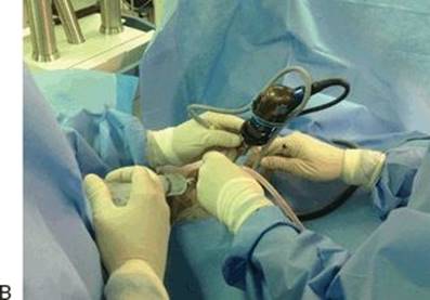
FIGURE 3.1 A. Drawing of binostril endoscopic approach to sella with endoscope in right nostril and additional instruments in each nostril. The inset drawing shows convergence of suction and ring curette in lower sella for adenoma removal as viewed by 0-degree endoscope placed in sphenoid sinus (A, adenoma; P, compressed pituitary gland; D, dura). B. Intraoperative photograph of hand positions and endoscope with otolaryngologist holding endoscope and irrigation while neurosurgeon has suction in left hand (in right nostril) and ring curette in right hand (in left nostril). (Image copyright Daniel Kelly Brain Tumor Center 2012.)
Instrumentation
Endoscopic equipment includes 4-mm rigid endoscopes (18 cm in length) with 0-, 30-, and 45-degree angled lenses and high-definition (HD) camera and two flat panel monitors (Karl Storz, Tuttlingen, Germany). Given the narrow working space afforded by the binostril endoscopic approach, all instruments should be as thin and low profile as possible. They may be straight or bayoneted depending upon the surgeon's preference. After many years of performing endonasal microscopic pituitary surgery, We prefer bayoneted microinstruments. Microdissectors, ring curettes, and microblades are on bayoneted handles. Similarly, microscissors, tumor grasping forceps (both straight and up angled), and the bipolar cautery are used in a single-shaft pistol-grip design to minimize visual obstruction. High-speed drills, microdebriders, and ultrasonic aspirators also are of the lowest possible diameter with angled handpieces. Straight and curved variable suctions should also be available. A micro-Doppler (Koven, Inc. or Mizuho, Inc.) probe is also used for all cases to localize the cavernous carotid arteries prior to opening the dura. Warm (99°F/37°C) sterile saline is used for irrigation both to clean the tip of the scope and to promote local hemostasis. A 50-cc syringe with a curved irrigation tip is used to deliver the irrigation into the operating field when necessary.
Preoperative Medications
Preoperative antibiotics (typically cefazolin) are given and continued for 24 hours. In patients with normal preoperative adrenal function or those with Cushing's disease, no perioperative glucocorticoids are administered. Those with adrenal insufficiency or borderline adrenal function are given 100 mg of hydrocortisone intravenously.
Positioning, Room Setup, and Prep
Following induction of general anesthesia with the patient in the supine position, the endotracheal tube emerges from the left corner of the mouth, and the anesthesiologists and anesthesia equipment are positioned on the patient's left side. For patients with Cushing's disease, acromegaly, other significant medical co-morbidities, or a large and vascular tumor, an arterial line and Foley catheter are placed; for other patients with microadenomas, small macroadenomas and typical RCCs, an arterial line and Foley catheter are not used.
An ergonomically efficient operating room setup is essential to ensure comfort of both surgeons especially during lengthy procedures. Two video monitors are positioned at almost 90-degree angles to each other: one above the patients' head and one to the left of the chest; the neuronavigation monitor is placed in between the two video monitors. Our current operating room configuration is depicted in Figure 3.2. The patient's head is placed in a horseshoe head holder and angled approximately 30 degrees toward the left shoulder. This arrangement allows both surgeons to stand comfortably on the patient's right side, one at the head and one immediately below the head, and able to comfortably view their respective video monitors. The head is inclined in a neutral plane (0 degree) relative to the floor for sellar lesions; for suprasellar lesions, 10 to 15 degrees of neck extension is used, and for infrasellar and clival lesions, 10 to 15 degrees of neck flexion is used. The surgical navigation mask (Stryker Navigation) is placed on the face, and the system is registered to the preoperative MRI and/or CT angiogram. Only in prolonged cases is the head pinned in 3-point fixation, for example, with a craniopharyngioma or tuberculum sella meningioma in which the operative time may exceed 6 hours and there is risk of pressure necrosis with the horseshoe head holder.


FIGURE 3.2 A. Diagram of operating room setup for endoscopic endonasal surgery for two surgeons with two HD video monitors, neuronavigation monitor, and ancillary equipment. (Image copyright Daniel Kelly Brain Tumor Center 2012.) B. Photograph of operating room setup showing HD monitors, neuronavigation monitor, and patient positioning prior to final draping. C. Intraoperative photograph of two surgeons looking toward their respective monitors.
The nasal cavity is prepped with decongestant (oxymetazoline 0.05%)-soaked Cottonoids placed in both nares for several minutes. The face, perinasal area, and right lower abdominal area (for a possible adipose tissue graft) are sterilely prepped and draped. A clear drape is used to allow visualization of the navigation mask during surgery. If the patient is fixed in pins and the tracking unit for navigation is attached to the Mayfield, a clear drape is not needed. Xylocaine 1% with 1:100,000 epinephrine is injected into the inferior and middle turbinates and lateral nasal walls bilaterally.
Approach to the Sphenoid Sinus
The initial approach through the nasal cavity uses a 0-degree 4-mm rigid endoscope and includes handling of the turbinates, raising of bilateral nasoseptal (NS) mucosa-preserving rescue flaps, wide sphenoidotomy, posterior septectomy, and posterior ethmoidectomies. As described below, we rarely use a vascularized nasoseptal flap in the skull base reconstruction and CSF leak repair for pituitary adenomas and RCCs and instead use a NS flap only in larger extended transplanum or transclival approaches. Using a Cottle elevator, the inferior and middle turbinates are out-fractured bilaterally, and the sphenoid ostia are identified. The middle turbinates are not routinely resected. We reserve the resection of the middle turbinate for lateral skull base pathologies such as Meckel's cave and pterygopalatine fossa lesions. It has been suggested that postoperative debridements are more difficult if the middle turbinate is not resected, but this has not been our experience. Bilateral NS “rescue flaps” based on the posterior nasal artery are created (Fig. 3.3). An extended shaft, manually bent, microtip Bovie (Megadyne E-Z Clean 6.0″/152 mm ref: 0016M, Draper, Utah) is used to incise the mucoperiosteum inferior to the sphenoid ostium preserving the posterior septal artery pedicle, which comprises two arterial branches in 80% of cases and is located 8 to 9 mm below the ostium. The incision is then extended anteriorly using the inferior aspect of the superior turbinate as a horizontal guide to preserve the septal olfactory strip (SOS mucosal flap) for approximately 2 cm along the vomer and posterior nasal septum. This maneuver is performed bilaterally. These bilateral mucoperiosteal “rescue flaps” are then pushed inferiorly toward the nasopharynx with Cottonoids to minimize obstruction and provide access into the sphenoid sinus (Fig. 3.4). The SOS mucosal flaps are pushed laterally and superiorly onto the superior turbinate where they usually become adherent and out of the surgical field. These mucosa-preserving flaps obviate the need to transect the sphenopalatine artery, thereby greatly reducing the potential for postoperative sphenopalatine hemorrhage and epistaxis. With the majority of the endonasal mucosa preserved and out of the surgical field, any remaining superior mucosa may be excised with the microdebrider but removal of this mucosa is infrequently needed. The sphenoid keel, vomer, and posterior nasal septum are demucosalized and in view. A wide sphenoidotomy is then performed with up and down biting Kerrison rongeurs or the drill. This bone removal can be done from ostia to ostia superiorly and inferiorly to preserve a large piece of keel that can be harvested and retained for possible use in reconstruction of the floor of the sella after removing the tumor. A posterior septectomy of approximately 15 to 20 mm is then performed with a backbiter typically placed through the left nostril. Care should be taken to not extend the septectomy too far superiorly or anteriorly, which can increase the risk of anosmia and nasal deformity. The sphenoidotomy is then further refined based upon the pathology being addressed in the sella. In general, however, removal of bone and mucosa should extend beyond the lateral edges of the ostia bilaterally to allow visualization of the tuberculum sella, floor of the sella, opticocarotid recesses, clival recess, and lateral sphenoid recesses. Posterior ethmoid air cells are also opened and removed to facilitate maneuverability of the endoscope and instrument superiorly.
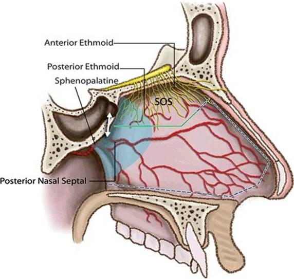
FIGURE 3.3 Illustration of NS rescue flap concept with preservation of sphenopalatine and posterior NS arteries and SOS. The mucosal incisions are started several millimeters below the inferior aspect of the sphenoid ostia and carried anteriorly as shown by the green line. The dotted line shows mucosal incisions if a complete NS flap is needed. The double arrow denotes the 9-mm distance typically between the inferior edge of the ostium and posterior nasal septal artery. The blue shading indicates the extent of the posterior septectomy. (Image copyright Daniel Kelly Brain Tumor Center 2012.)
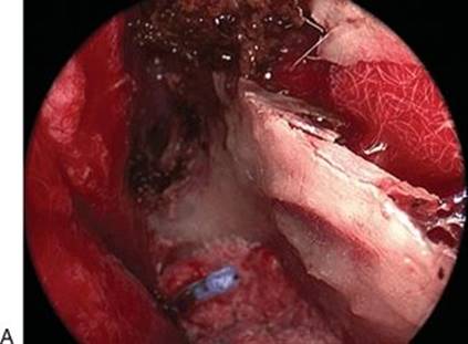
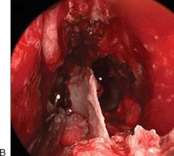
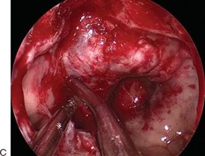
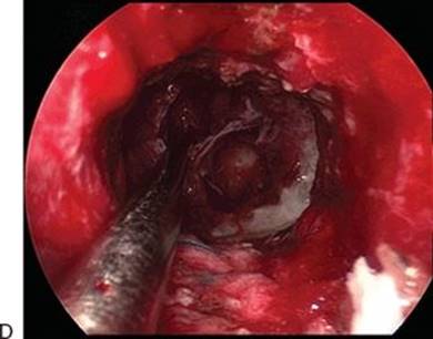


FIGURE 3.4 Intraoperative photographs with 0-degree endoscope of bilateral nasoseptal “rescue flaps” being created and preserved during tumor removal and at procedure completion. A. Sphenoid keel exposed and right rescue flap pushed down by long Cottonoid; left rescue flap being pushed downward as Cottonoid is being placed. B. Posterior septectomy completed, and both rescue flaps pushed downward by bilateral Cottonoids. C. Sphenoidotomy completed and Doppler probe localizing right cavernous carotid artery. D. After removing the tumor, view from posterior nasal cavity showing no obstruction from rescue flaps. E. Adipose tissue graft being placed into sella for reconstruction for CSF leak. F. After reconstruction is completed with adipose tissue graft and collagen sponge, Cottonoids are removed, and rescue flaps are replaced.
Sellar Exposure
During the sphenoid and sellar portion of the procedure, when the otolaryngologist is driving the endoscope and the neurosurgeon is operating, a sterile-draped pillow on a Mayo stand is positioned just above the head as an elbow rest to reduce arm fatigue while driving the endoscope. After the sphenoidotomy is completed, the face of the sella is identified, and intrasphenoidal bony septations are correlated with the patient's preoperative MRI. Particular note is made on the coronal images of where these septations reach the posterior wall of the sphenoid sinus relative to the carotid arteries, pituitary gland, and tumor and on sagittal views where such septations reach the planum or sella. Septations that end on the face of the sella are removed with a rongeur or high-speed drill down to the sella; those that end over a carotid artery should be removed with care, and excessive torquing of these septations should be avoided. The mucosa over the sella is removed, but the remaining mucosa of the sphenoid sinus is left undisturbed. The bony face of the sella is then removed from cavernous sinus to cavernous sinus and from the floor of the sella inferiorly to the tuberculum sella superiorly with a Kerrison rongeur or in some instances a high-speed hybrid-diamond bit drill. With large invasive tumors, the bone of the sella may be markedly thinned or absent, and the tumor may be directly under the mucosa or attenuated dura.
Localization of the Cavernous Carotid Artery
After opening the bony sella, although the carotid protuberances should be readily visible as should the lateral opticocarotid recesses, reaffirming the course of the carotid arteries with the micro-Doppler probe is recommended. The probe (10-MHz ES-100X MiniDop®with NRP-10H bayonet probe, Koven, St. Louis, MO, or 20-MHz Surgical Doppler, Mizuho America, Beverly, MA) is placed initially at the edge of the bony opening at 90 degrees to the dura. If faint or no audible flow is present, the probe is angled more laterally aiming under the bone edge, and in most cases, the carotid flow will become louder (Fig. 3.5). The probe is then moved superiorly and inferiorly to define the course of the carotid arteries, which typically have their most medial course superiorly and distally near the tuberculum sella before they pass through the dural ring to enter the subarachnoid space. If no Doppler flow is evident, then additional bone can be removed laterally to maximize exposure of the sella. If audible flow is still not evident and visual anatomy and navigation images indicate that the carotid has been exposed, consideration should be given to whether there is a technical problem with the probe.
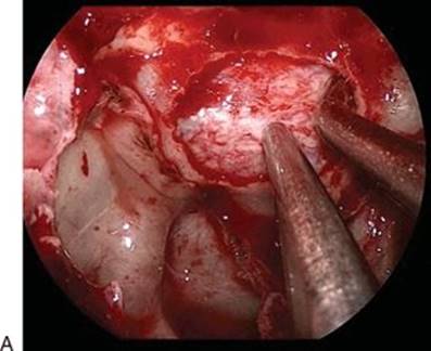

FIGURE 3.5 A. Intraoperative photograph of Doppler probe along left cavernous sinus after removal of the bony wall of the sella and before dural opening. B. Drawing of Doppler probe for localizing left cavernous carotid artery with medial edge of cavernous sinus exposed (shaded in blue) (A, adenoma; P, pituitary gland; CC, cavernous carotid artery; OC, optic canal). (Image copyright Daniel Kelly Brain Tumor Center 2012.)
Dural Opening
A wide U-shaped, superiorly based opening is made in the dura of the sella using a straight microblade (Mizuho Inc.). The initial opening in the dura should not transgress the pituitary gland or adenoma if possible. Angled microdissectors are then used to separate the dura from the underlying tumor and pituitary gland. The opening in the dura is enlarged superiorly, inferiorly, and laterally as needed with the use of a right-angled microhook blade or curved microscissors, which allow the cutting force of the blade to be directed away from the sella and cavernous sinus. Care should be taken in extending the opening in the dura too far superiorly in patients with microadenomas who often have a shallow sella and low-lying diaphragma sellae; such an opening can cause an early CSF leak. Laterally, the opening should generally extend to within 1 to 2 mm of the medial wall of the cavernous sinus. Low-pressure cavernous sinus venous bleeding is generally easily controlled using Surgifoam (Ethicon Inc., Johnson & Johnson Co., Piscataway, NJ) or Gelfoam (Pfizer Inc., New York, NY).
Tumor Removal
The binostril endoscopic approach is depicted in Figure 3.1A and B. A selective and complete removal of the tumor with preservation or improvement of pituitary gland function should be the goal for patients undergoing removal of an adenoma. In many instances, the tumor pseudocapsule can be identified and a plane established between the adenoma and the normal gland. Using microdissectors, irrigation, and gentle traction on the pseudocapsule, such adenomas can often be removed completely with preservation of the pseudocapsule as described by Oldfield et al. However, many if not most large macroadenomas are quite soft and require initial internal debulking with ring curettes and suction. After so doing, the tumor “rind” with an intact pseudocapsule can be gently separated away from the normal gland and diaphragma sellae (Fig. 3.6). For firm or rubbery adenomas, initial tumor debulking with curved and straight microscissors may be needed. Adenomas with suprasellar extension should be debulked inferiorly first followed by the suprasellar component. This sequence allows the suprasellar tumor, in part, to deliver itself from above and may minimize the chances of an early CSF leak. For large macroadenomas, it is essential to confirm descent of the diaphragma sellae as an indication that complete tumor removal has been accomplished. Probing the folds of the diaphragma with 45- and 90-degree up-angled ring curettes bilaterally, posteriorly, and anteriorly will help dislodge residual tumor when present in these areas. To further encourage downward descent of a suprasellar tumor, the anesthesiologist can induce a Valsalva maneuver to transiently increase intracranial pressure. Since most macroadenomas, even with a large suprasellar extension, are contained by a thinned but largely intact diaphragma sellae, the diaphragma sellae should completely evert and fall into the enlarged sella once a complete tumor removal is accomplished. If a full “mirror image” descent of the diaphragma sellae is not seen and only a partial descent of the diaphragma is noted, residual tumor is likely present.
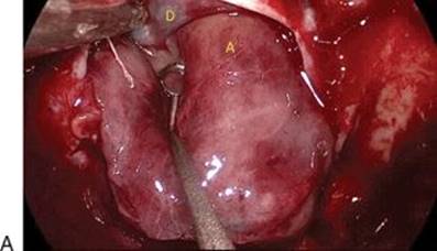
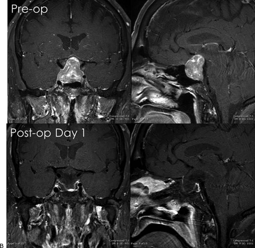
Figure 3.6 Large endocrine-inactive macroadenoma with visual loss and anterior pituitary gland failure. A. Intraoperative photograph with 30-degree endoscope of pseudocapsular dissection for large macroadenoma using ring curette and suction (D, diaphragma sella; A, pituitary adenoma pseudocapsule). B. Preoperative and postoperative coronal and sagittal postgadolinium MRIs demonstrating complete tumor removal.
For tumors with obvious or possible invasion of the cavernous sinus, visualization of the medial wall of the cavernous sinus is essential. Under direct visualization, what appeared to be invasion of the cavernous sinus on MRI may only be tumor compression of the medial wall of the cavernous sinus. However, in many cases, after the tumor in the sella has been removed, a defect in the medial wall of the cavernous sinus is seen. Tumor in the medial cavernous sinus can be removed or at least effectively debulked using gentle suction and angled ring curettes that have smooth outer edges (Fig. 3.7). Given the potential for injury to the cavernous carotid and the abducens nerve lateral to the carotid, aggressive curetting or grasping of tumor tissue along or lateral to the carotid artery should be avoided.
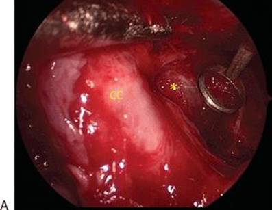

Figure 3.7 Invasive macroadenoma with invasion of the right cavernous sinus. A. Intraoperative photograph with 45-degree endoscope showing tumor removal in right medial cavernous sinus using ring curette and suction. B.Preoperative (top row) and postoperative day 1 (bottom row) MRIs showing gross total tumor removal of sellar and right cavernous sinus and repair of grade 2 CSF leak (CC, cavernous carotid artery; asterisk, retro-genu space in medial cavernous sinus; arrow, adipose tissue graft in right sella and medial cavernous sinus).
Once the tumor has been removed as completely as possible with a 0-degree endoscope, 30- and 45-degree angled endoscopes can be used to search further for residual tumor. This angled visualization is especially helpful in cases where the diaphragma sellae may not have descended fully into the sella. Removal of such tumor remnants can often be performed with angled suctions, tumor grasping forceps, or ring curettes.
Rathke's Cleft Cysts
Since intrasellar and intrasellar/suprasellar RCCs are typically located posterior to the anterior pituitary gland, removing them generally involves a direct approach through the anterior–inferior pituitary gland via a low midline vertical glandular incision or an approach under the gland avoiding an incision in the gland. Through this small working corridor, the cysts are usually easily removed with suction, ring curettes, and gentle irrigation. A 30- or 45-degree endoscope facilitates excellent visualization of the cyst cavity with minimal manipulation of the gland. After complete removal of the cyst, the cavity is inspected for residual cyst contents and cyst lining. Given that the resection cavity generally consists of a normal-appearing anterior and posterior pituitary gland, no attempt is made to vigorously strip the cyst wall off of these normal structures given the risk of worsening gland function. For purely suprasellar RCCs that are typically intimately attached to the pituitary stalk and often embedded within the superior aspect of the anterior pituitary gland, an extended transtubercular approach is often needed to allow maneuvering over the normal pituitary gland.
Intrasellar Hemostasis
After tumor removal, hemostasis is obtained with Surgifoam (Ethicon Inc., Johnson & Johnson Co., Piscataway, NJ) and full-strength hydrogen peroxide. The peroxide is irrigated directly into the sphenoid sinus and sella for 1 to 2 minutes. We have recently demonstrated that its use is safe from the standpoint of pituitary hormonal function, and it may have additional tumoricidal effects on residual microscopic foci of adenoma. However, it should not be used if there is a large diaphragmatic defect, which could allow it to track into the subarachnoid space. In cases in which there is persistent oozing, one should further inspect the sella with the endoscope to look for residual tumor.
Skull Base Reconstruction and CSF Leak Repair
Skull base reconstruction and CSF leak repair can be tailored to the size of the CSF leak and the bony and dural defects. Prior to reconstruction, an assessment of the size of diaphragmatic defect is performed. We grade CSF leaks as grade 0 (no leak), grade 1 (small), grade 2 (moderate), and grade 3 (large). If no obvious defect is seen, a Valsalva maneuver is induced to help visualize an occult or small (grade 1) CSF leak emanating through a small diaphragmatic defect.
All reconstruction involves the use of collagen sponge (Duragen, Helistat, or Instat) that acts as a scaffolding for fibroblast ingrowth and a vascularized dural replacement. In patients with no CSF leak (grade 0), a single layer of minimally moistened collagen sponge placed over the exposed diaphragma sellae, pituitary gland, and sellar dura is typically used as the only repair material. For most small (grade 1) CSF leaks, the repair includes intrasellar collagen sponge with an intrasellar, extradural buttress of previously harvested sphenoid keel bone or synthetic or absorbable plate and less frequently use of titanium mesh. A second outer layer of collagen is placed over the buttress and adjacent sellar and sphenoid bone. The repair is typically held in position with a small amount of tissue glue (DuraSeal, Confluent Surgical, Inc. or Tisseel, Baxter, Inc.). For medium (grade 2) CSF leaks or grade 1 leaks with a large intrasellar dead space, the repair includes an intrasellar abdominal adipose tissue graft, a layer of collagen sponge followed by a buttress of bone, synthetic plate, or titanium mesh wedged into the intrasellar extradural space. Additional adipose tissue is typically placed over the sella followed by another layer of collagen; the reconstruction is held in position with tissue glue (Fig. 3.8). In some grade 1 leaks in which the sella is filled with adipose tissue and the floor of the sella is intact with at least some anterior lip remaining to help hold the adipose tissue graft within the sella, only collagen sponge is layered over the adipose tissue followed by tissue glue without a rigid buttress. Finally, in cases of large tumors with grade 1, 2, or 3 leaks and a large dead space, in which the bony edges of the sella have been removed or eroded by tumor precluding wedging of a rigid buttress, single or binostril Merocel (Medtronic Mystic, Connecticut) nasal packs are passed into the sphenoid under direct endoscopic visualization up to the sellar reconstruction as a soft temporary buttress for 5 days (Fig. 3.9). All patients with an intraoperative CSF leak are placed on Diamox (acetazolamide) 250 mg, every 8 hours for 48 hours after surgery to diminish CSF production. To further evaluate the adequacy of the repair, prior to placing tissue glue, a Valsalva maneuver is performed to raise the patient's intracranial pressure; should there be CSF streaming around the repair or movement of the buttress, the repair should be revised. It is important to note that use of tissue glue in this repair paradigm is not for stopping egress of CSF per se but to prevent migration of the reconstruction materials (adipose tissue and collagen) away from the sella. For the large (grade 3) defects, typically seen with extended approaches, consideration should be given to use of a NS flap. However, it is unusual to have a grade 3 leak after removal of pituitary adenoma or RCC. In the minority of patients with a grade 3 leak, a lumbar drain for CSF diversion can be placed for 48 to 72 hours.
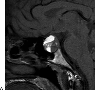

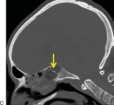
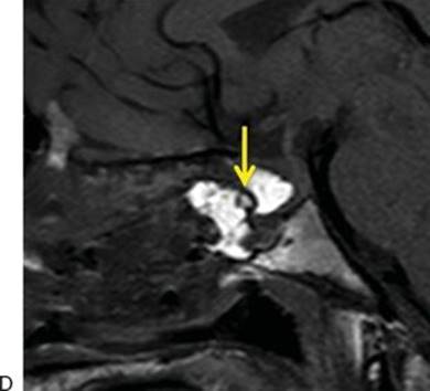
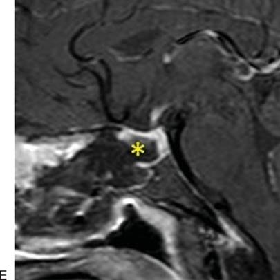
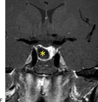
Figure 3.8 Macroprolactinoma with pituitary apoplexy. A. Pregadolinium sagittal MRI showing subacute hemorrhage. B. Postgadolinium coronal MRI showing severe compression of the gland and elevation by hemorrhagic tumor. C and D. Immediate postoperative CT with sagittal reconstruction and post-op day 1 sagittal MRI without gadolinium showing intrasellar and sphenoid sinus adipose tissue grafts with intervening bony buttress (arrow) for grade 2 CSF leak repair. E and F. Post-op day 1 sagittal and coronal postgadolinium MRIs with adipose tissue suppression sequence (asterisk) showing gross total tumor removal with reexpanded pituitary gland. Patient's prolactin fell from 286 to 16 ng/mL on postoperative day 2.
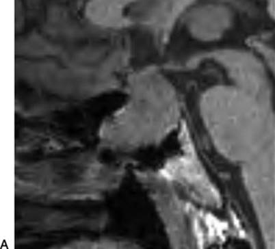
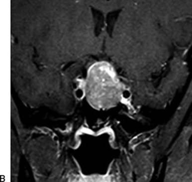
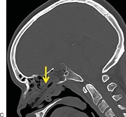
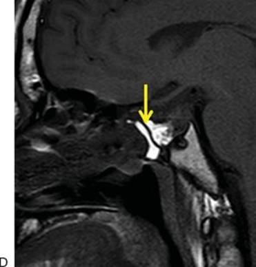
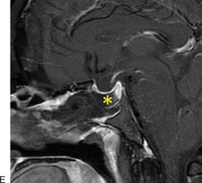
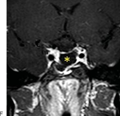
Figure 3.9 Invasive endocrine-inactive macroadenoma with large extension into the sphenoid sinus. A and B. Preoperative sagittal and coronal MRIs. C. Grade 1 CSF leak repair—immediate postoperative CT with sagittal reconstruction showing intrasellar and sphenoid sinus adipose tissue grafts held in position by binasal Merocel buttress (arrow). D. Post-op day 1 sagittal MRI without gadolinium showing intrasellar and sphenoid sinus adipose tissue grafts with intervening collagen sponge (arrow). E and F. Post-op day 1 sagittal and coronal postgadolinium MRI with adipose tissue suppression sequence (asterisk) showing gross total removal of the tumor with reexpanded pituitary gland.
Closure
Blood is suctioned from the sphenoid sinus, nasal cavity, and nasopharynx. Nasal hemostasis should be relatively complete to minimize blood being swallowed after extubation, which can cause nausea and vomiting and dislodgement of the materials used for skull base reconstruction. The previously created mucoperiosteal “rescue flaps” including the NS and SOS are returned to their normal anatomic positions. The NS flaps are gently elevated back along the residual sphenoid keel and inferior anterior nasal septum (Fig. 3.4). The middle turbinates are repositioned anatomically if they remain out-fractured. Nasal packing is not used unless a Merocel is needed to help hold the sellar floor reconstruction in position. To minimize chances of a postoperative CSF leak, nasal epistaxis, or intrasellar bleeding, excessive coughing on the endotracheal tube should be avoided during extubation, and blood pressure should be carefully monitored and controlled in the early postoperative period.
POSTOPERATIVE MANAGEMENT
Most patients undergoing removal of an adenoma or RCC are admitted to a non-ICU bed; their arterial line is removed before leaving the recovery room. For the first postoperative night, patients are given a humidified face tent, and decongestants are available upon request. Saline nasal spray is provided for a week after surgery and used based on patient preference. The Foley catheter is removed on the morning of postoperative day 1, and patients are encouraged to ambulate. An early postoperative MRI or CT scan is typically performed within 2 days of surgery for patients with macroadenomas or other larger tumors. All patients with pituitary-related lesions are followed in-hospital by an endocrinologist. Patients are monitored for diabetes insipidus based on urine output and urine specific gravity. Adrenal function is monitored by measuring AM serum cortisol and adrenocorticotropic hormone (ACTH) levels on the mornings of postoperative days 1 and 2. For patients with acromegaly, prolactinoma or Cushing's disease, growth hormone, prolactin, and cortisol/ACTH levels are followed on postoperative days 1 and 2 to document early remission based on subnormal hormone levels.
Most patients are discharged home on postoperative day 2 and have a serum sodium level checked on postoperative day 4 or 5 to monitor for delayed hyponatremia. Antibiotics are continued for only 48 hours (until discharge) if no nasal packs are in place or for 5 days when nasal packs are removed. Outpatient endoscopic sinonasal debridements are typically performed 3 times after surgery on approximately post-op days 10, 24, and 38. Neurosurgical follow-up is at approximately day 10 and again 3 months after surgery, including a complete pituitary hormonal evaluation. The next follow-up MRI is typically performed at 3 months after surgery and then again at 6- or 12-month intervals depending upon the pathology and patient specifics.
COMPLICATIONS
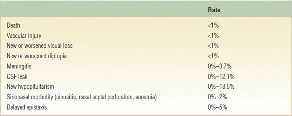
In experienced hands, endoscopic endonasal removal of sellar tumors has become increasingly safe and effective. However, serious complications can occur. Table 3.1 summarizes rates of major complications from several recent studies at experienced centers, including death, carotid or other vascular injury, visual loss from chiasmal or optic nerve injury, diplopia, meningitis, CSF leak, new pituitary gland failure, epistaxis, sinusitis, nasal septal perforation, and anosmia.
Table 3.1 Complication Rates of Endoscopic Endonasal Surgery for Pituitary Adenoma and RCCs
RESULTS
Outcomes after endoscopic endonasal tumor removal are generally equivalent with microscopic transsphenoidal series, but more recently, improved rates of remission are being reported with the fully endoscopic approach. This trend would not be surprising given the enhanced visualization afforded by the endoscope. Table 3.2 lists expected remission and resection rates for pituitary adenomas and RCCs based upon recent publications.
Table 3.2 Remission and Resection Rates for Pituitary Adenoma and RCCs

a Rates are for biochemical remission using standard contemporary criteria. Some for Cushing's disease include microscopic and endoscopic series.
b Rates are for gross total resection based on postoperative sellar MRI.
PEARLS
· Carefully review preoperative imaging with attention to pituitary gland location, tumor extension, course of parasellar carotid arteries, exposure needed, and anticipated skull base reconstruction.
· Perform a wide and tall sphenoidotomy, generous posterior septectomy, and wide sellar bony opening to allow maximal instrument maneuverability and access to the tumor.
· To maximize chances of preserving the gland and recovery, use gentle manipulation of the gland, and if need be, incise or sharply remove the attenuated component of the gland to allow better intrasellar access and tumor exposure.
· Obtain complete hemostasis in the sella with hemostatic agents and brief irrigation with hydrogen peroxide.
· Carefully perform repair of CSF leak and reconstruction of the skull base in multilayered fashion; perform a Valsalva maneuver prior to exiting the sphenoid sinus and closure to evaluate the integrity of the repair.
· Gently handle sinonasal tissues, and use bilateral NS rescue flaps to diminish chances of postoperative sphenopalatine artery bleeding, increase likelihood of preserved olfaction, and simplify postoperative nasal debridements.
· Maximize the safety of the procedure and efficiency using teamwork and communication.
PITFALLS
· A misguided trajectory with resultant injury of the carotid artery can be avoided with the use of neuronavigation and Doppler probe in each case.
· Failure to thoroughly explore sellar, suprasellar, and medial cavernous sinus regions as well as diaphragmatic folds with 0-degree and angled endoscopes can result in leaving residual tumor.
INSTRUMENTS TO HAVE AVAILABLE
· Standard endoscopic sinus surgery set
· Endoscopic skull base surgery instruments
· Endoscopic drill with 3- and 4-mm coarse diamond bits
· Doppler probe
· Dural knife
· Hemostatic materials
· Reconstructive materials
SUGGESTED READING
Esposito F, Dusick JR, Fatemi N, et al. Graded repair of cranial base defects and cerebrospinal fluid leaks in transsphenoidal surgery. Neurosurgery 2007;60:295–303; discussion 303–294.
Fatemi N, Dusick JR, de Paiva Neto MA, et al. The endonasal microscopic approach for pituitary adenomas and other parasellar tumors: a 10-year experience. Neurosurgery 2008;63:244–256; discussion 256.
Tabaee A, Anand VK, Barrón Y, et al. Endoscopic pituitary surgery: a systematic review and meta-analysis. J Neurosurg 2009;111:545–554.
Madhok R, Prevedello DM, Gardner P, et al. Endoscopic endonasal resection of Rathke cleft cysts: clinical outcomes and surgical nuances. J Neurosurg 2010;112:1333–1339.
Rotenberg B, Tam S, Ryu WH, et al. Microscopic versus endoscopic pituitary surgery: a systematic review. Laryngoscope 2010;120:1292–1297.