
Carl H. Snyderman
INTRODUCTION
Reconstruction of defects of the cranial base has been one of the greatest challenges of endoscopic endonasal surgery. Over the last decade, there has been an evolution of reconstruction techniques with a progressive decrease in the incidence of postoperative cerebrospinal fluid (CSF) leaks. A reconstructive algorithm provides a graded approach to reconstruction ranging from nonvascularized tissue grafts to local vascular flaps to regional vascular flaps to microvascular free flaps. Most large defects of the ventral skull base are effectively reconstructed with a vascularized septal mucosal flap or extracranial pericranial flap. When a septal mucosal flap is not available due to prior surgery or by involvement of tumor, an inferior turbinate flap (ITF) is a viable alternative for suprasellar and clival defects.
The ITF is pedicled on the lateral nasal branch of the sphenopalatine artery (Fig. 44.1). The sphenopalatine artery exits the sphenopalatine foramen at the posterosuperior corner of the maxillary sinus and branches into multiple arteries including the posterior septal branch and lateral nasal branches to the middle and inferior turbinates. The artery to the inferior turbinate courses along the inferior turbinate in a posterior to anterior direction and also sends minor branches to the inferior meatus.

Figure 44.1 Vascular anatomy of the lateral nasal wall. The inferior turbinate flap is supplied by a branch of the sphenopalatine artery that courses along the length of the inferior concha.
Several variations of the ITF are available (Fig. 44.2). A standard ITF consists of the mucosa covering the inferior concha and inferior meatus. An extended ITF (EITF) includes the mucosa of the floor of the nose. The ITF can be extended further by including the mucosa of the nasal septum. This creates a flap that is comparable in size and reach to a nasoseptal flap. The septal extension probably has a random blood supply and is at greater risk of distal ischemia than is a typical nasoseptal flap.

Figure 44.2 Variations of the inferior turbinate flap (ITF). A standard ITF consists of the mucosa covering the inferior concha and inferior meatus (green). An extended ITF (EITF) includes the mucosa of the nasal floor (blue). The EITF can be extended further by incorporating the mucosa of the nasal septum (blue dotted line).
HISTORY
It is important to inquire about prior nasal surgery including nasal septoplasty, turbinate reduction, endoscopic sinus surgery, treatment of epistaxis, and endoscopic or microscopic skull base surgery. Prior surgery may have sacrificed the vascular pedicle to the nasoseptal flap or ITF and compromised the nasal mucosa. Rarely, chronic inflammatory disease such as granulomatous disease may damage the mucosa.
PHYSICAL EXAMINATION
Nasal endoscopy includes an assessment of mucosal surfaces and integrity of the vascular pedicles. A large maxillary antrostomy that extends to the sphenopalatine foramen may have compromised the vascular supply. Similarly, a large sphenoid antrostomy with sacrifice of the posterior septal artery precludes the use of a nasoseptal flap.
Deviation of the nasal septum will make the dissection of the flap more difficult on the convex side and increases the risk of tearing the mucosa. Generally, a flap will be performed on the side of the nasal cavity with the most room for instrumentation. Increased tumor dissection on one side may necessitate using a flap where the pedicle is based on the contralateral side.
INDICATIONS
An ITF is indicated for large dural defects when a nasoseptal flap is unavailable or for coverage of exposed vascular structures (internal carotid artery). The ITF has a limited arc of rotation due to the orientation of the vascular pedicle, and the mucosa of the inferior concha does not conform well to a new surface. An EITF is used in most cases but may include a septal extension if a longer flap is needed and the septal mucosa is available. Due to the limited arc of rotation, an ITF is ideally suited for clival defects and provides optimal coverage in a horizontal orientation.
CONTRAINDICATIONS
Contraindications to the use of an ITF include the absence of a vascular pedicle (prior surgery or embolization of the internal maxillary artery) or prior resection of the inferior turbinate. Adhesions between the inferior turbinate and the nasal septum can be lysed prior to elevation of the flap. A severely deviated nasal septum is a relative contraindication and may need to be corrected to allow room for dissection. If bilateral transpterygoid approaches are necessary, exposure may be limited by the flap pedicle. In such cases, mobilization of the pedicle into the pterygopalatine space may be necessary.
A defect that is too large or distant to be completely covered by an ITF is a relative contraindication. Alternative flap reconstructions should be considered first. If the ITF is insufficient to provide complete coverage, it can be used in combination with multilayered fascial grafts. A deep clival defect may need to be filled with adipose tissue grafts deep to the flap so that the ITF can provide complete mucosal coverage. Tumor involvement of the nasal mucosa is a contraindication, especially for high-grade malignancies such as squamous cell carcinoma, adenocarcinoma, or melanoma or when final tumor margins are difficult to assess such as with adenoid cystic carcinoma.
PREOPERATIVE PLANNING
The surgical team should discuss the surgical approach, extent of exposure, and reconstructive needs prior to the surgery. Large dural defects should be reconstructed with vascularized tissues whenever possible. Every operation should have a backup plan for reconstruction if the primary reconstruction is not possible due to loss of the vascular pedicle or involvement by tumor.
Preoperative imaging of the vascular supply to the flap is not necessary. If preoperative embolization of a tumor is performed, communication with the interventional radiologist will ensure that vessels to reconstructive flaps will be preserved when feasible.
SURGICAL TECHNIQUE (VIDEO 44.1)
It is important to preserve the vascular supply to the ITF as part of the surgical approach and resection. The flap is usually harvested at the time of dural reconstruction unless the vascular pedicle is blocking access (transpterygoid approach). If the flap is elevated early in the procedure, the flap is protected by passing it through the antrostomy into the maxillary sinus. Vasoconstriction of the nasal mucosa is achieved by inserting cottonoid pledgets soaked in 0.05% oxymetazoline for several minutes and injection of local anesthetic (0.5% Xylocaine, 1:200,000 epinephrine) into the surrounding mucosa.
Standard Inferior Turbinate Flap
A generous middle meatal antrostomy is performed with removal of bone posteriorly to the sphenopalatine foramen and inferiorly to the attachment of the inferior turbinate. Care should be taken to avoid injury to the sphenopalatine artery and its branches through excessive removal of bone or electrocautery.
Using an insulated needle-tip electrocautery bent at a 45-degree angle, a mucosal incision is made through the mucosa to the underlying bone starting at the anterior margin of the antrostomy. The incision is extended along the anterior tip of the inferior turbinate in an S-shaped line that follows the curvature of the turbinate. It then curves around the anterior margin of the inferior meatus to the floor of the nasal cavity just inside of the pyriform aperture. An incision is made posterior to the sphenopalatine foramen on the medial surface of the medial pterygoid plate. The incision courses inferiorly and anteriorly to encompass the mucosa of the inferior meatus (Figs. 44.2 and 44.3).
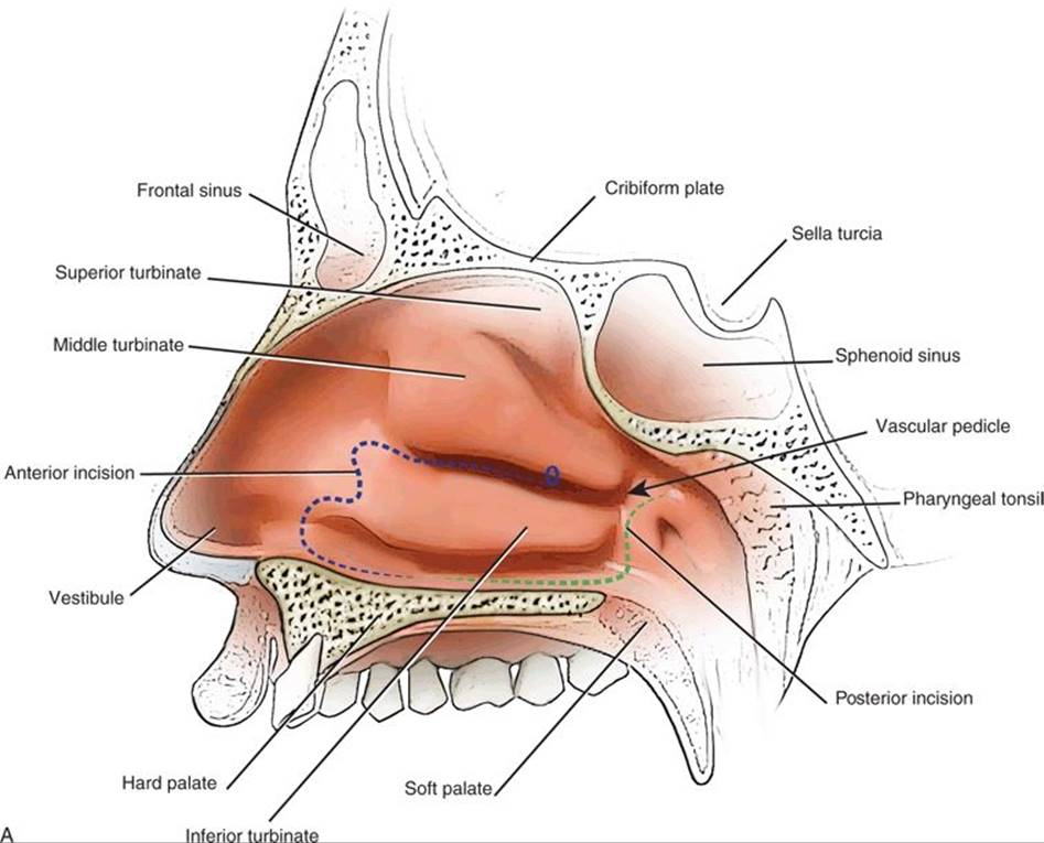
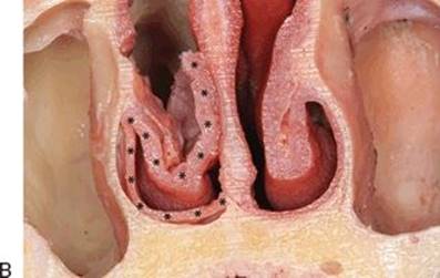
Figure 44.3 Incisions for a standard ITF. A. The anterior incision starts at the middle meatal antrostomy (circle) and follows an “S”-shaped course along the anterior projection of the inferior turbinate to expose the concha. The posterior incision is between the vascular pedicle (arrow) and eustachian tube. The two incisions connect along the nasal floor. B. Coronal view of anatomical dissection demonstrates elevation of the flap (asterisk) from the inferior concha with inclusion of mucoperiosteum from the floor of the nasal cavity.
A Cottle elevator is then used to elevate the flap in a subperiosteal plane. The bone of the inferior concha is very coarse and adherent to the flap, making it very difficult to elevate the flap from the inferior concha. Dissection begins at the anterior tip of the inferior turbinate, both medial and lateral to the conchal bone. The S-shaped incision is followed to elevate the mucosa superiorly in the inferior meatus. The bony canal of the nasolacrimal duct is identified, and the mucosa is sharply transected with fine-tip scissors (Kurze) in the subperiosteal pocket. The remaining mucoperiosteum of the inferior meatus is easily elevated in an anterior to posterior direction toward the vascular pedicle. The vascular pedicle can be further mobilized with careful dissection.
The bone of the inferior concha is removed with rongeurs, and small fragments remaining are dissected free from the flap if possible. The flap is then ready to be used for reconstruction. It is rotated inferomedially to the site of the defect. A standard ITF provides minimal coverage of small midclival defects (Fig. 44.4). The flap retains the shape of the inferior concha, and it can be difficult to position properly. The flap must be in contact with bone or dura for healing to occur; all surrounding mucosa should be removed first.

Figure 44.4 Standard left ITF used to cover an exposed aneurysm clip following endoscopic endonasal clipping of a vertebral artery aneurysm. (MS, maxillary sinus.)
The edges of the flap are covered with postage-stamp size pieces of Surgicel to anchor the flap, followed by similar-sized pieces of Gelfoam soaked in thrombin solution. Nasal packing consisting of a Foley catheter inflated with saline or nasal tampons (Merocel) is carefully placed to support the reconstruction.
Extended Inferior Turbinate Flap
Due to the limited size of a standard ITF, an EITF that includes the mucosa of the floor of the nasal cavity is used most commonly. The posterior mucosal incision extends across the nasal floor at the posterior edge of the hard palate. The medial limit is the junction of the nasal septum and premaxilla. This results in a flap that is wider than the standard ITF and adds an additional vertical dimension when it is transposed horizontally over the clival defect (Figs. 44.2 and 44.3B).
When maximal coverage is needed, the septal mucosa can be included in the design of the flap (Fig. 44.2). The anterior and posterior mucosal incisions are continued with parallel vertical incisions up the nasal septum, following the septal incisions for a nasoseptal flap. The superior septal incision parallels the skull base approximately 1 cm below the olfactory sulcus and includes the septal mucosa anterior to the olfactory sulcus and deep to the nasal bones. The septal mucoperichondrium/mucoperiosteum is elevated from the septal cartilage and bone with a Cottle elevator. At its junction with the premaxilla, the flap is adherent and sharp dissection may be necessary to avoid tearing the flap.
The flap is then rotated posteriorly to cover the skull base defect as described above (Fig. 44.5). The donor site on the cartilaginous nasal septum can be covered with a free mucosal graft (mucoperiosteum) from the resected middle turbinate. It is secured with a single suture and then covered with Silastic Doyle splints. This speeds the mucosalization of the nasal septum and minimizes long-term problems with septal crusting.
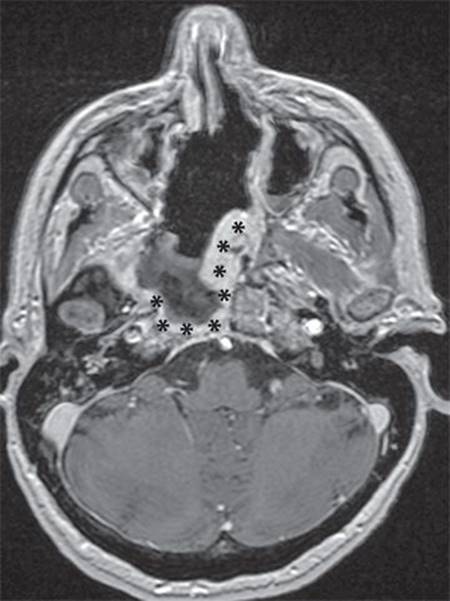
Figure 44.5 Postoperative MRI of an extended left ITF (asterisk) for a transclival defect. Note that the extended portion of the flap provides coverage of both paraclival internal carotid arteries.
POSTOPERATIVE MANAGEMENT
Nasal packing is removed after 5 to 7 days. Antibiotic prophylaxis is maintained while the packing is in place. If an EITF is performed, the Silastic splints are maintained for 3 weeks to enhance mucosalization of the septum and to prevent postoperative synechiae. Nasal saline rinses are started after the splints are removed and are used daily to minimize crusting and promote cleansing of the nasal cavity. Endoscopic debridement of postoperative debris and crusts is carefully performed periodically over the first several months until healing is complete.
COMPLICATIONS
Complications may be related directly to the reconstructive site or the flap donor site. Necrosis of the flap is rare but usually indicates a problem with the vascular pedicle. Direct injury to the vessels can occur from dissection at the sphenopalatine foramen or excessive cauterization of the stump of a resected middle turbinate. Torsion of the pedicle or compression from nasal packing or balloon may compromise blood flow. Ischemia of the distal portion of the flap could result from perforation of the flap or narrowing of the pedicle, especially when an EITF is employed.
A postoperative CSF leak is usually related to technical factors of the reconstruction rather than flap necrosis. Possible reasons include a flap that is too small for the defect, inadequate length with tension of the flap pedicle, poor apposition to underlying dura and bone, and elevated CSF pressure. If a leak develops, prompt endoscopic repair in the operating room is recommended to avoid the risk of meningitis. Most postoperative CSF leaks involve a small area of the reconstruction and can be repaired with repositioning of the flap edge or augmentation with fascia or adipose tissue grafts.
An expected sequela of surgery is crusting of the nasal cavity as mucosalization of the donor site occurs. It can take several months for healing to be complete and crusting to diminish. A minority of patients may suffer from chronic dryness from altered airflow due to loss of the inferior concha and associated crusting. Mucosal grafting of the donor site using a free mucoperiosteal graft from a resected middle turbinate can help minimize long-term crusting.
Temporary edema and stenosis of the nasolacrimal duct can result in epiphora. If it persists, consultation with an ophthalmologist is recommended for evaluation of the entire lacrimal drainage system and placement of lacrimal stents.
RESULTS
ITFs have a very reliable vascular pedicle and provide a good mucosal barrier for reconstruction of the dura or coverage of the internal carotid arteries. They are best suited for infrasellar defects that don’t require extensive rotation of the flap pedicle.
Examples of clinical applications include coverage of an exposed vertebral artery aneurysm clip (Fig. 44.4), closure of a dural defect following a transclival approach, coverage of exposed paraclival internal carotid arteries (Fig. 44.5), and protection and revascularization of clival bone following debridement for radionecrosis and osteomyelitis.


FIGURE 44.6 Anatomical dissection with comparison of flap sizes: extended ITF (A) and extended ITF with septal extension (B). (IT, inferior turbinate mucosa; F, nasal floor mucosa; S, septal mucosa.)
The size of the ITF has been reported to be 5.4 cm in length and 2.2 cm in width. The length of the EITF is consistent with the previously described ITF, while the width is 250% that of the ITF. This difference is accounted for by inclusion of the mucosa of the nasal floor, thus increasing its mean surface area to 27.26 cm2 (Fig. 44.6). The surface area was sufficient to cover defects spanning the clivus from one paraclival internal carotid artery to the other. The EITF compares favorably to the nasoseptal flap for reconstruction of infrasellar defects. The nasoseptal flap has a mean surface area of 25 cm; the EITF is slightly larger. When the septal mucosa is available for use with the EITF, the mean surface area increases to 40.53 cm2, which is significantly larger than the mean surface area reported for the nasoseptal flap. The addition of the nasal mucosa also decreases the arc of rotation by providing a longer flap with greater reach.
PEARLS
· The superior incision should start at the anterior margin of the maxillary antrostomy. The inferior incision begins posterior to the sphenopalatine foramen and runs between the posterior tip of the inferior turbinate and the eustachian tube. Narrowing of the pedicle will increase the arc of rotation of the flap.
· The flap is ideally suited for clival defects and is best oriented horizontally.
· The flap should be dissected from the inferior concha while it is still attached; otherwise, it is very difficult to remove the bone fragments from the flap.
PITFALLS
· The distal end of the nasolacrimal duct is sharply transected to prevent stenosis.
· With an extended flap, the septal mucosa may receive a random blood supply and be susceptible to ischemia.
· The conchal part of the flap is difficult to position properly since it retains its original shape and does not lie flat.
INSTRUMENTS TO HAVE AVAILABLE
· Needle-tip monopolar electrocautery (insulated) bent to 45 degrees
· Cottle elevator
· Fine-tip scissors (Kurze): straight or curved
· Silastic Doyle splints
· Merocel nasal tampons
· Silastic Crawford lacrimal stents (optional)
SUGGESTED READING
Padgham N, Vaughan-Jones R. Cadaver studies of the anatomy of arterial supply to the inferior turbinate. J Royal Soc Med 1991;84:728–730.
Fortes FS, Carrau RL, Snyderman CH, et al. The posterior pedicle inferior turbinate flap: a new vascularized flap for skull base reconstruction. Laryngoscope 2007;117(8):1329–1332.
Harvey RJ, Parmar P, Sacks R, et al. Endoscopic skull base reconstruction of large dural defects: a systematic review of published evidence. Laryngoscope. [Meta-Analysis Review]. 2012;122(2):452–429.
Choby GW, Pinheiro-Neto CD, de Almeida JR et al. Extended inferior turbinate flap for endoscopic reconstruction of skull base defects. J Neurol Surg B. 2014 May 6 [Epub ahead of print].