
C. Arturo Solares
INTRODUCTION
The wide popularization of endoscopic endonasal skull base surgical techniques has taken place in the past decade. This is due not only to advances in technology but also to the improved understanding of endoscopic skull base anatomy and the expanding expertise of skull base surgeons. One of the greatest challenges in mastering endoscopic skull base surgery has been developing techniques to prevent cerebrospinal fluid (CSF) leaks. The introduction of pedicled, vascularized flaps for use in cranial base surgery has met with great success. The middle turbinate flap serves as one of the latest additions to the armamentarium of vascularized flaps accessible to the skull base surgeon.
Vascularized pedicled flaps in endoscopic endonasal surgery became widely accepted after the advent of the Hadad-Bassagasteguy (HBF) or nasoseptal flap (NSF). Advantages provided by the use of vascularized flaps include restoration of local blood flow to the wound site, improved healing, decreased rates of surgical infection, decreased graft migration, and most importantly, decreased rates of CSF leak.
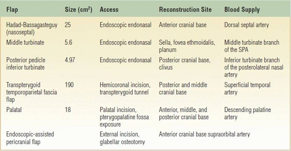
Since the nasoseptal flap was first described, a number of different pedicled flaps for reconstruction in endoscopic endonasal surgery have been described. These include the transpterygoid temporoparietal fascia flap, the endoscopic-assisted transglabellar pericranial flap, the posterior pedicle inferior turbinate flap, and the palatal flap. More information with regard to these flaps may be found in Table 43.1 of this chapter.
Table 43.1 Vascularized Flaps Available for Endoscopic Endonasal Cranial Base Surgery
Prevedello et al. first described the middle turbinate flap for skull base reconstruction in a cadaveric feasibility study in 2009. In most cases, the middle turbinate flap serves as an alternative to the nasoseptal flap for reconstruction of small defects of the sella, fovea ethmoidalis, and planum. Although small defects of the skull base have a 95% chance of successful closure with free tissue, vascularized repair is believed to improve healing rates and decrease the chance of CSF leak.
HISTORY
It is understood that a patient being considered for a middle turbinate flap will be undergoing a skull base procedure. The issues specific to this type of reconstruction are any history of previous nasoseptal surgery (septoplasty, partial septectomy) and nasoseptal deformities (i.e., known septal deviation) that may inhibit the ability of the surgeon to perform a nasoseptal flap. Previous sinus surgery should alert the surgeon to examine for the presence of large sphenoidotomies that could have compromised the distal sphenopalatine vasculature. Also, a history of previous skull base surgery will prepare the surgeon for the possible absence of certain vascularized reconstructive options. Lastly, the history and nature of the skull base pathology in question will alert the surgeon to perform a thorough physical examination to determine which reconstructive options are available in order to maintain oncologic principles (i.e., involvement of the septum by malignancy).
PHYSICAL EXAMINATION
As with all skull base pathology, a thorough head and neck examination as well as assessment of cranial nerve function should be performed. Nasal endoscopy is necessary to further assess the individual patient’s anatomy. Attention should be placed on identifying signs of previous nasoseptal surgery (septoplasty, partial septectomy) and nasoseptal deformities (large septal spurs, septal perforations) that may inhibit the ability of the surgeon to perform a nasoseptal flap. Previous sphenoid and pterygoid surgery should also be noted, as the distal sphenopalatine vasculature may have been compromised. Anatomical anomalies of the middle turbinate (paradoxical middle turbinate, concha bullosa, and unilateral hypoplasia) exist in 25% of the population and should be identified as they may contribute to increased difficulty in raising a middle turbinate flap.
INDICATIONS
The middle turbinate flap is most often used for reconstruction of skull base defects when the nasoseptal flap is unavailable. This may occur in patients with septal defects, previous septal surgery, and history of sphenoidotomy with possible compromise of the posterior septal artery and patients in whom the nasoseptal flap has been tried and failed. The middle turbinate flap may also be used for repair of posttraumatic or spontaneous CSF leaks. It has recently gained popularity for use in situations in which the need for a combination of flaps is anticipated. Some authors have also described the use of the middle turbinate flap for reconstruction following endoscopic endonasal nasopharyngectomy.
CONTRAINDICATIONS
Contraindications include an anticipated large defect to repair. Prevedello et al. found the average length and width of the middle turbinate flap to be 4.04 and 2.8 cm, respectively. Relative contraindications include anatomical variance of the middle turbinate and ability to perform a nasoseptal flap, which remains the gold standard vascularized flap for endoscopic anterior skull base reconstruction.
PREOPERATIVE PLANNING
Preoperative planning includes a thorough physical examination with diagnostic nasal endoscopy. CT images should be reviewed. It has been suggested that the middle turbinate length should be measured since the ability of the flap to reach the sella may be less likely in turbinates measuring less than 4 cm in length.
SURGICAL TECHNIQUE
The nasal cavity is decongested with oxymetazoline 0.05%. Figure 43.1 demonstrates the endonasal anatomy prior to flap elevation. The middle turbinate and septum are infiltrated with 1% lidocaine with 1:100,000 epinephrine. A vertical incision is then made through the mucosa at the anterior aspect of the middle turbinate. This is followed by a horizontal incision through the mucosa at the superomedial aspect of the middle turbinate. Care is taken to preserve the attachment of the bony middle turbinate to the cribriform plate. The mucoperiosteum is then elevated from the underlying bony middle turbinate (Fig. 43.2A and B). The bone of the middle turbinate is then removed in piecemeal fashion (Fig. 43.3A and B). An incision is then made through the axilla of the middle turbinate to detach the turbinate from the skull base. This incision is carried in a dorsal direction. Once the entire turbinate bone has been excised, the mucoperiosteum can be unfolded or “opened like a book.” If a need for extra length is anticipated, the pedicle may be dissected to the level of the sphenopalatine foramen; this increases both length and mobility. The flap may then be protected within the nasopharynx (Fig. 43.4) or maxillary sinus for the remainder of the procedure. Fibrin glue is applied once the flap has been appropriately positioned. Gelfoam is then packed around the site, and supporting nondissolvable packing is placed.
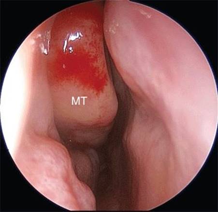
Figure 43.1 Endoscopic view of the right nasal cavity. The middle turbinate (MT) is in center view.


Figure 43.2 An incision has been made at the anterior aspect of the middle turbinate. The mucoperiosteum is being elevated on the medial aspect (A) and lateral aspect (B) of the middle turbinate.


FIGURE 43.3 A. The bone of the middle turbinate is being removed. B. View after the bone has been removed.

Figure 43.4 The mucoperiosteum has been unfolded or “opened like a book” and is being stored in the nasopharynx for later use.
POSTOPERATIVE MANAGEMENT
Postoperatively, the patient is instructed on maintenance of CSF leak precautions. This includes no nose blowing, no straining, no heavy lifting (>15 lb), stool softeners, and open mouth sneezing. Although no clear evidence exists as to efficacy, a third-generation cephalosporin is routinely administered in the perioperative period. An MRI or CT may be obtained to confirm tumor resection and evaluate for evidence of intracranial bleeding or pneumocephalus. Also, adequate perfusion of the flap can be confirmed on MRI. The packing is left in place until postoperative day 5. Following removal of the packing, the patient is asked to use nasal saline spray. Two weeks following surgery, the patient returns to the office for routine sinonasal debridement.
COMPLICATIONS
Complications are rare in skilled hands. Unintentional fracture of the cribriform plate may occur while the turbinate is being excised. Injury to the pedicle may devascularize the graft. As with all vascularized repairs, there is potential for the reconstruction to be inadequate and CSF leak to manifest itself.
RESULTS
The middle turbinate flap is relatively newly described and most often serves as an alternative for reconstruction when it is not feasible to use the nasoseptal flap. There are few data in regard to its use in the literature. In Prevedello’s initial cadaveric feasibility study, the flap was shown to have good coverage of all defects in the fovea ethmoidalis and planum. However, the reach to the sella was more variable, with 10 of 12 flaps being deemed adequate for reconstruction of the sella. A recent retrospective review by Julian et al. showed 100% success rate of the middle turbinate flap with no CSF leak in a series of 10 patients. Most authors note that it takes, on average, 30 minutes to complete the flap.
PEARLS
· Anatomical variation of the middle turbinate exists in 25% of patients (concha bullosa, paradoxical middle turbinate, unilateral hypoplasia) and should be considered in preoperative planning.
· Identification and preservation of the sphenopalatine artery branch to the middle turbinate is essential to maximize the length of the middle turbinate flap.
PITFALLS
· The middle turbinate flap may be technically challenging.
· Destabilization of the middle turbinate bone prior to elevation of the mucoperiosteal flaps may significantly increase the difficulty of the procedure.
INSTRUMENTS TO HAVE AVAILABLE
· Oxymetazoline-soaked pledgets
· 1% lidocaine with 1:100,000 epinephrine
· Bayonet forceps
· Nasal speculum
· 0-degree endoscope, light source, and camera
· Freer elevator
· Cottle knife
· Sickle knife
· Rongeurs
· Turbinate scissors
· Bipolar electrocautery device
· Nondissolvable packing
SUGGESTED READING
Lee HY, Kim CH, Kim JY, et al. Surgical anatomy of the middle turbinate. Clin Anat 2006;19(6):493–496.
Prevedello DM, Barges-Coll J, Fernandez-Miranda JC, et al. Middle turbinate flap for skull base reconstruction: cadaveric feasibility study. Laryngoscope 2009;119(11):2094–2098.
Simal Julián JA, Miranda Lloret P, Cárdenas Ruiz-Valdepeñas E, et al. Middle turbinate vascularized flap for skull base reconstruction after an expanded endonasal approach. Acta Neurochir (Wien) 2011;153(9):1827–1832.
Chen MY, Hua YJ, Wan XB, et al. A posteriorly pedicled middle turbinate mucoperiosteal flap resurfacing nasopharynx after endoscopic nasopharyngectomy for recurrent nasopharyngeal carcinoma. Otolaryngol Head Neck Surg 2012;146(3):409–411.