
Adam M. Zanation
INTRODUCTION
Dr. Alfred Ketchum in the 1960s best described the craniofacial resection as a combination of both transfrontal and transfacial approaches to tumors from the sinonasal cavity that involve the skull base and the intracranial cavity. The initial reconstructions were done with avascular onlay grafts using either temporalis fascia or tensor fascia lata. While some of these were successful, cerebrospinal fluid (CSF) fistula in the postoperative period was still commonplace. As more advanced lesions and higher-grade tumors were being removed with craniofacial resections, the use of adjuvant therapy in the form of radiation or chemoradiation became standard. This adjuvant therapy placed the patient at an even greater risk for disruption and necrosis of avascular skull base repairs; therefore, the importance of meticulous skull base reconstructions using vascularized free tissue transfer became routine in the 1970s and the 1980s.
Cranial base surgery has multiple goals and challenges as they relate to the intricate anatomy around the brain, eyes, and facial skeleton. Beyond the primary tumor resection (whether it be to decompress a cranial neuropathy or obtain negative margins for a sinonasal cancer removal) is the other primary goal of the operation which is reconstruction which includes cosmetic goals, such as reconstructing the bony vault around the orbit or the appropriate contour around the zygomatic or malar complex. However, beyond the cosmesis is the more important and potentially lifesaving reconstruction of a dural defect and closure of the CSF leak after tumor removal. Following a craniofacial resection, the standard technique is direct suture repair of the dural defect with an overlay reconstruction using a vascularized pericranial scalp flap. The goal of the pericranial flap is to cover the dura and to separate the sinonasal tract from the intracranial space with a robust vascular tissue barrier (Fig. 46.1).

FIGURE 46.1 Pericranial flap and other scalp flap options. (TPFF, temporoparietal fascia flap [superficial temporal artery]; TEMP M, temporalis muscle flap [deep temporal artery].)
The introduction of endoscopic endonasal skull base techniques has created new challenges in the reconstruction of the anterior cranial fossa. While a dural defect similar to an open craniofacial resection can be achieved endoscopically (with a dural defect from the meridian of the orbit to the meridian of the opposite orbit and from the optic chiasm to the frontal sinus), the ability to reconstruct such a defect is more challenging than in open surgery. The pedicled nasoseptal flap has produced excellent outcomes as a primary reconstructive option for endoscopic intradural skull base surgery. It has an axial blood supply based on the posterior nasoseptal artery and is readily available in the sinonasal cavity; therefore, it does not create a separate donor site defect. However, most sinonasal cancers have midline and septal involvement, and therefore the need to take widely negative margins often precludes the use of a nasal septal flap in this setting. Recently, the pericranial flap has become a useful and novel flap for endoscopic reconstructions. Operative techniques for both open and endoscopic pericranial flap use require an intimate knowledge of the vascular anatomy of the anteriorly based pericranial flap, as well as an understanding of the complex layers of the frontal and temporal scalp.
The pericranial flap is supplied by the supraorbital and supratrochlear arteries (Fig. 46.2). The main trunk and superficial branches of these arteries course from the orbit into the galea and frontalis muscle layer and give rise to deep branches that supply the pericranium. These deep branches can arise at the level of the orbit or within 10 mm of the orbital rim. It is important to understand that the deep branches that supply the pericranium may exit up to 1 cm above the exit point of the supraorbital and supratrochlear foramina; dissection of the flap beyond this level can injure the blood supply.

FIGURE 46.2 Supraorbital arterial supply to the pericranial flap.
HISTORY
Once the patient has been appropriately evaluated for the presence of a skull base tumor of the cribriform area or anterior cranial base, surgical planning proceeds around the goals for the primary tumor resection. Staging for distant and regional metastatic disease in the setting of sinonasal cancer is performed, and a firm histologic diagnosis is required for optimal treatment planning. Following a thorough discussion with the patient regarding operative options (whether it be an open or endoscopic craniofacial resection), the surgeon must inquire about prior procedures and prior trauma to the scalp area to know whether a pericranial flap is a viable reconstructive option. Patients with prior bicoronal approaches often have a truncated flap due to the low placement of the coronal incision. In the setting of revision surgery, the vascular pedicles of the pericranial flap may have been interrupted, or the scalp flap was elevated without regard to the potential future use of a pericranial flap; therefore, it can be scarred and/or have a significant numbers of holes. Poorly vascularized flaps that are not fully intact make for a suboptimal skull base reconstruction. It should also be noted that tumors of the sinonasal region and skull base can involve the orbit, as well as the soft tissues of the face and glabella. If an orbital exenteration has to be undertaken, sacrifice of the flap pedicle necessarily occurs on that side. If a significant amount of facial soft tissue has to be resected from the periorbital region or frontal scalp, the pedicle of the flap may be compromised. A history of prior radiation therapy is not a contraindication to the use of a pericranial flap.
PHYSICAL EXAMINATION
Physical examination should proceed to evaluating any prior trauma or scarring, as well as the presence of tumor protruding into the soft tissues of the glabella, scalp, forehead, or orbits. The ophthalmic division of the trigeminal nerve as well as the frontal branches of the facial nerve are at risk with harvesting a pericranial flap. A detailed examination of the cranial nerves, paying close attention to these two cranial nerves, should be performed. If the patient has had prior surgery or trauma, the flap pedicles can be evaluated with Doppler ultrasound to see if they are intact. This confirms vascularity at the supratrochlear and supraorbital exit points; however, it does not fully confirm vascularity of the entire axial flap.
Indications
In my opinion, a large dural defect with a sinonasal fistula is an absolute indication for reconstruction of the skull base with a vascular flap. Indications for reconstruction with a pericranial flap include an anterior skull base defect with resection of the dura resulting in an intra-arachnoidal and sinonasal CSF fistula. Relative indications include extradural resection without intraoperative CSF leak but in a patient undergoing radiation therapy.
Contraindications
Contraindications to the use of pericranial flaps include patients without an intact flap pedicle or suitable flap quality.
PREOPERATIVE PLANNING
Imaging studies, whether it be CT or MRI, should be devoted to the evaluation of the extent of the primary tumor. There are no special imaging studies that need to be devoted to planning the reconstruction with the pericranial flap. Next is preoperative planning of the primary route of access for resection of the tumor. An open craniofacial resection is going to require coronal access to the scalp in a very wide plane, and therefore the pericranial flap and the pedicles would need to be actively protected during the exposure and resection of the tumor. However, if an endoscopic transcribriform resection is planned, then the endonasal resection can proceed endoscopically until negative margins are obtained. If the operation must be converted to an open craniofacial resection to obtain clear margins, then the above principles still apply. However, if that is not the case, the operative surgeon should understand that the endonasal defect must include a Draf III frontal sinusotomy to allow for wide frontal sinus access and drainage since the pericranial flap is going to be transposed across the midline of the floor of the frontal sinus. With an endoscopic transcribriform approach, I have a separate instrument table and endoscope set that are clean for the scalp portions of the case.
SURGICAL TECHNIQUE (VIDEO 46.1)
Surgical techniques include the open extracranial pericranial flap as well as the endoscopically assisted pericranial flap for endonasal reconstruction; these will be discussed separately.
Description of the Open Anteriorly Based Pericranial Flap
The patient is placed under general anesthesia in a supine position. The patient is often placed in Mayfield head pins or on a horseshoe. If the patient is to be pinned, the pins should be placed posteriorly along the occiput or into the posterior temporal bone to allow for closure of the bicoronal defect, as well as to allow for posterior extension of the pericranial flap harvest. The coronal incision is made through the skin, the deep dermal tissues, and the galea. At this point, the galea separates superiorly, and Raney clips can be placed to control bleeding from the scalp edges, or selected bipolar cautery of the galea can be performed. The loose areolar tissue beneath the galea and superficial to the periosteum is identified. If an extremely long pericranial flap is to be taken, the dissection should proceed posterior to the scalp incision from the temporal line on one side to the opposite temporal line in this loose areolar plane up to 10 cm; this allows for extra posterior length of the pericranial flap that can be folded in at the time of the reconstruction. At this point, using a guarded-tip electrocautery, the pericranial flap is separated from the lateral margin of the temporalis muscle from the coronal incision to the posterior limit of the pericranial flap dissection.
The frontal branches of the facial nerve must be protected as the scalp flap is turned down. The frontal branches of the facial nerve run in the temporoparietal facial layer at the midportion of the zygoma. In order to protect the facial nerve, the dissection is carried down to the deep layer of the deep temporal fascia and the temporoparietal fascia is elevated anteriorly. The temporal line of fusion is where the superficial and deep layers of deep temporal fascia split around the superficial temporal adipose tissue pad on top of the zygomatic arch. Once the superficial layer of the deep temporal fascia is incised, dissection can proceed within the superficial temporal adipose tissue pad to the level of the zygomatic arch, and the frontal nerve within the temporoparietal fascia is elevated with the scalp flap as it is retracted inferiorly. This protects the facial nerve overlying the zygoma and provides a fascial plane along the deep surface of the scalp during the dissection. The periosteum of the scalp is then elevated along the zygoma to the lateral orbit. This should be done on both sides to allow for a maximum exposure of the frontal bones and orbital bar anteriorly. The pericranial flap can now be incised along the lateral aspect of the frontal bone, preserving the anteriorly based supraorbital and supratrochlear arteries on both sides. The thinning of the flap from the galea should be discontinued approximately 10 mm above the orbits, and if more rotation is needed, then a portion of the galea should be left on the flap above this to protect the deep branches (see Introduction). Now, the pericranial flap is pedicled anteriorly for use after the tumor is removed. If a subfrontal or supraorbital bar approach is to be performed (Fig. 46.3), then the supraorbital and supratrochlear foramina need to be fractured inferiorly to allow for mobilization of the pedicle down from the orbit while the orbital roof is lifted superiorly. Protection of the pedicle during this portion of the case is of paramount importance.


FIGURE 46.3 Subfrontal approach and reconstruction. A. Supraorbital (SOA) and supratrochlear (STrA) arterial bundles fully separated from the orbital bar in anticipation of a subfrontal approach during a revision craniofacial resection. B. Bone removal for subfrontal approach with frontal bone flap and orbital bar.
The frontal craniotomy is now opened, the dura is incised, and the frontal lobes are elevated, margins are taken, the transfascial portion of the tumor resection is taken up to the level of the cribriform and roof of the ethmoid, and incisions are made from the cranial and facial sides, excising the dura, roof of the ethmoid, and cribriform areas. The specimens are then sent to pathology to evaluate the surgical margins, and the wound is then copiously irrigated. Reconstruction of the dura is performed with a dural patch sutured to the dorsal edges. The pericranial flap is then rotated into the defect overlying the orbital bones and tucked above the planum sphenoidale. If the flap is long enough, it can be folded on itself to provide a thicker layer. The flap on the facial side is examined to make sure that it is tucked into appropriate position within the bony defect. Once it is in appropriate position, the brain is allowed to rest on the flap. The frontal bone is replaced leaving enough space to avoid compression of the axial blood supply to the flap. The scalp flap is replaced, and the incision is closed. Drains are not needed. The facial defect is then packed, usually with Vaseline-coated 1/2- to 1-inch packing brought out through the nostril, and then the facial incisions are closed in a cosmetically acceptable fashion. A head wrap is placed on the patient for compression of the scalp dissection making sure to not compress the pedicles.
Description of the Endoscopically Harvested Pericranial Flap for Endonasal Reconstruction
The endoscopically harvested pericranial flap uses similar methods; however, instead of a full coronal incision and being pedicled on both sides, the endoscopic pericranial flap is pedicled on one side and is harvested endoscopically through a 5-cm incision that is in the plane of the coronal incision (Fig. 46.4). A 5-cm incision is marked and then incised down through the galea into the loose areolar tissue. This is then back elevated to approximately the midline of the scalp and laterally to the temporal line. Using lighted and malleable retractors, an assistant retracts and a scope is introduced through the defect, and the loose areolar tissue is dissected anteriorly to the level of the supraorbital and supratrochlear arteries. The supraorbital arteries are located with a Doppler probe at the beginning of the operation and marked because the flap pedicle is going to be narrowed to within a 3-cm window around the Doppler signal. Usually, this corresponds to the medial canthal area to approximately 3 cm lateral to this. Once the subgaleal dissection has been completed, an extended guarded needle tip Bovie bent to a 45-degree angle is used to incise the pericranium under endoscopic assistance with an assistant retracting and suctioning smoke. The incision extends from the 3-cm pedicle and the supraorbital rim laterally along the temporal line and posteriorly at least 4 to 5 cm posterior to the coronal incision. It then travels medially to the midline scalp, and the incision continues along the midline to the glabellar area and then courses back into the medial brow. At this point, an elevator is used to elevate the pericranial flap from the underlying scalp. This is elevated from the cranium to the level of the supraorbital rim.
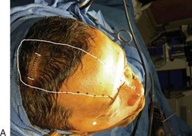


FIGURE 46.4 Endonasal pericranial flap. A. A 5-cm coronal plane port with outlines for planned unilateral endoscopically harvested pericranial flap. B. Dopplered supraorbital artery (SOA) and planned 3-cm pedicle. Nasion incision is shown in the concavity between the nasal and frontal bones. C. Transposed pericranial flap through the glabellar incision. Within the glabellar incision, the nasionectomy is shown. D. Endoscopic view from the nasal side looking into the endonasal defect through the nasionectomy. E. Endoscopic endonasal view of a transcribriform defect resulting from resection of an esthesioneuroblastoma from orbit to orbit and from planum to frontal sinus. F.Endoscopic view of pericranial flap skull base reconstruction of the defect (edges marked with dots) shown in (E).
Once the flap has been harvested, it then has to be transposed into the endonasal defect. Transposition occurs through a 1-cm glabellar incision. This is made in the glabellar concavity for appropriate cosmesis and healing with monopolar electrocautery. The nasion is identified, and a 4-mm rough diamond drill bit is used to drill an osteotomy across the nasion from the medial canthus to the medial canthus protecting the medial canthal tendons and the lacrimal system, approximately 1.5 cm wide and 4 mm in height. A subperiosteal dissection is then performed from the glabellar side connecting it to the midline pericranial dissection. This is then widened to the side of the flap to allow for an appropriate tunnel for the flap to be transposed. The flap is then transposed through the tunnel and then passed through the glabellar defect to cover the endonasal defect. First, a collagen graft is placed intradurally. The flap overlays the dural defect and the bone defect. The Draf III frontal sinusotomy is left open via both lateral ports, while the flap is brought down the middle of the frontal sinus floor. The flap is placed over the lateral orbits and back into the sphenoid sinus and then is packed into place with Surgicel followed by glue or sealant and absorbable and nonabsorbable packing, such as collagen or Gelfoam, and finally Merocel sponges. These are left in place for 5 to 7 days. The glabellar incision is closed with absorbable sutures, and the 5-cm port incision in the coronal plane is closed in a standard fashion. The routine use of drains is not needed. A head wrap is placed on the patient for compression of the scalp dissection making sure to not compress the pedicles.
POSTOPERATIVE MANAGEMENT
A head wrap is left on the patient for the first 2 postoperative days. If the patient has a coagulopathy or a bloody dissection, a JP suction drain can be placed at the flap donor site. If a lumbar drain is to be used, it should be monitored carefully, often in an ICU setting. I use lumbar drains when the defect includes the suprasellar cistern but not if solely confined to the cribriform area. Patients are placed on stool softeners and told not to blow their nose and to sneeze with their mouth open for 6 weeks. They have bed rest for approximately 3 days with subcutaneous heparin for prophylaxis against deep venous thromboembolism. Facial sutures and coronal sutures are removed on postoperative days 7 to 10 or if the patient has been irradiated up to day 14. Packing is removed on days 5 to 7, and the patient is told to start nasal saline rinses on day 7, three times a day. I do not routinely use airway diversion with tracheostomy.
COMPLICATIONS
Complications include hematoma at the donor site, nerve injury (sensory or motor), necrosis of the skin flap, hair loss at the incision site, infection, and contour deformity in the area of the flap, as well as flap necrosis and postoperative CSF leakage. If the patient does develop a small low-flow CSF leak in the postoperative period, it can be managed with a lumbar drain and expectant management; however, I take all of these patients back to the operating room for reexploration from the nasal side to see if this is related to death of the flap, malpositioning of the flap, or presence of a small fistula within the flap. If it is related to death of the flap, then a secondary flap such as a free flap may have to be placed into the defect; however, if the flap is viable and the leak is due to a malpositioning or a small hole in the flap, then this can be patched with adipose tissue, mucosal grafts, or an inlay/onlay with acellular dermis allograft. Lumbar drainage is used for 3 days if a postoperative CSF leak occurs. The rate of postoperative CSF leak should be less than the accepted historical standard of 10%.
RESULTS
Many studies examining the use of vascularized reconstruction of skull base defects, both open and endoscopic, have shown that the pericranial flap is an excellent vascularized flap to reconstruct the cribriform area. The donor site defects are minimal, the risks to cranial nerves are minimal, and the prevention of fistula in the postoperative setting is excellent. CSF leak rates are less than 10% in large series of craniofacial resections, and a high-volume referral center should strive for CSF leak rates of less than 5%.
PEARLS
· Prior frontal and scalp surgery should alert the surgeon to have secondary options for skull base reconstruction.
· Tumor invasion into pedicles or frontal areas is a contraindication to pericranial flap reconstruction.
· Understanding the relationship of the facial nerve, zygomatic arch, and temporoparietal fascia and deep temporal fascia planes is important to preserve frontal facial nerve function when elevating the flap.
· Understanding the relationship of the supraorbital and supratrochlear arteries and the deep branches to the pericranium in the 1-cm area above the orbital rim is important to preserve the blood supply to the flap.
· Extracranial pericranial flap dissection for endonasal reconstruction is a complex technique that requires time and a steep learning curve. Having patience and practicing in the laboratory are recommended.
PITFALLS
· Postoperative CSF leakage is often from the suprasellar cistern over the planum sphenoidale secondary to malpositioning of the flap. It is this area where the optic nerves are coursing medially and the optic chiasm is difficult to see and the surgeon does not perform an adequate inlay of the flap in this area. This is also the area that is most difficult to perform primary dural repair. To minimize this risk, transfacial examination of the flap inset once the frontal bone flap and scalp are closed should be performed. The use of an endoscope (even in the setting of open surgery) can provide excellent visualization of the flap in this area. If the flap has slipped forward, it should be repositioned over the planar bone and supported with a bolster.
INSTRUMENTS TO HAVE AVAILABLE
· Craniotomy tray or large plastic tray
· Drill
· Doppler probe
SUGGESTED READING
Snyderman CH, Janecka IP, Sekhar LN, et al. Anterior cranial base reconstruction: role of galeal and pericranial flaps. Laryngoscope 1990;100(6):607–614.
Ganly I, Patel SG, Singh B, et al. Complications of craniofacial resection for malignant tumors of the skull base: report of an International Collaborative Study. Head Neck 2005;27(6):445–451.
Yoshioka N, Rhoton AL Jr. Vascular anatomy of the anteriorly based pericranial flap. Neurosurgery 2005;57(1 Suppl):11–16; Discussion 11–16.
Zanation AM, Snyderman CH, Carrau RL, et al. Minimally invasive endoscopic pericranial flap: a new method for endonasal skull base reconstruction. Laryngoscope 2009;119(1):13–18.
Patel MR, Shah RN, Snyderman CH, et al. Pericranial flap for endoscopic anterior skull-base reconstruction: clinical outcomes and radioanatomic analysis of preoperative planning. Neurosurgery 2010;66(3):506–512; Discussion 512.