
Carl H. Snyderman
INTRODUCTION
The primary options for reconstruction of defects of the dura of the anterior cranial base now include multilayer fascial grafts, the nasoseptal flap, and the pericranial flap. The pericranial flap has been the preferred form of reconstruction following craniofacial resection for multiple decades. It is a versatile and reliable vascularized flap that can be employed in a variety of surgical situations.
The scalp has five layers corresponding to the acronym SCALP: skin, subcutaneous tissue, aponeurosis, loose areolar tissue, and periosteum. The pericranial flap consists of the inner two layers of the scalp: the loose areolar and periosteal layers. A pericranial flap may be combined with the galeal layer (aponeurosis) to form a galeopericranial flap.
The pericranial flap is pedicled on the supraorbital and supratrochlear vessels, branches of the ophthalmic artery (Fig. 47.1). Of these, the supraorbital vessel is dominant and exits from the supraorbital foramen. It divides into superficial and deep branches to supply the galeal and pericranial layers, respectively. The pericranial flap merges with the galeal layer near the orbital rim. The sensory innervation of the frontal scalp also arises from the supraorbital and supratrochlear neurovascular bundles (ophthalmic division of the trigeminal nerve) and perforates the galeal layer. The motor supply of the frontalis muscle is provided by the temporal branches of the facial nerve. The anatomy of the frontal scalp allows a flap to be elevated that is pedicled on the vascular supply of the supraorbital and supratrochlear vessels with preservation of the sensory and motor innervation of the scalp.

FIGURE 47.1 The pericranial scalp flap derives its primary blood supply from deep branches of the supraorbital and supratrochlear vessels.
HISTORY
A detailed history regarding prior cranial surgery is important to determine reconstructive options. Prior scalp incisions may interrupt the vascular supply and limit the length of the pericranial flap. Prior reconstruction with a pericranial flap may preclude the use of a new pericranial flap although it is often possible to harvest one from the other side if only one pedicle was used previously. It is also possible that a galeal flap could be used if the pericranium is not available. Caution is advised when reading operative reports since many surgeons incorrectly use the term galeopericranial flap to describe flaps that are actually pericranial flaps. Prior orbital surgery may also compromise the vascular anatomy of the pericranial flap.
A history of prior radiation is also important to ascertain since the vascularity of the scalp may be compromised. Dissection of a pericranial or galeopericranial flap may further compromise the vascularity and result in necrosis of the scalp. A history of trauma to this area is important in decision making.
PHYSICAL EXAMINATION
The scalp is examined to note the location of prior incisions or trauma (Fig. 47.2). These may limit the length and design of the pericranial flap. The sensory and motor innervation of the frontal scalp is assessed. Intact vasculature (supraorbital and supratrochlear vessels) can be confirmed using Doppler sonography.

FIGURE 47.2 This patient has prior temporal and bicoronal scalp incisions that limit the placement of scalp incisions and the length of an anteriorly based pericranial flap.
INDICATIONS
The primary use of the pericranial flap is for the repair of large defects of the dura following a craniofacial resection using either a transcranial or an endonasal approach. Neoplasms commonly encountered include sinonasal malignancy (olfactory neuroblastoma, squamous cell carcinoma, adenocarcinoma, sinonasal undifferentiated carcinoma), olfactory groove meningioma, and suprasellar neoplasms (craniopharyngioma, extrasellar giant pituitary adenoma). Cerebrospinal fluid (CSF) leaks resulting from surgery or trauma or arising spontaneously can also be repaired with a pericranial flap. The pericranial flap can reach as far as the lower clivus.
Pericranial flaps can be used to provide soft tissue coverage of exposed bone following excision of a tumor or resulting from radionecrosis. The vascularity of a pericranial flap is sufficient to support a skin graft. Pericranial flaps may be used to provide protection of critical tissues such as the dura and the carotid arteries. This not only protects the structures from desiccation but also promotes vascularization and healing. Similarly, a pericranial flap can be used to cover exposed hardware or to separate the sinuses from the deep tissues.
A pericranial flap is a superior alternative to adipose tissue grafts for obliteration of the frontal sinus following excision of tumors, debridement of infected bone, or comminuted fractures. The anterior wall of the frontal sinus is excised, and all mucosa is drilled from the walls of the sinus. The pericranial flap is placed into the sinus providing vascularized tissue that facilitates rapid healing and avoids infection. In contrast to adipose tissue, there is no loss of tissue volume. The anterior wall of the sinus can be replaced with titanium mesh.
CONTRAINDICATIONS
The only absolute contraindication to the use of a pericranial flap is the absence of a blood supply. This may result from prior surgery or trauma or the needs of the current surgery (e.g., orbital exenteration). Small defects of the dura can be repaired effectively using alternative reconstructive methods. A relative contraindication is a scalp with compromised vascularity from prior radiation therapy. Lateral defects, especially those requiring bulk, are better repaired using a temporalis transposition flap.
PREOPERATIVE PLANNING
Every cranial base surgery requiring reconstruction should have a backup plan including one or two alternatives, in case the blood supply to the flap is sacrificed or the flap provides insufficient coverage. If the vascular supply to the pericranium is in doubt, Doppler sonography can be used to confirm blood flow through the vascular pedicle. The surgical approach should be planned in a manner such that the reconstructive tissues are not compromised.
SURGICAL TECHNIQUE
The patient's head should be accessible for a bicoronal scalp incision from ear to ear. The head can be supported on a padded horseshoe headrest or immobilized with a Mayfield head holder. The pins should be placed posterior to the bicoronal plane and allow subgaleal dissection posterior to the incision. A thin strip of hair (2 cm) is shaved along the incision line, and the proposed incision is infiltrated with approximately 20 mL of 0.5% Xylocaine/1:100,000 epinephrine. Temporary tarsorrhaphy sutures are placed, and the field is prepped and draped. A large Ioban plastic drape (3 M Company) covers the entire surgical field (Fig. 47.3). This avoids the placement of bulky towels over the eyes and compression of the orbital tissues when the scalp is reflected inferiorly. Staples are placed parallel to both sides of the bicoronal incision to prevent movement of the drape and towels.
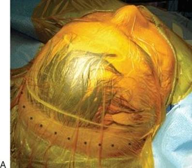

FIGURE 47.3 A thin strip of hair is shaved along a bicoronal line that extends from ear to ear. A. The entire surgical field is covered with a plastic drape to minimize orbital compression. B. Staples are placed parallel to the incision to secure the drapes.
A bicoronal scalp incision is made over the vertex of the scalp from ear to ear. This incision provides the greatest exposure and is cosmetically favorable, even in balding individuals. The incision extends through all layers of the scalp to the underlying bone; laterally, the incision extends to the deep temporal fascia enveloping the temporalis muscle. If a longer pericranial flap is needed, subgaleal dissection posterior to the incision is performed. The pericranial layers are then incised to the underlying bone.
The pericranial flap may be dissected at the time of the scalp elevation or delayed until the time of reconstruction. An advantage of initial dissection is an easier plane of dissection from the galea. Disadvantages include a thinner pericranial flap due to failure to include all of the loose areolar layers of tissue and desiccation of the flap during a long surgery. For these reasons, I prefer to dissect the flap at the end of the surgery.
The scalp is elevated from the underlying cranium; laterally, this requires separation of the pericranium from the deep temporal fascia at the margin of the temporalis muscle using a broad periosteal elevator. The periosteum is then elevated to the level of the superior orbital rims. Laterally, the pad of adipose tissue between the superficial and deep layers of the deep temporal fascia is dissected to protect the temporal branches of the facial nerve. The supraorbital neurovascular bundle is identified at the supraorbital notch and carefully dissected free. In a minority of patients, the supraorbital neurovascular bundle is enclosed by a bony foramen up to 1 cm from the orbital rim. In such cases, it is necessary to release the neurovascular bundle by performing lateral osteotomies with a small osteotome (Fig. 47.4). The periorbita is then elevated from the roof of the orbit bilaterally and to the level of the nasion in the midline. If traction sutures are placed in the scalp, they should be placed at the midline and lateral to the orbit to avoid inadvertent injury to the vascular supply to the pericranium. The scalp should be kept moist during the remainder of the surgery with moistened gauze.
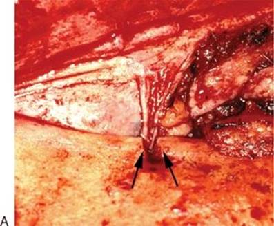

FIGURE 47.4 The supraorbital neurovascular bundles are identified. If enclosed by a foramen, the neurovascular bundle is released by small osteotomies (arrows) with an osteotome (A). The periorbita is elevated from the roof of the orbits with preservation of the supraorbital neurovascular bundles (arrows), and the scalp is dissected to the level of the nasion in the midline (B).
The pericranium and its blood supply are now protected, and the surgical team can proceed with a craniotomy and tumor resection. If a subfrontal approach is performed, orbital osteotomies are performed, and the supraorbital bar is removed. Following repair of the dural defect with primary closure or a fascial graft, a pericranial flap is harvested (Fig. 47.5). The flap is dissected from the undersurface of the galea using sharp tenotomy scissors. The flap is dissected transversely so that the scissors do not perforate the flap. Constant traction on the pericranial flap using a moistened sponge helps maintain the proper plane of dissection. The flap should be retracted at 90 degrees from the plane of dissection to prevent perforation (“buttonholing”) of the flap. Laterally, the pericranium is incised vertically; the incision should be lateral to the vascular pedicle. Elevation of the flap continues to the frontalis muscle where the base of the pericranial flap merges with the galea. Dissection usually stops at this point unless additional flap length is needed. Branches of the nerves and vessels are at risk of injury in this area, and cauterization of bleeders should be done cautiously with bipolar electrocautery. If any perforations of the flap have occurred during elevation, they can be individually closed with 4-0 chromic sutures. For midline defects, bilateral vascular pedicles are usually preserved. This maximizes the blood supply but limits the mobility and reach of the flap. The pedicle of the flap can be sacrificed on one side without jeopardizing the vascularity of the flap (Fig. 47.6). The remaining pedicle should be at least 3 cm wide.

FIGURE 47.5 Following completion of the intracranial dissection and primary dural repair, a pericranial scalp flap is sharply dissected from the inner surface of the galea.

FIGURE 47.6 A pericranial scalp flap based on a unilateral vascular pedicle.
The pericranial flap should separate the nasal cavity from the bone flaps; otherwise, infection of the bone flap can occur. For this reason, the flap should be placed inferior to the supraorbital bar (Fig. 47.7). The flap is rotated posteriorly to cover the dura to the posterior margin of the defect. Small tacking sutures can be placed between the flap and the dura around the periphery of the flap to prevent migration and reinforce the dural closure (Fig. 47.8). In order to prevent compression of the flap, a 2- to 3-mm margin of bone is drilled from the inferior margin of the bone flap in the midline. This gap is undetectable at the level of the nasion and does not result in a cosmetic deformity. The flap pedicle should not be under tension since replacement of the scalp at the end of the operation may pull on the flap and disrupt the repair. The intranasal surface of the pericranial flap can be covered with fibrin glue or Gelfoam and supported with nasal packing (Merocel tampons or a Foley balloon catheter inflated with saline) if desired.

FIGURE 47.7 The pericranial flap is rotated posteriorly to cover the entire dural repair. It should be placed inferior to the supraorbital osteotomy to separate the bone grafts from the nasal cavity.

FIGURE 47.8 If possible, the edges of the pericranial flap are sutured to the dural margins to provide separation of the nasal cavity and cranial cavity.
The scalp incision is closed in the usual manner following replacement of the craniotomy bone flap. A pressure dressing or gauze wrap is not placed on the scalp since this may further compromise the vascularity of a thinned scalp and contribute to scalp necrosis.
POSTOPERATIVE MANAGEMENT
No special care is necessary postoperatively. The intranasal surface of a pericranial flap is kept moist with saline nasal spray until mucosalization is complete. The patient is monitored for signs of a CSF leak or scalp infection.
COMPLICATIONS
Injury to the vascular pedicle of the pericranial flap can occur during dissection (laceration or cauterization) or from compression or torsion of the pedicle during placement. This can result in ischemic necrosis of the flap and delayed healing with CSF leak.
Complications of flap dissection include injury to sensory and motor nerves innervating the scalp, resulting in frontal hypesthesia and paresis of the frontalis muscle. Dissection in the wrong plane, superficial to the galeal layer, can also result in injury to hair follicles with hair loss and excessive thinning of the scalp. Devascularization of the superficial layers of the scalp, especially in patients who have received radiation therapy, can cause ischemic necrosis of the skin with exposure of cranial bone and hardware.
Failure to eliminate dead spaces and to maintain contact between the pericranial flap and underlying dura delays healing and provides a space for accumulation of fluids (hematoma, seroma, or subgaleal CSF collection). This, in turn, increases the risk of infection and CSF leak.
RESULTS
Pericranial flaps are the most frequently used flaps for reconstruction of defects of the anterior cranial base and have been used successfully for decades. An analysis of a large series of craniofacial resections reported a CSF leak rate of less than 10% using vascularized flaps for reconstruction.
A pericranial flap can also be used in endoscopic endonasal surgeries of the anterior cranial base by introducing it extracranially at the level of the nasion (see Chapter 46). In this situation, the flap is exposed with a bicoronal scalp incision, and a bone window is created at the level of the nasion. The pericranial flap is dissected in the usual manner and can be pedicled unilaterally or bilaterally. The flap is inserted through the bone defect below the level of the frontal sinuses to cover the defect in the dura.
PEARLS
· The length of the pericranial flap can be extended by dissecting posterior to the bicoronal scalp incision in the subgaleal plane.
· A moist gauze provides gentle but secure traction of the pericranial flap during dissection without tearing the flap.
· The pericranial flap can be pedicled on one or both supraorbital arteries.
· The flap is dissected at the end of the surgery to avoid thinning and desiccation of the flap.
PITFALLS
· Damage to the blood supply to the pericranium may occur with excessive dissection or cauterization of the flap pedicle.
· Compression of the pericranial flap pedicle can occur unless additional bone is removed from the inferior margin of the bone flap.
· Compressive scalp dressings should be avoided postoperatively since they may compromise the vascularity of the frontal scalp.
INSTRUMENTS TO HAVE AVAILABLE
· Tenotomy scissors
· Bipolar electrocautery
SUGGESTED READING
Cantù G, Solero CL, Pizzi N, et al. Skull base reconstruction after anterior craniofacial resection. J Craniomaxillofac Surg 1999;27(4):228–234.
Parhiscar A, Har-El G. Frontal sinus obliteration with the pericranial flap. Otolaryngol Head Neck Surg 2001;124(3):304–307.
Yoshioka N, Rhoton AL Jr. Vascular anatomy of the anteriorly based pericranial flap. Neurosurgery 2005;57(1 Suppl):11–16; Discussion 11–6.
Patel MR, Shah RN, Snyderman CH, et al. Pericranial flap for endoscopic anterior skull-base reconstruction: clinical outcomes and radioanatomic analysis of preoperative planning. Neurosurgery 2010;66(3):506–512; Discussion 512.
Patel MR, Stadler ME, Snyderman CH, et al. How to choose? Endoscopic skull base reconstructive options and limitations. Skull Base 2010;20(6):397–404.