
Edward R. Laws, Jr.
INTRODUCTION
The traditional approach to midline anterior intracranial skull base lesions such as pituitary macroadenomas, meningiomas, and craniopharyngiomas has been through a craniotomy. Although results with craniotomy in experienced hands have generally been satisfactory, a less invasive and strictly midline approach has the potential advantages of less brain retraction, the ability to devascularize the tumor from below, and the avoidance of inadvertent damage to the optic nerves and optic chiasm.
The evolution of the transsphenoidal approach to the anterior skull base has been a major contribution in the development of neurosurgery. Building on the revelation that transsphenoidal microsurgery could deal with pituitary lesions in a highly effective manner, technologic advances and conceptual advances together have allowed us to extend our surgical skills beyond the sella and into the suprasellar space (Fig. 6.1). Critical to this advance has been introduction of the operating endoscope, image guidance, and the maintenance of the basic principles of skull base surgery including the use of microneurosurgical technique.

FIGURE 6.1 Diagram of the exposure for the extended approach. The bone has been removed from the posterior planum to the sella.
HISTORY
Suprasellar tumors can present with a wide range of symptoms, from vision loss, to mental changes from secondary hydrocephalus, to pituitary hormonal abnormalities. For example, a typical adult patient with a craniopharyngioma may or may not have normal visual function but also may have headache, memory difficulties, fatigue, and mild sexual dysfunction. They will usually not have symptoms of diabetes insipidus.
PHYSICAL EXAMINATION
The physical examination in patients with suprasellar lesions concentrates on disorders of pituitary function and physiology and on abnormalities related to visual function. If the lesion has produced hypopituitarism from compression of the normal gland, physical signs may be those of pallor, generalized weakness, and changes in the texture of the skin. There may be associated cognitive deficits and occasional psychological disturbances.
The visual field examination may reveal a typical bitemporal hemianopsia, decreased visual acuity, enlargement of the physiologic blind spot, or scotomas. These findings may be quite subtle in the early stages of compression of the optic nerves and chiasm. Ocular computed tomography may be useful, as it can demonstrate the thinning of the retinal fiber layer that accompanies chiasmal compression.
INDICATIONS
Ordinarily, the major indication for surgery is progressive visual loss. Patients may also have intractable headache, pituitary hormonal failure (hypopituitarism), mental or memory changes from compression of the hypothalamus, or hydrocephalus, usually from obstruction of the foramina of Monro.
CONTRAINDICATIONS
The relative contraindications to the endoscopic endonasal transsphenoidal microsurgical approach to the suprasellar space are dependent upon the anatomy of the lesion and the anatomy of the skull base. If the sella turcica is small and the lesion is primarily suprasellar, the limiting factors of exposure are the distances between the cavernous carotid arteries and the optic canals. If these are narrow, and not expanded by the lesion, maneuverability may be compromised. More importantly, if the lesion engulfs arteries of the circle of Willis, or if it extends into the lateral optic canals intracranially, then a craniotomy approach may be a wiser strategy. These assessments depend upon neuroradiologic imaging with MRI and CT scans, which are essential to the preoperative planning. The position of the optic chiasm in relation to the sella and the lesion is often a major determinant of the surgical approach. The endoscopic transsphenoidal approach is best suited to the removal of retrochiasmatic lesions as the chiasm and optic nerves do not need to be displaced to expose the lesion.
PREOPERATIVE PLANNING
The majority of the preoperative planning should include a complete endocrine laboratory evaluation and extensive, sophisticated imaging studies. The endocrine evaluation includes measurement of serum levels of the primary hormones of the anterior pituitary and assessment of the patient for diabetes insipidus. It is imperative to normalize any preexisting hormonal deficits and to use perioperative corticosteroids to protect the patient and his/her vision when necessary. A high-resolution pituitary-centered MRI study should be performed to evaluate the lesion and the anatomic distortions produced by it. The position of the optic nerves, optic chiasm, and optic tracts should be carefully evaluated with imaging, as well as the relationship of the dorsal aspect of the tumor to the ventricular system. The sella itself can be enlarged, providing a natural pathway for the extended transsphenoidal approach. A CT scan should also be done to evaluate the presence of calcifications within a suprasellar tumor. An MRI may be adequate for the evaluation of the vessels of the circle of Willis and their tributaries. If not, image guidance using high-resolution CT angiography can be incorporated into the surgical planning and the operative procedure.
SURGICAL TECHNIQUE (VIDEO 6.1)
The image-guidance system is calibrated and used to determine the trajectory of approach and the important anatomic landmarks.
Ideally, a nasal septal flap (see Chapter 42) based posteriorly upon the sphenopalatine artery is raised and placed in the nasopharynx safely away from the operative pathway, without kinking its blood supply. This is an important step for craniopharyngiomas and other lesions where a large intraoperative cerebrospinal fluid (CSF) leak is created.
Through the right and left nostrils, using the operating endoscope, the ostia of the sphenoid sinus are identified by gently laterally displacing the middle and superior turbinates. In my experience, it is rarely necessary to resect a turbinate in order to achieve satisfactory exposure for midline lesions.
The mucosa around the ostia is cauterized, and the ostia are enlarged using bone punches. After a submucosal injection of Xylocaine and epinephrine, the mucous membrane over the posterior septum is incised and a submucosal flap can be raised to expose the vomer. Using a Cottle elevator, the posterior nasal septum is crossed and the mucosa over the anterior wall of the sphenoid sinus on the opposite side is elevated. Careful dissection will mobilize the mucosa away from the inferior aspect of the anterior wall of the sphenoid sinus, diligently protecting the sphenopalatine arteries. The mucosa posteriorly can be resected using a microdebrider.
The anterior wall of the sphenoid sinus is then removed with appropriate bone punches and rongeurs. The sphenoidotomy is enlarged to accommodate the operating endoscope and to provide a satisfactory panoramic view of the sella, the clivus, and the planum sphenoidale. The bony sella is then opened with a chisel (or a drill), and careful resection of bone is carried out from one cavernous sinus to the other laterally and from the tuberculum down to the junction of the sella with the clivus in the superior–inferior plane. Wide exposure of the dura is necessary, and using the high-speed drill and appropriate bone punches, a portion of the planum can be resected in order to allow access to the suprasellar compartment anteriorly. The limiting factors are the carotid arteries and the optic canals, so careful attention must be paid to imaging studies and the anatomic details as the bone is resected and the dura exposed.
Next, the superior intercavernous sinus should be carefully uncovered and the dura opened superior and inferior to this structure. Bipolar cautery can then be used to obliterate the superior intercavernous sinus. The dural openings are connected so that the dura can be opened like a book, exposing the arachnoid of the chiasmatic cistern (Fig. 6.2). Tumors such as meningiomas will present directly arising from the dura, whereas encapsulated craniopharyngiomas will lie beneath the arachnoid. Careful resection in the subarachnoid plane should then be carried out, with cauterization of the lateral vessels feeding the capsule of the lesion. It is important to identify and preserve the superior hypophyseal artery and its branches feeding the inferior aspect of the optic chiasm. Cystic portions of craniopharyngiomas can be drained by entering through the capsule, further decompressing the lesion. Following decompression, mobilization of the lateral walls, and the superior aspect of the tumor, where it is connected to the optic chiasm with arachnoidal adhesions, can be performed. These are taken down with sharp dissection, freeing the chiasm and allowing it to move superiorly. In the sella, the superior aspect of the pituitary gland is dissected free from the inferior aspect of the tumor and dissection posteriorly will reveal the relationship of the tumor to the pituitary stalk. When the stalk is intimately involved, as in many craniopharyngiomas, sharp sectioning of the stalk to free the inferior border of the tumor is advisable. The decompressed tumor can then be carefully mobilized, and attached or incorporated cysts can often be delivered through the transsphenoidal aperture, which usually measures approximately 2 cm in diameter. This aperture is almost always larger than the exposure one achieves through the lamina terminalis using a craniotomy. Additionally, it allows removal of the tumor from behind the optic chiasm, preventing excessive manipulation of the chiasm and the optic nerves and tracts.
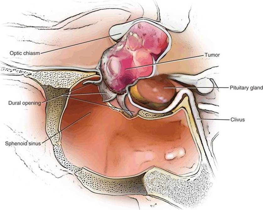
FIGURE 6.2 The extended approach for retrochiasmatic suprasellar tumors. The dura has been opened in a cruciate fashion to expose the suprasellar tumor.
Once the tumor is removed and thorough endoscopic evaluation using angled endoscopes shows no remnants intracranially, hemostasis is assured, and a multilayer closure can be constructed. A free graft of fascia lata or rectus abdominis fascia can be used as an inlay graft, and it is my practice to plug the aperture in the skull base with a suitably tailored adipose tissue graft placed in a “collar stud” fashion and buttressed by a carefully tailored plastic plate. The previously raised septal flap is then placed over the exposed sphenoid bone at the margins of the skull base aperture and held gently in place with Gelfoam. Nasal packs are used when necessary. It is not my usual practice to use lumbar drainage pre- or postoperatively; however, this can be done in select cases.
POSTOPERATIVE MANAGEMENT
Because most craniopharyngiomas directly involve the pituitary stalk, the surgeon must anticipate the development of diabetes insipidus if it was not present before surgery. This requires careful monitoring of fluid balance, electrolytes, and replacement therapy, along with the judicious use of DDAVP desmopressin. Periodic imaging may be necessary to trace the absorption of intracranial air and the status of the operative site. The anterior pituitary hormones must be measured and replaced if abnormal.
COMPLICATIONS
The most common complication of the extended endoscopic transsphenoidal approach is that of postoperative CSF leak. The necessarily large aperture at the base of the skull requires careful multilayer closure to provide a secure barrier against CSF rhinorrhea. The relatively recent adoption of the nasal septal flap technique has significantly lowered the risk of this complication.
The one complication that is more common following endoscopic transnasal procedures than the former microscopic technique is that of postoperative epistaxis. This usually is related to branches of the sphenopalatine artery that are frequently encountered in the endoscopic dissection of mucosa away from the vomer. Intracranial hemorrhage can also occur if vessels related to the suprasellar lesion are violated. This includes the feeding vessels and branches of the circle of Willis.
If the suprasellar tumor is intimately associated with or adherent to the optic chiasm, then visual loss can be a complication of the procedure. Every effort should be made to visualize the intracranial structures clearly and to use sharp dissection under endoscopic view to remove fragments of tumor.
Nasal airway complications including sinusitis, sinus occlusion, mucocele formation, inflammatory change, adhesions, and synechiae may also occur, and appropriate postoperative nasal care is necessary to prevent these problems.
RESULTS
In general, the results of this procedure in experienced hands are very good and provide outcomes that are at least comparable, and in many cases superior, to a variety of open craniotomy techniques (Fig. 6.3). One may anticipate recovery of vision in up to 87% of patients. The rate of preservation of normal pituitary hormonal function varies with the nature of the lesion—very good for meningiomas and pituitary adenomas but poor for craniopharyngiomas.

FIGURE 6.3 (A) Pre- and (B) postoperative, post-contrast MRI, sagittal view, of the endonasal removal of suprasellar craniopharyngioma.
PEARLS
· If the sella is enlarged and the lesion is a craniopharyngioma, one can conclude that the origin of the tumor was infradiaphragmatic.
· The optic chiasm and hypothalamus are not adherent to most of these tumors, and a gross total removal can be accomplished by resecting the diaphragm and tumor capsule from below.
· Maximizing transsphenoidal exposure by removing bone between the carotid arteries, over the superior intercavernous sinus, between the optic canals, and ascending forward along the planum sphenoidale is important in obtaining the necessary visualization and exposure of the tumor.
· Angled endoscopes can provide a superior view of the pathologic anatomy.
· Careful occlusion and opening of the intercavernous sinus is often a key to maintaining the proper trajectory and exposure.
· Effective bipolar cautery may be necessary to control intracranial bleeding sites, and angled instruments may be necessary to reach fragments of tumor that are not in a direct line of sight.
PITFALLS
· Multilayered closure and bone reconstruction of the operative defect must be done carefully and securely, and the nasal flap needs to be approximated to bare bone surrounding the defect.
· Because we still lack truly effective bipolar cautery instruments, bleeding from the intracranial space may be difficult to control. Hemostasis must be assured in an incremental fashion as the surgeon progressively devascularizes the lesion.
· Regular monitoring of serum sodium is necessary since symptomatic postoperative hyponatremia can occur as a result of surgery for any suprasellar lesion.
INSTRUMENTS TO HAVE AVAILABLE
Surgical Instruments for Endoscopic Transsphenoidal Operation
1.Short (18 cm) and long (30 cm) 0-degree endoscopes, 4-mm diameter
2.Long (30 cm) 30-degree endoscope, 4 mm diameter
3.Standard sinonasal instruments
4.Bipolar cautery Aesculap GK560R
a.Pistol grip with variable tips
5.Pituitary rongeurs: Codman 53-1230; Miltex 20-572; Miltex 20-570
6.Kerrison punches: Aesculap FF724R
a.1- to 3-mm Storz 662121–23
b.Down-biting (2 mm) Storz 662132
7.Fukushima suction tips—6F (Ruggles R-8994), 7F (Ruggles R-8995), and 8F (Ruggles R-8996)
8.Frazier suction tips—7F, 9F, and 10F Codman 70-1088
9.Drill
a.High-speed nasal extension (70,000 RPM)
b.3-mm diamond bit
10.Chisels (Mueller [4 mm] B04-RH1400, Aesculap [7 mm] OL302R) and mallet (Codman 88-2520)
11.Straight and angled ring curettes
12.Microscissors (pistol grip)
13.Micro-Doppler
14.Stammberger punches
15.Nerve hook Codman 381030
16.Hardy: Boss 72-2150; 72-2155
17.Micro hook Storz 28164H
SUGGESTED READING
Laws ER Jr. Transsphenoidal microsurgery in the management of craniopharyngioma. J Neurosurg 1980;52:661–666.
Kaptain GJ, Vincent DA, Sheehan JP, et al. Transsphenoidal approaches for the extracapsular resection of midline suprasellar and anterior cranial base lesions. Neurosurgery 2001;49:94–101.
Dumont AS, Kanter AS, Jane, JA Jr, et al: Extended transsphenoidal approach. In: Sheehan JP, Laws ER, eds. Frontiers of Hormone Research, Vol. 34: Pituitary Surgery – A Modern Approach. New York: Springer, 2006:29–45.
Cavallo LM, Prevedello DM, Esposito F, et al. The role of the endoscope in the transsphenoidal management of cystic lesions of the sellar region. Neurosurg Rev 2008;31:55–64.
Kassam AB, Thomas A, Carrau RL, et al. Endoscopic reconstruction of the cranial base using a pedicled nasoseptal flap. Neurosurgery 2008;63:ONS44–ONS52; discussion ONS52–ONS53.