
Charles Teo
INTRODUCTION
Approaches to the anterior skull base have evolved radically over the past three decades. With dramatic technologic innovations in neuroendoscopy, endoscopic endonasal approaches (EEAs) to the anterior skull base have emerged as a viable and less invasive alternative to open transcranial or transfacial approaches. While endonasal approaches to the sella have been successfully performed for decades with microscopic visualization, endoscopy offers several advantages, including improved up-close visualization, superior illumination, and the option to use an angled line of sight. This final advantage allows surgeons to visualize and operate in regions beyond the straight line of sight provided by the operating microscope.
One such region is the medial cavernous sinus. Due to its association with numerous critical neurovascular structures, the cavernous sinus continues to present serious surgical challenges. Nevertheless, with the use of an angled endoscope in experienced hands, selected lesions of the medial cavernous sinus can now be addressed safely through the endonasal corridor. This approach relies exclusively on the endoscope for visualization. Therefore, familiarity with endoscopic-controlled endonasal surgery with angled endoscopes is a prerequisite to performing this approach.
HISTORY
There are two lines of questioning that are important when evaluating a patient for an EEA to the cavernous sinus. Firstly, it is important to ascertain if the patient has had previous surgery or sinus pathology that may complicate the approach. Previous surgery may have exposed the carotid artery, optic nerve, or pituitary gland. Previous pathology, such as sinusitis, or procedures, such as rhinoplasty, may have vital bearing on the preferred surgical corridors. Secondly, preoperative documentation of cranial neuropathies may give some indication of the pathology of the lesion and location. For example, if there is significant facial dysesthesia, then the lateral wall of the cavernous sinus is likely involved. Given the close proximity of the optic nerves and pituitary gland, visual function may be affected.
PHYSICAL EXAMINATION
Preoperative nasal endoscopy will yield important information about which nostril to use, the potential mucosal flaps that may need to be elevated, and the presence or absence of infection. A complete neurologic examination should focus on cranial nerves II to VII. Preoperative formal visual field examination and endocrinology consultation are mandatory.
INDICATIONS
Lesions of the cavernous sinus are notoriously challenging to treat. The indications for treatment depend upon several factors. These include the location and size of the lesion, suspected pathology, need for tissue for diagnosis, growth patterns of the lesion, compression of surrounding neurovascular structures, symptoms, and preexisting cranial nerve deficits. The three most common pathologies encountered in the medial cavernous sinus are meningiomas, pituitary adenomas, and schwannomas.
In general, the surgical treatment of cavernous sinus meningiomas is associated with a high rate of cranial nerve palsies when the cavernous sinus is entered surgically. For this reason, many surgeons reserve surgery for growing or symptomatic cavernous sinus meningiomas and only remove the extracavernous portion of the tumor. In general, I agree with this conservative approach for suspected cavernous sinus meningiomas, especially those that are asymptomatic, stable in size, and with a significant intracavernous portion. At the same time, in cases where the medial cavernous sinus is involved, the endonasal corridor is well suited to biopsy, debulking, and possible gross total resection if the degree of intracavernous involvement is limited. Because meningiomas of the cavernous sinus tend to surround the carotid artery and adjacent cranial nerves, complete removal is generally not advisable if a substantial degree of intracavernous involvement exists.
In contrast to meningiomas, pituitary adenomas are usually amenable to surgical treatment even when they invade the cavernous sinus. The typical soft consistency and medial-to-lateral spread (from the pituitary gland outward) of these lesions make the endonasal corridor an ideal approach for addressing pituitary adenomas with cavernous sinus extension. An advantage of this pattern of growth is that the carotid artery and cranial nerves are typically displaced laterally by the tumor. In such cases, the tumor plane offers a safe surgical corridor into the medial cavernous sinus, and a soft, “suckable” adenoma can be removed with much less risk to neurovascular structures.
The indications for an EEA to the medial cavernous sinus include a lesion that is localized to the medial wall of the cavernous sinus or one that has grown in a medial-to-lateral fashion to displace the carotid artery laterally. Other anatomic features that must be considered prior to selecting this approach include the degree of pneumatization of the sphenoid sinus, the presence of intradural spread or subpial invasion by the tumor, and the position of cranial nerves III through VI. The ideal conditions for an EEA to the medial cavernous sinus include a large pneumatized sphenoid sinus, an entirely extraaxial pathology, and lateral displacement of all cavernous sinus neurovascular structures. A crucial tenet of this approach is that the angled endoscope offers a direct view of the medial wall of the cavernous sinus that is not achievable with standard microscopic visualization. Thus, familiarity with the nuances of endoscopic-controlled surgery is required when planning to address this region via the endonasal corridor.
In summary, indications for the EEA to the medial cavernous sinus include
· Lesions based on the medial wall of the cavernous sinus
· Medial-to-lateral extension of a pituitary adenoma
· Lateral displacement of the carotid artery and cranial nerves
Another approach to the cavernous sinus is a paramedian, transmaxillary approach that involves expert knowledge and familiarity with the structures within the pterygopalatine fossa. This approach is mostly used to remove lesions of the lateral cavernous sinus but may be used with medial lesions and as such will be discussed below in some detail.
CONTRAINDICATIONS
The primary contraindications to this approach reflect anatomic constraints that would place the carotid artery and cranial nerves at high risk. Lesions based within the cavernous sinus, which encase the carotid artery and cranial nerves or displace them medially, are not well suited to an endonasal approach. An asymptomatic benign meningioma with such characteristics may best be observed rather than treated surgically. Other lesions with significant involvement of the surrounding neurovascular structures whose diagnosis is unclear may best be treated with a biopsy rather than complete removal. The goal of treatment for any cavernous sinus lesion involves a thoughtful analysis of the need for complete removal and the degree of risk of neurovascular injury that is justified. Finally, the presence of active sinus infection is a relative contraindication to surgery. Of course, if the operation is considered urgent, it may be done under antibiotic coverage, especially as most lesions are extracranial. If there is no urgency, then time should be spent treating the active infection in the sinuses. As mentioned previously, the endonasal approach to the medial cavernous sinus relies on angled endoscopic visualization. Therefore, lack of familiarity with endoscopic techniques is a contraindication to this approach.
Common contraindications to this approach include the following:
· Encasement of the neurovascular structures in the cavernous sinus
· Medial displacement of the carotid artery and cranial nerves
· Lack of familiarity with endoscopic-controlled techniques
PREOPERATIVE PLANNING
Prior to surgery, a careful review of all pertinent imaging is mandatory. This includes a preoperative MRI, preferably one with thin coronal cuts through the pituitary fossa and adjacent cavernous sinus. The location and extent of the lesion of interest should be assessed, in addition to possible involvement or displacement of surrounding neurovascular structures. These include the carotid arteries, the optic nerves, the pituitary gland and stalk, and the cranial nerves of the cavernous sinus. Normal anatomy should be carefully evaluated. An assessment of the position of the carotid arteries is critical. The flow voids of the carotid arteries are readily noticed on T2-weighted sequences. Special consideration should be taken to assess the position and medial extent of the cavernous portion of the carotid arteries, which may approach the midline in up to 8% of patients. The extent of cavernous sinus involvement by the tumor should be assessed, in addition to the pattern of displacement of all cavernous sinus neurovascular structures.
A CT scan provides an excellent overview of the bony anatomy. An MRI may also provide invaluable anatomical information and is used by many instead of CT. The size and position of the sphenoid sinus as well as the presence and location of septations within the sinus should be noted. Importantly, many sphenoid sinus septations are not in the midline and are often based over one of the carotid protuberances laterally. Lesions that involve the sella demand a full preoperative endocrinologic workup. Likewise, formal visual field testing is necessary for lesions compressing the optic nerves, even in the absence of frank visual complaints. Before surgery, patients should be counseled regarding the risk of nasal deformity, CSF leak, endocrine disturbances, and injury to the carotid artery and cranial nerves.
SURGICAL TECHNIQUE (VIDEO 5.1)
The endonasal approach to the medial cavernous sinus is an endoscopic-controlled technique, meaning that all operative visualization relies on the endoscope and surgical instruments are manipulated outside of the endoscope, but in its field of view. This is in contrast to endoscopic-assisted surgery, where an endoscope is temporarily introduced to inspect a surgical field following a standard microscopic approach, or purely endoscopic surgery, where all instruments are manipulated through a working channel attached to the endoscope. I prefer high-definition endoscopes offering 0, 30, and rarely 70 degrees of angled viewing. While bayoneted instruments are useful in microsurgery because they prevent the surgeon's hand from blocking the microscope's line of sight, they are unnecessary and even detrimental in EEAs. Bayoneted instruments quickly crowd the working area of the surgeon's and assistant's hands, and attempts to rotate bayoneted instruments may cause the surgeon's hand to bump the endoscope. For this reason, straight endonasal instruments should be used. Angled nonbayoneted instruments, such as the angled bipolar and suction, allow the surgeon to access lateral structures when viewed with a 30-degree endoscope. Also, the use of multifunctional “hybrid” instruments greatly improves the efficiency of the EEA by allowing an instrument in one hand to perform multiple functions. Examples include suction bipolars, debriders with built-in irrigation and suction, and some instruments with multiple functions including suction, irrigation, bipolar, and tissue ablation. For the endonasal approach, I prefer a two-surgeon method where one surgeon holds and guides the endoscope, allowing the operating surgeon bimanual control of all endonasal instrumentation. In addition, collaboration with an experienced otolaryngologist is critical in optimizing the safety and efficacy of the approach.
Because the endonasal approach involves multiple surgeons and a diverse array of equipment, including high-definition endoscopes and monitors, an image-guidance system, and specialized endonasal instrumentation, the proper setup of the operating room is critical in optimizing the flow of the procedure. The high-definition monitor is positioned behind the head of the bed, allowing the surgeon to look directly at the image while maintaining a natural ergonomic operating stance. The image-guidance system is placed just to the side of the monitor, also at the head of the bed. Both surgeons are positioned to the right of the supine patient, while the scrub nurse is positioned to the left of the patient. This allows the nurse and surgeon to efficiently transfer instruments without the surgeon turning from the monitor. The anesthesiologist is positioned toward the foot of the bed on the patient's left side.
The patient is positioned in the supine position. The abdomen is prepped and draped in preparation for harvesting adipose tissue, muscle, or fascia. The head is fixated in a Mayfield holder, extended and turned laterally so that the surgeon has a direct view through the nares. The frameless image-guidance system is registered and confirmed. Frameless stereotaxy is used to confirm the surgical trajectory, to assess normal anatomic landmarks during the operation, and to identify structures that should be avoided during the course of surgery.
The patient's nose is prepared with Cottonoid strips soaked in 1:2,000 adrenaline. After a few minutes, the strips are removed and the middle turbinate and septal mucosa are infiltrated with 1% bupivacaine with 1:100,000 adrenaline. In general, I prefer a two-nostril two-surgeon approach. The lower half of the middle turbinate is resected or in some circumstances simply displaced laterally. If intradural extension of the tumor is suspected, a vascularized nasoseptal flap is prepared. This flap is elevated from the nasal septum based on a pedicle containing the posterior septal artery. A partial posterior septectomy is made, and both sphenoid ostia are identified. Using a combination of Kerrison rongeurs, high-speed drill, and endonasal debrider, a wide bilateral sphenoidotomy is performed. The sphenoid sinus is entered, and after careful removal of sinus septations, the relevant anatomy is inspected, including the carotid and optic prominences, the medial and lateral opticocarotid recesses, and the sella. Image guidance and ultrasound Doppler can be useful adjuncts in identifying the at risk anatomic structures. The surgeon should be aware of possible bony dehiscence over the carotid arteries. The position of the carotid arteries can be confirmed with image guidance or ultrasound. In cases where large tumors distort the surrounding anatomy, image guidance is indispensable.
Once within the sphenoid sinus, the bony opening depends on the size and location of the lesion of interest. For pituitary adenomas with lateral extension into the cavernous sinus, the floor of the sella is first removed, followed by extension of the bony opening to the lateral wall of the sphenoid sinus. The position of the carotid artery must be carefully assessed prior to drilling laterally. Adenomas, which displace the carotid artery laterally, create a zone of safety for drilling of the lateral wall of the sphenoid sinus. This maneuver and all subsequent work performed laterally are enhanced with the use of an angled endoscope and angled instruments to provide good visualization of the lateral wall and medial cavernous sinus. Depending on the location of the lesion and the trajectory offered by the nasal sinuses, the use of the contralateral nostril may offer a more direct approach to the ipsilateral wall of the medial cavernous sinus. Once the lateral wall of the sphenoid sinus is removed, the medial cavernous sinus is visible and the pathology of interest is addressed. Often, a natural cleavage plane may be exploited on the outer margin of the tumor. Respecting this plane during surgery significantly reduces the risk of injury to the neurovascular structures of the cavernous sinus. In the case of pituitary adenomas, entrance into the cavernous sinus may be achieved by using the natural fenestration(s) that exists between the pituitary fossa and the sinus, usually behind the anterior genu of the carotid artery. If there are no windows into the sinus, then direct access through the anterior face of the cavernous sinus may be performed safely using frameless stereotactic guidance and intraoperative Doppler ultrasound to ensure that the anterior loop of the intracavernous carotid artery is not inadvertently damaged. Bleeding from the cavernous sinus is usually minimal. However, once the tumor has been removed, the sinus may reopen and bleeding may be copious. The first thing to remember about venous bleeding is that it is readily controlled. Therefore, do not panic. The initial maneuver is to elevate the head of the bed. This simple step is sometimes enough to control the hemorrhage. The next maneuver is to inject hemostatic agents such as Floseal (Baxter) or Surgiflo (Johnson and Johnson) directly into the sinus and then cover this with a Cottonoid and apply gentle pressure. If hemostatic agents are not available, then compressive agents such as Gelfoam and Surgicel may be used. In the case of other lesions such as schwannomas and meningiomas, this more direct approach is augmented by a transmaxillary dissection. Instead of lateralizing the turbinate, this is medialized and the hiatus semilunaris is identified and used as a landmark to gain entry into the maxillary sinus. Once the medial wall of the maxillary sinus is opened, the lateral part of the face of the sphenoid sinus is opened, lateral to the ostium. To gain more exposure inferiorly and to identify the V2 branch of the trigeminal nerve, the posterior wall of the maxillary sinus may be removed. This will reveal the contents of the pterygopalatine fossa.
Once the lesion has been addressed, a careful closure is of utmost importance. I prefer a layered reconstruction of the sphenoid sinus, with use of an abdominal adipose tissue graft and/or vascularized nasal septal flap if CSF is encountered during the operation. The basic tenets of closure include the following:
· A multilayered closure is superior to a single layer.
· A vascularized graft is superior to a nonvascularized one.
· An autologous graft is superior to a synthetic graft.
· Counter pressure may be applied via an intranasal balloon or nasal packing.
· Glue or dural sealant is a useful adjunct to seal the defect.
· Routine lumbar drainage is discouraged although single lumbar puncture serves to reduce CSF pressure immediately and for several days after surgery. CSF will continue to leak out of the dural hole into the epidural space, acting as a temporary “escape valve.”
POSTOPERATIVE MANAGEMENT
The endonasal approach to the medial cavernous sinus is, in general, associated with less morbidity and shorter hospital stays than open cranial approaches to this region. Postoperative evaluation of pituitary function is essential for lesions involving the pituitary gland or stalk. All patients should be assessed for a possible CSF leak. In the absence of endocrine dysfunction or CSF leak, most patients are discharged home within 48 hours of surgery. I typically do not use lumbar drainage to prevent a CSF leak, as appropriate skull base reconstruction is the mainstay of avoidance of a CSF leak.
COMPLICATIONS
Potential complications of the endonasal approach to the medial cavernous sinus include the following:
· CSF leak/meningitis
· Pituitary dysfunction
· Visual loss
· Ophthalmoplegia
· Injury to the carotid artery
· Approach morbidity such as synechia, poor humidification, and sinus ostia obstruction
RESULTS
Between 2006 and 2010, I have treated 160 pituitary adenomas. Of the 160 cases, 22 were recurrent tumors, 76 lesions were endocrinologically active, and 34 lesions extended laterally into the cavernous sinus. Using an endoscopic-controlled technique, without an operating microscope, I achieved a 78% cure rate for active tumors (defined as gross total resection and biochemical remission) and an 88% cure rate for inactive tumors (defined as gross total resection). In a logistic regression analysis, cavernous sinus invasion was not a negative predictor of cure. These results demonstrate that an excellent outcome is achievable using the EEA for pituitary adenomas regardless of extension into the cavernous sinus (Fig. 5.1). For appropriately selected cases, cavernous sinus involvement should not be considered a contraindication to curative surgery.


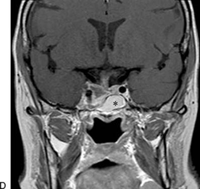
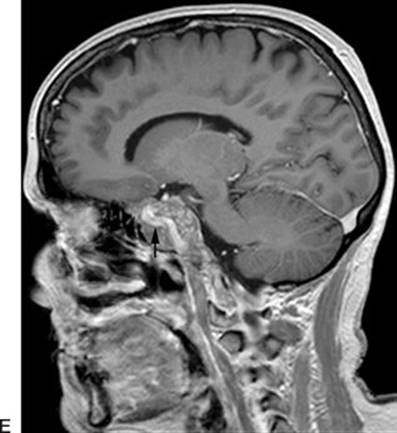

FIGURE 5.1 A–F. Preoperative (A,C,E) and postoperative (B,D,F) MRI of a patient with a prolactin-secreting pituitary macroadenoma extending laterally into the right cavernous sinus. The preoperative images demonstrate the extent of the tumor and the lateral displacement of the cavernous portion of the internal carotid artery (arrow). Given the size of the tumor and lack of response to medical treatment, the patient underwent a midline EEA. After removal of a portion of the lateral sphenoid sinus wall, medial to the ICA, the medial wall of the cavernous sinus was entered. The tumor was removed with angled instruments under visualization with a 30-degree-angled endoscope. The postoperative MRI demonstrates an excellent resection. The abdominal adipose tissue graft is demonstrated in the sphenoid sinus (asterisk). Following surgery, the patient's prolactin level returned to well within normal range, indicating a biochemical cure.
PEARLS
· The angled endoscope and angled instruments are essential to visualize and reach the medial cavernous sinus from a midline transsphenoidal approach. Conversely, 0-degree scope and straight instruments are used for the more lateral transmaxillary approach.
· Special consideration of the position of the contents of the cavernous sinus is critical. The ideal lesion for this approach displaces the carotid artery and cranial nerves in a medial-to-lateral fashion.
· The texture and consistency of tumors dictates the ease and safety of removal. Soft pituitary adenomas may be completely removed from the cavernous sinus with gentle suction, whereas radical resection of fibrous meningiomas may not be possible.
· Venous bleeding should be expected and is generally easily controlled by head-up positioning of the patient, hemostatic agents, and gentle pressure.
· The surgeon should be aware of possible dehiscent bone over the cavernous carotid arteries.
· When operating under endoscopic visualization, the surgeon should be cognizant of more superficial structures out of the view of the scope that may be restricting the operative corridor and limiting surgical freedom.
· A 2-surgeon, 3- or 4-handed approach allows the surgeon to employ standard bimanual microsurgical techniques. This requires an assistant to hold and guide the endoscope.
· Meticulous and durable reconstruction is absolutely essential to avoid CSF leak.
PITFALLS
· Vascular injury. The most feared vascular complication is injury to one of the carotid arteries. This may be avoided by proper planning and recognition of the position of the cavernous carotids on preoperative imaging. The surgeon should be wary of possible dehiscent bone over the carotid prominences when drilling in the sphenoid sinus. In addition, firm and fibrous tumors should not be pulled from the cavernous sinus without first carefully and completely dissecting the lesion from surrounding structures. When operating within the cavernous sinus, “blind” pulling of tumor from a region beyond the view of the endoscope should be avoided. If injury to the carotid artery occurs, the technique that appears to be most effective is direct packing with macerated muscle obtained from the patient's abdominal wall or temporalis muscle.
· Inadequate visualization. The midline endonasal approach to the medial cavernous sinus relies on an angled line of sight; therefore, the use of angled endoscopes and instruments is essential to visualize the region of interest.
· CSF leak. Meticulous layered skull base reconstruction is essential to avoid the most common complication of this approach, which is CSF leak.
INSTRUMENTS TO HAVE AVAILABLE
· Endoscopic shaver
· Endonasal drill with cutting and diamond bits
· Micro–Doppler ultrasound with malleable probe
· Selection of ring curettes
· Angled suctions with smooth atraumatic tips
· Endonasal bipolar forceps
SUGGESTED READING
Dehdashti AR, Ganna A, Karabatsou K, et al. Pure endoscopic endonasal approach for pituitary adenomas: early surgical results in 200 patients and comparison with previous microsurgical series. Neurosurgery 2008;62:1006–1017.
Ceylan S, Koc K, Anik I. Endoscopic endonasal transsphenoidal approach for pituitary adenomas invading the cavernous sinus. J Neurosurg 2010;112:99–107.
Lindley T, Greenlee JD, Teo C. Minimally invasive surgery (endonasal) for anterior fossa and sellar tumors. Neurosurg Clin N Am 2010;21(4):607–620.
Teo C, Wait S. Endonasal approach to tumors of the pituitary fossa: a shift in the treatment paradigm. Clin Neurosurg 2011;58:79–83.