
Douglas J. Mathisen
Bronchoscopy is an essential part of thoracic surgery. All those who perform general thoracic surgery should be well versed in the use of a variety of bronchoscopes. Flexible bronchoscopy has supplanted rigid bronchoscopy as the most common way to perform bronchoscopy. Because of the frequent use of the flexible bronchoscope, rigid bronchoscopy is somewhat of a lost art. Many have never done a rigid bronchoscopy. There are certain indications where rigid bronchoscopy is the preferred method of choice and a busy thoracic surgeon should be familiar in the use of a rigid bronchoscope. We will consider each separately.
Flexible Bronchoscope
There are a variety of flexible bronchoscopes available. They come in pediatric sizes, adult bronchoscope size, a larger channeled bronchoscope, video bronchoscopes, and bronchoscopes that only have optics and no capability for suctioning. The adult bronchoscopes have port instruments pass through. These instruments include biopsy forceps, brushes, snares, aspirating needles, and balloon dilators, allowing a variety of procedures to be done through the flexible bronchoscope. There are a variety of ways the flexible bronchoscope can be inserted.
Flexible bronchoscopy is typically done in the awake patient. Some patients may require gentle sedation while others may be able to have the procedure performed only with topical anesthesia. My preferred route and method is through the mouth. The patients gargle 4% Xylocaine and expectorate without swallowing. Once they have gargled sufficiently, they are then sprayed with an atomizer with 1% Xylocaine in the pharynx to improve anesthesia. As long as we are not evaluating for the source of hemoptysis, I frequently will use a butterfly with 4 cc of 1% Xylocaine passed through the cricothyroid membrane (Figs. 1.1 and 1.2). The patient should be instructed to try not to cough, not to swallow, and not to speak during this installation. The butterfly is inserted at a 45-degree angle through the cricothyroid membrane to avoid laceration of the membranous wall. Once the operator is confident they are in the airway, a syringe filled with Xylocaine is aspirated to be certain you are in the airway. Once that has been established, 4 cc of 1% Xylocaine is slowly instilled into the airway. If it is done slowly enough, typically, patients will not cough. If they do start to cough, the Xylocaine should be instilled quickly and the butterfly removed. Gentle pressure should be held over the insertion site. Patients are instructed to cough to spread the Xylocaine throughout the airway and especially on the underneath surface of the vocal cords. To insert the flexible bronchoscope through the mouth, the assistant holds the tongue with a gauze and gently retracts. This allows the operating surgeon to insert the flexible bronchoscope from the head of the table into the mouth, identifying the epiglottis and inserting the bronchoscope through the vocal cords. Once through the vocal cords, the tongue can be released. If the patient is not adequately anesthetized and continues to cough or gag, more sedation may be required or more topical anesthesia applied through the flexible bronchoscope to the cords themselves by placing the bronchoscope just above the vocal cords and gently instilling a small amount of Xylocaine onto the vocal cords. Usually, I use 2 to 3 cc of 1% Xylocaine. This technique has been very effective, and when combined with gentle sedation, is well tolerated by virtually everyone.
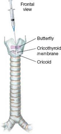
Figure 1.1 Frontal view.
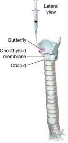
Figure 1.2 Lateral view.
Others prefer passing the flexible bronchoscope through the nose. Sometimes this is difficult due to abnormalities in anatomy or bleeding that may ensue. To prepare the patient for transnasal insertion, it is best to spray the nose with 1% or 2% Xylocaine; 4 cc is the amount used. Sometimes epinephrine should be included with this to constrict blood vessels and reduce the risk of bleeding. Cotton swabs soaked in Xylocaine can be placed into the nasal passage to aid in anesthetizing the area. Patients are still required to gargle with Xylocaine and spray the back of the throat as they will be sensitive when the scope enters the pharynx. The same procedure for the vocal cords, either using the butterfly technique or directly applying Xylocaine through the bronchoscope to the cords, is necessary.
The flexible bronchoscope has many other ways in which it can be effectively used. Passing it through the rigid bronchoscope allows careful inspection of the distal airway while still maintaining ventilation of the patient. Placing an uncuffed endotracheal tube over the flexible bronchoscope allows it to be inserted after proper topical anesthesia, control of the airway and repeatedly removing the bronchoscope as specimens are obtained (Fig. 1.3). The uncuffed or cuffed endotracheal tube can be connected to oxygen and supplement the patient’s oxygen in that fashion. A small adaptor can be connected to tubing with oxygen and the cap allows insertion of the bronchoscope. Another effective means of inserting the flexible bronchoscope is with a laryngeal mask airway that has been inserted at the beginning of anesthesia. With this same connecting tubing, the patient can be ventilated while working through the connector. It has been my experience that a little topical anesthesia is oftentimes effective in allowing the flexible bronchoscope to be inserted easily under the epiglottis and into the airway. Even though the patient may be deeply anesthetized, their cough reflex is somehow maintained. With patience and a little topical Xylocaine, this reflex is eliminated. In emergencies, a small endotracheal tube can be threaded over a bronchoscope, inserted through an laryngeal mask airway (LMA) and inserted into the airway. This may be useful in certain emergencies where control of the airway is difficult and/or the operator is inexperienced with other methods of controlling the airway.
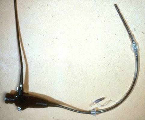
Figure 1.3 A well-lubricated endotracheal tube is threaded over an adult flexible bronchoscope.
In some cases, evaluating and controlling the airway is best done with a flexible bronchoscope with an endotracheal tube threaded over it (Figs. 1.1 and 1.2). This can be done under local anesthesia. This can be done as an awake intubation. It can be done when there is suspected trauma and concern exists about the status of the airway. The operating surgeon should choose a flexible bronchoscope that has adequate suctioning as oftentimes secretions or blood is a part of the problem. It should be well lubricated to pass easily through a no. 7 endotracheal tube although smaller sizes can be used depending upon the circumstances. Pediatric scopes can also be used; however, they provide little resistance to guide a tube into the airway and are inadequate for suctioning in most cases. Once the airway has been safely entered, the endotracheal tube can be advanced over the scope while continuously observing the airway through the bronchoscope to be certain it has not come out of the airway. It sometimes is required to have a little gentle jaw thrust to allow the endotracheal tube to pass easily. Once the airway is secured, suctioning and anesthesia can be done.
Once the flexible bronchoscope has been inserted, it can be used for many purposes. Its simplest use is to make careful observations, locate tumors, strictures, or the source of bleeding. If biopsies are required, bronchoscopic flexible forceps can be passed through the working channel and biopsies obtained under direct vision. Some foreign bodies can be removed with the biopsy forceps; however, a snare is sometimes more efficacious in trying to remove a foreign body with the flexible bronchoscope. Adult flexible bronchoscopes with an adequate channel can be used to pass balloon dilators and dilate strictures or tamponade areas of bleeding. Bronchoscopic washings and brushings can be performed through the flexible bronchoscope.
 COMPLICATIONS
Bronchoscopy done under awake anesthesia has as its most common complication hypoxia and arrhythmias. Therefore, patients should be monitored and receive supplemental oxygen to minimize this risk. The most serious complication occurs in those patients who have tracheal stenosis and flexible bronchoscopy is used in the outpatient setting. Repeated attempts at intubating the patient can convert a marginal airway into a critical airway and should be avoided at all costs. If one encounters an unsuspecting stenosis, it is best to view it from above the stenosis and not manipulate the bronchoscope across it and thereby reducing the chance for bleeding or edema and loss of the airway. Special note should be made of those patients who have tracheobronchial trauma. Repeated attempts at trying to intubate a patient over a flexible bronchoscope should be avoided. If this approach does not work at first, a plan should be made for an emergency tracheostomy to secure the airway. Bleeding can occur when transbronchial biopsies are attempted or biopsies in the subsegmental airways. The subsegmental airways are quite delicate and can be easily penetrated by the flexible bronchoscope biopsy forceps. Small amounts of bleeding can usually be managed with cold saline irrigation or the use of dilute 1:10,000 solution of epinephrine. One should monitor the amount of Xylocaine used to avoid Xylocaine toxicity and its side effects. Another indirect complication of flexible bronchoscopy is the persistent use of the bronchoscope to aspirate large quantities of blood, which are inadequately removed by the flexible scope. This may lead to hypoxia, inability to ventilate the patient, and cardiac arrest.
Other uses of the flexible bronchoscope include positioning endotracheal tubes, double-lumen tubes, aspirating through the tubes during the course of an operation, and use of the flexible scope through an endotracheal tube to identify intraluminal pathology that is not apparent to the surgeon outwardly. Bronchial blockers can be positioned with the use of the flexible bronchoscope. The bronchial blocker ideally is passed around a single-lumen tube but sometimes can be passed through an endotracheal tube by scoring the rubber gasket on the connecting tubing between the anesthesia tubing and the endotracheal tube. A needle can be used to lacerate the cap, the bronchial blocker passed through that with the stopper in the end of the cap, and then positioned with the use of a flexible bronchoscope. The same is true for the blocker, which is passed around the endotracheal tube, advanced under direct vision with the flexible scope, and then guided into the proper mainstem bronchus. This can sometimes be aided by using a biopsy forceps through the flexible bronchoscope, grasping the balloon bronchial blocker, and advancing it in that fashion. Difficult intubations are made much easier if either the endotracheal tube or double-lumen is inserted over a flexible bronchoscope; the airway can be visualized, the tube advanced under direct vision, and can be done in a much safer fashion.
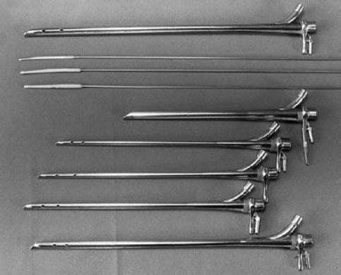
Figure 1.4 Series of graduated rigid bronchoscopes to allow dilation under direct vision and gradual dilation.
Rigid Bronchoscopy
Rigid bronchoscopes are much less commonly used in today’s contemporary practice of thoracic surgery. There are many types of rigid bronchoscopes and a busy thoracic surgeon should be familiar with all of them. First, rigid bronchoscopes come in different sizes from pediatric bronchoscopes up to the adult rigid bronchoscope (Fig. 1.4). The availability of multiple different sizes of rigid bronchoscopes allows for placement in difficult airways, dilating tracheal strictures, and performing biopsies. For difficult airways where tracheal stenosis might present a problem, having pediatric bronchoscope all the way through adult bronchoscopes available is essential. There are rigid bronchoscopes available that have magnifying telescopes that are able to be passed through the channel along with a rigid biopsy forceps, allowing very careful biopsies under direct vision through the rigid bronchoscope (Fig. 1.5A,B). The old rigid bronchoscopes that required very long instruments to be passed down the operating channel were always very challenging due to the lack of light, optics, and the inability to see around the biopsy forceps, which were being passed.
 TECHNIQUE
The most common technique of inserting a rigid bronchoscope is to place the middle finger on the upper teeth and pull back. The index finger is used to push the lower lip out of the way and the bronchoscope is inserted using the thumb to lever it off the teeth. People with prominent teeth or small mouths present a challenge. It may be necessary to insert the rigid bronchoscope laterally in the area of the molars rather than anteriorly where the teeth are most prominent. Those who are unskilled or having to use small, rigid bronchoscopes may find the use of the laryngoscope helpful. The curved laryngoscope is inserted allowing visualization of the epiglottis. The rigid bronchoscope can then be inserted underneath the epiglottis under direct vision. Sometimes it requires taking the laryngoscope out at this point as it is difficult to pass the bronchoscope with the laryngoscope in place. If the operator is underneath the epiglottis, it would be easy to identify the vocal cords and pass the scope. Small, rigid bronchoscopes have challenging optics and light sources. The use of a 2-mm telescope inserted into the lumen of the pediatric rigid bronchoscope allows for better visualization and adequate light, allowing a more safe passage of the small scopes into the airway. Indications for the rigid bronchoscope over the flexible bronchoscope are thought to be assessing for bleeding, removal of foreign bodies, evaluating tracheal strictures or tumors, dilating the trachea, or coring out tumors. The rigid telescopes that are passed through the bronchoscope come in 0-degree, 30-degree and 45-degree angulations, allowing assessment of the mainstem and the lobar orifices. The most common mistakes in the technique are inability to find the epiglottis, finding the epiglottis but advancing the scope too far beyond and elevating the larynx and intubating the esophagus, and failure to rotate the rigid bronchoscope 90 degrees, allowing it to enter with the tapered end between the vocal cords.
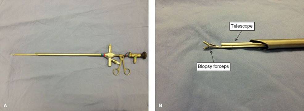
Figure 1.5 A,B: An example of a rigid bronchoscope with telescope near the biopsy forceps allowing excellent visualization for biopsy.
The most common use for the rigid bronchoscope is careful assessment of the airway, especially in patients who have tumors or stenoses. The ability to use high-quality optics to inspect the mucosa and the ability to make very precise measurements is invaluable to the tracheal surgeon. Measurements are generally taken from fixed points such as the carina, the bottom and top of a tumor or stenosis, and the vocal cords. This gives a very precise estimate of the degree of involvement, either by benign stenosis or malignant tumor. The rigid bronchoscope can be used to core out obstructive malignant tumors. It is best to visualize beyond the tumor to know exactly what you are dealing with beyond the obstruction. It oftentimes is preferable to pass a flexible bronchoscope through the rigid scope, passing by the side of the tumor, and aspirating any retained secretions or blood that might exist, allowing for more effective ventilation and a greater margin of error. It is ideal to biopsy the tumor to determine its consistency and vascularity. Once the operator is satisfied with that, further removal of tumor can be done with rigid biopsy forceps. The bronchoscope that allows visualization through a telescope inserted into the rigid bronchoscope to the end of the bronchoscope and then biopsy forceps being passed next to them, allows for direct vision and is preferable in these circumstances (Fig. 1.5A,B). Once it has been determined that the tumor is of certain consistency and vascularity that allows proceeding with coring out of the tumor, this can be done with multiple bites with the biopsy forceps or using the tip of the rigid scope to corkscrew and gently “core out” the tumor. It can then be retrieved with the biopsy forceps or with the suction catheter and removing the entire rigid bronchoscope with the tumor attached to the suction. Any bleeding that ensues can be managed with tamponade by the rigid bronchoscope. Topical application of 1:10,000 dilute epinephrine, cold saline, or the placement of a balloon to temporarily tamponade it. This has to be done in short intervals because of the inability to ventilate a patient with the balloon inflated.
When rigid bronchoscopy is done to assess for massive hemoptysis, it is best to assess quickly the origin of the bleeding, use the large suction catheter to remove blood, and irrigate as needed. Once the laterality of bleeding has been determined, the rigid bronchoscope can be inserted into the opposite mainstem bronchus and the patient ventilated safely.
Foreign bodies are best retrieved with the rigid bronchoscope. There are a variety of biopsy forceps, alligator forceps, and duckbill forceps that can be passed through the rigid scope to grasp a foreign body and remove it. It sometimes can be removed through the lumen of the bronchoscope. Other times, it has to be held securely against the end of the bronchoscope and then removed with the bronchoscope.
 COMPLICATIONS
There are complications that can occur with rigid bronchoscopy. Certainly, breaking teeth or dentures must be avoided at all costs. People who have bad dentition, multiple caps, or bridges are at greater risk. Those who have a small mouth and prominent teeth are also at great risk. This should be an uncommon complication. Inserting the bronchoscope can injure the patient by entering into the esophagus and perforating it, choosing too large a scope and damaging the vocal cords. Not staying in the proper axis of the airway risks perforation. When dilating tracheal stenosis, it is important to have the proper-sized scope to initially insert through the stenosis, passing under direct vision through the stenosis, gently corkscrewing it, while applying gentle pressure. Once through the stenosis, the patient can be safely and more effectively ventilated. It is important to stay in the axis of the airway to avoid perforation. It is important to have good visualization to avoid the elevation of mucosal flaps. Careless insertion of the rigid scope can damage the vocal cords and must be avoided. Bleeding that occurs from biopsying or coring out tumors, again, can be managed with topical epinephrine, tamponade with the scope, placement of balloon catheter through the rigid scope, and insufflating it to tamponade the bleeding. There must be close cooperation between the operating surgeon and the anesthesiologist to avoid hypoxia and long periods of time without ventilation. If there is a large air leak around the rigid bronchoscope, manually closing the mouth and nose allows a seal. This can be aided by the use of lap pads to give some substance to the attempted occlusion of the nose and mouth. The flexible scope can be quite effectively used when passed through the lumen of the rigid bronchoscope. This can be done with an adaptor to create a seal and allow the operating surgeon time to perform whatever is needed with the flexible scope. The rigid bronchoscope, in certain circumstances, may be used as a primary method of controlling the airway when ventilating a patient. Certain conditions such as large goiters that distort the anatomy may only be managed with a rigid bronchoscope left in place. The surgeon must be well versed in the use of rigid bronchoscopes.