
Andrew J. Kaufman and Raja M. Flores
 INDICATIONS
Pulmonary segmentectomy is most commonly used to treat patients with peripheral primary bronchogenic lung cancers less than 2 cm in size. Historically, segmentectomy was first described as a surgical treatment for bronchiectasis and tuberculosis. In 1939, Churchill and Belsey described anatomic segmentectomy for bronchiectasis of the lingua of the left upper lobe. These benign disease processes tended to occur bilaterally in the sublobar segments of the lung and were therefore well suited to a lung-sparing approach to resection. Over time, the procedure of segmentectomy became defined as the operative ligation of each individual bronchopulmonary segmental artery, vein, and bronchus, division of the lung parenchyma along the intersegmental plane, and in oncologic cases, the clearance of segmental, hilar, and mediastinal lymph nodes.
In the United states, by the end of the 1960s in men and the 1990s in women, lung cancer had become the most common form of cancer related death. Until the 1960s, pneumonectomy was regarded as the standard surgical treatment for lung cancer. However, the mortality rate associated with pneumonectomy for non-small cell lung cancer, first described by Graham in his groundbreaking series from 1933, approached 30% to 40%. In addition, the significant morbidity of pneumonectomy spurred interest in lesser more anatomic resections. Shimkin’s landmark comparison in 1962 of the Ochsner and Overholt clinics’ results comparing pneumonectomy and lobectomy for the treatment of lung cancer, clearly illustrated that patients with localized lung cancer had improved survival compared to those with more advanced disease, regardless of whether lobectomy or pneumonectomy was performed. Lobectomy with systematic lymph node dissection then became the standard operation for lung cancer. Controversy regarding the extent of resection and interest in lung-sparing procedures continued into the 1970s when Jensik et al. first established that anatomic segmentectomy provided an equivalent 5-year survival compared to lobectomy in stage I lung cancer patients. In response to this controversy, during the 1980s, the Lung Cancer Study Group (LCSG) performed the only randomized controlled trial to-date comparing survival outcomes after lobectomy or sublobar resection, which included segmentectomy and nonanatomic wedge resection in the treatment of early (cT1N0, stage IA) nonsmall cell lung cancer. This seminal study, published in 1995, concluded that lobectomy offered statistically significant superior control of local recurrence; yet no statistically significant survival benefit. However, the threefold increase in local recurrence rate found in the sublobar group and the trend toward decreased survival established lobectomy as the gold standard. Segmentectomy was considered a “compromise” operation for patients with limited cardiopulmonary reserve.
More recently, the controversy regarding sublobar resection and segmentectomy has again resurfaced as the diagnosis of small (less than 2 cm), peripheral, multiple, and subsolid nodules has increased dramatically with the advent and common use of CT scans of the chest. Furthermore, the diagnosis of smaller and less solid nodules has increased with the implementation of lung cancer screening programs. The characteristics of lung nodules diagnosed presently are significantly different than the lung cancers diagnosed in the LCOG study which relied on chest x-ray for diagnosis and allowed lesions up to 3 cm in size to be included in the study. Recent changes in the WHO classification of lung adenocarcinomas into adenocarcinoma in situ (AIS), minimally invasive adenocarcinoma (MIA), and solid invasive carcinoma, reflect the profound heterogeneity in size, density, and invasive behavior seen in lung cancers diagnosed today using more modern approaches. In Japan, the early use of lung cancer screening programs and the adoption of sublobar resection for detected early stage lung cancers have provided significant data illustrating equivalent survival outcomes with segmentectomy and lobectomy. The Japanese experience illustrates that in appropriately selected patients, even those eligible for lobectomy, intentional sublobar resection, and in particular, segmentectomy, can provide equivalent long-term survival compared to lobectomy. Wedge resection has consistently been shown to offer inferior results in terms of local control and survival compared to both lobectomy and segmentectomy.
Currently, segmentectomy, either performed as an open or VATS procedure, is most indicated in patients with small, less than 2 cm (T1aN0M0 stage IA) peripheral lung cancers with limited pulmonary reserve who cannot tolerate formal lobectomy. In these patients, a segmentectomy will provide the most accurate oncologic staging and best overall outcomes compared to wedge resection, radiofrequency ablation, and radiation therapy. Recent studies have shown the feasibility, safety, and efficacy of segmentectomy as a primary treatment for early lung cancers regardless of cardiopulmonary risk. Furthermore, patients with synchronous primary lung cancers or metachronous cancer after previous lung resection should be considered for segmentectomy.
VATS segmentectomy is a technically challenging and demanding operation. The technical difficulty of performing a segmentectomy, either open or VATS, compared to a lobectomy, has likely contributed to the lack of widespread adoption of the procedure. In addition, the complexity of performing a VATS segmentectomy is considerable compared to open segmentectomy and VATS lobectomy. Some studies have shown higher complication rates; further limiting its use outside of expert centers with high-volume and high-expertise VATS programs. Over the past two decades, VATS Lobectomy has been proven to offer equivalent oncologic outcomes compared to open lobectomy. The long-term safety and equivalence in outcomes of a VATS approach for treating lung cancer has increased interest in VATS segmentectomy as a definitive treatment for early stage lung cancer. The benefit of a minimally invasive approach includes a shorter hospital length of stay, improved cosmesis, decreased postoperative pain, better postoperative pulmonary function and pulmonary toilet, and an increased tolerance of adjuvant chemotherapy, compared to open thoracotomy. In addition, VATS segmentectomy preserves healthy lung tissue and reduces the loss of pulmonary function. This is critical for patients who have undergone previous lung resection, have minimal pulmonary reserve, and otherwise would not be candidates for resection, or in patients who have or are at risk of developing multiple lung cancers.
 CONTRAINDICATIONS
VATS segmentectomy should not be considered in patients with tumors larger than 2 cm (any T1b or greater T stage tumor) or in whom there is evidence of nodal disease, in effect, precluding any lung cancer staged greater than AJCC seventh edition T1aN0M0, stage IA. VATS segmentectomy should not be considered in patients with central tumors close to the origin of the lobar bronchi, or in tumors with endobronchial invasion, as an appropriate margin will require a more extensive resection that is accomplished more readily with an anatomic lobectomy. One can consider performing an extended VATS segmentectomy, that includes additional parenchyma from a neighboring segment without hesitation, or a formal segmentectomy of multiple segments. However, the amount of viable lung tissue preserved after removing multiple segments would be questionable and the risk of complications, such as torsion of the unresected segment, higher compared to a lobectomy. Hilar and mediastinal lymph nodes must be examined at the time of surgery and any lymph node metastasis is an indication to convert the operation to a formal lobectomy.
 PREOPERATIVE PLANNING
All patients must have dedicated imaging of the chest with a CT scan, including the adrenal glands, using 3-mm intervals or less per image. Contrast enhanced scans are not necessary but can be helpful in providing specific information regarding the size and location of lymph nodes, the anatomic location of critical vessels, and the relationship of the tumor to vascular structures that is helpful for preoperative planning and segmental localization. In addition, reformatted images of axial, sagittal, and coronal views are imperative for determining the exact segmental location of the lesion. Margins of at least 2 cm must be planned for adequate oncologic outcomes and the precise location of the tumor is essential for guaranteeing an appropriate surgical approach. In addition, variations in the anatomy of the right upper lobe are common, and detailed preoperative knowledge of the arterial, venous, and bronchial anatomy is required for a safe and effective operation. A preoperative CT-guided biopsy is helpful for lesions more centrally located, are nonsolid, or are located deeper than 2 cm from the pleural edge because a wedge resection for diagnosis may alter the lung parenchyma to a degree that VATS segmentectomy will be difficult to complete. If the lesion is superficial, or located within a large segment with enough surrounding parenchyma, an intraoperative wedge resection for diagnosis is commonly performed to confirm the diagnosis of cancer followed by a completion segmentectomy done in the same sitting for definitive therapy. A diagnostic and therapeutic VATS segmentectomy is also acceptable, but VATS segmentectomy is considerably more technically challenging than wedge resection or lobectomy, and the risk of complications intraoperatively or postoperatively may not justify this as a common approach to the diagnosis and treatment of lung nodules.
A preoperative PET/CT scan must be done to clinically stage the patient as accurately as possible. PET uptake in hilar or mediastinal lymph nodes should be evaluated with Endobronchial ultrasound—transbronchial needle aspiration (EBUS-TBNA) or mediastinoscopy as up to 7% of lung cancers less than 1 cm in size will have lymph node metastasis. One must bear in mind that most tumors considered for segmentectomy are small and subsolid. These lesions may not show PET avidity and a negative PET must not be considered proof of lack of malignancy as the resolution of PET is diminished in small and less dense lesions.
Many patients considered for segmentectomy will have marginal cardiopulmonary function and a complete cardiopulmonary evaluation must be completed prior to surgery. Pulmonary function tests (PFTs) with forced expiratory volume in 1 second (FEV1) and DLCO (diffusion of carbon monoxide) are essential for documenting the patient’s respiratory reserve. A calculation of postoperative predicted FEV1 and DLCO can be made by assigning roughly 5% of perfusion to each segment. However, attention should be paid to the degree of pulmonary dysfunction because conversion to lobectomy may become necessary in some patients at the time of surgery. In general, most patients will be evaluated with a cardiac stress test to quantify their cardiac risk for the procedure. Additional investigation with split lung function and ventilation–perfusion testing, may help in the accurate calculation of expected loss of pulmonary function. Patients with borderline data points should undergo cardiopulmonary exercise testing (CPET) to determine the VO2 max. Patients with VO2 max values greater than 20 mL/kg/min are considered safe for pulmonary surgery.
Although not necessary in many cases, preoperative tumor localization and marking with methylene blue dye or metallic fiducial markers can be helpful in locating small, nonsolid, or deep nodules at the time of surgery. Navigational bronchoscopy technology that merges GPS capabilities with thin-cut CT imaging provides a useful method for marking small and subsolid tumors that may be difficult to palpate intraoperatively using a VATS technique. Fiducial markers and the injection of blue dye using navigational bronchoscopy can add tactile and visual aids for localizing and successfully removing these lesions. In addition, the three-dimensional software used for navigational bronchoscopy planning enables the most accurate determination of the segmental location. The ability to view the three-dimensional location of the nodule is especially helpful for lesions that exist close to the border of adjacent segments because it allows for the planning of surgical margins and an extended resection. CT-guided marking with fiducials and dye is also feasible but requires a separate outpatient procedure with added patient inconvenience.
 SURGERY
Positioning
Following the induction of general anesthesia, a single-lumen endotracheal tube is placed first. A diagnostic bronchoscopy is performed to confirm the bronchial anatomy and reveal any variations that will affect either the operative plan or control of the airway for single lung ventilation. Next, a double-lumen endotracheal tube is used to establish single lung ventilation. This allows for bronchoscopic confirmation of airway anatomy during the case with a pediatric bronchoscope. An arterial line is placed along with two large-bore peripheral IVs for access and beat-to-beat monitoring. EKG pads are placed strategically away from the sterile field. The patient is placed in the left lateral decubitus position with the right side up. The patient is placed in a flexed position to increase the aperture of the rib spaces. An axillary roll is placed and all pressure points adequately padded to avoid nerve injury. The right arm is placed on a padded arm board with the elbow flexed gently toward the head to open the space under the axilla.
Port Sites
A standard three port VATS technique is used for each right upper lobe segment. The camera port consists of a 10-mm incision in the seventh intercostal space, midaxillary line. A posterior 10-mm port is placed inferior and posterior to the scapular tip and is used for retraction of the lung and the stapling device for all of the hilar structures. A 3- to 4-cm utility incision is placed in the fourth intercostal space, starting from the anterior border of the latissimus dorsi muscle toward the ventral chest wall in the direction of the interspace (Fig. 22.1). Port placement can be varied as needed to accommodate each individual patient’s anatomy and surgeon’s comfort. The correct angle of approach and distance from the right upper lobe hilum for VATS instruments is critical for a safe, efficient, and reproducible operation and the exact location of port sites may change with differing patient anatomy and body habitus. In general, the camera port site should allow for a panoramic view of the entire chest but be located anteriorly enough that the entire anterior aspect of the hilum can be visualized during the dissection of each anatomic structure. At the same time, visualization of the posterior hilum is also necessary for posterior segmentectomy. Either a 10- mm or 5-mm 30-degree–angled scope is used for the case. The angled scope is essential for providing safe visualization of the hilar structures. The utility incision should be placed directly above the superior pulmonary vein and should allow for a slight, 10-degree tilt, of the instruments when approaching the hilum from the operating surgeon’s side, which is the patient’s anterior chest.
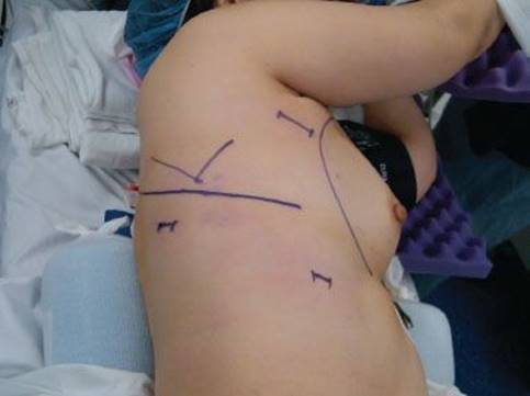
Figure 22.1 Patient positioning and port site placement (JPEG): The patient is placed in the left lateral decubitus position. The camera port is placed in the seventh intercostal space, midaxillary line. The posterior port site, used for lung retraction and stapling devices, is placed below and posterior to the tip of the scapula. The utility incision is 3 to 4 cm in length and starts just anterior to the border of the latissimus dorsi muscle near the axillary crease.
Technique
Anatomy
A thorough and complete knowledge of the segmental anatomy of the right upper lobe is essential for performing a safe and effective VATS segmentectomy. The right upper lobe is divided into three individual segments, an apical, a posterior, and an anterior segment. To perform each segmentectomy with a VATS approach, the surgeon must have a detailed understanding of each individual segmental bronchial, arterial, and venous anatomy and their common variations. Segmental anatomy is less commonly explained or detailed in most anatomy textbooks or surgical atlases and the reader’s knowledge of the three-dimensional positions of these structures may not be as substantial as lobar anatomy. From the lateral surgical view of the lung, the surgeon can visualize each of the three right upper lobe segments, the posterior, apical, and a portion of the anterior segment of the right upper lobe (Fig. 22.2A). The posterior segment has a substantial apical portion that is located posterior to the apical segment. If the lung is retracted posteriorly, the view from the anterior or medial aspect of the hilum will show the anterior and apical segments only (Fig. 22.2B).
The bronchial anatomy is most commonly illustrated as three separate and distinct segmental airways: Apical (B1), posterior (B2), and anterior (B3), arranged in a three-point star formation similar to the Mercedes-Benz logo when viewed from an endobronchial position (Fig. 22.3A,B). While this configuration is common, it occurs in less than 50% of patients. The second most frequently seen anatomic variation is two branches of the right upper lobe, with both the apical (B1) and the anterior (B3) bronchi sharing the same origin with a separate posterior (B2) segmental bronchus. Rarely, the apical and posterior will share a common bronchus, with the anterior bronchus emanating separately.
The right pulmonary artery most frequently has two main branches: A truncus anterior that divides again into the apical segmental arteries and the anterior segmental arteries, and a posterior ascending artery that leaves the ongoing pulmonary artery further distally after the takeoff of the truncus anterior. The truncus anterior is a broad, prominent artery, and can have a variable takeoff position from the right main pulmonary artery, sometimes very proximal with early division into the anterior and apical branches. Frequently, the apical and anterior divisions divide off the main pulmonary artery almost completely separately, as if they were two distinct arterial branches of the main pulmonary artery. In most patients, the posterior segment of the right upper lobe receives pulmonary blood from both the ascending posterior branch of the pulmonary artery and a separate recurrent branch that originates from the common arterial branch to the apical segmental arteries after they have divided from the truncus anterior (Fig. 22.4).
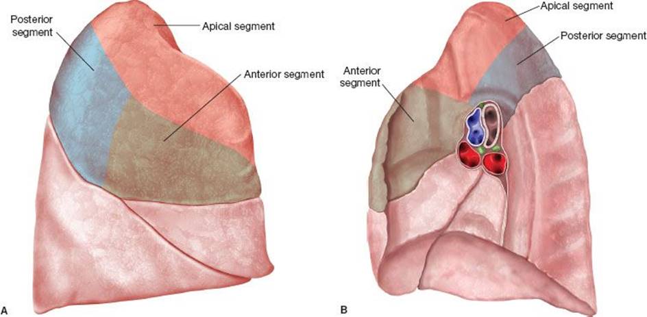
Figure 22.2 A: Lateral view of the right upper lobe segmental anatomy. (1) Apical segment, (2 ) Posterior segment, (3 ) Anterior segment. B: Medial view of the right upper lobe. (1) Apical segment, (2 ) Posterior segment, (3 ) Anterior segment.
The superior pulmonary vein normally has three divisions that are easily seen from the anterior view of the hilum. There is a superior branch, a central branch, and the middle lobe veins. The most superior vein drains the apical segment. The central vein has a deeper posterior portion that drains the posterior segment. The most inferior branch of the central vein is most commonly the anterior segmental vein (Fig. 22.5).
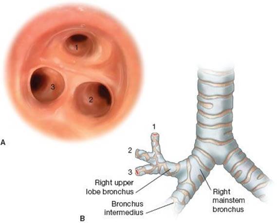
Figure 22.3 Right upper lobe segmental bronchial anatomy. A: Endobronchial anatomy of the right upper lobe. (1) Apical segment, (2 ) Posterior segment, (3 ) Anterior segment. B: (1 ) Apical segment, (2 ) Posterior segment, (3 ) Anterior segment.
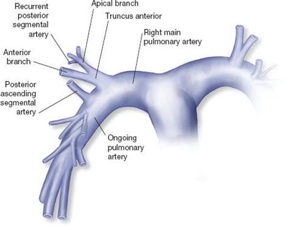
Figure 22.4 Pulmonary artery anatomy of the right upper lobe: The truncus anterior, the first branch of the right pulmonary artery, supplies the apical and anterior segments. A recurrent branch to the posterior segment commonly arises from the apical branch. A posterior ascending artery to the posterior segment branches from the ongoing pulmonary artery and supplies the posterior segment.
General Approach to VATS Segmentectomy
The entire operation is performed extracorporeally, without direct visualization of the operative field or rib spreading. The segmental structures, bronchus, artery, and vein, are individually ligated. A complete systematic lymph node sampling of the mediastinum and hilar nodes must be completed and sent for frozen section as any lymph node metastasis will require a formal lobectomy. A pediatric bronchoscope is used to help identify the correct bronchial anatomy through the double-lumen endotracheal tube. Selective and differential inflation of the segments demarcates the intersegmental plane, or fissure between each segment, which is then divided by a stapling device to create the fissure.
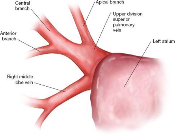
Figure 22.5 Right superior pulmonary vein anatomy: The superior pulmonary vein drains into the left atrium. The upper division drains the apical, anterior, and posterior segments. The right middle lobe has separate branches that drain into the superior pulmonary vein.
Prior to commencing the operation, bronchoscopy is performed to confirm the bronchial anatomy. Bronchoscopic findings are compared with the preoperative CT scan to help orient the surgeon to the overall size and shape of the upper lobe and especially the position and pattern of arterial and venous branches. After positioning and port placement, the entire thorax is inspected and all three lobes of the lung are inspected for lesions with direct finger palpation.
VATS Right Upper Lobe Posterior Segmentectomy
The right upper lobe is retracted posteriorly with a lung grasper placed from the posterior port site to expose the anterior hilum. A flexible tip argon beam coagulator, set to 100, is then used to score the mediastinal pleura anteriorly and superiorly around the entire hilum. The superior pulmonary vein is dissected completely so that all three divisions of the vein, superior, central, and middle lobe branches, are clearly visible (Fig. 22.6). The path of the central vein branch is critical to dissect and identify. The dissection is brought superiorly above the superior pulmonary vein and truncus anterior until the right upper lobe bronchus is encountered. Blunt dissection with a sponge stick or peanut sponge to expose the underlying airway and push the peribronchial tissue toward the lung parenchyma should be done.
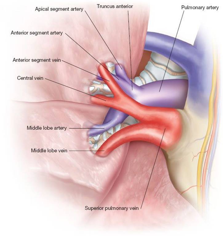
Figure 22.6 Anterior view of the right upper lobe hilum: After dissection of the mediastinal pleura, the entire superior pulmonary vein is exposed. The truncus anterior will lie just superior to the upper division of the superior pulmonary vein.
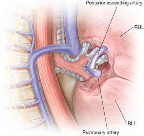
Figure 22.7 Posterior ascending artery: Dissection of the fissure and retraction of the lower lobe and upper lobes help identify the position of the posterior ascending artery. Lymph nodes are frequently encountered and are sampled.
The lung is then retracted medially toward the mediastinum to expose the posterior hilum. The posterior segment of the right upper lobe is retracted superiorly and anteriorly while a separate lung grasper pulls the most apical portion of the right lower lobe superior segment inferiorly, thus exaggerating the superior aspect of the major fissure. The major fissure may be complete or incomplete and comfort dissecting within fissure thoracoscopically is essential to performing a VATS segmentectomy. The entire procedure can be accomplished mainly from a posterior approach. However, if the fissure is incomplete and thick, a central dissection within the fissure may be necessary to identify the posterior ascending artery. The major fissure is dissected until the ongoing pulmonary artery is identified (Fig. 22.7). The plane of Leriche is entered which allows for safe and controlled pulmonary artery dissection. The dissection is brought posteriorly until the posterior ascending branch is visualized. Small lymph nodes are commonly found at the junction of the posterior ascending artery and the ongoing PA. These nodes should be carefully grasped within a Singley forceps and dissected for frozen section analysis. Circumferential dissection of the ascending branch is performed with Pearson scissors and a Harken clamp with the area of dissection focused distally, away from the origin of the ascending branch, to avoid the injury prone “crotch” of the arterial vessels. A vascular load stapler is used to divide the artery.
The upper lobe bronchus is now more accessible for dissection. Blunt and sharp dissection on the posterior hilum with a Pearson scissors will define the upper lobe bronchus and its three segmental bronchi. From the posterior view, the posterior segmental bronchus will course horizontally toward the mediastinum. The apical bronchus will be angled superiorly. The anterior bronchus will be the most inferior bronchial structure seen and will be a few millimeters deeper toward the mediastinum compared to the other two segmental bronchi (Fig. 22.8) and often times obscured from view by overlying lung parenchyma. Previous division of the posterior ascending pulmonary artery permits more aggressive retraction of the upper lobe superiorly and anteriorly which allows the best visualization of the segmental bronchi. The argon beam is used to score the pleura overlying the bronchi and a peanut sponge forcefully dissects the soft tissue upward into the specimen. The posterior segmental bronchus is then circumferentially dissected with Pearson scissors and a Harken clamp. Aggressive dissection on the medial (deep side) of the bronchus by spreading the Harken clamp while pulling posteriorly against the posterior segmental bronchus will create enough room to pass a stapler. Care must be taken because a recurrent arterial branch originating from the superior branch of the truncus anterior passes just behind the posterior segmental bronchus to supply the posterior segment in addition to the previously ligated posterior ascending branch. An endoscopic stapler (Covidien Purple Load) is placed across the posterior segmental bronchus and closed (Fig. 22.8). The stapler is then taken of all tension and “relaxed” into the patient so that the posterior segmental bronchus lay in its correct anatomic position and to avoid inadvertent external occlusion of the other bronchi. Bronchoscopy can be performed to confirm the correct bronchus is occluded if necessary.
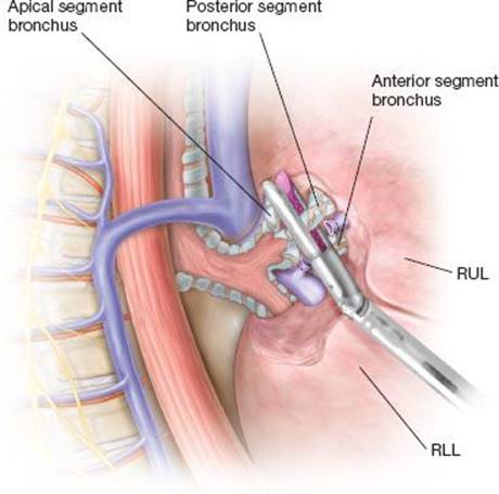
Figure 22.8 Right upper lobe segmental anatomy, posterior view: With the lung retracted medially, after division of the posterior ascending artery, the individual branches to the right upper lobe are easily visualized. With this exposure, a stapler is placed across the posterior segmental bronchus and closed to allow for ventilation to the remaining segments.
The anesthesiologist is then instructed to give a gentle breath to the right lung and the remaining apical and anterior segments are visualized inflating, further confirming that the correct bronchus is occluded. A line of demarcation between the deflated posterior segment and the inflated apical and anterior segments will be created (Fig. 22.9). The stapler is then fired ligating the bronchus. The bronchial stump is then grasped with an empty ring forceps and forcefully retracted superiorly and laterally toward the posterior chest wall. The recurrent posterior segmental artery is then identified and ligated. The venous branches to the posterior segment are now more easily approached as they run just in front of the anterior segmental bronchi. Their path is easily seen running along the divided bronchial stump retracted superiorly. These venous branches can be dissected and ligated in the standard fashion or taken en bloc when the fissure is created.
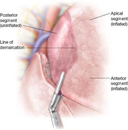
Figure 22.9 Ventilation to the apical and anterior segments created a line of demarcation between the posterior segment and the other segments. A stapler is guided along the line of demarcation to create the anatomic fissure.

Figure 22.10 The bronchial stump to the posterior segment is pulled into the specimen side. The stapler is placed superior to the stump to ensure that the entire lymphovascular pedicle to the posterior segment is included in the resection.
If air begins to cross the segmental fissure into the posterior segment, the argon beam coagulator can be used to score the surface of the lung directly on the line of demarcation just after ligating the segmental bronchus. This will provide a visual line of resection for creating the fissure. With the bronchial stump retracted superiorly and toward the patient’s back, an endoscopic stapler is placed across the demarcation line and fired. The stapler must be placed with the tips traveling along the path of the ligated posterior segmental vein and behind and deep to the ligated bronchial stump (Fig. 22.10). The bronchial stump must be pulled into the specimen side of the resection to ensure adequate lymphatic vessel clearance. The posterior segment travels apically almost parallel to the spine toward the thoracic inlet. Once the dissection proceeds past the ligated bronchial stump, there are no structures that can be injured and the line of resection can be as wide as desired into the apical segment toward the apex of the lung. The specimen is placed in a sterile bag and removed for pathologic review with frozen section confirmation of the bronchial margin. A standard lobe-specific or complete mediastinal nodal sampling or dissection is then completed.
VATS Right Upper Lobe Apical Segmentectomy
Patient positioning, bronchoscopy, and review of imaging, and port placement are identical to the previous description. This is mostly an anterior to posterior dissection and superior hilar dissection. The lung is grasped with a VATS lung retractor and retracted posteriorly toward the patient’s back. The anterior hilar pleura is scored with an argon beam coagulator to decrease nuisance bleeding during the blunt and sharp dissection of the mediastinal pleura. The superior pulmonary vein is completely dissected with care to identify the apical, central, and middle lobe branches. The apical segmental pulmonary vein most commonly is the most superior branch of the superior pulmonary vein and runs superiorly and anteriorly over the truncus anterior branch of the pulmonary artery (Fig. 22.6). The apical vein is dissected circumferentially and divided with a vascular load stapler placed from the posterior port site while lung retraction is transferred to a grasper placed in the utility incision. The truncus anterior is then dissected until its origin off the right main pulmonary artery is identified and the distal divison into the apical and anterior branches is visualized. This requires significant dissection along the vessel into the lung parenchyma. Lymph nodes are often encountered along the artery and bleeding can occur if the lymph nodes are injured. Argon Beam Coagulation will easily control bleeding from dissected lymph nodes. Dissection of the mediastinal pleura superiorly above the hilum underneath the azygos vein enhances the visualization of the lateral and apical aspect of the truncus anterior and the apical segmental bronchus.
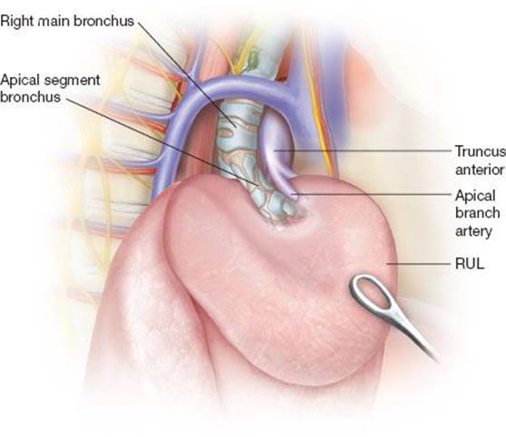
Figure 22.11 Retraction of the apical segment to expose the apical segmental bronchus. The apical segmental artery coursed just medially to the bronchus.
The lung is retracted inferiorly putting tension on the right upper lobe bronchus. The most superior and vertically oriented bronchus is the apical bronchus and the apical branches of the pulmonary artery will be seen coursing just medially along this airway (Fig. 22.11). This view will enable better understanding of the arterial anatomy and allow the surgeon to return to an anterior approach to the hilum. With the lung retracted posteriorly, the apical segmental bronchus is identified just superior to the truncus anterior. The dissection is brought deeper into the lung parenchyma to visualize the bifurcation of the artery into the apical branches and the anterior branches. The apical branches are circumferentially dissected and ligated with a vascular load stapler placed through the posterior port site. The lung is again retracted in an inferior direction. The medial and posterior sides of the apical bronchus are now dissected with a Pearson scissors. With the apical arterial branches already ligated, there is little concern for injuring the PA as the dissection moves under the apical segmental bronchus. Either a right-angle clamp or Harken clamp is passed under the apical segmental bronchus and a monofilament tie passed for additional retraction. An endovascular stapler is then passed across the bronchus from the posterior port site and closed. Anesthesiology is instructed to give a controlled test breath until the posterior and anterior segments are seen inflating. A line of demarcation will form between the apical segment and the other two segments. The stapler is fired and the bronchial stump is grasped in an empty ringed forceps and retracted forcefully toward the posterior and apical chest wall from the front utility incision. This allows an anterior approach creating the fissure. A “V”-like resection will be performed, starting from the line of demarcation between the anterior segment and the apical segment (Fig. 22.12). The stapler is placed along the line of demarcation of inflation and deflation toward the bronchial stump, which will act as the target for creating the apex of the “V”, from the utility incision. Multiple firings of the stapler may be necessary to reach the bronchial stump. Again, the staple line must include the bronchial stump into the specimen side of the resection. The stapler can continue in an apical direction once it passes the bronchial stump along the line of demarcation between the apical and posterior segments. Or, if the tissue is too thick, the apical segment can be retracted inferiorly to allow the stapler to begin its division of the fissure from the apical direction, with the bottom of the “V” meeting just below the bronchial stump, also passed from the utility incision. The specimen is placed in a sterile bag and removed. Lymph nodes from along the hilar dissection are sent for frozen section analysis along with a standard mediastinal lymph node dissection or sampling.
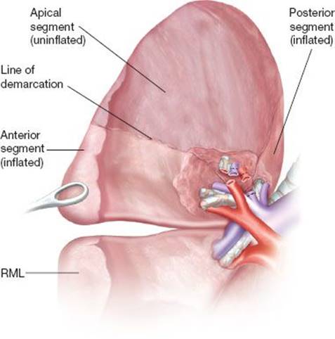
Figure 22.12 Creation of the fissure to the apical segment: The dotted lines illustrate the line of demarcation after selective ventilation. The stapler follows the line of demarcation in a “V”-like pattern of resection. The bronchial stump is retracted to allow the stapler to pass deep and central to the bronchial stump to ensure that the lymphovascular pedicle is removed with the specimen.
VATS Right Upper Lobe Anterior Segmentectomy
This procedure is almost entirely an anterior approach to the hilar structures. Preoperative planning, port sites, and patient positioning are as described previously. To provide more flexibility in retraction and visualization of the segmental structures, the minor fissure between the anterior segment of the upper lobe and the right middle lobe is divided to a point just superficial to the underlying pulmonary artery. The middle lobe can now be retracted out of the operative field. The anterior hilum is opened in the standard fashion with argon beam coagulator and blunt and sharp dissection. Great care must be taken to identify and protect the middle lobe pulmonary vein. Most commonly, a superior venous branch and a central venous branch will be seen draining the right upper lobe. An empty ringed forceps is placed on the medial edge of the anterior segment and the segment is retracted inferiorly and posteriorly exposing anterior segmental vein. This vein will be the most inferior branch of the central upper lobe vein and directly superior to the middle lobe vein (Fig. 22.6). This is easily divided with a vascular load stapler placed from the posterior port site. The remainder of the superior pulmonary vein can now be dissected off the ongoing pulmonary artery that lies just deep to these vessels. Coursing behind the apical segmental vein and above the posterior segmental vein will be the anterior segmental branch of the truncus anterior. Dissection on the ongoing PA can be done with these structures identified. The fissure between the middle lobe and the anterior segment should now be completed to open the view of the arterial and bronchial structures. A stapler should now be passed above the ongoing pulmonary artery just inferior to the posterior segmental vein to open the remainder of the minor fissure.
Retraction of the anterior segment toward the apex will accentuate the venous branches to the apical and posterior segments. The anterior segmental arteries can now be dissected and ligated with a vascular load stapler passed from the posterior port site. Just deep to these arterial branches lay the anterior segmental bronchus. Significant lymphadenopathy is frequently encountered at this point of the dissection and meticulous dissection is needed to remove these nodes. The anterior segmental bronchus will be the most vertical airway and the posterior bronchus will be coursing posteriorly toward the back of the patient. Dissection of the crotch between these two airways is done with a Pearson scissors. The apical segmental bronchus is superior and deep to this dissection. The proximal stump of the ligated arterial branches to the anterior segment will guide the surgeon to the medial border of the anterior segmental bronchus. A Harken clamp is passed behind the bronchus. A stapler is passed from the posterior port site across the bronchus and closed. Bronchoscopic confirmation can be performed at this time. Anesthesia is instructed to provide inflation to the right lung. The apical and posterior segments should be seen inflating. The anterior segmental bronchus is now divided. The line of demarcation between inflated and deflated lung will serve as the guide for creating the fissure (Fig. 22.13). The bronchial stump is grasped in an empty ringed forceps and retracted inferiorly toward the middle lobe. From the utility incision, a stapler is placed along the line of demarcation between the anterior and apical segments. The tip of the stapler is aimed at the bronchial stump. The minor fissure has already been divided to a point behind the bronchial stump. Thus the stapler can be placed with its tips aiming for the point of a “V” just behind the bronchial stump to ensure that the bronchial stump is included in the specimen (Fig. 22.12). The specimen is removed in a sterile bag and a standard lymph node dissection or sampling is performed.
In all cases, a single 28-French chest tube is placed in a posterior apical position. The lungs are reinflated under direct camera visualization. The wounds are closed with 2-0 Vicryl sutures for the muscular layers and 3-0 Monocryl sutures for the skin. Sterile dressings are applied. The patient is extubated in the operating room and recovered in a postanesthesia care unit before transfer to an intermediate level care unit.
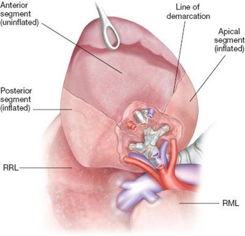
Figure 22.13 Creation of the fissure anterior segment: The dotted lines illustrate the line of demarcation of the anterior segment after ligation of the anterior segmental bronchus. The anterior segmental vein and artery have already been divided. The bronchial stump is retracted to allow the stapler to include the lymphovascular pedicle in the specimen.
 POSTOPERATIVE MANAGEMENT
Patients spend the first postoperative day in an intermediate level care unit that provides continuous telemetry, arterial line measurements, and a low nursing-to-patient ratio. Prophylactic beta-blockers may be used in patients at risk for atrial fibrillation. The patient is advanced to a diet as soon as possible. By the first postoperative day, patients are ambulating. Pain is controlled with a patient-controlled analgesia (PCA) epidural pain catheter device placed preoperatively. Chest tubes are placed on low continuous suction to 20 cm of water pressure for the first 24 hours then placed to water seal. Chest drains are removed once significant air leaks have stopped and drainage is less than 300 cc over a 24-hour period. Incentive spirometry, chest physiotherapy, and deep coughing exercises are provided by respiratory therapists. Daily ambulation is strongly encouraged. Patients are moved to standard patient rooms on the first postoperative day.
 COMPLICATIONS
The risks of intraoperative or postoperative complications for VATS segmentectomy are similar to those of VATS lobectomy. Multiple studies have documented 30-day mortality rates for both procedures at less than 1%. Overall, both VATS segmentectomy and VATS lobectomy carry a roughly 15% to 25% risk of morbidity. The most common postoperative complications for both VATS lobectomy and VATS segmentectomy include pneumonia, respiratory failure with hypoxemia, atrial fibrillation, and prolonged air leak. The rates of each complication are not significantly different between techniques.
 RESULTS
The results of the randomized LCSG published in 1995 established lobectomy as the gold standard surgical therapy for stage I nonsmall cell lung cancer. The LCSG trial, conducted in the 1980s, enrolled 276 patients diagnosed by chest x-ray with clinical T1N0 tumors less than or equal to 3 cm in size to receive either lobectomy or sublobar resection. Sublobar resection included both wedge resection and anatomic segmentectomy. The study demonstrated an increased rate of recurrence in the sublobar group with an observed tripling of locoregional recurrence rate (p = 0.008, two-sided) without a significant difference in the rate of distant recurrence. The rates of death and death with cancer were lower in the lobectomy group. They observed a 30% increase in death rate and a 50% increase in death with cancer rate in the sublobar patients, p = 0.088. The authors reported a 75% increase rate of overall recurrence when eligible patients were included, a rate that decreased to 50% when the entire 276 cohort was included. However, these statistical findings are open to question because the analysis included a one-sided test and reached p values above the customary p = 0.05 standard for significance.
Currently, many factors have caused the re-evaluation of lobectomy as the optimal treatment for early stage lung cancers that may be amenable to a sublobar resection. Advances in diagnostic capability with high-resolution multidetector computed tomography and staging modalities including PET/CT and EBUS-TBNA have increased the ability to diagnose and accurately stage small lung cancers. Furthermore, our understanding of the importance of tumor size and the risk of regional or distant metastasis has led many experts to question the validity of the LCSG findings since tumors larger than 2 cm were included in the sublobar resection group. Similarly, the natural history of subsolid nodules, detectable with CT but not x-ray, and likely excluded from the LCSG trial, has been shown to follow a more indolent growth pattern and thus more amenable to lesser resection. Furthermore, the proportion of patients within the general population of advanced age with significant comorbidities is increasing compared to previous decades and the need for less invasive, less morbid, lung-sparing techniques is more imperative.
In the last decade, data from many studies have suggested equivalent outcomes between segmentectomy and lobar resection for peripheral, early stage lung cancers. In 2005, Okada et al. reported a large multicenter retrospective study of 1,272 patients who underwent sublobar or lobar resection for nonsmall cell lung cancer. The 5-year cancer-specific survival after segmentectomy for tumors less than 2 cm was 96.7% compared to 92.4% for the lobectomy group and 85.7% in the wedge resection group. For tumors between 2 and 3 cm, 5-year cancer-specific survival was 84.6% in the segmentectomy group versus 87.4% in the lobectomy group and 39.4% in the wedge resection cohort. The differences in survival between techniques in tumors less than 3 cm were not statistically significant. However, in tumors greater than 3 cm, lobectomy was superior to both segmentectomy and wedge resection. In tumors greater than 3 cm, the 5-year cancer-specific survival for lobectomy was 81.3% compared to 62.9% and 0%, for segmentectomy and wedge resection, respectively. In each of the three groupings, the survival differences between lobectomy and segmentectomy were not significant. However, 5-year cancer-specific survival was significantly worse for wedge resection in both the 2-to 3-cm group and the greater than 3-cm group when compared to segmentectomy and lobectomy, suggesting that wedge resection is associated with decreased survival in tumors greater than 2 cm.
A propensity matched retrospective analysis by Wisnivesky et al. of the SEER database for patients >65 years of age with tumors ≤2 cm in diameter showed that both the overall survival and cancer-specific survival of patients treated with sublobar resection were not significantly different than those treated with lobectomy. Interestingly, in tumors between 2 and 3 cm in size, sublobar resection was associated with significantly increased overall and cancer-specific mortality. This finding mirrors the results of the Japanese data. Most recently, Altorki et al. reported their analysis of the International Early Lung Cancer Action Program (I-ELCAP) database comparing sublobar resection for clinical stage IA lung cancers. In this propensity matched analysis, the authors showed no significant survival difference between sublobar resection and lobectomy. In this study, 347 patients were treated with surgery: 294 lobectomies, 16 segmentectomies, and 37 wedge resections. The majority of these tumors were <2 cm in diameter. Recurrence was higher in the sublobar group than in the lobectomy group (20% vs. 10%, p = 0.21), however, this was not statistically significant. Of concern, all of the recurrences occurred in those patients who underwent wedge resection while no recurrence was seen in those who underwent segmentectomy.
 CONCLUSIONS
The role of VATS segmentectomy in the treatment of early stage lung cancer remains controversial. At present, lobectomy is considered by many as the gold standard treatment for stage I non-small cell lung cancers. However, as CT screening becomes more prevalent, the incidence of small lung cancers and lung cancers of differing invasive potential will increase. In addition, as the population ages, the numbers of lung cancer cases diagnosed in the elderly, with multiple comorbidities, will also increase. Many people may survive a previous lung cancer and require definitive treatment of metachronous disease. Thus segmentectomy may become more appealing as a definitive treatment. Many recent studies suggest that VATS segmentectomy for peripheral small lung cancers is equally effective as lobectomy. Ongoing randomized trials such as the Cancer and Lymphoma Group B trial (CALGB 140503) and the Japanese Clinical Oncology Group trial (JCOG0802/WJOG4607 L) will help define the role of sublobar resection and segmentectomy versus lobectomy in the treatment of early stage lung cancer.
Recommended References and Readings
Churchill ED, Belsey R. Segmental pneumonectomy in bronchiectasis. Ann surg. 1939;109:481–489.
Ginsberg RJ, Rubenstein LV. Randomized trial of lobectomy versus limited resection for T1N0 non-small cell lung cancer. Lung Cancer Study Group. Ann Thorac Surg. 1995;60:615–622.
Graham EA, Singer JJ. Successful removal of an entire lung for carcinoma of the bronchus. JAMA. 1933;101:1371–1374.
Jensik RJ, Faber LP, Milloy FJ, et al. Segmental resection for lung cancer. A fifteen-year experience. J Thorac Cardiovasc Surg. 1973; 66:563–572.
Kodama K, Higashiyama M, Yokuochi H. Intentional limited resection for selected patients with T1N0M0 non-small cell lung cancer: A single institution study. J Thorac Cardiovasc Surg. 1997; 114:347–353.
Landreneau RJ, Sugarbaker DJ, Mack MJ, et al. Wedge resection versus lobectomy for stage 1 (T1N0M0) non-small cell lung cancer. J Thorac Cardiovasc Surg. 1997;113:691–698.
Okada M, Nishio W, Sakamoto T, et al. Effect of tumor size on prognosis in patients with non-small cell lung cancer: The role of segmentectomy as a type of lesser resection. J Thorac Cardiovasc Surg.2005;129:87–93.
Shimkin MB, Connelly RR, Marcus SC, et al. Pneumonectomy in bronchogenic carcinoma. A comparison of end results of the Overholt and Ochsner clinics. J Thorac Cardiovasc Surg. 1962; 44:503–519.
Travis WD, Brambilla E, Noguchi M, et al. International Association for the Study of Lung Cancer/American Thoracic Society/European Respiratory Society International Multidisciplinary Classification of Lung Adenocarcinoma. J Thorac Oncol. 2011;6:244–285.
Wisnivesky JP, Henschke CI, Swanson S, et al. Limited resection for the treatment of patients with stage IA lung cancer. Ann Surg. 2010;251:550–554.