
Laureano Molins, Jean Philippe Berthet, Juan J. Fibla, David Sánchez, and Abel Gómez-Caro
 INDICATIONS/CONTRAINDICATIONS
General indications for segmentectomy include indeterminate solitary pulmonary nodules, limited infectious or inflammatory disease and selected early stage nonsmall cell lung cancer (NSCLC). Segmentectomy should not be performed in patients who do not fulfill standard criteria for conventional lung resection. Relative contraindications include the following: Coagulopathy, skin infection over the site, diffuse lung disease, and extensive metastases.
In 1995, the Lung Cancer Study Group performed a randomized controlled trial demonstrating that sublobar resections for NSCLC smaller than 3 cm resulted in increased rate of locoregional recurrences compared with lobectomy (8.6% vs. 2.2%, respectively). Subsequently, segmentectomy has generally been restricted to patients with marginal cardiopulmonary function. However, proponents of segmentectomy have maintained that the results of this trial were inconclusive because a third of all patients underwent nonanatomic resections, and the trial included patients with tumors up to 3 cm. In several large, nonrandomized studies from the United States, Japan, and Europe, anatomic segmental resection techniques have been shown to achieve comparable rates of recurrence and survival for stage IA NSCLC.
It has been suggested that segmentectomy, in addition to achieving a complete (R0) resection with adequate surgical margins and systematic nodal staging, may be associated with reduced morbidity and mortality as compared with lobectomy, as it can preserve pulmonary parenchyma and lung function and may be the only feasible surgical option in patients with severe pulmonary impairment that could otherwise not tolerate lobectomy. However, phase III studies are lacking in patients with diffuse emphysema, and some studies have shown better postoperative 3-month functional results with lobectomy versus sublobar resections in patients with poor lung function related to diffuse emphysema. In stage IA NSCLC with diffuse emphysema (ppoVEMS <40%), lobectomy demonstrated a double therapeutic role because of lung reduction effect, especially with upper lobectomies.
Optimal Cases for Segmentectomy
 Well-centered, solitary pulmonary nodule in the targeted segment peripheral and 2 cm or less.
A solitary pulmonary nodule in conjunction with other lesions that need to be resected. During cases of suspected metastases, it is sometimes difficult to distinguish a metastatic lesion from a new lung primarily at the time of frozen section. The use of segmentectomy affords a complete, yet parenchymal-preserving, anatomic resection that may provide definitive management in either circumstance.
Small (<1 cm) ground-glass opacities detected on computed tomography imaging (CT scan) that are frequently associated with early cancers (Noguchi A and B adenocarcinoma) and highly amenable to anatomic segmentectomy. These can be associated with a 5-year survival greater than 90%, and a 6-year survival rate of 100% when associated with frozen section.
 PREOPERATIVE PLANNING
General: The preoperative workup for patients undergoing anatomic segmentectomy for a stage I NSCLC should mirror that of patients undergoing conventional lobectomy. Lung cancer staging typically includes a clinical history, physical examination, standard blood work, CT scan, as well as 18F-fluoro-D-glucose positron emission tomography (PET/CT) imaging. Suggestion of N2 (usually 2R, 4R, 7 stations) nodal disease on a CT imaging or PET/CT (short axis >1 cm and uptake SUVmax >2.5, respectively) needs histologic confirmation using endobronchial ultrasound (EBUS) techniques or mediastinoscopy. Suggestion of N1 (10R, 11R, 12R stations) disease may offer guidance for the highest yield targets to be assessed by frozen section during the dissection allowing for reconsideration of lung-sparing strategy.
A dedicated chest CT scan with intravenous contrast of “topographic quality” (high resolution) is critical to determine lesion size, segment location (regarding intersegmental planes and fissure), and to properly identify anatomic structures including intersegmental veins that need to be preserved. We found a CT with 3D reconstruction particularly helpful in performing a safer dissection of pulmonary artery (PA) branches especially when the fissure is fused, by ascertaining the number and direction of right superior segmental arteries (diameter >2 mm). This technique has been reported to identify 95% of PA branches in patients undergoing segmentectomy. An accurate preoperative marking (injection of radiotracer, hook wire, coil markers, radiopaque markers) during CT imaging may be indicated in indeterminate impalpable solitary pulmonary nodules (i.e., ground-glass opacities) to help intraoperative decision regarding the segment to resect.
Bronchoscopy: The most consistent landmarks of upper right lobe segmental anatomy are segmental bronchi that should be accurately identified during preoperative bronchoscopy to rule out anatomical or endobronchial abnormalities.
Lung function: Resection is functionally possible in patients belonging to “good risk” group with predictive postoperative FEV1 >40% of the predicted value (calculated on the basis of spirometry and isotopic scanning), with no major hypoxemia (<60 mm Hg) or hypercapnia (>46 mm Hg). In “high-risk patients” segmentectomy is considered functionally possible when VO2 max during exercise is >10 mL/kg/min.
 SURGERY
Surgical anatomy: (Fig. 21.1) The right upper lobe includes three segments: Superior (apical) S1, posterior S2, and anterior (ventral) S3. Theoretically, it is possible to achieve segmentectomies of S1, S2, and S3. However, ventral segmentectomy is typically not performed because of its technical complexity (challenging exposure of anterior bronchus (SB3) and ventral artery (SA3) located behind the apical vein (SV1)) and difficulty with preservation of the anterior vein (SV3). In case of a stage 1 NSCLC of S1 or S2, bisegmentectomy S1 + S2 are most often performed together to achieve sufficient disease-free margins.
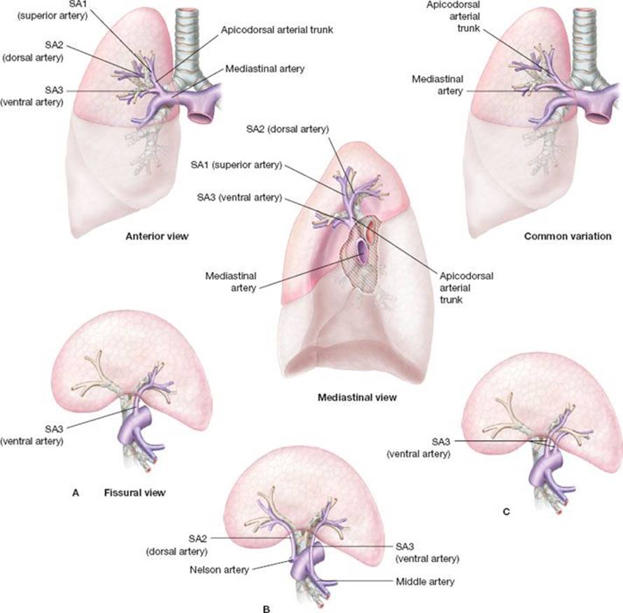
Figure 21.1 Basic anatomy of right superior segments. Upper images: Anterior view; mediastinal view; common variation; mediastinal artery; apicodorsal arterial trunk; SA1 (superior artery); SA2 (dorsal artery); SA3 (ventral artery) arising from mediastinum. Lower images: Different examples of fissural artery (fissural view); A: SA3 (single ventral artery). B: Unusual origin of SA2 and SA3 arising from pulmonary artery or middle lobe artery respectively. C: 2 type of ventraal artery arising from mediastinum or fissura.
Veins: The upper root of the right superior pulmonary vein is made of the superior (SV1), posterior (SV2), and anterior segmental veins (SV3) union. The superior and the posterior vein may receive collaterals from the inferior and middle lobe veins. SV1 is easily recognized and controlled and SV2 is sometimes controllable in the fissure. Usually, veins should be carefully spared during bronchial and arterial dissection and divided as the last step during intersegmental plane management.
Arteries: SA1 and SA3 arise from truncus anterior branch of the PA. The mediastinal artery typically divides into two or three branches: Superior artery (SA1) and anterior artery (SA3). S2 is usually vascularized by SA2, arising from the intralobar PA. SA2 is located at the posterior part of the PA and ascends in front of the right superior lobar bronchus. In some patients, SA2 originates from SA6 (10%) and occasionally can be mistaken for SA3 (when SA3 also come off the intralobar PA).
Bronchus: When it enters lung parenchyma, right upper lobe bronchus divided into three segmental bronchi, SB1 (superior or apical bronchus), SB2 (posterior or dorsal bronchus), and SB3 (anterior or ventral bronchus). SB2 and SB3 may originate from a common “apicodorsal,” “apicoventral,” or “dorsoventral” bronchial trunk or originate separately and thus may be stapled together or separately depending on the required resection.
Positioning and approach: All procedures are performed under general anesthesia and epidural or paravertebral catheters should be available to all patients. A double-lumen endotracheal tube is preferred to a bronchial blocker to enable intraoperative bronchoscopy during one-lung ventilation. The patient is positioned in the lateral decubitus with table slight flexion at the level of scapula tip improving access and exposure. The chest is entered through a standard muscle-sparing posterolateral or anterior thoracotomy in the fourth or fifth intercostal space according to associated lesions and local anatomy. A 5-cm thoracotomy combined with a thoracoscope placed through an anterior single port may be a feasible alternative. The thoracic cavity should be inspected to rule out signs of unexpected advanced disease such as mediastinal node positivity or pleural involvement, and the tumor palpated to reaffirm the planned segmental resection.
Technique
General consideration: We use a peanut dissector, scissors, and suction tip to facilitate gentle distal dissection of segmental pedicle. Precise dissection of fissures requiring punctate electrocoagulation and isolated application of ligatures can be conducted with the help of magnifying loupes. Control of large vessels is accomplished with vascular staplers or nonabsorbable 3/0 to 4/0 ligatures while hemostasis of small caliber vessels is accomplished with clips or a bipolar vessel-sealing device (LigaSure, Valleylab, Boulder, CO, USA).
Surgical steps:
1. (Fig. 21.2). After opening the perihilar mediastinal pleura and removing the 10R lymph node that lies between the right main bronchus and the azygos, we usually perform an extended distal exposure of the truncus anterior artery. Because the arterial supply to the right upper lobe has several variations, the origins of SA2 and SA6 should be identified in the posterior part of interlobar fissure, and the middle lobe artery, truncus anterior artery with SA1/SA3 origins should be fully exposed anteriorly. We achieve full exposure of the upper lobe pedicle and assess bronchial direction before any arterial division.

Figure 21.2 Bronchial steps. Perihilar mediastinal pleura has been opened and 10R has been removed. Right superior lobe is gently pulled anteriorly and distal full exposure of SB1 and SB2 is generally easier after dividing posterior part of fissure.

Figure 21.3 Targeted SB2 has been ascertained by 23-gauge butterfly needle before division. If arising from posterior part of fissure, SA2 has to be divided first. Intersegmental plane identification is facilitated by using selective jet ventilation in the distal bronchial stump (distal SB2) after division (upper right lobe deflated).
2. Bronchial exposure requires retraction of the right upper lobe superiorly and anteriorly. A safe and easy dissection of the distal right upper lobe bronchus typically requires division of the posterior part of the fissure (often using a stapler). The dissection of SB1, SB2, and origin of SB3 are greatly facilitated by the division of SA2 arising from the fissure in case of S2 segmentectomies.
3. The ligation of segmental vein is best performed last, after the intersegmental plane has been defined, as the venous drainage might not be actually apparent.
Before division (Fig. 21.3), we gently cross-clamp the targeted segmental bronchus proximally and puncture the airway distally with 23-gauge butterfly needle, instillating 0.5 L of 100% oxygen. This technique avoids whole lung inflation where the lung can be easily overinflated and the surgical view impaired (particularly in cases of severe emphysema). Usually, S1 and S2 segmentectomies are accomplished almost completely from a posterior approach, working in the posterior aspect of the fissure. After visualization of the bronchus, an encircling silk suture may help with dissection before ligature and division. BS1 and BS2 may be divided together with a stapler or individually according to segmentectomy type and anatomy. Thanks to initial full dissection (mediastinal and in the fissure), SA1 and SA2 (if not already done) are easily divided at this step.
An S3 segmentectomy typically begins with isolation and division of the anterior segmental vein that allows the identification, exposure, and division of the anterior segmental artery coming from the lower portion of the truncus anterior artery. Completion of the anterior part of the horizontal fissure is generally required before BS3 division.
Intersegmental Plane (Identification and Division)
This step is facilitated by manual palpation (Fig. 21.4). However, several methods have been used for the demonstration of intersegmental plane and technique of parenchymal division. The standard technique involves creation of a ventilated–deflated line by reventilating temporarily the ipsilateral lung once the targeted segmental bronchus has been stapled. Failure in identifying a clear demarcation line (related to collateral canals) and limitation of surgical field (related to overinflation of emphysematous lung) lead to implementation of novel methods that are mandatory in VATS and sometimes useful in conventional segmentectomies.

Figure 21.4 If arising from apicodorsal arterial trunk, SA2 is divided at this step with mechanical stapler. Intersegmental plane division is easier when using manual palpation. It may be initiated from central or peripheral part of lung parenchyma using combination of electrocautery and gentle blunt dissection. Ligation of segmental vein is best performed last.
Several methods have been suggested, one is reventilating the whole lung and then collapsing it once the targeted bronchus has been ligated, leaving only the diseased segment inflated. A second is using selective jet ventilation to the bronchus to be divided through the channel of a 3.5 mm flexible bronchoscope when appropriate. Finally, instillating intravenous isocyanine green (3 mg/kg) can help demarcate the devascularized segment under infrared light. Our preferred method is to jet ventilate the targeted segment through a catheter inserted in the distal segmental bronchial stump once the targeted bronchus has been divided and mark the segmental delineation with an operative marking pen or electrocautery during ventilation testing (guiding division of the segment once atelectasis returns).
For parenchymal division, we use a combination of blunt dissection and electrocautery in the peripheral intersegmental plane, and staplers for the central portion of the fissure (last 2/3 cm). This method has the advantage of sparing parenchyma and avoiding the use of many stapler cartridges without major adverse event such as prolonged air leaks. Management of small air leaks and bleeding may be improved by applying hemostatic agents on the intersegmental plane (Fig. 21.5). Microscopic assessment of margins is essential intraoperatively to accomplish a R0 resection.

Figure 21.5 Management of small air leaks and bleeding may be improved by applying hemostatic patch on the intersegmental plane.
Lymph node dissection: Smaller tumors have a lower rate of lymph node metastasis. However in up to 11% of cases, a positive intersegmental or segmental lymph node may be encountered and the operation should be converted to a lobectomy. Thus, in all oncologic cases the segmental lymph nodes should be analyzed intraoperatively and in addition, a full hilar and mediastinal lymph node dissection (2R, 4R, 7, 8, 9) should accompany any segmental resection mirroring that of patients undergoing conventional lobectomy. In most cases, a full hilar, segmental and intersegmental lymph node resection is performed to facilitate bronchial and arterial dissection, providing enough length to facilitate stapling or ligature. Some groups successfully use indocyanine green fluorescence image-guided surgical procedures for sentinel lymph node biopsy to eliminate unnecessary lymph node dissection in patients with lung cancer. We usually perform systematic lymphadenectomy (intersegmental, hilar, and mediastinal) as the first step of pulmonary segmental resection.
In cases of chronic infectious lesions, dissection is completed cautiously given the hypertrophied bronchial circulation. Individual bronchial arteries of significant size should be controlled with clips. Macroscopic assessment of margins is usually sufficient and the removed specimens are cultured with samples sent to microbiology laboratory.
 POSTOPERATIVE MANAGEMENT
The postoperative management of a segmental resection does not differ much from the lobectomy. Depending upon patient’s comorbidities and the complexity of surgery, decision regarding postoperative care should be made well in advance.
Pain Control
Management of postoperative pain includes epidural or paravertebral catheter. The addition of a local anesthetic provides a more rapid onset of action and may help localize correct catheter placement. Cuschieri et al. reported a postoperative pulmonary complication rate of 24% in postoperative patients receiving epidural analgesia, compared with a rate of 64% in those randomized to receive intramuscular morphine. Perioperative use of nonsteroidal anti-inflammatory drugs may complement other pain management strategies.
Physiotherapy
Lung expansion techniques include incentive spirometry, deep breathing exercises, postural drainage, percussion and vibration, cough, suctioning, mobilization, intermittent positive pressure breathing, and continuous positive airway pressure (CPAP). A recent preintervention/postintervention trial attributed a significant reduction in postoperative complications following thoracotomy to the introduction of a perioperative chest physiotherapy program.
Fast-track Protocol
A fast-track regimen that included early mobilization and enteral feeding and epidural/paravertebral analgesia resulted in fewer postoperative pulmonary complications, especially atelectasis and pneumonia, than standard care.
Prevention of Thromboembolism
Surgery and neoplasm are a well-recognized risk factor for the development of deep vein thrombosis and subsequent pulmonary embolism. Much as postoperative pulmonary complications, the risk of venous thromboembolic (VTE) disease is influenced by patient- and procedure-related factors. Risk assessment and recommendations for prevention of VTE in surgical patients have recently been updated.
Air Leak and Management of Chest Tubes
Some degree of postoperative leak is generally unavoidable in segmentectomy resection, and reflects an alveolopleural fistula arising from exposed alveoli after intersegmental division. A careful intraoperative inspection for air leaks after intersegmental division should be performed. Pulmonary sealants have been the focus of a large amount of research in the area of intraoperative prevention of air leaks; however, the overall results of these studies so far have found no clear advantage in their routine use. Thus, the use of sealants should best be reserved for patient at highest risk for developing postoperative prolonged air leak.
Placing chest tubes to water seal does not stop particularly large expiratory leaks and pneumothorax may occur when chest tubes are placed on seal with large air leaks. In a recent meta-analysis examining the effect of suction compared with water seal alone, no differences were identified in terms of the duration of air leaks, duration of chest tubes, and length of stay. Placing the chest tubes to suction appears to be superior to water seal in reducing the incidence of pneumothorax, however, the clinical significance of this finding is unclear.
Radiologic Studies
Periodic chest radiographs should be performed to evaluate the parenchymal and the pleural space. Due to the complexity and the anatomical variations in vein drainage of lung segments, any parenchymal infiltrate in the residual lobe without signs of infection should lead to the suspicion of parenchymal infarction. A contrast CT scan would assist in confirming this complication, leading to a completion lobectomy.
 COMPLICATIONS
Morbidity: Complication rates after segmentectomy vary between 6.6% and 46% depending on the series. Jones et al. analyzed 62 cases and found an overall complication rate of 39%, Shuchert et al. analyzed 785 procedures and found a rate of 34.9%, American College of Surgeons Oncology Group Z0030 trial with 70 procedures found 4.6% of patients with complications and Okada et al. with 230 cases found a rate of 6.6%. A retrospective review of 785 consecutive patients undergoing anatomic segmentectomy from 2002 to 2010 analyzing perioperative course, reported an overall morbidity of 34.9%, with a major morbidity rate of 9.3%. Pulmonary complications were encountered in 17.3% of patients. The most common pulmonary complications were respiratory failure (5.5%), pneumonia (4.5%), and persistent air leak greater than 5 days (3.8%). The most common minor complications were atrial fibrillation or supraventricular tachycardia (6.5%).
The series by Jones et al. with 62 cases reported the most frequent pulmonary complication as the need for postoperative bronchoscopy for retained secretions and atelectasis (16%), followed by pneumonia (14%), supraventricular dysrhythmias (10%), air leak >7 days (8%), and reoperation (5%).
 RESULTS
Survival
Okada et al. compared the 5-year cancer-specific overall survival of patients with a pathologic stage I disease finding 92.4% survival after lobectomy and a 96.7% after segmentectomy with no statistical difference.
Recurrence and Resection Margin
In a multi-institutional, prospective Japanese study evaluating 55 patients with peripheral tumors less than 2 cm who underwent segmentectomy, a mean margin of 2.3 cm was obtained, and local recurrence at 5 years was 1.8%. Similarly, in another series of 182 patients who underwent segmentectomy for stage I NSCLC, the mean tumor size was 2.3 cm (mean margin was 1.82 cm) with a local recurrence of 7.7%. These findings suggest that a margin of at least 1 cm is necessary to limit local recurrence.
Schuchert introduced the concept of the surgical margin to tumor size ratio. Whereas 65% of recurrences had a surgical margin less than or equal to 2 cm, 78.3% of these patients were found to have a margin to tumor size ratio less than 1, suggesting that a ratio of margin to tumor size may be a better indicator of recurrence risk when performing a segmentectomy resection.
Single segmentectomy sometimes may not be sufficient for those tumors located in the intersection of several segments as the resection margin to the tumor cannot be guaranteed. Consequently, multisegmentectomies are occasionally needed to achieve adequate margins.
Lung Function
The major advantage of segmentectomy over lobectomy is that it spares pulmonary parenchyma and therefore can correspondingly decrease perioperative morbidity and preserves long-term respiratory function. Ginsberg et al. found a statistically significant difference in the decline in FEV1 when comparing segmentectomy and lobectomy. FEV1 decreased by 5.2% after segmentectomy as compared with 11.1% after lobectomy.
Prognostic Predictors
Some variables have been identified as prognostic indicators. Suzuki et al. proposed the concept of consolidation/tumor size ratio and demonstrated an accurate correlation between low consolidation/tumor size ratio and noninvasive lung adenocarcinoma. Koike et al. examined 223 patients with T1a peripheral NSCLC who underwent segmentectomy and found with a multivariate analysis that lymphatic permeation was the only independent predictor for both poor prognosis and recurrence.
Currently there are two prospective randomized trials of lobar versus sublobar resection in patients with T1aN0 NSCLC. One, the Cancer and Leukemia Group B (CALGB) 140503, and the other, the Japan Clinical Oncology Group (JCOG) 0802/West Japan Oncology Group (WJOG) 4607L.
Recommended References and Readings
Allen MS, Darling GE, Pechet TT, et al. Morbidity and mortality of major pulmonary resections in patients with early-stage lung cancer: Initial results of the randomized ACOSOG ZOO30 trial. Ann Thorac Surg. 2006;81:1013–1020.
Belda-Sanchis J, Serra-Mitjans M, Iglesias Sentis M, et al. Surgical sealant for preventing air leaks after pulmonary resections in patients with lung cancer. Cochrane Database Syst Rev. 2010; (1):CD003051.
Brunelli A, Belardinelli R, Refai M, et al. Peak oxygen consumption during cardiopulmonary exercise test improves risk stratification in candidates to major lung resection. Chest. 2009;135(5):1260–1267.
Brunelli A, Varela G, Refai M, et al. A scoring system to predict the risk of prolonged air leak after lobectomy. Ann Thorac Surg. 2010;90(1):204–209.
Cordier GJ, Cabrol C. Les pédicules segmentaires du poumon. - Tome I - Poumon Droit - Ed. L’expansion scientifique française - 1952.
Coughlin SM, Emmerton-Coughlin HM, Malthaner R. Management of chest tubes after pulmonary resection: A systematic review and meta-analysis. Can J Surg. 2012;55(4):264–270.
Crabtree T, Puri V, Timmerman R, et al. Treatment of stage I lung cancer in high-risk and inoperable patients: Comparison of prospective clinical trials using stereotactic body radiotherapy (RTOG 0236), sublobar resection (ACOSOG Z4032), and radiofrequency ablation (ACOSOG Z4033). J Thorac Cardiovasc Surg. 2013;145(3):692–699.
Cuschieri RJ, Morran CG, Howie JC, et al. Postoperative pain and pulmonary complications: Comparison of three analgesic regimens. Br J Surg. 1985;72(6):495–498.
Fukuhara K, Akashi A, Nakane S, et al. Preoperative assessment of the pulmonary artery by three-dimensional computed tomography before video-assisted thoracic surgery lobectomy. Eur J Cardiothorac Surg. 2008;34(4):875–877.
Ginsberg RJ, Rubenstein LV. Randomized trial of lobectomy versus limited resection for T1N0 non-small cell lung cancer. Ann Thorac Surg. 1995;60:615–623.
Jones DR, Stiles BM, Denlinger CE, et al. Pulmonary segmentectomy: Results and complications. Ann Thorac Surg. 2003;76(2):343–348.
Keenan RJ, Landreneau RJ, Maley RH Jr, et al. Segmental resection spares pulmonary function in patients with stage I lung cancer. Ann Thorac Surg. 2004;78(1):228–233; discussion 228–233. Review.
Kishi K, Homma S, Kurosaki A, et al. Small lung tumors with the size of 1 cm or less in diameter: Clinical, radiological, and histopathological characteristics. Lung Cancer. 2004;44(1):43–51.
Klok FA, Mos IC, Nijkeuter M, et al. Simplification of the revised Geneva score for assessing clinical probability of pulmonary embolism. Arch Intern Med. 2008;168(19):2131–2136.
Kodama K, Higashiyama M, Takami K, et al. Treatment strategy for patients with small peripheral lung lesion(s): Intermediate-term results of prospective study. Eur J Cardiothorac Surg. 2008;34:1068–1074.
Koike T, Yamato Y, Yoshiya K, et al. Prognostic predictors in non-small cell lung cancer patients undergoing intentional segmentectomy. Ann Thorac Surg. 2012;93(6):1788–1794.
Kushibe K, Takahama M, Tojo T, et al. Assessment of pulmonary function after lobectomy for lung cancer–upper lobectomy might have the same effect as lung volume reduction surgery. Eur J Cardiothorac Surg. 2006;29(6):886–890.
Leshnower BG, Miller DL, Fernandez FG, et al. Video-assisted thoracoscopic surgery segmentectomy: A safe and effective procedure. Ann Thorac Surg. 2010;89(5):1571–1576.
Martin-Ucar AE, Nakas A, Pilling JE, et al. A case-matched study of anatomical segmentectomy versus lobectomy for stage I lung cancer in high-risk patients. Eur J Cardiothorac Surg. 2005; 27:675–679.
Moriya Y, Iyoda A, Hiroshima K, et al. Clinicopathological analysis of clinical N0 peripheral lung cancers with a diameter of 1 cm or less. Thorac Cardiovasc Surg. 2004;52(4):196–199.
Moroga T, Yamashita S, Tokuishi K, et al. Thoracoscopic segmentectomy with intraoperative evaluation of sentinel nodes for stage I non-small cell lung cancer. Ann Thorac Cardiovasc Surg. 2012;18(2):89–94.
Muehling BM, Halter GL, Schelzig H, et al. Reduction of postoperative pulmonary complications after lung surgery using a fast track clinical pathway. Eur J Cardiothorac Surg. 2008;34(1):174–180.
Nakamura K, Saji H, Nakajima R, et al. A phase III randomized trial of lobectomy versus limited resection for small-sized peripheral non-small cell lung cancer (JCOG0802/WJOG4607 L). Jpn J Clin Oncol.2010;40(3):271–274.
National Cancer Institute. Phase III Randomized Study of Lobectomy Versus Sublobar Resection in Patients With Small Peripheral Stage IA Non-Small Cell Lung Cancer (CALGB-140503). In: ClinicalTrials.gov. Bethesda (MD): National Library of Medicine (US). 2000- [cited 2014 Jul 29]. Available at: http://clinicaltrials.gov/show/NCT00499330 NLM Identifier: NCT00499330.
Novoa N, Ballesteros E, Jimenez MF, et al. Chest physiotherapy revisited: Evaluation of its influence on the pulmonary morbidity after pulmonary resection. Eur J Cardiothorac Surg. 2011; 40(1):130–134.
Okada M, Koike T, Higashiyama M, et al. Radical sublobar resection for small-sized non-small cell lung cancer: A multicenter study. J Thorac Cardiovasc Surg. 2006;132:769–775.
Okada M, Mimura T, Ikegaki J, et al. A novel video-assisted anatomic segmentectomy technique: Selective segmental inflation via bronchofiberoptic jet followed by cautery cutting. J Thorac Cardiovasc Surg. 2007;133(3):753–758.
Okada M, Nisho W, Sakamoto T, et al. Effect of tumor size on prognosis in patients with non-small cell lung cancer: The role of segmentectomy as a type of lesser resection. J Thorac Cardiovasc Surg.2005;129:87–93.
Pettiford BL, Schuchert MJ, Santos R, et al. Role of sublobar resection (segmentectomy and wedge resection) in the surgical management of non-small cell lung cancer. Thorac Surg Clin. 2007; 17:175–190.
Schuchert MJ, Abbas G, Awais O, et al. Anatomic segmentectomy for the solitary pulmonary nodule and early-stage lung cancer. Ann Thorac Surg. 2012;93(6):1780–1785.
Sekine Y, Iwata T, Chiyo M, et al. Minimal alteration of pulmonary function after lobectomy in lung cancer patients with chronic obstructive pulmonary disease. Ann Thorac Surg. 2003;76(2):356–361; discussion 362.
Suzuki K, Koike T, Asakawa T, et al. A prospective radiological study of thin-section computed tomography to predict pathological noninvasiveness in peripheral clinical IA lung cancer (Japan Clinical Oncology Group 0201). J Thorac Oncol. 2011; 6(4):751–756.
Swanson SJ. Video-assisted thoracic surgery segmentectomy: The future of surgery for lung cancer? Ann Thorac Surg. 2010; 89:S2096–S2097.
Varela G, Brunelli A, Rocco G, et al. Evidence of lower alteration of expiratory volume in patients with airflow limitation in the immediate period after lobectomy. Ann Thorac Surg. 2007; 84(2):417–422.
Warren WH, Faber LP. Segmentectomy versus lobectomy in patients with stage I pulmonary carcinoma. Five-year survival and patterns of intrathoracic recurrence. J Thorac Cardiovasc Surg.1994;107(4):1087–1093; discussion 1093–1094.
Yang CF, D’Amico TA. Thoracoscopic segmentectomy for lung cancer. Ann Thorac Surg. 2012;94(2):668–681.