
M. Blair Marshall and Dominic Emerson
 INDICATIONS/CONTRAINDICATIONS
This chapter deals with the technique for video-assisted right superior and basilar segmentectomy. The indications for video-assisted segmentectomy are similar to open segmentectomy and are the same for all segments. Video assistance refers to the visual guidance used to perform the procedure.
Segmentectomy is a useful option in the management of benign disease limited to either the superior or basilar segments that can be managed with segmental resection. Metastatic disease, unable to be managed with a wedge resection can also be treated with segmentectomy, as long as oncologic principles are followed. There continues to be debate as to the utility of segmentectomy in the management of primary lung cancer. The largest randomized controlled trial to date examining the outcomes of patients treated with segmentectomy versus lobectomy for early (T1N0M0) non-small cell lung cancer (NSCLC) concluded that segmentectomy resulted in higher local recurrence and lower overall survival (Ginsberg, 1995), a result which has also been observed in a later study examining the SEER database (Whitson, 2011). Variations in technique and margins of resection may account for this variability. In contrast, individual studies have demonstrated similar outcomes between segmentectomy and lobectomy for nonsmall cell lung cancer (Okada, 2006; Tsutani, 2013), although none of these were randomized prospective studies.
Although segmentectomy is typically offered to individuals who would not tolerate lobectomy, currently we offer it to individuals with small (<3 cm) peripheral lesions. As the debate over the oncologic acceptability of segmental resection continues, we follow select criteria for the use of this limited resection in patients with nonsmall cell lung cancer. It is our practice to perform segmentectomy in patients with typical carcinoid tumors and small cancers when in favorable anatomic locations. Although some may consider previous thoracotomy or other extensive adhesions as a contraindication to VATS segmentectomy, we have not found this to be the case.
Indications for Segmental Resection
 Benign disease: Pathology within segment
Metastatic disease: Pathology inadequately resected with a wedge resection
Primary lung cancer: Early stage disease following sound oncologic principles
Absence of lymph node metastases
Adequate parenchymal margins
Unable to tolerate a lobectomy
Contraindications for Segmental Resections
Pathology not adequately treated with a segmental margin
Lymph node metastases
Adhesions (relative)
 PREOPERATIVE PLANNING
Workup of pathology being treated with a segmental resection should follow guidelines for the management of the suspected or proven pathology and will not be reviewed.
For video-assisted segmental resections, a careful review of the bronchial and vascular anatomy pertaining to the planned segmental resection is important to identify any aberrant vasculature that may complicate the resection. In particular, one should also pay close attention to the vascular anatomy that will be left behind to the remaining lobar segments, as inadvertent division would prevent sparing these segments.
Critical three-dimensional knowledge of the anatomy is crucial to safely and successfully performing these types of procedures. For superior segmentectomy and basilar segmentectomy, the surgeon should have an intimate understanding of the three-dimensional anatomy. The knowledge should be such that one can mentally visualize the bronchovascular structures through the parenchyma (Fig. 24.1).
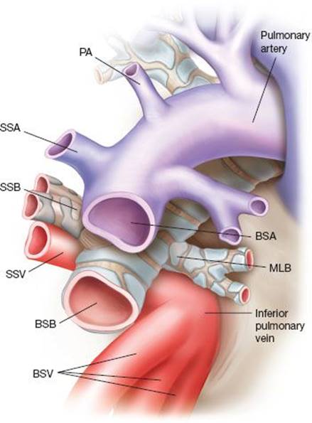
Figure 24.1 Three-dimensional anatomy of the right lower lobe artery, vein, and bronchus as viewed between the fissure of the upper, middle and lower lobes. The location of the pulmonary artery (PA), superior segmental artery (SSA), superior segmental vein (SSV), superior segmental bronchus (SSB), middle lobe bronchus (MLB) basilar segmental artery (BSA), basilar segmental vein (BSV), and basilar segmental bronchus (BSB) are marked.
 SURGERY
As with any pulmonary resection, detailed discussion regarding operative approach, ventilation needs, planned procedure, fluid management, and patient-specific factors must occur with the anesthesia team prior to the start of the case. The surgeon should perform a detailed flexible bronchoscopy prior to the procedure to evaluate the extent of endobronchial lesions or anatomic variations in the bronchial anatomy.
Positioning
Following bronchoscopy, the patient is positioned in a left lateral decubitus position with the table broken at the level of the hip. Rather than position as one would for a posterolateral thoracotomy, we tend to modify positioning for more anterior exposure. The patient’s shoulders are rotated exposing the anterior axillary line. The right elbow is also rotated cephalad to encourage the latissimus to a more posterior location. This allows the utility incision to be made without going through the latissimus dorsi muscle and keeps the thoracodorsal nerve out of the way of the utility port (Fig. 24.2).
Technique
The approach for video-assisted segmentectomy is the same that one would use for video-assisted lobectomy. Our approach is somewhat different from others in that we do not use the posterior port for anything more than 5-mm instruments. Given that the posterior intercostal spaces are narrower than the anterior spaces, placing staplers or other larger instruments through this port may increase trauma to the intercostal nerve.
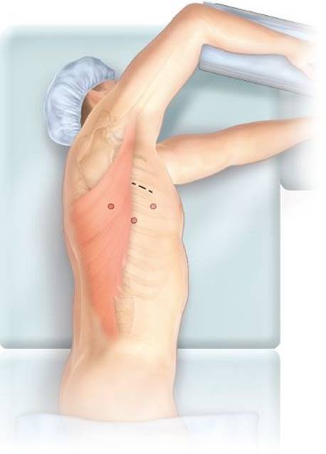
Figure 24.2 Patient in a left lateral decubitus position. Left shoulder rotated under and right elbow rotated in the cranial direction to force the border of the latissimus dorsi to a more posterior position and expose the axilla.
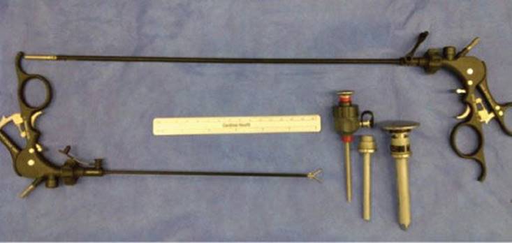
Figure 24.3 Standard laparoscopic instrument at top of image compared with the pediatric laparoscopic instrument. Metal ports 3 mm, 5 mm, and 10 mm in diameter as seen from left to right.
Instruments
We frequently use the 3-mm pediatric laparoscopic instruments. These are much shorter than the standard laparoscopic instruments, eliminating the ergonomic challenges of using traditional laparoscopic instruments. As well, they allow one to finely dissect structures when needed. Also one should have a laparoscopic needle driver on hand to repair small vessels or for other suturing needs. We also use a laparoscopic knot pusher to tie sutures on vessels or other knots, as when sewing the bronchus closed, as an alternative to stapling. All of our resections are performed with a 5-mm 30-degree thoracoscope.
We use two 5-mm metal ports, a single 10-mm port, and a soft tissue retractor for the utility port (Fig. 24.3). At times, we may only use three ports, but have found the addition of a fourth 5-mm port useful as we do not have to move the camera from the anterior to the posterior port as frequently. To use the staplers from the anterior port, it is necessary to use articulating staplers.
The first site of entry into the chest is in the sixth or seventh rib space at the anterior axillary line with a 10-mm port. A second 5-mm port is placed posteriorly below the scapula, at the seventh intercostal space. A third 3- to 5-mm port is placed either between these two, usually a rib space or lower or alternative location as needed. The optimal location for the location of the utility port is identified with the camera in the chest, positioning the port directly over the location of the artery in the fissure. This incision is typically a 3- to 4-cm incision that allows extraction of the specimen and placement of multiple instruments (Fig. 24.4).
For pulmonary surgery in general, some advocate a specific sequence for ligation and division of the bronchovascular structures. We typically dissect and divide the artery, bronchus, and vein in that order but on occasions find other sequences more preferable based on anatomic issues, pathology, etc. It is critical to identify all structures, be aware of aberrancies, and perform a thorough anatomic investigation prior to division. Three-dimensional reconstruction of the bronchovascular structures using a fine-cut CT may assist in identifying aberrancies in advance, although we have not found this to be necessary.
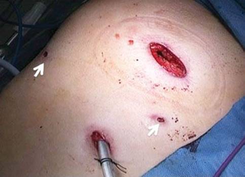
Figure 24.4 Incisions at completion of right basilar segmentectomy. A utility port and three additional ports (arrows). The chest tube is exiting the single 10-mm port site.
One cannot stress enough the extent of dissection and mobilization of the vessels. Typically, in patients who have a wedge resection for diagnosis prior to the segmentectomy, we use the time waiting for the frozen section diagnosis to begin the dissection. The pleura along the anterior, inferior, and posterior aspects of the lung is divided and the parenchyma swept up and away from the inferior vein both anteriorly and posteriorly.
Superior Segmentectomy: Bronchovascular Division
The interlobar pulmonary artery is first addressed. For patients with complete fissures, the dissection is technically much simpler. Patients with incomplete fissures create more of a technical challenge. We will highlight some of the strategies we use to overcome the challenges related to these situations.
If the fissure is not complete, an energy-sealing device is used to divide the parenchyma down to the artery along the line of the confluence of the fissures. Once the artery is identified in the fissure, the branches of the artery are dissected. Dissecting and resecting lymph nodes at the branch points helps in mobilizing the artery (Fig. 24.5). Early branching or multiple takeoffs of the superior segmental artery are common (Fig. 24.6). Occasionally, the posterior ascending branch (recurrent), which typically takes off proximal to the superior segmental artery, may instead branch off of the superior segmental artery. In this setting one may divide the superior segmental branch distal to the takeoff of the posterior ascending branch. After careful identification of the desired segmental artery is accomplished, we clip the proximal side and use a sealing device or ligation with silk tie for the distal side. When using a sealing device, it is important to seal the distal artery for several millimeters to prevent back bleeding during the remainder of the dissection.
Once the arterial branch is divided, the bronchus can be dissected. The superior segmental bronchus dives posterior and when the fissure is incomplete, dissection may be a challenge. To facilitate the exposure, we will complete the fissure between the upper lobe and superior segment, first. This is performed by dissecting between the upper lobe bronchus and bronchus intermedius posteriorly (Fig. 24.7A). The posterior aspect of the vein is also simultaneously dissected during this view (Fig. 24.7B). The three-dimensional relationship of the vein, artery, and bronchus to these segments should be well understood (Fig. 24.7C). Lymph nodes are typically cleared from the subcarinal space between the upper lobe takeoff and the bronchus intermedius. One must recognize that the superior segmental artery will run just deep to these. Careless dissection in this area can lead to injury.
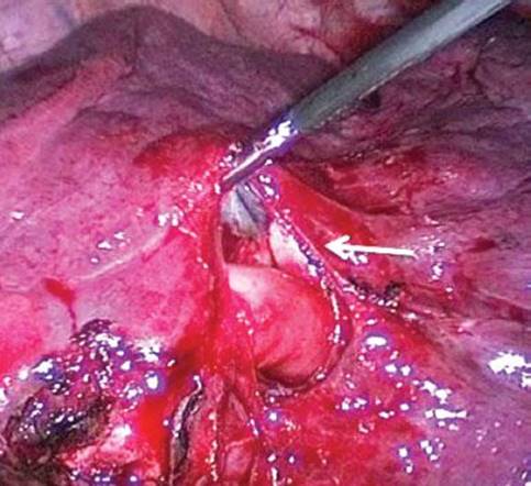
Figure 24.5 Interlobar pulmonary artery in the fissure with a lymph node being dissected. The superior segmental bronchus can be seen coursing behind the superior segmental artery (arrow).
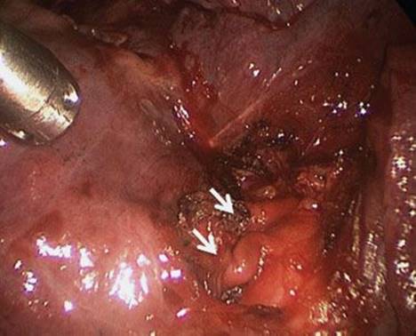
Figure 24.6 Two superior segmental branches of the pulmonary artery (arrows).

Figure 24.7 A: The posterior dissection of the bronchus with the vertebral column to the left and the lung reflected anteriorly: Azygos vein (A), right upper lobe bronchus (RUL), and bronchus intermedius (BI). B: Dissection of the superior segmental vein (SSV) from the posterior view. The basilar segmental venous trunk can be seen as well (BSV). C: Illustration depicting the three-dimensional relationships between the bronchus, pulmonary artery, and inferior pulmonary vein: Right upper lobe (RUL), basilar segmental vein (BSV) and basilar segmental bronchus (BSB), and superior segmental vein (SSV), superior segmental artery (SSA), and superior segmental bronchus (SSB).
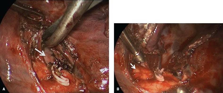
Figure 24.8 A: Dissection along the border of superior segmental bronchus through to communicate with the posterior dissection. The pleura overlying the spine is seen through this “window” (arrow). The posterior venous branch to the upper lobe can be seen below the scissors. B: Superior segmental bronchus (arrow) seen in the bed of the divided superior segmental arteries after completing the fissure between the superior segment and upper lobe.
From the anterior view, a dissecting instrument, such as a Maryland, is then passed from the interlobar fissure, posterior and lateral to the superior segmental bronchus through to the posterior space between the upper lobe takeoff and the bronchus intermedius. The fissure can then be completed with a stapler placed through this window (Fig. 24.8A,B). During dissection of the bronchus, a branch of the superior segmental vein can be seen (Fig. 24.9A). This is a branch and ligation here is an error as one would not have the entire trunk. The trunk of the superior segmental vein lies further caudal and unlike the artery is not in as close proximity to the bronchus (Fig. 24.9B). The superior segmental bronchus can then be ligated with a stapler or transected and oversewn (Fig. 24.10). The vein is typically ligated with a clip or silk tie (Fig. 24.11).
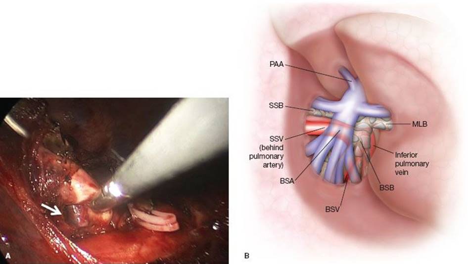
Figure 24.9 A: Superior segmental bronchus being dissected with view of superior segmental vein running along the posterior wall of the bronchus (arrow). B: Illustration depicting three-dimensional interlobar bronchovascular anatomic relationships: posterior ascending artery (PAA), superior segmental bronchus (SSB), superior segmental vein (SSV), basilar segmental artery (BSA), basilar segmental vein (BSV), basilar segmental bronchus (BSB), and middle lobe bronchus (MLB).
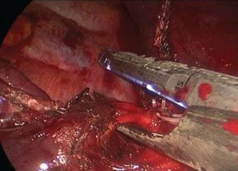
Figure 24.10 Ligation and division of the superior segmental bronchus from the anterior port.
Basilar Segmentectomy: Bronchovascular Division
The basilar artery is initially exposed in the fissure (Fig. 24.12). Once it is dissected, a stapler is used to ligate and divide the vessel. At times, getting the stapler to the appropriate angle on the artery may be a challenge, especially in the setting of incomplete fissures. Dividing the parenchyma in the fissure between the lower lobe and middle lobe will increase the exposure for the basilar segmental dissection (Fig. 24.13A,B). As well, to prevent traction injury to small branches, in attempting to mobilize the basilar segmental artery, we may ligate the medial basilar segmental branch first (Fig. 24.14). Once the arterial branch is divided, the bronchus can be dissected.
The view of the basilar segmental bronchus is usually excellent with the camera in the anterior port, however, to divide the bronchus with a stapler, once it has been dissected, we will move the camera to the posterior port when there are only three ports (Fig. 24.15).
Next the vein is divided. Failure to adequately mobilize the vein may result in the adventitia being caught in the bronchial staple line obscuring the anatomy (Fig. 24.16A). To insure that the superior segmental vein is preserved, the anatomy is clarified by dissecting the vein away from the bronchial anastomosis (Fig. 24.16B). The basilar segmental vein is clipped or ligated.
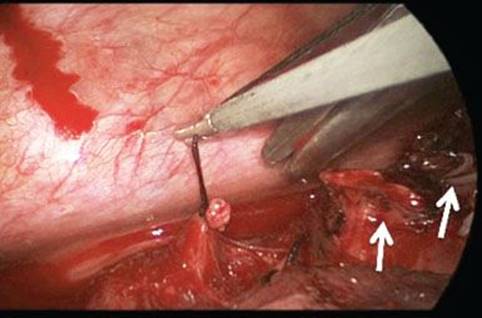
Figure 24.11 Silk tie on superior segmental vein with the lung retracted anteriorly. Divided superior segmental bronchus and artery are seen to the right (arrows).
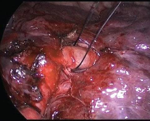
Figure 24.12 Basilar segmental artery as seen in the interlobar fissure.
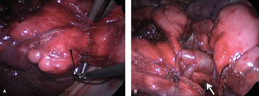
Figure 24.13 A: Right-angled clamp creating the window between the medial aspect of the basilar artery and the hilar space between the superior and inferior veins. B: View of basilar artery and underlying bronchus (arrow) after division of the fissure.
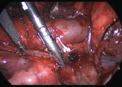
Figure 24.14 Energy-sealing device on the distal medial basilar segmental artery.
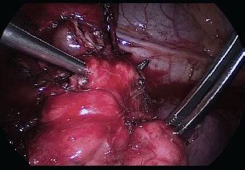
Figure 24.15 Right-angled clamp around basilar segmental bronchus as seen with the camera through the posterior port. Looking medially at the pericardium and phrenic nerve. The divided basilar segmental artery is seen above the bronchus.
Parenchymal Division Between Superior and Basilar Segments
Once the bronchovascular structures are ligated, division of the parenchyma between the superior segment and basilar segments is performed. Identification of the anatomical division of the superior segment and basilar segments can be aided by partially reinflating the lung, although we typically do not rely on this technique. Occasionally, one observes a cleft between these segments (Fig. 24.17A). The stapler is placed just beyond the expected anatomic border of the desired segment, and fired to divide the lung (Fig. 24.17B). The camera is used to view the hilar structures to insure that the stapler is not injuring the structures to be preserved (Fig. 24.18). The completed resection identifies ligation of the superior segmental bronchovascular structures for the superior segmentectomy (Fig. 24.19A,B), or preservation of the same for a basilar segmentectomy (Fig. 24.20A,B). Once the segment(s) in question are entirely detached, they are placed in a sac and removed through the utility port. Removing the soft tissue retractor prior to removing the larger basilar segments is helpful.
A lymph node dissection is performed if indicated and intercostal blocks are placed by injecting the intercostal spaces from third to the tenth. Typically a 20- to 24-Fr chest tube is placed at the conclusion of the case through the anterior port site, and the incisions are closed in the standard fashion.
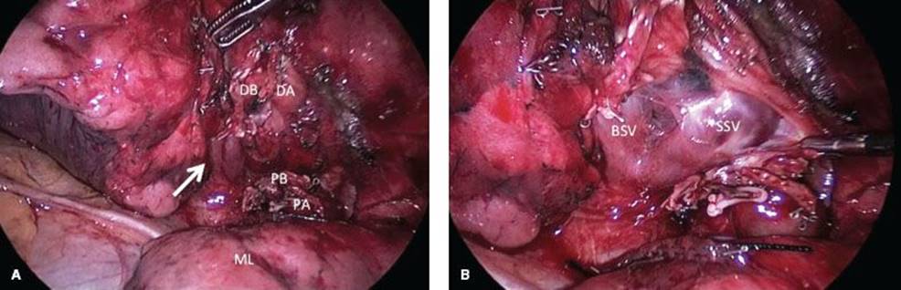
Figure 24.16 A: Basilar segmental bronchial staple line obscuring the anatomy of the inferior vein (arrows). Proximal bronchial and arterial staple line (PB and PA) and distal bronchial and arterial staple line (DB and DA). The middle lobe (ML) is viewed in the foreground. B:Basilar segmental staple lines on basliar segmental parenchyma and middle lobe as well as basilar segmental bronchus. The basilar segmental vein (BSV) and superior segmental vein (SSV) are highlighted.
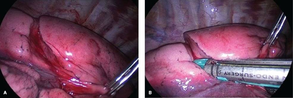
Figure 24.17 A: Cleft defining the superior from basilar segments. B: Stapler coursing along the segmental fissure.
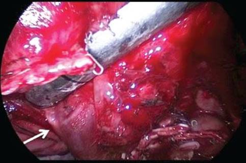
Figure 24.18 Hilar view with divided proximal segmental bronchus and artery in foreground and superior segmental vein (arrow). The clips mark the artery.
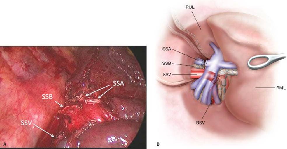
Figure 24.19 A: Completed superior segmentectomy with ligated superior segmental vein (SSV), superior segmental arteries (SSAs), and superior segmental bronchus (SSB). B: Illustration demonstrating the three-dimensional relationship between the artery, bronchus, and vein. The unseen right middle lobe bronchus and arteries are highlighted.

Figure 24.20 A: Basilar segmentectomy completed with intact superior segmental vein (SSV), superior segmental bronchus (SSB), and superior segmental artery (SSA). Bronchial and arterial anastomoses are seen in the foreground. B: Illustration demonstrating the three-dimensional relationship between the artery, bronchus, and vein following basilar segmentectomy.
 POSTOPERATIVE MANAGEMENT
Postoperatively, one should expect the vast majority of these patients to go to a surgical floor after the recovery room. As with any major thoracic procedure, optimizing residual lung function with pulmonary toilet, adequate pain control, and appropriate fluid management is essential. We typically immediately place these individuals on oral pain medication.
The chest tube can typically be removed the following day, assuming there is no air leak. These patients are then readied for discharge, with the majority leaving the hospital on the first postoperative day.
 COMPLICATIONS
Right lower lobe segmentectomy carries with it the potential for complications that are similar to other pulmonary resections. As the patient population who are typically offered segmentectomy have a high rate of baseline pulmonary disease, these individuals are at a higher risk for complications such as prolonged air leak, pneumonia, and supplemental oxygen requirement. In addition, in patients with emphysematous changes, resection of a section of lung base may alter residual lung function more than would be predicted, as the lung base is often contributing more to pulmonary reserve than is the apex.
Ultimately, the majority of air leaks will close with time, and reoperation is uncommon. Pneumonia risk can be lessened by encouraging pulmonary toilet and early ambulation. Finally, preventing the inadvertent creation of a new pulmonary cripple can be achieved with detailed operative planning, including PFTs, V/Q scans, and fine-cut CT.
 RESULTS
Long-term results for segmentectomy must be compared to the current gold standard of resection, lobectomy. Multiple authors have examined segmentectomy versus lobectomy in early cancer, which, as mentioned above, has produced conflicting results. When considering a patient for sublobar resection, there is a theoretical advantage to formal segmentectomy rather than a nonanatomic wedge resection, however, this has not been proven. As a consequence, though segmentectomy is a much more challenging operation, but given the dissection down to the hilum and the incorporation of the draining lymphatics, we favor this over a simple wedge resection.
 CONCLUSIONS
Video-assisted right lower lobe segmentectomy is uncommonly performed due to the potential inferiority as compared to lobectomy, as well as the high technical demand of the procedure. These factors lead to it being underutilized. With earlier detection of disease using fine-cut CT scans, which are able to detect smaller and smaller lesions, the role for video-assisted segmentectomy may increase, and the ability to apply this technique effectively will continue to be essential to the training of future thoracic surgeons.
Recommended References and Readings
Fell SC. Segmental resection. In: Patterson GA, Cooper JD, Deslauriers J, et al., eds. Pearson’s Thoracic and Esophageal Surgery. Philadelphia, PA: Churchill Livingstone; 2008:887–893.
Keenan RJ, Landreneau RJ, Maley RH Jr, et al. Segmental resection spares pulmonary function in patients with stage I lung cancer. Ann Thorac Surg. 2004;78(1):228–233.
Schuchert MJ, Pettiford BL, Keeley S, et al. Anatomic segmentectomy in the treatment of stage I non-small cell lung cancer. Ann Thorac Surg. 2007;84(3):926–932.
Tsutani Y, Miyata Y, Nakayama H, et al. Oncologic outcomes of segmentectomy compared with lobectomy for clinical stage IA lung adenocarcinoma: Propensity score-matched analysis in a multicenter study. J Thorac Cardiovasc Surg. 2013;146(2):358–364.