
N. Golan and Thomas K. Waddell
Introduction
Segmentectomy is a sublobar yet anatomical resection since it involves removing the segmental bronchus down to its primary branch along with all the lung parenchyma, lymph node grouping draining the segment and the segmental pulmonary artery (PA) (Table 25.1). It should be well differentiated from a wedge resection, which is a nonanatomical sublobar resection. Unfortunately a substantial amount of clinical research has grouped the two together thus obscuring the results and true benefits of a formal segmentectomy. For primary lung cancer, formal lobectomy or pneumonectomy still remains the gold standard for curative intent procedures. Segmentectomy emerged as a lung parenchyma-sparing technique for treating infectious pulmonary conditions; however, in view of the anatomical principles of the resection, it may serve as an adequate oncologic lung-sparing resection for carefully selected patients or as a compromise resection for those with limited lung function. Segmentectomy is technically more demanding and challenging than lobectomy and requires a thorough understanding of the lung anatomy and its variations. However, the left upper lobe (LUL) is usually well suited to an anatomical sublobar resection. We highly recommend reviewing Chapter 15 outlining open left upper lobectomy and the detailed discussion of anatomy in particular. We will review a standard straightforward open approach based on a staple-assisted extended resection as opposed to the “finger fracture” technique. Thereafter will review different problems/difficulties which one should recognize, different solutions which can be efficiently applied, and discuss specific scenarios for which an open approach may be required.
 INDICATIONS/CONTRAINDICATIONS
One perspective on the question of “Indications” for an open segmental resection is when to choose open rather than VATS. A second perspective is when to select segmental resection compared to lobar resection or wedge resection. For the most part the first question, open versus VATS segmentectomy, is similar to the open versus VATS lobectomy question. VATS segmental resection is technically more challenging but is our preference whenever possible. Uncertainty of anatomic location of the lesion either before or during a VATS procedure is a common reason for deciding to perform an open procedure. Regarding the question of extent of resection, current evidence supports the use of lobectomy for most patients. When anatomically favorable, in patients with adequate lung function, we would preferentially choose segmental resection for solid tumors less than 1 cm, or for ground glass (GGO) lesions less than 2.5 cm. With GGO lesions, one must take extra care planning the line of resection, as the lesion may not be palpable to guide adequacy of surgical margin, even during open resection. When otherwise suitable for VATS resection, we have found the CT-guided placement of a wire coil for fluoroscopic assurance of resection margin to be helpful. For patients with compromised lung function, our preferred approach is to use VATS surgery to minimize short-term risk. The long-term benefits of segmental resection over lobectomy are less than one might expect, at least in terms of FEV1. An increasingly common indication for segmental resection is the patient with multiple lung tumors, either synchronous or metachronous. In this setting, it is wise to consider segmental resection if adequate margins (at least equivalent to the size of the tumor) can be obtained. A wedge resection, by comparison, for nonsmall cell lung cancer (NSCLC) is inadequate as it carries a higher local recurrence rate, higher cancer-associated mortality, and worse overall survival.
TABLE 25.1 Choosing an Open Approach—Common Indications and Contraindication

No randomized controlled trial demonstrated a benefit for metastasectomy, nevertheless the cumulative experience supports resection of oligometastatic disease. An open approach has been advocated in this specific setting to increase the yield of detecting additional small metastases. As imaging studies improve, the yield decreases and the significance of these additional tiny lesions is unknown. Thus our preference in this setting is VATS wedge resection where possible. However, for more centrally located metastases we favor segmental resection over lobectomy wherever possible in view of the possibility of future recurrence.
Other common indications for segmentectomy include congenital, infectious, and inflammatory disorders.
 PREOPERATIVE PLANNING
Diagnosis
It is recommended to strive for a tissue diagnosis before going ahead with a segmentectomy, as dissection of the vessels does carry some risk. When tissue diagnosis was not achieved in advance, in some cases it may be possible to perform wedge resection as diagnostic biopsy during the operation. But in some cases the better judgment is to simply proceed with segmental resection, as a preliminary wedge biopsy may cause significant distortion.
Staging
Preoperative staging should be undertaken for any resection for malignancy. Intraoperative staging should be especially thorough with a liberal use of intraoperative quick sections to determine if segmental resection is still an appropriate choice.
Is the Disease Technically Resectable by Segmentectomy and at What “Price”?
Prior to the procedure the imaging should be reviewed to assess the tumor, the anatomical structures, and their relations to each other. Special attention must be paid to the location of the lesion in the lung to assess whether segmentectomy can be performed with adequate margins and without compromising adjacent segments. An attempt to assess function of the segment to be removed should be made, with attention to features such as high diaphragm, regional emphysema or atelectasis, and correlation with V/Q scans.
Physiologic Competence
The inability to comfortably walk up a single flight of stairs is the point where one wonders if the patient can tolerate any more than segmentectomy from a respiratory point of view. Lung perfusion studies, formal pulmonary function tests, and extensive cardiopulmonary exercise testing can further assess marginal patients. The final assessment of ability to withstand limited lung resection is a nuanced decision with no absolute cutoffs. It should take into consideration the location of the tumor, the function of that segment and function of both lungs, and intangibles such as patient’s personality type. The potential need for an extended resection/possible lobectomy or pneumonectomy and the ability of the patient to tolerate it should be specifically evaluated. All other comorbidities should be assessed and optimized, such as a short course of steroids for COPD and smoking cessation. The need for an ICU bed and special postoperative issues such as mobility and social issues should be assessed in advance.
A thorough discussion with the patient/family detailing the risks and benefits of the proposed procedure should be held. The patient should be well educated about the procedure and perioperative period. There should be clear communication with anesthesia, ICU, nursing, and other support caregivers for best patient care.
 SURGERY
Anatomical Considerations
The relevant lobar anatomy is described in Chapter 15. Within the lobe, the anatomic unit of the lung is the bronchopulmonary segment (Fig. 25.1). Each possesses its own bronchus, pulmonary arterial, venous, and lymphatic system and as such it can be removed individually without disturbing the function of adjacent segments. The segments are constant in their topographic shape and each is pyramidal in shape with the apex toward the center and base toward the pleural surface. They are surrounded by the connective tissue septa, which are continuous with the pleural surface. At the center of the segment runs a segmental bronchus, which usually further divides distally. A corresponding segmental PA branches to accompany the segmental bronchus. One main segmental vein drains each segment but as opposed to the bronchus and artery the veins run in an intersegmental plane. Thus, the veins mark the boundaries of each segment. The drainage pathway of the lymphatic is from subpleural lymphatic to larger channel running along the segmental arteries and bronchi and then to subsegmental and segmental lymph nodes. The most reliable landmark of the segment is the bronchus; it is rarely anomalous.
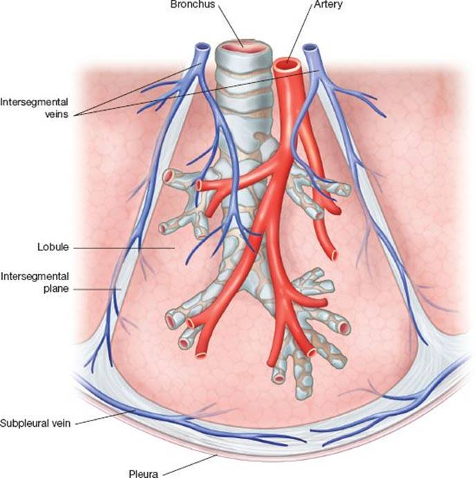
Figure 25.1 Anatomy of the bronchopulmonary segment The bronchopulmonary segment is pyramidal in shape. The apex of the pyramid tip is oriented to the center of the lung. The bronchus runs at the center of the segment and is accompanied by a pulmonary artery segmental branch, which runs along its posterior surface. Each segment is drained by one major venous segmental branch. The segmental veins follow the intersegment plane, which marks the boundaries of each individual segment. An open fracture technique takes advantage of this nature to allow dissection along the intersegment plane. There is substantial collateral drainage for each segment allowing for an extended stapler-assisted segmentectomy.
Equipment
An open segmentectomy requires a standard surgical tray identical to a lobectomy tray. While performing a VATS segmentectomy adequate equipment for open thoracotomy should still be available in case of an emergency conversion. Due to the nature of operating in a deep cavity we cannot overemphasize the need for a good headlight. Using the thoracoscopic equipment in an open procedure can facilitate lighting of the surgical field and can enable viewing difficult areas such as the apex the of the chest wall. We never pass off the thoracoscopic equipment after conversion to open thoracotomy and respectively have available a separately wrapped flexible pleuroscope during open cases.
Anesthesia and Preparation for Surgery
A team briefing is performed with the patient awake to review side, planned surgery, and preoperative medications. Lung isolation is preferably achieved by a double-lumen tube, although a bronchial blocker is acceptable and easier to place in smaller patients. We prefer a left-sided double-lumen tube and verify its position by a pediatric bronchoscope after intubation and again after positioning the patient. The remainder of the preoperative preparation is similar to that for upper lobectomy (Chapter 15).
Positioning and Incision
Posterolateral thoracotomy is our standard approach. We strive to perform a “semimuscle-sparing thoracotomy” by dividing latissimus but mobilizing the serratus anterior muscle from the chest wall. Making an effort to preserve the muscle allows better chest wall integrity and early perioperative shoulder function and it may become beneficial if a muscle flap will be considered, however, the rate of seromas is reported to be increased. Depending on the magnitude of the operation, the latissimus can be retracted, divided, or mobilized for possible use as a major flap. The patient is positioned in the right lateral decubitus position, an axillary roll is placed just caudal to axilla to protect the brachial plexus, the table is flexed between the 12th rib and the upper pelvis and reverse Trendelenburg positioning is used to flatten the flank and open the intercostal spaces. The patient is stabilized by a bean bag covered superficially by a gel pad and stiffened by applying suction. It should be hugging the patient bilaterally, away from the axilla and support the pelvis as well. All pressure points should be well padded. The sterile field should include the lateral border of the spine up to level of the neck allowing the extension of the thoracotomy posteriorly if required. At the anterior border it should include the sternum. Care must be taken to ensure the epidural catheter is not directly caught by the sterile covers or it can be inadvertently removed at the end of the case.
The chest is entered at the level of the fifth intercostal space. This correlates with the oblique fissure. For a better exposure one can shingle a posterior segment of the rib or completely resect a rib using the subperiosteal plane. This can facilitate entering a complicated pleural space such as when the lung is severely adherent to the chest wall. Occasionally starting with an extrapleural dissection can further facilitate this. If the use of an intercostal muscle flap is to be expected than harvesting the muscle should be done at this stage allowing safe handling and protecting its blood supply.
After final positioning, verify that all lines, tubes, and devices are well placed and still working.
Intraoperative Assessment
Generally speaking, we approach most patients with either planned thoracotomy or planned VATS and a conversion rate, perhaps somewhat higher than for lobectomy (<5%). However, thoracoscopy can be performed before going ahead with a thoracotomy. It is less invasive and may reveal findings encouraging for proceeding with VATS or alternatively may clearly mandate the need for an open procedure. On rare occasions it unfortunately might reveal advanced disease where segmentectomy is not appropriate. Once thoracoscopy equipment is added, it can be helpful in an open approach for lighting and overcoming blind spots.
Full assessment is usually achieved after lung mobilization. The extent of the disease and location should be assessed visually, by palpation and with the use of quick section as needed. For curative intent NSCLC resection lobar, hilar, and mediastinal nodes should be addressed and sent intraoperatively for pathology if suspicious. It should be emphasized that intraoperative lymph nodes’ sampling is one of the advantages and benefits of a surgical approach as opposed to other local treatments such as external radiation. The intraoperative finding of lobar/N1 disease should strongly advocate for performing a lobectomy. However if the patient is unable to tolerate a lobectomy a less favorable alternative would be performing segmentectomy with radical lymphadenectomy and consideration of post-op adjuvant treatments. Another possible alternative would be to abort surgery and refer for chemo and possible radiation treatments. The intraoperative finding of N2 disease should either suggest lobectomy, lymphadenectomy, and adjuvant treatment or aborting and referring to chemo and possible radiation. For patients with metastatic disease the finding of positive mediastinal lymph nodes might exclude the patient from surgery.
Stapler-assisted Segmentectomy
In the searching for ways to avoid complications (air leak) and to facilitate a minimally invasive approach the use of stapler-assisted segmentectomy has become more common. The older technique, the so-called “open fracture technique,” involved following the intersegmental vein using blunt and blind digital dissection. In some hands, this has been replaced by precision cautery dissection of the same plane. This latter technique can be performed by VATS as well. In this approach, it is technically important to maintain the integrity of the vein, both to prevent bleeding and to preserve the adjacent segmental venous drainage as the segments share a common intersegmental venous anatomy. However, stapler-assisted segmentectomy is technically much easier and more common. Although stapler-assisted segmentectomy carries the potential for an increased risk of venous congestion and parenchymal dysfunction, this generally remains theoretical since the collateral intersegmental circulation is very reliable. In fact for a stapler-assisted approach the venous segmental branch should be divided early as it allows better lung manipulation and tailoring of the margins.
The primary advantages of the stapler-assisted approach are the potential reduction in bleeding and prolonged air leak from the raw residual segmental surface. The use of staples also allow for an extended segmentectomy as the margins can be deliberately increased to accommodate wider margins than the segment itself. This may be important if the tumor ends close to or just at the intersegmental border. Generally speaking the margins should be at least 1 cm from the tumor edge. Some studies advocate for the margins to be at least of same diameter as the tumor.
Defining the Intersegmental Border
There are several simple approaches to define the intersegmental border without dissecting along the intersegmental vein. One of the best is to use selective ventilation of either the target segment or the rest of the lung thus forming a demarcation line between the inflated and deflated segments. A few techniques can be used, although in all approaches one must be certain that the correct segmental bronchus is targeted.
 Clamp the target segmental bronchus and inflate the lung, this segment will remain atelectatic while the rest inflates. However, due to the excellent intralobar collateral circulation it may inflate as well.
Alternatively, the segmental bronchus can be clamped after full lung ventilation, the relevant segment will remain inflated for longer as the rest of the lung deflates.
Although rarely performed the target bronchus can be closed or divided, cannulating its distal stump and directly inflating to selectively inflate the target segment.
Bronchoscopic examination can help define the relevant bronchial anatomy. The use of segmental ventilation by positioning the bronchoscope in the relevant segment and jet ventilating through the working channel may also selectively inflate this segment.
Other hints and innovative ways for finding the correct intersegmental plane:
An accessory fissure may exist between the lingula and the upper division, which can help to demarcate the border.
A novel technique is based on fluorescence imaging after ligating the dominant PA and injecting indocyanine green intravenously. The indocyanine will selectively integrate to the nontarget segments and serve as the basis for the different visualization. Alternatively, the identification of intersegmental planes can be based on an infrared thoracoscopy after transbronchial injection of indocyanine green. Both techniques have primarily been described for VATS surgery but are theoretically achievable with open surgery as well, provided a fluorescence camera is available.
As an advantage of the stapler-assisted technique, when in doubt make sure the tumor margins are sufficient and cheat toward the adjacent segments. As the parenchymal division extends toward the adjacent segment it should not include the relevant supplying bronchus, artery, or vein, which is the basis of keeping the adjacent segments viable.
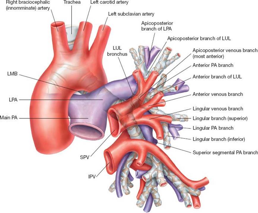
Figure 25.2 Pulmonary artery, veins, and airway. A schematic figure depicts the anatomical relations between the left bronchus and LPA and SPV branches. Notice the direction of the PA as it comes above and around the LMB. Pay attention to the different PA branches as they bifurcate from the LPA. In the larger figure the PA branches directly from the LPA and runs from posterior to anterior as it follows the anterior segmental bronchus.
Straightforward upper division segmentectomy (lingular-sparing upper lobectomy)
1. Open the anterior mediastinal pleura to isolate and divide the upper lobe branches of the superior pulmonary vein (SPV).
Open the anterior mediastinal pleura (Figs. 25.2 and 25.3); watch for the phrenic nerve, identify the inferior pulmonary vein (IPV), SPV, and lingular branches. Make sure the SPV is well separated from the IPV (look for a common confluence), verify that the lingular branches are draining directly to the SPV. Isolate and divide the upper division portion of the SPV while preserving the lingular veins, the vein wall can be grasped directly assuming that a broad grasp is used. Keep in mind the PA and its branches at the posterior deeper aspect of the vein.
2. Open the posterior pleura, the fissure, and divide the PA branches.
Retract the lung anteriorly and identify the PA as it is about to become interlobar and use this as an entrance point for the subadventitial plane (Fig. 25.4). Open the posterior pleura along the anterior border of the esophagus from the level of the IPV to the level of the PA as it comes around the bronchus, then curve anteriorly opening the roof of the aortopulmonary window, this plane should run along the superior border of the proximal PA branches. Continue this dissection to connect with the mediastinal pleura opened previously from the front. Coming toward the aortopulmonary window you may need to retract lung laterally and inferiorly, staying close to the PA and away from the aorta to avoid injuring the recurrent laryngeal nerve. Retract the lung anteriorly again, identify the PA as it comes around the left main bronchus (LMB), at this level it should lie most superficial, just below the parietal pleura. Enter the subadventitial plane and dissect anterograde along the PA in direction of fissure. This will facilitate identification of the interlobar PA in the fissure. The next steps are greatly facilitated by division of the lung parenchyma of the posterior aspect of the fissure, either with a stapler or sharp dissection. To safely exclude the lingular branches, the interlobar PA anatomy and divisions should be well identified, just enough to identify the last posterior PA branch to LUL, the first superior segmental PA branch of the left lower lobe (LLL), and the lingular branches. Start ligating and dividing the arterial branches to LUL from the level of the interlobar PA, excluding the lingular PA branches.
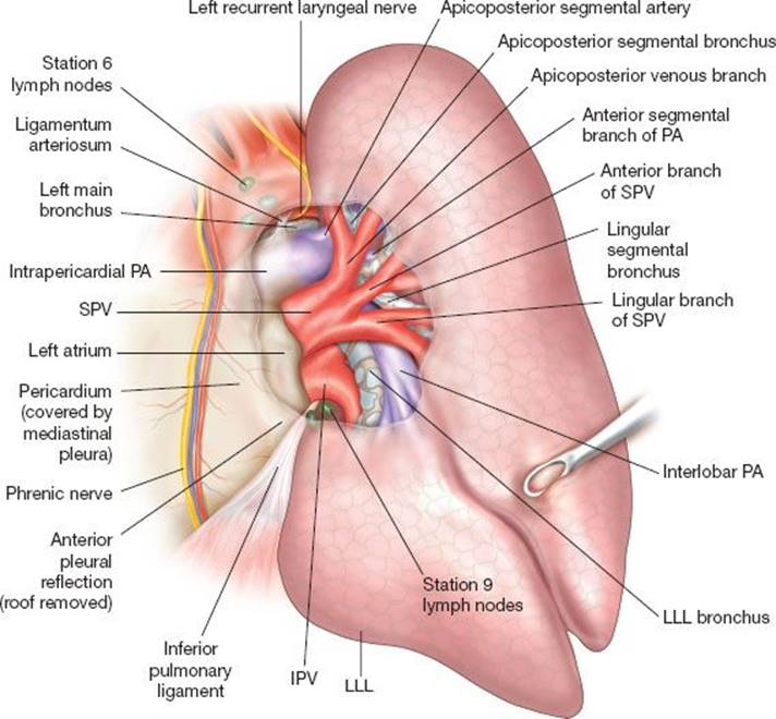
Figure 25.3 Anterior view: Mediastinum and lung—intra- and extrapericardial anterior view while the lung is retracted posteriorly and laterally, showing the mediastinal intra- and extrapericardial relations of the PA, SPV, and IPV. Notice that the IPV is completely omitted beyond its proximal part, allowing for better understanding of the airway and PA anatomy. Pay attention to the ligamentum arteriosum and recurrent laryngeal nerve.
3. Dissection and division of the upper division bronchus from within the fissure.
Look for the bronchus in the fissure, above the interlobar between the divided PA branches (Fig. 25.5). Gently dissect this plane, searching and feeling for the bronchus while using a “peanut” (a small rounded gauze, slightly bulging from a tip of an instrument). This maneuver allows for better definition of the bronchial anatomy as the goal is to identify and solely isolate the upper proper bronchus. Care should be taken not to over strip and be aware you are facing and dissecting along the posterior bronchial membrane, which is very vulnerable. Care should also be taken to avoid damaging the lingular PA branches at the lateral, anterior border of this dissection, which also mark the direction and location of the lingular bronchus. Advancing distally along the LUL bronchus, the lingular, anterior, and apicoposterior bronchi should reveal themselves. Although the lingular and upper divisions are the first bifurcation from the LUL bronchus, at this level all distances between the bronchial bifurcations are very short, thus one can easily become mistaken as to the identity of a bronchus. An intersegmental lymph node, between the lingular and upper division bronchi, marks the lateral border of the upper proper bronchus. This node should be dissected and serve as the exit point for the encircling instrument. One should be certain to encircle the entire upper proper bronchus and rely on thorough and multidirectional assessment. From this posterior perspective, the anterior bronchus is deep and heading away from the surgeon. To further verify, close the upper proper bronchus and inflate the left lung, the upper division should remain atelectatic while the lingula inflates. However, due to collateral circulation, which is usually excellent within the lobe, the upper division might inflate despite an adequate closure of its bronchus thus making it difficult to judge. Sometimes it is instructive to keep the upper lobe proper bronchus closed and allow the left lung to deflate, air will be trapped for longer in upper division while the lingula will deflate. We always use a flexible bronchoscope to confirm correct lobar identification. Notice that you might encounter resistance while encircling the upper lobe proper bronchus as coming close to the exit point. The cause can be the result of the direction of the anterior bronchi. Attempting to encircle the upper proper bronchus more proximally (just a millimeter or two) or even just changing the direction of the instrument to take a more medial course can potentially overcome this problem. The back wall of the instrument lies against the SPV upper branches. However, these branches should already be divided. If this step was not taken yet or the instrument encircling the bronchus is aimed too medially, the SPV might be injured. The bronchus can be divided using a stapler, we usually prefer a TA-type with larger staples but it can also be divided sharply and closed with interrupted sutures.
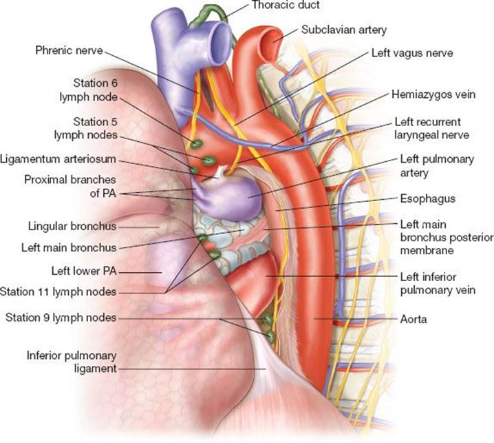
Figure 25.4 Posterior view: Mediastinum and lung. Posterior view of the posterior mediastinum and the left lung. The lung is retracted anteriorly. Notice the order of the PA, LMB, and IPV as they present from superior to inferior. Pay attention to the very proximal bifurcation of the first PA branches, which are prone to traction injury. As the LPA turns around the LMB to become superior and superficial to the PA, it lies in a subpleural position. This point is at the upper posterior edge of the left fissure and can be comfortably utilized to expose the PA and enter into the subadventitial plane.
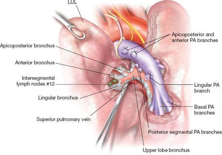
Figure 25.5 Dividing the left upper lobe proper bronchus. Posterior view from a lateral decubitus position showing the interlobar PA and the bronchi to the LUL. All PA LUL branches, excluding the lingular, are already divided. To safely exclude the lingular branches, the entire interlobar PA anatomy and divisions should be well identified. This requires dissecting the PA along its course. The upper division bronchus can be exposed at the deeper interval between the divided PA vessels. The interlobar PA may be rolled inferiorly to allow for better exposure of the LUL bronchus. An intersegmental lymph node, between the lingular and upper division bronchi, marks the lateral border of the upper proper bronchus. This node should be dissected and serve as the exit point for the encircling instrument.
4. Complete the parenchymal boundary of the resection.
Use the transected bronchus as the base of the segmental resection during the division of the lung parenchyma in the intersegmental plane, advancing from lateral to the hilum. The lung can be inflated again to refresh your memory as to where the intersegmental plane is. Make sure not to compromise the SPV as approaching the hilum, avoiding the other PA branches and lingular bronchus. Once close to the hilum, consider changing the direction of the stapler to come from anterior medial (hilum) to posterolateral (previous stapled line). Take care to ensure adequate margins exist even if this translates to extending the resection across the segmental plane. For the finger fracture technique, place a grasping instrument on the divided intersegmental vein and retract very firmly from medial (hilum) outward. Strip and pull with the fingers of the other hand from medial to lateral until the pleural surface is reached. There will be bleeding so continue quickly and then pack the raw oozing surface. After a few minutes the surface will be remarkably dry, free of bleeding and much of the air leak. The remaining bleeding points can be cauterized and the air leaks sutured.
5. Completion of staging and closure.
Verify at least sampling of lymph node stations 4L and 7 either now or previously via mediastinoscopy. Perform full lymphadenectomy either routinely but definitely if there is evidence of stage II or higher disease and remove or sample nodes from stations 5, 6, 8, 9, 10, and 11. Most will have been sampled during the course of the dissection. Station 7 is approached from behind while retracting the lung anteriorly, opening the anterior esophageal plane between the LMB and the esophagus and dissecting the preesophageal space at this level watching deeper and medially for the right main bronchus and above for the carina, paying careful attention to avoid injuring the posterior bronchial membrane. Station 4 can be approached anteriorly by retracting the left main pulmonary artery (LPA) inferiorly and exposing the tracheobronchial angle. Dividing the ligamentum arteriosum can facilitate dissection of station 4. Division of the inferior pulmonary ligament can be performed but becomes less critical as smaller amounts of lung tissue are removed. Hemostasis should be reviewed. Check for air leak under saline irrigation with controlled testing of the bronchial stump up to 20 cm of H2O pressure. If an epidural has not been placed, intercostal blocks or paravertebral catheter can be placed. One, or occasionally two, chest tubes are placed. Hemostasis should be reviewed again and sponges counted. The ribs are approximated with two interrupted, figure-of-eight, heavy absorbable pericostal sutures, taking care to set the tension in both sutures equally. The lingula is inflated taking care to ensure it is not rotated. The muscular layers are closed in layers.
Other Segmental Resections of the Left Upper Lobe
Other common resections include lingulectomy, anterior segmentectomy, and apicoposterior segmentectomy. It is possible although not common to subdivide the latter into apical or posterior segments. Each can be completed following the same basic steps in almost the same order. For lingulectomy, for example, the same structures are dissected but the lingular branches of vein, PA, and bronchus are divided rather than protected (Fig. 25.6). One major difference is the dissection of the vein required for apicoposterior (or the subsegmental resections) segmentectomy. Division of the artery is straightforward by retracting the lung laterally and inferiorly and approaching from the top of the aortopulmonary window. Division of the bronchus is also straightforward from the posterior perspective. Division of the apicoposterior segmental vein can be left to last and the anatomy sorted out after division of the artery and bronchus. Similarly, division of the bronchus to the anterior segment may be approached from the front as opposed to the posterior perspective when completing an anterior segmentectomy.
In general, one can alter the stages according to convenience and personal ease of dissection. If the different PA branches are not well identified as to the segment they serve then before division a complete dissection to expose the full extent of the PA should be done. This will reveal all branches allowing for better orientation and avoiding irreversible mistakes. Some advocate taking the bronchus as early as possible when dealing with infectious etiologies since the lung manipulation may expose the rest of the lung to contamination. Table 25.2 lists some of our practice experience recommendations per different aspects of the surgery as we aim to prevent expected difficulties or complications. Following we discuss possible tactics to better confront some of the common difficulties and scenarios that the surgeon may face advancing through the procedure.
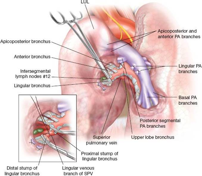
Figure 25.6 Dividing the lingular bronchus. Posterior view from a lateral decubitus position showing the interlobar PA and the bronchus. The lingular segmental PA branches (two separate segmental arteries) are already divided. The lingular bronchus can be discovered deep to the divided lingular PA vessels and is the most lateral (distal) bronchus to branch from the LMB. The PA may be rolled inferiorly to allow for better exposure of the LUL bronchus.
TABLE 25.2 Avoiding Problems
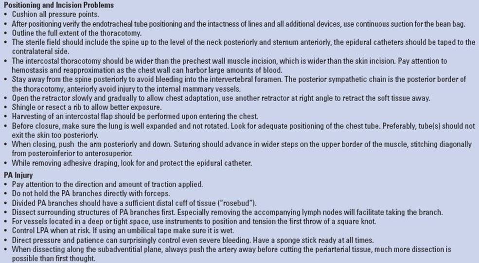

Dealing with Specific Difficulties
Chapter 15 highlights some special approaches when encountering technical difficulties, for example, dissecting and controlling the PA and how to deal with PA bleeding. These are occasionally needed during segmental resection, but much more common is minor tear of a PA branch. If sufficiently distal and small, it can often be controlled with gentle packing. If well controlled, turn your attention elsewhere and upon returning the problem has often coagulated. There are some useful techniques for dealing with short PA branches but more commonly if the branches are that short, then extending the resection to lobectomy is probably the most appropriate course of action.
Other strategies, such as approaches for a difficult fissure or advancing through difficult planes are described in detail in Chapter 15.
 POSTOPERATIVE MANAGEMENT
There is nothing unique about the postoperative management following anatomic segmentectomy. In general the course should be a bit easier, but it is often counterbalanced as it is more often used in a higher risk population.
 COMPLICATIONS
The overall mortality in most high-volume centers range from 1% to 3% and should not exceed lobectomy. The more common early complications involve the cardiopulmonary system and include atrial fibrillation, prolonged air leak, pneumonia, and an atelectasis requiring bronchoscopy. Prolonged air leak is the most common complication of segmental resection occurring in approximately 10%. The treatment depends on the severity, extent of lung expansion and the patient’s clinical situation. Small alveolar fistula may seal spontaneously. One of the concerns of prolonged air leak is the development of empyema. The treatment may include careful surveillance, removing the tube despite a small air leak after confirming with tube clamping that the lung does not collapse further. Pleurodesis, for example, by applying a blood patch or doxycycline, may seal the leak. Applying a viable flap to close a fistula and fill the pleural space is a last resort. The overall intraoperative bleeding rate for pulmonary resections is 5% and for significant postoperative bleeding around 3%. Other risks are similar to those following lobectomy and are described in Chapter 15.
Stapler-assisted segmentectomy does carry a specific risk of compromising the venous drainage of neighboring segments especially in the setting of an extended resection, this is usually a theoretical rather than a clinical concern due to the well-developed collateral drainage. Yet it is a concern and there are reports of the development of a pseudotumor of the adjacent segments as a consequence of vascular infarct. If the segmental major blood supply and drainage is severely compromised, the affected segment should be removed. For the tissue left behind necrosis of the remaining part can occur. This can often resolve with conservative treatment, but when it fails completion lobectomy might be needed. Dissecting the LUL and leaving an anatomical subdivision might also result in torsion; this can simply be avoided by fixation of the remaining LUL. The most common long-term complication is postthoracotomy pain.
 RESULTS
Although lobectomy or pneumonectomy is still the gold standard as curative intent procedures, for physiologically borderline patients a sublobar resection may be the only possible safe alternative when considering resection. Whether segmentectomy may be comparable to lobectomy small peripheral NSCLC is the subject on ongoing clinical trial. Previous clinical research has often grouped anatomical and nonanatomical sublobar resections thus obscuring the results and true benefits of a formal segmentectomy. The only randomized prospective trial comes from the Lung Study Group and published by Ginsberg et al. in 1995. This study also combined segmentectomy with wedge resection for the overall survival but did report a break down of locoregional recurrence rate. They found a rate per person per year of 0.022 for lobectomy, 0.044 for segmental resection, and 0.086 for wedge resection. There was a trend toward longer survival in the lobectomy arm (p = 0.08). Most of the nonrandomized clinical data also suggest a favorable overall survival for lobectomy and lower cancer-associated mortality than for segmentectomy. However, these differences are less prominent in different subgroups of patients and the survival may be comparable in appropriate patient subgroups. Thus, there is still a role for consideration of segmentectomy for small peripheral early stage NSCLCs, patients older than 75 years, tumors size <2 cm, and that the expected pathologic margins are greater than 1 cm especially if an extended resection was performed.
No randomized controlled trial has examined the benefits of metastasectomy. Nevertheless the cumulative experience support resection in some circumstances such as a few or preferably a single metastasis, the primary site under good control, and a long disease-free interval. Nearly all reports indicate that complete resection of metastatic disease is associated with longer life expectancy and is potentially curative. The prognosis differs according to the above parameters. A review from the International Registry of Lung Metastases identified 5,206 patients with a variety of primary metastatic tumors (epithelial carcinomas, sarcomas, germ cell tumors, and melanomas) who underwent a pulmonary metastasectomy; this series included 4,572 (88%) in whom complete resection was carried out. The overall 5-, 10-, and 15-year survival rates were 36%, 26%, and 22% respectively.
 CONCLUSIONS
Segmentectomy is a sublobar yet anatomical lung resection. It should be well differentiated from a wedge resection and care should be taken when evaluating the results reported in the literature.
Surgical lung resection remains the primary and preferred approach to the treatment of early stage NSCLC. In selected patients segmentectomy may offer equivalent or potentially better long-term outcomes compared to lobectomy or pneumonectomy and in many may be the only option for a safe lung resection. Segmentectomy can also be indicated in cases in which a wedge resection could have been sufficient yet it may offer better lung preservation.
As minimally invasive techniques have less perioperative complications and offer a safer and smoother course, with equivalent oncologic outcome, the role of an open procedure should be reserved for candidates who can benefit for its advantages comparing to a minimally invasive approach. Moreover minimally invasive approach can well suit physiologically borderline and sicker patients and therefore patient criteria for the resection should be adjusted to match the applied approach.
Mediastinal lymph node staging should follow the guidelines for invasive staging as practiced in any NSCLC workup. However, a thorough lymph node sampling which includes the intersegmental stations up to the mediastinal lymph nodes, should be performed during the segmentectomy. The results should tailor the ongoing procedure and future treatment. The initial reflux should include gentle and direct pressure. This may be the sole measure needed to overcome the bleeding, however, LPA control should be pursued. Some PA injures might mandate the need for an extended lung resection and the surgeon should value the case-specific risks and the physiologic capability to withstand such, in advance.
Stapler-assisted segmentectomy involves dividing the intersegmental plane using a stapler and relying on differential ventilation (as new techniques are evolving) to outline the intersegmental border. It reduces bleeding and air leak, which is the commonest early perioperative complication. Despite a potential risk to compromise the adjacent venous drainage especially when practicing an extended margins approach, these concerns are usually not clinically significant.
Patients can benefit from a structured peri- and postoperative protocol. This should deal with all aspects of patient care by all care providers. The most common early post-op complications involve the cardiopulmonary system and mortality should not exceed lobectomy.
Recommended References and Readings
Carballo M, Maish MS, Jaroszewski DE, et al. Video-assisted thoracic surgery (VATS) as a safe alternative for the resection of pulmonary metastases: A retrospective cohort study. J Cardiothorac Surg.2009;4:13.
Cerfolio RJ, Bryant AS, McCarty TP, et al. A prospective study to determine the incidence of non-imaged malignant pulmonary nodules in patients who undergo metastasectomy by thoracotomy with lung palpation. Ann Thorac Surg. 2011;91(6):1696–1700.
Ginsberg RJ, Rubinstein LV. Randomized trial of lobectomy versus limited resection for T1 N0 non-small cell lung cancer. Lung Cancer Study Group. Ann Thorac Surg. 1995;60(3):615–622.
Hornbech K, Ravn J, Steinbrüchel DA. Outcome after pulmonary metastasectomy: Analysis of 5 years consecutive surgical resections 2002–2006. J Thorac Oncol. 2011;6(10):1733–1740.
Howington JA, Blum MG, Chang AC, et al. Treatment of stage I and II non-small cell lung cancer: Diagnosis and management of lung cancer, 3rd ed: American College of Chest Physicians evidence-based clinical practice guidelines. Chest. 2013;143(5 suppl):e278S–e313S.
Kasai Y, Tarumi S, Chang SS, et al. Clinical trial of new methods for identifying lung intersegmental borders using infrared thoracoscopy with indocyanine green: Comparative analysis of 2- and 1-wavelength methods. Eur J Cardiothorac Surg. 2013;44(6):1103–1107.
Leshnower BG, Miller DL, Fernandez FG, et al. Video-assisted thoracoscopic surgery segmentectomy: A safe and effective procedure. Ann Thorac Surg. 2010;89(5):1571–1576.
Odell DD, Kent MS, Fernando HC. Sublobar resection with brachytherapy mesh for stage I non-small cell lung cancer. Semin Thorac Cardiovasc Surg. 2010;22(1):32–37.
Pastorino U. Long-term results of lung metastasectomy: Prognostic analyses based on 5206 cases. The International Registry of Lung Metastases. J Thorac Cardiovasc Surg. 1997;113(1):37–49.
Rami-Porta R, Tsuboi M. Sublobar resection for lung cancer. Eur Respir J. 2009;33(2):426–435.
Sekine Y, Ko E, Oishi H, et al. A simple and effective technique for identification of intersegmental planes by infrared thoracoscopy after transbronchial injection of indocyanine green. J Thorac Cardiovasc Surg. 2012;143(6):1330–1335.
Smith CB, Swanson SJ, Mhango G, et al. Survival after segmentectomy and wedge resection in stage I non-small-cell lung cancer. J Thorac Oncol. 2013;8(1):73–78.
Ugalde P, Camargo Jde J, Deslauriers J. Lobes, fissures and bronchopulmonary segments. Thorac Surg Clin. 2000;17:587–599.
William H, Milloy FJ. Pulmonary vascular system and pulmonary hilum. Thorac Surg Clin. 2007;17:601–617.