
Hugh G. Auchincloss and Christopher R. Morse
 INDICATIONS/CONTRAINDICATIONS
Segmentectomy, a sublobar anatomotic lung resection, as a treatment for primary lung malignancy remains a somewhat controversial topic. The findings of the Lung Cancer Study Group in 1995 suggested that rates of local recurrence and death were considerably higher with sublobar resection as compared to lobectomy; however, the sublobar resection group included nonanatomic wedge resections. Subsequently, a mounting body of evidence mainly from single-institution trials has demonstrated that for stage IA NSCLC segmentectomy is indistinguishable from lobectomy with regard to oncologic outcome and may be better tolerated by patients. Aggressive lung cancer screening programs could lead to a rise in the number of patients with early-stage cancers for whom segmentectomy should be considered.
Less controversially, segmentectomy remains a good oncologic operation for patients with early-stage lung cancer who, because of limited pulmonary reserve, would not tolerate a formal lobectomy. Segmentectomy is also indicated in a variety of benign conditions including bronchiectasis, tuberculosis, and lung abscess. The decreasing incidence of these benign conditions coupled with the growing interest in segmentectomy as a first-line oncologic operation means that in the future most segmentectomies will be performed for cancer.
There are no indications or contraindications that apply to the left lower lobe specifically. Typically the superior segment of the left lower lobe and the composite basilar segments are amenable to segmentectomy. The decision about whether to proceed with a thoracoscopic resection depends on the anticipated difficulty of the dissection and ability to localize the lesion with confidence. Patients with a history of prior chest surgery or radiation may benefit from proceeding directly to thoracotomy.
Indications
Resectable cancer in a patient with insufficient pulmonary reserve to tolerate a lobectomy
Multiple synchronous lesions in different lobes
Lung resection in a central location where wedge resection would be impossible, either for metastatic disease or for diagnosis of an unknown lesion
Relative Indications
<2 cm primary lung cancer
<2.5 cm GGO
 PREOPERATIVE PLANNING
The preoperative workup prior to segmentectomy does not differ significantly from any other anatomic lung resection. All patients should undergo pulmonary function tests and basic cardiac risk screening. If pulmonary function is considered borderline then a ventilation–perfusion scan may be performed. We continue to use mediastinoscopy routinely as a method to stage the mediastinum. This can be performed concomitantly with lung resection; alternatively, scheduling mediastinoscopy as an earlier separate procedure may eliminate the risk of a false-negative frozen pathology result although this is extremely rare.
The larger question often remains whether a lesion is amenable to a segmental approach. A close review of all imaging is essential with particular attention paid to the location of the lesion and its position in regard to other anatomic landmarks, that is, blood vessels and segmental bronchi.
 SURGERY
Anesthesia
Our preference is for the use of epidural anesthesia for an intraoperative and postoperative pain management, particularly when the indication for segmentectomy is that the patient may lack sufficient pulmonary reserve to tolerate lobectomy. Standard induction is performed and after bronchoscopy to evaluate anatomy and secretions; the patient is intubated with a double-lumen endotracheal tube. Anesthesia is maintained during the operation with inhaled and intravenous agents.
Positioning
The patient is placed in the lateral decubitus position with the left side up. The operating room table is flexed to open the rib spaces while rolled towels placed anterior and posterior and 2-in cloth tape placed across the hips secure the patient in place. The legs are padded at all pressure points and placed in gentle flexion at the knee and hip, and the left arm is placed on a padded elevated armrest. The patient’s head is placed in the neutral position.
Port Sites
Three thoracoscopic ports are typically used for a left lower lobe segmentectomy (Fig. 28.1). Typically a 1-cm camera port is placed at the eighth interspace at the midaxillary line. A port is placed posterior and inferior to the scapular tip for lung retraction and an anterior access port is placed over the fifth interspace measuring 4 to 5 cm in length. This anterior port starts at the anterior aspect of the latissimus dorsi extending toward sternum. The access port is typically placed over the pulmonary artery in the oblique fissure. We prefer the use of either a 10-mm or 5-mm 30-degree HD scope.
Technique
Anatomy
An understanding of the segmental anatomy of the left lower lobe is essential. The left lower lobe bronchial anatomy includes the superior segment bronchus and the basilar segmental bronchi—anteromedial, lateral, and posterior. In regard to the pulmonary artery, the superior segmental supply for the left lower lobe originates from the interlobar pulmonary artery at or above the levels of the lingular artery, usually as a single trunk. Caudal to the origin of the superior segmental artery is the basal trunk. In approximately 70% of individuals this is a single common trunk. The left inferior vein runs below and slightly posterior to the superior pulmonary vein and distribution tends to follow the distribution of the bronchial tree.

Figure 28.1 Three thoracoscopic ports are used for a thoracoscopic left lower lobe segmentectomy. A 1-cm camera port is placed at the eighth interspace at the midaxillary line. A working port is placed posterior and inferior to the scapular tip for lung retraction and an anterior access port is placed over the fifth interspace measuring 4 to 5 cm in length.
VATS Left Lower Lobe Superior Segmentectomy
The pulmonary artery is initially identified in the fissure with it being easier to identify with a complete fissure. The upper lobe is retracted cephalad with the lower lobe dropping caudally, exposing the artery in the fissure (Fig. 28.2). If the fissure is not complete, dissection will need to be started with cautery and possibly sharp dissection through the access port. Once the artery is identified, the individual branches are dissected out. If this proves difficult, resecting surrounding lymph nodes may help to clarify branch points of the artery. The superior segment artery is surrounded with a vessel loop and divided with a vascular stapler.
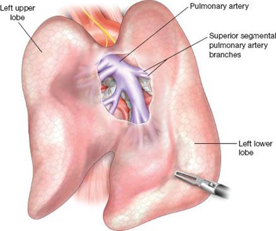
Figure 28.2 The upper lobe is retracted cephalad with the lower lobe dropping caudally, exposing the pulmonary artery in the fissure, and allowing for identification of the superior segment branch.
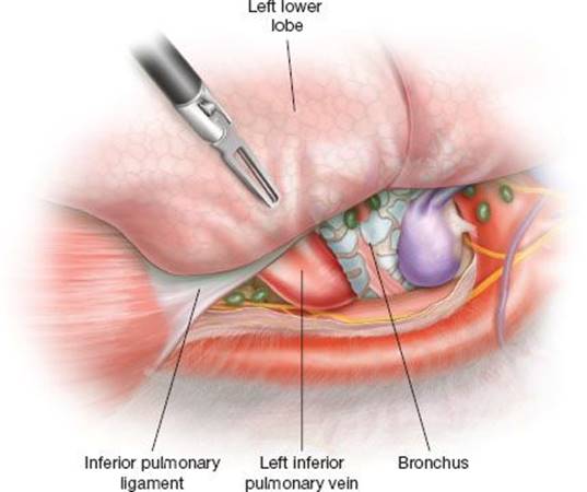
Figure 28.3 The left lower lobe is retracted cephalad and the inferior pulmonary ligament taken down. Dividing the pleura posteriorly, the inferior hilum is dissected revealing the inferior pulmonary vein. This can be further exposed with gentle blunt dissection along the vein.
Addressing the inferior pulmonary vein on the left side, the lung is retracted cephalad and the inferior pulmonary ligament divided. Any lymph nodes (Level 9) within the ligament should be harvested. After dividing the inferior pulmonary ligament, the pleura posteriorly and anteriorly in the inferior hilum is dissected revealing the inferior pulmonary vein. The vein is dissected out distally, often with a peanut dissector, which can facilitate the bronchial dissection, and the superior branch identified (Fig. 28.3).
With the superior segmental artery divided and the vein dissected, attention can turn to addressing the bronchus. Retracting the divided superior segment artery in a cephalad direction can make identifying the superior segment bronchus easier, particularly with the inferior and posterior dissection of the inferior pulmonary vein (Fig. 28.4). Often, completing of the oblique fissure superiorly can help identify the bronchus. The bronchus is surrounded, clamped and selective ventilation of the left lung can assist with demarcating between segments prior to division.
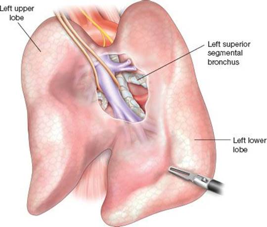
Figure 28.4 By retracting the divided superior segment artery in a cephalad direction identification of the superior segment bronchus is made easier, particularly with the inferior and posterior dissection of the inferior pulmonary vein. The can also be approached from the posterior, inferior aspect of the dissection. Often working from both directions can facilitate dissection of the airway and division.
VATS Left Lower Lobe Basilar Segmentectomy
The left lower lobe basilar artery is exposed in the fissure in a similar manner to the exposure for the left superior segmental artery. The artery is surrounded and adjacent lymph nodes harvested to ease exposure of the vessel. Dividing the basilar artery with a stapler can be challenging, as angles can be difficult particularly with incomplete fissures. Opening the fissure on either side of the artery with cautery can assist with placement of the stapler. If additional exposure is needed, completing the fissure in either direction can help as well. Again the inferior pulmonary ligament is divided in similar fashion as for the superior segmentectomy and the inferior pulmonary vein and its branches dissected out. The bronchial dissection is made easier by completing the inferior aspect of the oblique fissure with a thick tissue stapler. As mentioned previously, a difficult dissection can be made easier by completing a local lymph node harvest, exposing the branches of the airway. The bronchus is then clamped and the left lung selectively ventilated, allowing the surgeon to demarcate between segments for division with a thick tissue stapler.
A lymph node dissection is performed if indicated and should include all subcarinal and aortopulmonary (AP) window lymph nodes for a cancer operation.
Division of Parenchymal Plane
With the bronchus transected, division of the intersegmental plane can begin working toward center of hilum and the transected bronchus. Often there is a subtle accessory intersegmental fissure that can guide stapling. Occasionally, it is helpful to again inflate the lung slightly to demonstrate the line of division. Lung parenchyma is typically divided with a thick tissue stapler; we prefer the use of a 45 mm as the 60 mm can be difficult to maneuver in the chest.
An older “fracture technique” is performed with digital dissection following the intersegmental vein to demarcate segments. This has been replaced by cautery and certainly can be performed in a thoracoscopic procedure.
 POSTOPERATIVE MANAGEMENT
There are no significant differences in our management of patients after segmentectomy versus lobectomy. Perioperative antibiotics are continued for two doses and subcutaneous unfractionated heparin is started on the morning after surgery. Patients typically have a single chest tube placed to suction initially and then transitioned to water seal and removed on the first or second postoperative day if there is no air leak. The epidural catheter rarely remains beyond the third postoperative day provided the chest tube has been removed. The transition to oral narcotics begins when patients are tolerating a diet. Ambulation and pulmonary toilet are encouraged.
 COMPLICATIONS
Studies that have compared lobectomy and segmentectomy have typically been oriented toward oncologic outcomes rather than postoperative complications; however, it is general accepted that the morbidity and mortality following segmentectomy is no different or worse than for lobectomy. Some studies have suggested a lower rate of immediate pulmonary complication (e.g., reintubation or prolonged ventilation) following segmentectomy owing to the relative preservation of lung parenchyma, excluding patients whose indication for segmentectomy is poor pulmonary reserve. The incidence of prolonged air leak may be slightly higher following segmentectomy (∼10%) given the need for more difficult parenchymal dissection.
 RESULTS
The Lung Cancer Study Group in 1995 reported that limited lung resection as compared to lobectomy for lung cancer resulted in a 75% increase in local recurrence rate and a 50% increase in death with cancer rate. However, limited lung resection included wedge resection as well as segmentectomy and there was considerably heterogeneity in the study population. The ambiguity of these results has led investigators to seek out populations in whom segmentectomy is likely to be equivalent to lobectomy in terms of long-term survival and locoregional recurrence. Several studies have found that a tumor size of 2 cm appears to be the inflection point below which segmentectomy performs as well as lobectomy. Similarly, elderly patients and patients with adenocarcinoma may have equivalent outcomes with segmentectomy. A recent study by Altorki et al. looking specifically at lung tumors less than 3 cm discovered during screening found no significant difference in survival between lobectomy and segmentectomy (though there was a decreased rate of appropriate mediastinal nodal staging in the segmentectomy group). These results apply directly to the growing body of patients with early stage tumors discovered incidentally for whom segmentectomy may be considered as a first line operation. Ultimately, further randomized controlled trials are needed, several of which are ongoing in the United States and Japan.
The potential downside of segmentectomy is often debated; less commonly discussed but equally ambiguous are the benefits of segmentectomy with regard to pulmonary function. No study has conclusively demonstrated that patient undergoing parenchymal-sparing lung resections have durably superior postoperative pulmonary function compared to those undergoing lobectomy. Similarly no studies have demonstrated a decreased rate of pulmonary complications with limited resection. No data exist on long-term functional status or quality of life. Still, it is likely all patients, not just those with borderline pulmonary function, benefit from the minimum amount of parenchymal resection that is oncologically appropriate.
Recommended References and Readings
Altorki NK, Yip R, Hanaoka T, Sublobar resection is equivalent to lobectomy for clinical stage 1 A lung cancer in solid nodules. J Thorac Cardiovasc Surg. 2014;147(2):754–762.
Chamogeorgakis T, Ieromonachos C, Georgiannakis E, et al. Does lobectomy achieve better survival and recurrence rates than limited pulmonary resection for T1N0M0 non-small cell lung cancer patients? Interact Cardiovasc Thorac Surg. 2009;8(3):364–372.
De Zoysa MK, Hamed D, Routledge T, et al. Is limited pulmonary resection equivalent to lobectomy for surgical management of stage I non-small-cell lung cancer? Interact Cardiovasc Thorac Surg.2012;14(6):816–820.
Ginsberg RJ, Rubinstein LV. Randomized trial of lobectomy versus limited resection for T1 N0 non-small cell lung cancer. Lung Cancer Study Group. Ann Thorac Surg. 1995;60(3):615–622.
Gorenstein LA, Sonett JR. The surgical management of stage I and stage II lung cancer. Surg Oncol Clin N Am. 2011;20(4):701–720.
Nakamura H, Taniguchi Y, Miwa K, et al. Comparison of the surgical outcomes of thoracoscopic lobectomy, segmentectomy, and wedge resection for clinical stage I non-small cell lung cancer. Thorac Cardiovasc Surg.2011;59(3):137–141.
Okada M, Koike T, Higashiyama M. Radical sublobar resection for small-sized non-small cell lung cancer: A multicenter study. J Thorac Cardiovasc Surg. 2006;132(4);769-775.
Okumura M, Goto M, Ideguchi K, et al. Factors associated with outcome of segmentectomy for non-small cell lung cancer: Long-term follow-up study at a single institution in Japan. Lung Cancer.2007;58:231–237.
Rami-Porta R, Tsuboi M. Sublobar resection for lung cancer. Eur Respir J. 2009;33(2):426–435.
Sienel W, Stremmel C, Kirschbaum A, et al. Frequency of local recurrence following segmentectomy of stage IA non-small cell lung cancer is influenced by segment localisation and width of resection margins–implications for patient selection for segmentectomy. Eur J Cardiothorac Surg. 2007;31(3):522–527.
Tsutani Y, Miyata Y, Nakayama H, et al. Appropriate sublobar resection choice for ground glass opacity-dominant clinical stage IA lung adenocarcinoma: Wedge resection or segmentectomy. Chest.2014;145(1):66–71.
Tsutani Y, Miyata Y, Nakayama H, et al. Oncologic outcomes of segmentectomy compared with lobectomy for clinical stage IA lung adenocarcinoma: Propensity score-matched analysis in a multicenter study. J Thorac Cardiovasc Surg. 2013;146(2):358–364.
Tsutani Y, Miyata Y, Nakayama H, et al. Sublobar resection for lung adenocarcinoma meeting node-negative criteria on preoperative imaging. Ann Thorac Surg. 2014;97:1701–1707.