
Sean C. Grondin and Colin Schieman
 INDICATIONS/CONTRAINDICATIONS
Parenchyma-sparing lung resections were first described in the 1930s by Churchill for patients diagnosed with benign lung disorders such as bronchiectasis and tuberculosis. This pioneering surgeon described segmental resection of the lung, also known as segmentectomy, using anatomic planes to resect pulmonary tissue based on the bronchovasacular anatomy. It is important that a clear distinction be made between segmentectomy and wedge resection as the latter is a sublobar resection of the lung tissue without using intersegmental planes. Segmentectomy is generally thought to be superior to wedge resection for lung cancer resections because it allows isolation of the bronchovascular structures and improved resectability of regional lymph nodes. To be successful in performing a segmental resection, a surgeon requires a thorough understanding of the three-dimensional anatomic segmental anatomy of the lung. Segmentectomy is currently performed for a wide variety of benign and malignant lung disorders (Table 27.1) and may be completed using minimally invasive or open techniques. In nonsmall cell lung cancer (NSCLC) segmentectomy has traditionally been performed for patients with limited pulmonary function for whom lobectomy is not feasible, where a lesser resection offers a more suitable alternative. More recently, publications suggest that segmentectomy may be an acceptable alternative to lobectomy in the management of patients with small (<2 cm) peripheral NSCLC tumors with local recurrence rates and 5-year survival similar to lobectomy in carefully selected patients. Although, the benefits and role of segmentectomy in patients with imaging suggesting solitary or multiple foci of atypical alveolar hyperplasia (AAH), adenocarcinoma in situ (AIS—formerly bronchoalveolar carcinoma), or minimally invasive adenocarcinoma (MIA—<5-mm invasive adenocarcinoma) remains unclear it is an often utilized approach.
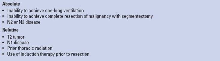
Contraindications to anatomic segmentectomy are listed in Table 27.2. Although some factors such as previous thoracic surgery, incomplete or absent lung fissures or bulky mediastinal adenopathy may make performing a segmentectomy technically more challenging, they are not considered contraindications to the procedure. Segmentectomy should not be performed in patients with a preoperative diagnosis of N2 disease. It is our opinion that patients with a preoperative diagnosis of N1 disease should preferentially undergo lobectomy. Some surgeons do, however, consider segmentectomy with complete nodal resection in select patients with known N1 disease who are not candidates for lobectomy due to limited cardiopulmonary reserve and who do not have evidence of bulky adenopathy.
TABLE 27.1 Indications for Anatomic Segmentectomy

 PREOPERATIVE PLANNING
As with any patient being considered for lung resection, a careful preoperative assessment should be performed to ensure suitability for surgery. After a thorough history and physical examination, an evaluation of the cardiac and pulmonary fitness should be performed using one or a combination of pulmonary function testing, low technology exercise testing with shuttle walk test (SWT) or a stair climb test (SCT). Quantitative ventilation–perfusion scans and cardiopulmonary exercise testing (CPET) is reserved for patients with significant cardiopulmonary impairment. Cardiac testing such as echocardiogram and exercise stress testing are reserved for symptomatic patients with a history of cardiac disease. For patients with suspected or confirmed lung cancer, computed tomography (CT) of the chest and upper abdomen, and positron emission tomography (PET) scanning is routinely used to assess the primary tumor as well as to rule out metastatic disease. Testing using magnetic resonance imaging or CT of the head is reserved for symptomatic patients to rule out neurologic metastases. Suspected mediastinal nodal disease identified on imaging studies is further assessed using endobronchial ultrasound and/or mediastinoscopy.
There is no universally accepted definition of what comprises a patient with poor cardiopulmonary function. At our institution, patients with predicted postoperative (PPO) forced expiratory volume in 1 second (FEV1) and/or PPO diffusion capacity (DLCO) less than 40% predicted, PCO2 >45 mm Hg, PO2 <50% on room air, ejection fraction <45%, and moderate-to-severe pulmonary hypertension are considered high risk for surgery. These marginal patients undergo CPET with measurement of maximal oxygen consumption (VO2max). If VO2max is <10 mL/kg/min (or 35% predicted) patients should be counseled about nonoperative treatment options for lung cancer and the increased risk of perioperative death and cardiopulmonary complications even with sublobar lung resection. Prior to surgery, high-risk patients with potentially resectable lung cancer should undergo smoking cessation counseling, be enrolled in a pulmonary rehabilitation program, and be evaluated by a multidisciplinary team, which includes radiation therapists, medical oncologists, pulmonologists, radiologists, and thoracic surgeons. Despite these diagnostic measures, however, it is often difficult to define a patient as medically inoperable for lung resection.
TABLE 27.2 Contraindications for Anatomic Segmentectomy
 SURGERY
In preparation for surgery, the patient is kept fasting for 8 hours prior to general anesthesia. The patient receives prophylactic antibiotics intravenously as well as a subcutaneous heparin injection (5,000 units) for deep vein thrombosis (DVT) prophylaxis within 60 minutes of skin incision. When the patient enters the operating room and prior to the induction of anesthesia, a detailed safety “time-out” is routinely performed involving the entire operating room team. Importantly, the correct side of the surgery is confirmed with the patient and surgeon, as well as by the anesthesia team and nursing staff. Preoperative CT or PET imaging is reviewed with the operative team prior to induction of anesthesia to check the size and segment location of the target lesion and for the presence of adenopathy.
For the open technique an epidural catheter or paravertebral catheter is often placed to optimize postoperative pain management. A left-sided double-lumen tube or bronchial blocker is used for lung isolation. Position of the double-lumen tube is confirmed using a pediatric bronchoscope.
Position
For open procedures patients are placed in the lateral decubitus position and secured in place using a bean bag or rolls. The bed is flexed at the level of the hip to extend the rib cage in the operative field. Careful padding of the extremities is undertaken to minimize the risk to the patient of developing pressure necrosis or neuromuscular injury. The top arm is carefully hung using a padded arm sling. Position of the double-lumen tube is always reconfirmed once final positioning of the patient is complete. The proposed chest incision (posterolateral thoracotomy) is then marked followed by a second safety check list review. This methodical review is done before prepping, draping, and making the incision to ensure the correct administration of medications (antibiotics and prophylactic heparin) and confirm the side of surgery and availability of blood products. At this point, the left lung is deflated and the patient prepped and draped.
Technique
Although it is possible to do a left lower lobe segmentectomy through a muscle-sparing incision, a posterolateral thoracotomy through the fifth intercostal space (ICS) is our preferred approach. The skin incision extends from the anterior axillary line approximately one finger breath below the tip of the scapula along the fifth ICS. Posterior to the scapula the incision turns slightly cephalad midway between the scapula and spine. Electrocautery with suction is used for hemostasis and to minimize smoke exposure to the operative team. The latissimus dorsi muscle is usually divided with electrocautery and the serratus anterior muscle exposed. The serratus muscle is mobilized along its inferior border and retracted anteriorly to expose the ribs. Once the ribs have been counted to ensure the appropriate identification of the fifth ICS, the thorax is entered using electrocautery. After the initial intercostal incision and exploration of the pleural cavity, placement of a rib spreader facilitates extension of the intercostal incision 3 to 4 cm posterior to the internal mammary vessels anteriorly and to the erector spinal muscle posteriorly. In carefully selected patients at high risk for a bronchopleural fistula (e.g., steroids, chronic infection, immunocompromised, etc.), an intercostal muscle bundle is mobilized prior to placement of a rib spreader for coverage of the bronchial stump at the completion of the segmentectomy.
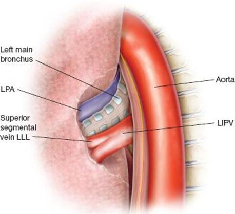
Figure 27.1 Posterior view of the hilum with a view of the left inferior pulmonary vein (LIPV), left lower lobe (LLL) mainstem bronchus, and left main pulmonary artery (LPA).
Once the chest is opened a thorough examination of the pleural space is done to rule out unrecognized disease and mobilize any pleural adhesions. The lung is palpated to confirm the location of the target lesion and to assess for any additional nodules or abnormalities. The surgeon must confirm that the target lesion is confined with a definable resectable segmental boundary. As is classically described, we advocate deliberate N1 nodal sampling of the intraparenchymal segmental nodes, which consistently arise adjacent to the origin of the segmental bronchus. These segmental N1 nodes will be positive in 2% to 6% of clinical T1N0 cases. Frozen-section analysis is performed on any suspicious N1 nodes. If the nodes are positive, we perform a lobectomy provided the patient is a candidate for the procedure. Interestingly, as cited in a recent series by Nomori, when completion lobectomy was performed for N1 positivity following segmentectomy there was no evidence of residual tumor or nodal metastases in the completion lobectomy specimen in 10 cases. These findings bring into question the true need for completion lobectomy in this specific clinical setting. Despite these findings, we would generally not perform a segmentectomy if metastatic nodal disease is identified unless the patient is clearly unable to tolerate lobectomy.
Whether doing a left lower lobe superior or basilar segmentectomy, the dissection begins with mobilization of the inferior pulmonary ligament superiorly to the inferior pulmonary vein. The pleura overlying the hilum is incised to identify and confirm the presence of a distinct inferior and superior pulmonary vein. Careful review of the pulmonary vein anatomy is important to ensure correct identification of the vein branches prior to ligation. Posteriorly the pleura is mobilized to fully visualize the inferior vein as well as to identify the left mainstem bronchus and pulmonary artery (Fig. 27.1). For a left lower lobe segmentectomy the inferior pulmonary vein is dissected so as to allow identification of its two main tributaries, the basilar segmental branch and the superior segmental branch (Fig. 27.2).
Left Lower Lobe Superior Segmentectomy
For a left lower lobe superior segmentectomy the superior segmental branch of the inferior pulmonary vein is isolated and divided typically with a stapler or alternatively it may be suture ligated. Care must be taken not to compromise the remaining inferior pulmonary vein trunk when dividing the segmental vein branch to avoid venous thrombosis. The superior segmental bronchus lies posterior to the vein and is isolated from the posterior direction (Fig. 27.3). Placement of a silk suture around the bronchus allows gentle traction on the bronchus and facilitates smooth placement of the stapler around the structure. Once the stapler is positioned and closed, the silk suture is removed and the lung is gently inflated to ensure the correct segment of lung will be removed. Bronchoscopic examination of the lower lobe through the double-lumen tube is used to confirm the segmental anatomy and ensure the endotracheal tube is away from the staple line. The bronchoscope will also transilluminate the airway in the operative field to ensure correct anatomic orientation of the bronchial segments.
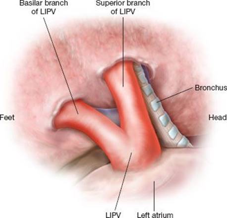
Figure 27.2 Posterior view of the left inferior pulmonary vein (LIPV) branches. For a left lower lobe superior segmentectomy, the superior vein branch is isolated and divided with a stapler to expose the segmental bronchus.
After the bronchus is stapled, the pulmonary artery to the superior segment is isolated and divided with a stapler (Fig. 27.4). The surgeon must be careful to identify each pulmonary artery branch prior to ligation due to the high degree of anatomic variation in the branches of the pulmonary artery. Occasionally, the major fissure is complete allowing for easy identification and isolation of the artery branches in the fissure. If the arterial dissection is difficult, proximal arterial control is instituted for safety.
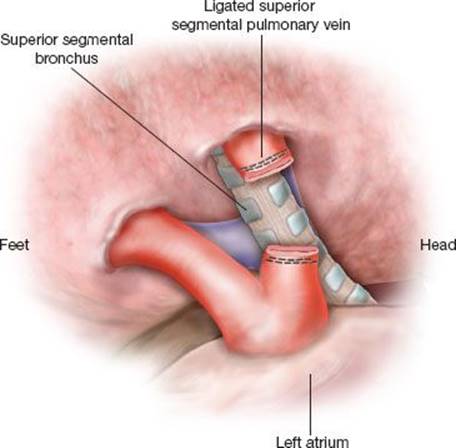
Figure 27.3 Posterior view of the left lower lobe superior segmental bronchus after division of the superior segmental vein. The segmental bronchus is isolated and divided with a stapler.
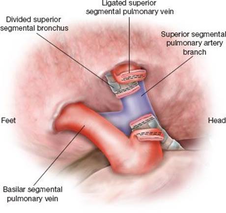
Figure 27.4 Posterior view of the left lower lobe superior segmental pulmonary artery branch after division of the segmental vein and bronchus. The segmental artery is isolated and divided with a stapler.
After division of the artery, the parenchymal dissection is then completed with a stapler using gentle partial inflation of the lung if necessary to demarcate the intersegmental plane of dissection (Fig. 27.5). Once removed, the specimen is examined to ensure negative margins of resection at the bronchus and lung parenchyma. Frozen-section analysis is used if margins are equivocal. At this point a thorough mediastinal nodal dissection of intraparenchymal, intersegmental, interlobar, and mediastinal nodes is performed and any suspicious nodes are sent for frozen-section analysis as well. If feasible, a lobectomy is performed if the parenchymal margins are positive or if N1 disease is identified. Because the remaining basilar segments are fixed at two distinct points, the inferior vein and the bronchopulmonary pedicle superiorly, torsion should be nearly impossible. To our knowledge torsion following segmentectomy of the left lower lobe has not been reported. If there is concern that the remaining segments show propensity for rotation or torsion, fixation of the remaining segments to the lingula can be easily performed.
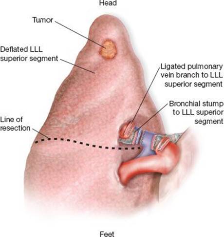
Figure 27.5 Once the artery, vein, and bronchus to the segment have been divided, a stapler is used to divide the lung parenchyma along the line of demarcation. In this illustration, the left lower lobe (LLL) superior segment is prepared for removal with gentle lung reexpansion used to identify the unventilated and unperfused segment of lung.
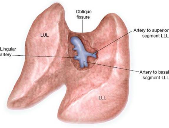
Figure 27.6 If the oblique fissure is complete, the pulmonary artery branches of the left lower lobe (LLL) can be approached through the major fissure prior to division of the segmental bronchus. Note that the artery to the superior segment is located at approximately the same level as the lingular artery.
Left Lower Lobe Basilar Segmentectomy
The left lower lobe basilar segments include the anterior, posterior, medial, and lateral segments. A basilar segmentectomy in many ways requires the same sequence of dissection as a superior segmentectomy. To perform a left lower lobe basilar segmentectomy, the basilar segmental pulmonary vein branch is approached and isolated inferiorly after division of the inferior pulmonary ligament. The vein is divided with a stapler to identify the basilar segmental bronchus. The bronchus is stapled after verifying the correct position of the stapler with insufflation and bronchoscopic examination. The artery can then be isolated and divided with a stapler. If the fissure is complete, the basilar arterial trunk can also be approached and divided through the oblique fissure after division of the vein branch (Fig. 27.6). Using this approach, the bronchus is stapled last. The nodes adjacent to the segmental bronchus are excised. Using partial lung inflation to guide the resection margin, the parenchymal resection margin is divided along the segmental fissure with a stapler. The specimen is assessed for margins and a mediastinal node dissection is completed. Although torsion of the superior segment has not been reported to our knowledge, depending on the propensity of the remaining superior segment to rotate, fixation of this segment to the posterior aspect of upper lobe may be performed using a stapler to prevent torsion.
 POSTOPERATIVE MANAGEMENT
The practice at our institution following open left lower lobe segmentectomy is to place a single 28-French chest tube at the apex on 20-cm suction for drainage. If there is concern postoperatively for excessive bleeding, prolonged air leak, or a possible pleural space problem, two chest tubes (one apical and one in the basal supradiaphragmatic region) are placed. Patients are monitored in a step down unit for 12 to 24 hours postoperatively with epidural anesthesia. Aggressive chest physiotherapy with pulmonary toilet is an important part of postoperative care. Risk of DVT is minimized by early mobilization of the patient, the use of subcutaneous heparin, and pneumatic stockings. Typically, two additional doses of antibiotic are given postoperatively and then discontinued. On the first postoperative day, the chest tube is switched to underwater seal unless there is an increasing pneumothorax or subcutaneous emphysema. On the second day, the chest tube is usually removed providing absence of air leak.
 COMPLICATIONS
Segmentectomy is a well-tolerated procedure with a mortality rate less than 1%. In 1995, the Lung Cancer Study Group reported no significant difference in postoperative morbidity or mortality between patients undergoing segmentectomy and lobectomy. The most common complications are persistent air leaks and persistent pleural spaces. Both are interrelated and the incidence varies based on the difficulty of dissection of the intersegmental planes. In most cases these two complications can be managed conservatively with only a small percentage of patients developing more serious complications such as bronchopleural fistula, empyema, or sepsis.
 RESULTS
The Lung Cancer Study Group demonstrated in a randomized prospective trial that lobectomy had a lower local recurrence rate and an increased 5-year survival compared to sublobar resection in good-risk patients undergoing surgery for early-stage NSCLC. Based on these data limited pulmonary resection with segmentectomy has generally been reserved for patients with poor pulmonary risk. With the advent of CT screening programs for the detection of lung cancer, the debate regarding the role of sublobar resection for the treatment of Stage I NSCLC in patients with good pulmonary function has been rekindled. Several retrospective studies have demonstrated no statistically significant difference in local and regional recurrences between segmentectomy and lobectomy for peripheral T1aN0M0 NSCLC. When a meta-analysis was performed of 14 articles an apparent advantage of lobectomy over segmentectomy was demonstrated for 5-year survival. This trend did not reach statistical significance. The role of sublobar resection as an alternative to lobectomy in good-risk patients with small peripheral NSCLC tumors will hopefully be clarified in the near future when two phase III randomized trials (CALGB 140503 and JCOG0802) are complete.
In patients with marginal pulmonary function and early-stage NSCLC, sublobar resection with segmentectomy has been demonstrated to be safe allowing for good preservation of lung function postoperatively. Furthermore, wedge resection has been shown to be inferior to segmentectomy when comparing sublobar resection techniques and should not be performed on patients with Stage I NSCLC when it can be avoided. At our institution, segmentectomy is performed on carefully selected high-risk patients with stage I NSCLC and also in good-risk patients with imaging suggesting multifocal disease and peripheral ground-glass opacities or small nodular abnormalities suspected to be malignant (e.g., AAH, AIS, MIA).
 CONCLUSIONS
Segmentectomy is currently performed for a wide variety of lung disorders and may be completed using minimally invasive or open techniques. In patients with suspected or confirmed NSCLC, segmentectomy was historically reserved for patients with significant cardiopulmonary insufficiency. Recent studies suggest, however, that a local recurrence rate and 5-year survival similar to lobectomy can be achieved for patients with T1aN0M0 (<2 cm) NSCLC. With the advent of CT screening programs for the early detection of lung cancer, further studies are needed to elucidate the role of segmentectomy in patients with solitary or multiple foci of AAH, AIS, or MIA.
Recommended References and Readings
Blasberg JD, Pass HI, Donington JS. Sublobar resection: A movement away from the Lung Cancer Study Group. J Thorac Oncol. 2010;5:1583–1593.
Brunelli A, Kim WA, Berger KI, et al. Physiologic evaluation of the patient with lung cancer being considered for resectional surgery: Diagnosis and management of lung cancer, 3rd ed: American College of Chest Physicians Evidence-Based Clinical Practice Guidelines. Chest. 2013;143(suppl 5):e166S–e190S.
Cerfolio RJ, Allen MS, Trastek VF, et al. Lung resection in patients with compromised pulmonary function. Ann Thorac Surg. 1996; 62:348–351.
Churchill E. Segmental pneumonectomy in bronchiectasis. Ann Surg. 1939;109:481–499.
Friedberg JS. Segmentectomy. In: Sugarbaker DJ, Bueno R, Krasna, et al., eds. Adult Chest Surgery. 1st ed. New York, NY: McGraw-Hill Companies Inc; 2009:561–566.
Ginsberg RJ, Rubinstein LV. Randomized trial of lobectomy versus limited resection for T1N0 non-small cell lung cancer. Lung Cancer Study Group. Ann Thorac Surg. 1995;60:615–623.
Keenan RJ, Landreneau RJ, Maley RH, et al. Segmental resection spares pulmonary function in patients with stage I lung cancer. Ann Thorac Surg. 2004;78:228–233.
LoCicero J III. Segmentectomy and lesser resections. In: Shields TW, LoCicero J III, Reed CE, et al., eds. General Thoracic Surgery. 7th ed. Philadelphia, PA: Lippincott Williams & Wilkins; 2009:479–485.
Martin-Uncar AE, Nakas A, Pilling JE, et al. A case-matched study of anatomical segmentectomy versus lobectomy for stage I lung cancer in high-risk patients. Eur J Cardiothorac Surg. 2005;27:675–679.
Mattioli S, Ruffato A, Puma F, et al. Does anatomical segmentectomy allow an adequate lymph node staging for cT1 a non-small cell lung cancer? J Thorac Oncol. 2011;6:1537–1541.
Nakamura H, Kawasaki N, Taguchi M, et al. Survival following lobectomy vs. limited resection for stage I lung cancer: A meta-analysis. Br J Cancer. 2005;92:1033–1037.
Nakamura K, Saji H, Nakajima R, et al. A phase III randomized trial of lobectomy versus limited resection for small-sized peripheral non-small cell lung cancer (JCOG0802/WJOG4607 L). Jpn J Clin Oncol.2010;40:271–274.
Nomori H, Mori T, Izumi Y, et al. Is completion lobectomy merited for unanticipated nodal metastases after radical segmentectomy for cT1 a N0 M0/pN1–2 non-small cell lung cancer? J Thorac Cardiovasc Surg. 2012;143:820–824.
Okada M, Yoshikawa K, Hatta T, et al. Is segmentectomy with lymph node assessment an alternative to lobectomy for non-small cell lung cancer of 2 cm or smaller? Ann Thorac Surg. 2001;71:956–960.
Schuchert MJ, Abbas G, Awais O, et al. Anatomic segmentectomy for the solitary pulmonary nodule and early-stage lung cancer. Ann Thorac Surg. 2012;93:1780–1785.
Stiles BM, Altorki NK, Segmentectomy versus lobectomy for stage I lung cancer in patients with good pulmonary function. In: Ferguson M, ed. Difficult Decisions in Thoracic Surgery. 2nd ed. London: Springer-Verlag; 2011:125–133.
Whitson BA, Groth SS, Andrade RS, et al. Survival after lobectomy versus segmentectomy for stage I non-small cell lung cancer: A population-based study. Ann Thorac Surg. 2011;92:1943–1950.
Whitson BA, Andrade RS, Maddaus MA. Lung segmentectomy for patients with peripheral T1 lesions. Op Tech Thorac Cardiovasc Surg. 2006;11:310–322.